Conservation of genome content and virulence
determinants among clinical and environmental
isolates of
Pseudomonas aeruginosa
Matthew C. Wolfgang*, Bridget R. Kulasekara*, Xiaoyou Liang*, Dana Boyd*, Kai Wu†, Qing Yang†, C. Garrett Miyada†, and Stephen Lory*‡
*Department of Microbiology and Molecular Genetics, Harvard Medical School, 200 Longwood Avenue, Boston, MA 02115; and†Affymetrix, Inc., Santa Clara, CA 95051
Communicated by John J. Mekalanos, Harvard Medical School, Boston, MA, April 23, 2003 (received for review March 24, 2003)
Pseudomonas aeruginosais a ubiquitous environmental bacterium capable of causing a variety of life-threatening human infections. The genetic basis for preferential infection of certain immunocom-promised patients or individuals with cystic fibrosis byP. aerugi-nosais not understood. To establish whether variation in the genomic repertoire ofP. aeruginosastrains can be associated with a particular type of infection, we used a whole-genome DNA microarray to determine the genome content of 18 strains isolated from the most common human infections and environmental sources. A remarkable conservation of genes including those en-coding nearly all known virulence factors was observed. Phyloge-netic analysis of strain-specific genes revealed no correlation be-tween genome content and infection type. Clusters of strain-specific genes in the P. aeruginosa genome, termed variable segments, appear to be preferential sites for the integration of novel genetic material. A specialized cloning vector was developed for capture and analysis of these genomic segments. With this capture system a site associated with the strain-specific ExoU cytotoxin-encoding gene was interrogated and an 80-kb genomic island carryingexoUwas identified. These studies demonstrate thatP. aeruginosastrains possess a highly conserved genome that encodes genes important for survival in numerous environments and allows it to cause a variety of human infections. The acquisition of novel genetic material, such as the exoU genomic island, through horizontal gene transfer may enhance colonization and survival in different host environments.
M
embers of the genusPseudomonasare some of the most diverse bacterial species in the environment. Particularly important are Pseudomonas aeruginosa, which are commonly encountered aerobic microbes that have evolved the remarkable capacity to inhabit diverse natural environments and infect higher organisms such as insects, plants, and animals (1). In humans,P. aeruginosacan colonize virtually any mucosal surface and invade tissues and blood (2). Its ability to thrive in a broad range of environments is in part caused by the fact that it possesses a large and diverse genome (3). Furthermore, P.aeruginosapossesses the largest proportion of regulatory genes
(⬇1 in 10) of all of the sequenced bacterial genomes (3). This striking feature likely provides a means for coordinating the expression of its many genes in response to a wide range of environmental demands.
Comparative genomics based on the analysis of complete genomes of different strains of the same species can provide valuable insights into the acquisition or loss of genes through horizontal gene transfer and evolution of genes through changes at the nucleotide sequence level. With the availability of DNA microarrays, a larger number of genomes can be compared (4). When representative subsets of strains are examined, useful evolutionary questions can be answered by genome content analysis. Particularly informative have been those studies that used genomic microarray hybridization technology to investigate the epidemiology of disease and correlate the loss of genes with
changes in virulence. Specifically, microarray analysis has been used to demonstrate a correlation between certain genes in
Vibrio cholerae and specific pandemics (5). In addition, a
mi-croarray hybridization-based study of Staphylococcus aureus
variants associated with toxic shock syndrome and methicillin resistance revealed that these strains evolved in parallel with changing conditions in the host environment and antibiotic treatment (6). The same approach was used to demonstrate the genetic basis for attenuation of virulence in strains of
Mycobac-terium tuberculosis including those used for vaccination (7).
Analysis of Helicobacter pylori genomes from a wide range of strains has resulted in the identification of a limited number of variable chromosomal segments and their association with spe-cific virulence factors (8). Further studies of this organism have provided evidence for the evolution of the genome of individ-ual clones (including deletions of genes) in a highly restricted niche (9).
To correlate the genome content ofP. aeruginosastrains with the various niches that it can inhabit and the different diseases that it can cause, we assembled a collection of strains and assessed their genomic repertoire by using a whole-genome DNA microarray. We have shown that the genome of this organism is highly conserved and that there exists a core set of genes, including nearly all known virulence factors, which are present in all strains regardless of disease source. Interestingly, the same genes are conserved among environmental isolates. We further developed a general strategy, including a specialized cloning vector, for the interrogation of chromosomal regions that show a high degree of polymorphism and often serve as sites of large deletions and insertions. The approach of combining microarray hybridization analysis and targeted capture of iden-tified variable chromosomal segments should facilitate the anal-ysis of many variants of a single species without the need for large-scale sequencing of entire genomes.
Materials and Methods
Genome Content Analysis.GeneChipP. aeruginosaGenome Ar-rays (Affymetrix) were used to assess genome content. Chro-mosomal DNA hybridizations were performed as described (10). Hybridization intensity data were extracted from the scanned array images, and intrachip normalizations were performed by using AffymetrixMICROARRAY SUITE 5.0software. The average signal intensity of the probe sets was scaled to 500 for interchip comparisons. A presence兾absence determination was made by comparing hybridization signal between strains. Values derived from the PAO1 genomic DNA hybridization were used as baseline. Probe sets with a ratio of ⬎0.25 were considered present (blue). Ratios between 0.1 and 0.25 were considered indeterminate (gray), and ratios⬍0.1 were designated absent (yellow).
Design and Construction of a Multifunctional Yeast Capture Vector.A detailed description of the construction of the yeast capture vector p0975–0989capture is provided in Supporting Materials
and Methods, which is published as supporting information on
the PNAS web site, www.pnas.org.
Recombinational Cloning. The variable genomic segments of P.
aeruginosastrains PAO1, PAK, CF127, 6077, and JJ692, flanked
by conserved genes PA0975 and PA0989, were cloned by co-transforming 200 l of competent Saccharomyces cerevisiae
strain CRY1–2 spheroplasts with 10g of unsheared chromo-somal DNA and 1 g of MluI-linearized plasmid p0975– 0989capture. Competent spheroplasts were prepared as de-scribed with the exception of using 10 units of Zymolyase 20T (ICN) instead of lyticase and 10 mM DTT instead of 2-mercap-toethanol (11). Transformation mixtures were plated on uracil-deficient media containing cycloheximide (2.5g兾ml).
Selection of Recombinant Clones. S. cerevisiae colonies were screened by PCR for the presence of captured P. aeruginosa
sequences. A portion of each colony was suspended in 15l of zymolyase buffer (10 mM sodium phosphate buffer, pH 7.5兾1 M sorbitol兾2.5 mg/ml zymolyase 20T) and incubated with shaking for 1 h at 37°C. Two microliters of suspension was used as template for each PCR. Primers used to detect an insert were
5⬘-GGCTCGACCTCAATGGCATGGGCG and 5⬘
-TCA-GAAATATGGCGTCGGGTCGGA, which amplified a 500-bp portion of PA0976 that is not included in the original vector but is predicted to be present in the captured sequences. Plasmid DNA from yeast colonies that produced a PCR product of the correct size was purified and electrotransformed intoEscherichia coliGenehogs (Invitrogen) for subsequent analysis.
Results and Discussion
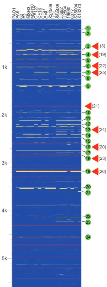
Strain Variability and Genome Conservation.We have examined the genome composition of a collection of 18P. aeruginosastrains by using a whole-genome DNA microarray consisting of probes for 5,549 nonredundant genes that constitute the genome of the sequenced strain PAO1 (3). The collection included four envi-ronmental isolates, a laboratory strain, and strains isolated from the most common human infections, including four respiratory isolates from children (⬍24 months) with cystic fibrosis, five strains associated with urinary tract infections, and two isolates each from ocular and blood infections (Table 1, which is published as supporting information on the PNAS web site). Fig. 1 indicates the predicted presence or absence of each PAO1 gene in the interrogated strains, organized relative to their chromo-somal location in the reference PAO1 genome. The presence or absence of these genes was predicted based on the relative hybridization of chromosomal DNA to the microarray (see
Materials and Methods). Detailed results for each gene are
provided in Table 2, which is published as supporting informa-tion on the PNAS web site.
The fraction of PAO1 genes detected in the various strains ranged from 96.1% to 97.7% (Fig. 4, which is published as
Fig. 1. Analysis of genome content indicates a high level of gene conserva-tion. The diagram indicates the presence and absence of genes found in the sequenced genome of strain PAO1 in clinical and environmental isolates ofP. aeruginosaas detected by microarray hybridization. Strains are indicated at the top. The source of each strain is as follows: PAO1, reference strain; PAK,
laboratory strain; environmental isolates (62, E2, MSH3, MSH10); respiratory isolates from children (⬍24 months) with cystic fibrosis (CF127, CF18, CF27, CF5); isolates from urinary tract infections (X24509, UDL, S54485, JJ692, U2504); strains isolated from ocular infections (19660, 6077), and blood iso-lates (S35004, X13273). Horizontal lines represent genes. Blue indicates that a gene is present, yellow represents absence, and gray indicates that presence was indeterminate. Red lines represent the location of tRNA genes in strain PAO1. The scale represents 5,613 genes (including 5,549 ORFs and 64 tRNA-encoding genes) organized according to the PAO1 chromosome. Green circles indicate variable segments in theP. aeruginosagenome as discussed in the text. Red triangles indicate known sites of insertion of horizontally acquired genetic material. References are indicated in parentheses.
supporting information on the PNAS web site), with 5,183 (93.4%) of the 5,549 PA01 genes present in all strains tested. These 5,183 genes represent the core set of genes, presumably encoding proteins that function in the diverse range of environ-ments that this organism can inhabit. This finding represents a conservative estimate because our analysis does not compensate for sequence polymorphisms or strain-specific genes (see below) that are unrelated at the nucleotide sequence level but encode proteins with conserved function.
The distribution of core genes based on functional classifica-tion relative to the PAO1 genome (annotaclassifica-tion tables available at www.Pseudomonas.com) indicates a broad conservation of func-tional diversity (Fig. 5, which is published as supporting infor-mation on the PNAS web site). The only functional class of genes that showed significant underrepresentation in all strains in-cluded genes associated with horizontal gene transfer (classified as related to phage, transposon, or plasmid). Only 8.6% of these genes were conserved in all strains examined. Of the 53 variable genes in this category 42 constitute two cryptic prophages identified in strain PAO1 but not found in most of the examined strains (Fig. 1, segments 3 and 4) (3).
Of particular interest are those genes associated with P.
aeruginosavirulence. These genes are almost exclusively found
in the category called secreted factors. Additional genes related to the expression of virulence determinants are found in the categories motility and attachment, protein secretion兾export apparatus, and transcriptional regulators. Examination of the genes that play a direct role or are predicted to play a direct role in virulence (267 genes; Table 3, which is published as supporting information on the PNAS web site) indicates a high level of conservation (⬇97%). The extensive conservation of virulence genes in the genomes of strains regardless of their clinical source suggests that the disease-causing ability of this opportunistic pathogen during human infections relies, in general, on a set of highly conserved pathogenic mechanisms. However, specific features of each disease may be influenced by phenotypic characteristics provided by individual or a small number of strain-specific virulence genes (for example,exoU and exoS, see below). The conservation of virulence determinants also extends to environmental isolates, suggesting that selection for the maintenance of such traits exists in the environmental reservoir. Based on this conservation it is likely that the environmental strains studied possess the ability to cause human infections despite the low probability of encountering a human host.
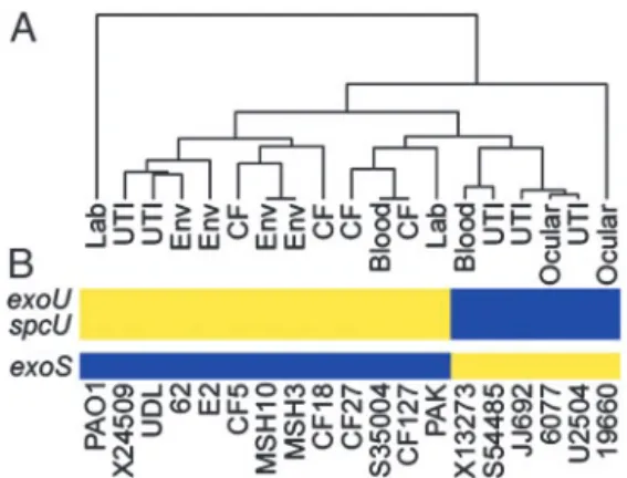
The 368 genes that are absent from the genome of at least one strain can be considered strain specific (Table 4, which is published as supporting information on the PNAS web site). This set of genes was subjected to a phylogenic analysis, which shows that strains from similar environmental or clinical sources are in general no more related than those from different sources (Fig. 2A). Not surprisingly, the most closely related strains are the only two isolates from the same locale, the two environmental strains isolated from Spirit Lake, Washington (MSH3 and MSH10). These strains were selected from a larger collection based on unique colony appearance and differential sensitivity to the pilus-specific phage PO4. The second most highly related strains are a pair of clinical isolates from a cystic fibrosis patient and a blood infection (strains CF127 and S35004, respectively) col-lected at hospitals in different cities (Table 1). Perhaps the most interesting relationship is demonstrated by a group of five strains, three of which were isolated from urinary tract infections (S54485, JJ692, and U2504), one from an ocular infection (6077), and the last from a blood infection (X13273). These strains share several epidemiological markers including the presence of genes for the O11-serotype capsular polysaccharide (data not shown). In addition, these strains are characterized by the presence of the
exoUgene and its cognate chaperonespcUin their genomes (Fig. 2B). ExoU is a cytotoxin secreted by the type III secretion system
ofP. aeruginosa(13). Its expression is associated with increased
virulence in model infection systems and poor clinical prognosis in patients (14, 15). A relationship between ExoU-expressing strains has been previously recognized among ocular isolates based on the coincidence ofexoUwith several phenotypic and genetic traits (16). It is apparent from our studies that this clonal relationship, based on gene content, includes isolates from a wider range of disease states. Another ocular isolate (19660) also carries theexoUandspcUgenes; however, it is phylogenetically unrelated to the rest of the strains in this survey. It was previously reported that carriage ofexoUis accompanied by the absence of
exoS, which encodes another type III secreted protein (17, 18). We also found that the six strains, in this study, withexoUin their genome all lackedexoS(Fig. 2B). The relative conservation of the genomes of these strains, including theexoU carriage and deletion of exoS (see below), implies that conserved selective pressures contributed to the evolution of these genomes in different environmental niches. What selective advantage arises from mutual exclusion of one of two genes encoding virulence factors with distinct activities is unclear. Because the host cell contact-dependent type III secretion system secretes both ExoU and ExoS, the advantage afforded by expression of either one of these genes almost certainly involves interaction with a target eukaryotic organism.
Distribution and Pattern of Strain-Specific Genes Relative to the Reference Genome.Distinct patterns of variability were evident when the genomes of multiple strains were compared (Fig. 1). Strain-specific genes were localized to 90 discrete regions rela-tive to the PAO1 genome (Table 2). Many of these regions are composed of small gene blocks (one to four genes) that showed variability in one or more strains. These variable blocks likely contain genes that are highly polymorphic at the nucleotide sequence level or are gained or lost through local recombination events (see below).
A second pattern, which is more readily apparent, is charac-terized by large clusters of tandem genes that show varying levels of polymorphism between strains. Twenty-four of these regions (termed variable segments) were identified (Fig. 1, Table 2). These variable segments are scattered throughout the genome; however, a significant number are immediately adjacent to
tRNA or tmRNA genes (Fig. 1, segments 2, 4–6, 16–19, and 24). The use of these genes as targeting sites for the integration of mobile genetic elements is well documented (19). In fact, four differentP. aeruginosatRNA genes have been previously shown to act as integration sites for bacteriophage, plasmids, and genomic islands (19–23) (Fig. 1). A number of additional genomic islands identified in P. aeruginosa are not directly associated with tRNA genes but do localize to regions of the chromosomal that show a high degree of variability (24–26) (Fig. 1). Based on the association of known genomic islands and other horizontally transferred genetic elements with regions of vari-ability, we predict that the variable segments identified here may represent additional sites for integration of novel genetic material.
Deviation in the percentage (G⫹C) content of genes from that of the genome average is another marker of horizontal gene transfer. Annotation of the sequenced PAO1 genome revealed 10 such regions (3). With the exception of a cluster of low percentage (G⫹C) content genes encoding ribosomal proteins, all were associated with regions ofP. aeruginosa genomes that contain strain-specific genes (Table 2). In fact, strain-specific genes as a group (Table 4) have an average percentage (G⫹C) of 61.8%, which is significantly lower than the average for core genes (67.1%). This finding suggests that many of these genes are associated with recently acquired genetic material.
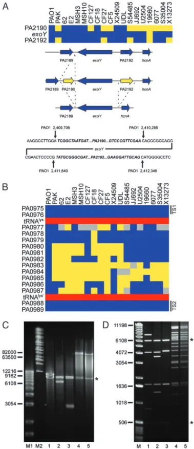
Interrogation of Strain-Specific Genes.An ‘‘absent’’ call based on the microarray hybridization results suggests that the particular locus in the test strain, relative to PAO1, either lacks the corresponding gene altogether (deletion) or it contains a gene or genes that significantly differ from that in the reference strain. To investigate several such loci we designed oligonucleotides that corresponded to conserved flanking genes and used them to prime PCRs such that the resulting amplification products would span the polymorphic regions. Sequence and size analysis of the products allowed us to delineate whether an undetected gene had been deleted or replaced by a small insertion. Two such examples involving genes encoding proteins secreted by the type III secretion system are presented. The example in Fig. 3Ashows that the exoY gene (PA2191), encoding a secreted adenylate cyclase (27), is flanked in PAO1 by two ORFs (PA2190 and PA2192) that encode conserved hypothetical proteins. These two flanking genes are absent from the chromosome of four isolates (PAK, CF27, S54485, and X13273). Six strains (CF127, CF5, JJ692, U2504, 6077, and S35004) lack only PA2190. Finally, one of the strains (19660) lacks all three genes (PA2190,exoY,
and PA2192). Sequence analysis allowed us to identify the
Fig. 3. Analysis of polymorphic genes and variable segments associated with type III secreted effector-encoding genes. (A) Loss of genes in the flanking region ofexoY. (Top) Diagram showing the presence (blue) and absence (yellow) ofexoYand flanking genes in the indicated strains. (Middle) Sum-mary of the specific deletion兾insertion events detected by PCR analysis. Not shown is the deletion of all three genes in strain 19660. (Bottom) Sequence analysis of theexoYflanking regions indicates the specific deletion兾insertion junctions for strains PAK, CF127, CF27, CF5, S54485, JJ692, U2504, 6077, S35004, and X13273. Sequences in bold兾italics are absent in the corresponding deletion strains indicated as yellow (Top). Coordinates of the junction sites relative to the PAO1 chromosome are given. (B) Diagram showing a variable
segment ofP. aeruginosagenomes associated with the acquisition ofexoU. Blue boxes indicate the presence of PAO1 genes in the interrogated strains. Yellow indicates undetected genes, and gray represents genes for which no definitive determination could be made. Red boxes show the location of an intact tRNAlysgene and a partial duplication of the same gene in the PAO1 genome. Black bars indicate the location of conserved targeting sequences (TS1 and TS2) used in the construction of the capture vector. (C) Size deter-mination of captured sequences between conserved genes PA0975 and PA0989 in fiveP. aeruginosastrains. DNA fragments were analyzed on a 1% agarose gel by pulsed-field gel electrophoresis using the Bio-Rad Chef Mapper System. Lane M1, 1-kb DNA ladder (Invitrogen); lane M2, Midrange I PFG Marker (New England Biolabs). Subsequent lanes contain plasmids with cap-tured sequences from the indicated strains. Inserts were separated from the vector (indicated by*) by digestion with I-SceI. Lane 1, PAO1; lane 2, PAK; lane 3, CF127; lane 4, 6077; lane 5, JJ692. (D) Restriction fragment fingerprinting of captured sequences. Lane M, 1-kb DNA ladder. The following lanes contain NcoI digested plasmids with captured sequences from the indicated strains: 1, PAO1; 2, PAK; 3, CF127; 4, 6077; and 5, JJ692. Asterisks denote the location of vector fragments.
precise site of deletion or insertion in each case relative to the PAO1 genome (Fig. 3A).
A similar analysis of theexoSregion confirmed that a number of strains lack this particular gene (Fig. 2Band Fig. 6, which is published as supporting information on the PNAS web site). In the PAO1 genomeexoSis flanked by a near perfect (9 of 10 bp) direct repeat (Fig. 6). Sequence analysis indicates that a recom-bination event between these repeats could account for the loss of exoS in strains S54485, JJ692, U2504, 19660, 6077, and X13273. In all cases the adjacent gene (orf1) is unaffected (Fig. 6). Interestingly,orf1encodes a specific chaperone essential for ExoS secretion from P. aeruginosa by the type III secretion system (28). It is therefore likely that the absence ofexoSis the result of a specific deletion event. Given that nearly all strains that lack exoS contain exoU (Fig. 2B) it is conceivable that acquisition ofexoUleads to the deletion ofexoSby an unknown mechanism.
Interrogation of Variable Segments in the Genomes ofP. aeruginosa by Recombinational Cloning.The inability to span a region iden-tified as absent by PCR may indicate the presence of a large insertion. Clusters of unique genes at a particular location in the genome called ‘‘genomic islands’’ are not uncommon. As de-scribed above, microarray analysis can be used to identify preferential sites of integration of such islands. We constructed a capture vector for yeast recombinational cloning of genomic islands. The vector is a modified version of the plasmid used for cloning P. aeruginosa LPS islands (26). It includes several features that will greatly facilitate the analysis of bacterial genomes and simultaneously provide a tool for further down-stream functional studies of the genes within the genomic islands. The salient features of the vector are outlined in Fig. 7, which is published as supporting information on the PNAS web site. PCR amplification of regions flanking a variable chromo-somal segment results in two targeting sequences that are cloned into the capture vector to provide specificity. After cotransfor-mation of competentS. cerevisiae with P. aeruginosa chromo-somal DNA and the capture vector, recombination mediated by endogenous yeast proteins facilitates cloning of theP. aeruginosa
chromosomal DNA flanked by the homologous targeting se-quences. The captured chromosomal DNA is bordered by a short sequence specifying the recognition site for the I-SceI restriction endonuclease. Because this sequence is absent from the P.
aeruginosagenome (and from the sequenced genomes of most
bacteria) it can be used in preliminary estimation of the size of the insert.
A single capture vector can be constructed and used to interrogate the same region in a large number of strains. The captured sequences can be mobilized into any recipient P.
aeruginosastrain by conjugation. Transfer of the captured island
into a recipient strain lacking the equivalent sequences will allow for functional assessment. After targeted disruption of a partic-ular gene or transposon mutagenesis of the entire segment, the inactivated gene(s) can be readily introduced onto theP. aerugi-nosachromosome by allelic exchange using the method of SCE jumping (29).
We used this vector to investigate whether acquisition ofexoU
and its linked chaperonespcUinvolves integration of a genomic island into a common locus in theP. aeruginosachromosome. The published sequence ofexoUfrom strain PA103 (GenBank accession no. AF027291) (30) contained a segment 3⬘ of the
spcU-coding sequence that was nearly identical to the position 1,069,669–1,069,934 bp of theP. aeruginosastrain PAO1 chro-mosome. This segment is located in a highly polymorphic region of the chromosome (Fig. 1, segment 6). Closer examination reveals that this variable segment (encompassing genesPA0977–
PA0987) is flanked by highly conserved genes (Fig. 3B). To
interrogate this region of the chromosome in differentP.
aerugi-nosastrains we designed a capture vector that included targeting sequences derived from the conserved flanking genes (Fig. 3B). As a control we cotransformed competent yeast with the capture vector and chromosomal DNA from strain PAO1. The resulting clone was analyzed by digestion with I-SceI, which released a 12-kb fragment from the capture vector, corresponding to the predicted size of the PAO1 sequence (Fig. 3C, lane 1). Restric-tion fragment fingerprinting of the cloned insert with theNcoI restriction endonuclease generated fragments that agreed with the predicted location ofNcoI recognition sites in this region of the PAO1 chromosome (Fig. 3D, lane 1). Chromosomal DNA from four additional strains was captured and the recovered plasmids were analyzed as above (Fig. 3 CandD). TwoexoU -carrying strains (6077 and JJ692) possessed a large insert of⬎80 kb (Fig. 3C, lanes 4 and 5). The NcoI digestion patterns confirmed that these inserts are very similar or perhaps even identical (Fig. 3D, lanes 4 and 5). PCR analysis confirmed that they indeed carried theexoUandspcUgenes (data not shown). Analysis of sequences captured from strains lackingexoU(PAK and CF127) indicated that these strains each carried a different-sized chromosomal segment (⬇7 and 3 kb, respectively; Fig. 3C, lanes 2 and 3). TheNcoI restriction pattern for these segments suggests that they may have arisen through the deletion of sequences from the larger PAO1 sequence. Because a tRNA gene flanks this region, integration of the acquired DNA in the
exoU-carrying strains may involve a site-specific recombination system (19). Interestingly, this locus has been previously de-scribed as one of two sites for semistable incorporation of a large plasmid present in many genomes of European isolates of P.
aeruginosa(22). Collectively, these data indicate that this region
of the P. aeruginosa chromosome may be a ‘‘hotspot’’ for integration of horizontally acquired DNA.
Conclusion
The work presented here shows that the genomes ofP. aerugi-nosa strains, representing distinct clinical or environmental sources, are highly conserved. These results support a basic model whereby conservation of a large genome allows this organism to inhabit the widest possible range of environments and confers the ability to cause human infections after relatively infrequent encounters. Although we cannot predict the total gene content of individual strains or whether detected genes are functional, the remarkable conservation of genes encoding proteins associated with virulence indicates that most strains, regardless of source, possess the basic pathogenic mechanisms necessary to cause a wide variety of human infections. The fact that this conservation extends to environmental strains indicates the existence of a natural environmental eukaryotic host and further suggests that environmental strains ofP. aeruginosacan serve as the source for human infections.
Analysis of the microarray hybridization patterns revealed a number of variable segments in theP. aeruginosagenome that may serve as sites of integration of horizontally transferred genetic material. Given the limitations of the microarray hybrid-ization technique we cannot determine which of these sites are occupied or the nature of the acquired sequences. To investigate these sites we developed a multifunctional capture vector and used it to identify an 80-kb genomic island carrying a known virulence gene (exoU). This approach will be useful for identi-fying additional virulence genes that may enhance the ability of
P. aeruginosa to survive in specific host niches; however, we
expect this number to be small, as extensive work over the past 25 years using a variety of clinical isolates has led to the discovery of very few virulence genes that are not part of the core set present in nearly everyP. aeruginosastrain analyzed.
useful tool for the isolation and interrogation of strain-specific islands in bacterial genomes. When these technologies are combined, they should provide essential tools for large-scale analysis of bacterial populations and lead to a better under-standing of the selective pressures that control genome mainte-nance and the spread of novel genetic material.
This work was supported by a grant from the Cystic Fibrosis Foun-dation and Grant DK53369 from the National Institutes of Diabetes and Digestive and Kidney Diseases. GeneChipP. aeruginosaGenome Arrays were subsidized by Cystic Fibrosis Foundation Therapeutics. M.C.W. was supported by a Cystic Fibrosis Foundation postdoctoral research fellowship.
1. Mahajan-Miklos, S., Rahme, L. G. & Ausubel, F. M. (2000)Mol. Microbiol.37, 981–988.
2. Bodey, G. P., Bolivar, R., Fainstein, V. & Jadeja, L. (1983)Rev. Infect. Dis.5, 279–313.
3. Stover, C. K., Pham, X. Q., Erwin, A. L., Mizoguchi, S. D., Warrener, P., Hickey, M. J., Brinkman, F. S., Hufnagle, W. O., Kowalik, D. J., Lagrou, M.,
et al. (2000)Nature406,959–964.
4. Fitzgerald, J. R. & Musser, J. M. (2001)Trends Microbiol.9,547–553. 5. Dziejman, M., Balon, E., Boyd, D., Fraser, C. M., Heidelberg, J. F. &
Mekalanos, J. J. (2002)Proc. Natl. Acad. Sci. USA99,1556–1561. 6. Fitzgerald, J. R., Sturdevant, D. E., Mackie, S. M., Gill, S. R. & Musser, J. M.
(2001)Proc. Natl. Acad. Sci. USA98,8821–8826.
7. Kato-Maeda, M., Rhee, J. T., Gingeras, T. R., Salamon, H., Drenkow, J., Smittipat, N. & Small, P. M. (2001)Genome Res.11,547–554.
8. Salama, N., Guillemin, K., McDaniel, T. K., Sherlock, G., Tompkins, L. & Falkow, S. (2000)Proc. Natl. Acad. Sci. USA97,14668–14673.
9. Israel, D. A., Salama, N., Arnold, C. N., Moss, S. F., Ando, T., Wirth, H. P., Tham, K. T., Camorlinga, M., Blaser, M. J., Falkow, S. & Peek, R. M., Jr. (2001)
J. Clin. Invest.107,611–620.
10. Wolfgang, M. C., Lee, V. T., Gilmore, M. E. & Lory, S. (2003)Dev. Cell4, 253–263.
11. Burgers, P. M. & Percival, K. J. (1987)Anal. Biochem.163,391–397. 12. Eisen, M. B., Spellman, P. T., Brown, P. O. & Botstein, D. (1998)Proc. Natl.
Acad. Sci. USA95,14863–14868.
13. Finck-Barbancon, V., Goranson, J., Zhu, L., Sawa, T., Wiener-Kronish, J. P., Fleiszig, S. M., Wu, C., Mende-Mueller, L. & Frank, D. W. (1997)Mol. Microbiol.25,547–557.
14. Allewelt, M., Coleman, F. T., Grout, M., Priebe, G. P. & Pier, G. B. (2000)
Infect. Immun.68,3998–4004.
15. Hauser, A. R., Cobb, E., Bodi, M., Mariscal, D., Valles, J., Engel, J. N. & Rello, J. (2002)Crit. Care Med.30,521–528.
16. Lomholt, J. A., Poulsen, K. & Kilian, M. (2001)Infect. Immun.69,6284–6295. 17. Feltman, H., Schulert, G., Khan, S., Jain, M., Peterson, L. & Hauser, A. R.
(2001)Microbiology147,2659–2669.
18. Yahr, T. L., Goranson, J. & Frank, D. W. (1996)Mol. Microbiol.22,991–1003. 19. Williams, K. P. (2002)Nucleic Acids Res.30,866–875.
20. Elsabbagh, H., Xiong, G. & Lutz, F. (1993)Mol. Gen. Genet.237,421–428. 21. Kropinski, A. M. (2000)J. Bacteriol.182,6066–6074.
22. Kiewitz, C., Larbig, K., Klockgether, J., Weinel, C. & Tummler, B. (2000)
Microbiology146,2365–2373.
23. Larbig, K. D., Christmann, A., Johann, A., Klockgether, J., Hartsch, T., Merkl, R., Wiehlmann, L., Fritz, H. J. & Tummler, B. (2002) J. Bacteriol. 184, 6665–6680.
24. Liang, X., Pham, X. Q., Olson, M. V. & Lory, S. (2001)J. Bacteriol.183, 843–853.
25. Arora, S. K., Bangera, M., Lory, S. & Ramphal, R. (2001)Proc. Natl. Acad. Sci. USA98,9342–9347.
26. Raymond, C. K., Sims, E. H., Kas, A., Spencer, D. H., Kutyavin, T. V., Ivey, R. G., Zhou, Y., Kaul, R., Clendenning, J. B. & Olson, M. V. (2002)J. Bacteriol.
184,3614–3622.
27. Yahr, T. L., Vallis, A. J., Hancock, M. K., Barbieri, J. T. & Frank, D. W. (1998)
Proc. Natl. Acad. Sci. USA95,13899–13904.
28. Yahr, T. L., Hovey, A. K., Kulich, S. M. & Frank, D. W. (1995)J. Bacteriol.177, 1169–1178.
29. Wong, S. M. & Mekalanos, J. J. (2000)Proc. Natl. Acad. Sci. USA 97, 10191–10196.
30. Hauser, A. R., Kang, P. J. & Engel, J. N. (1998)Mol. Microbiol.27,807–818.