© 2017, IRJET | Impact Factor value: 5.181 | ISO 9001:2008 Certified Journal
| Page 1572
Method of medical images Encoding and decoding in a designed
telemedicine system
Yasameen Alyouzbaki
1
Eng., Dept. of Computer Engineering, Al-Mustansiriya University ,Baghdad, Iraq
---***---Abstract -
In this paper we introduce a method for encodingand decoding the medical images which enables the users of our designed telemedicine system (researchers in the medical field, patients) to get the desired images and the useful information safely, easily and quickly via a web browser. Our system was tested over Wired and Wireless networks to evaluate its transmission of the original images or of a single wanted slice on demand. The users can get the desired images through a graphical user interface designed for easy access. Response time, slice position and the amount of data delivered were determined as part of the tests included in this paper.
Key Words
:
medical, images, encoding , decoding,telemedicine system
1. INTRODUCTION
Using electronic data and communication technologies to provide and support health care and medicine studies in case of difficult contact between the participants is known as telemedicine. The most used applications are in the fields of transmission of high-resolution X-rays, cardiology, orthopedics, dermatology and psychiatry. Groups of physicians, teachers and researchers are often discussing from large distances. Telemedicine also includes the management of electronic patient records, access to databases on the Web and on private networks, and use of e-mail by many of the medical specialists.
Telemedicine also helps students whose work involves developing medical devices or studying some medical specialties like Sonar, Doppler or X-Ray scanning; they always need to get images from different types of medical scanning devices for their diagnosis, as well as other important information for the patients [1, 2].
Remotely accessing and viewing medical images through the web is important for modern students or specialists. With a web browser, they can access and browse medical images from anywhere and at any time, via different devices, without installation and maintenance of additional client software. This can make medical image data sharing a lot easier. DICOM (Digital Imaging and Communications in Medicine) is the widely accepted standard for medical image storage and delivery among medical devices. In some cases we need medical images for remote diagnosis, in a different location than the one where the patient is hospitalized, for example due to the lack of a specialist doctor or of hardware necessary for treatment [2, 3]. The patient also may need to
refer to medical imaging at different times during the progress of his condition, to get accurate diagnosis and follow-up. For scientific purposes, scholars and researchers in the medical field may need sequential medical images for patients in a certain age or a certain sex and a profile of the diagnosis from the specialist doctor. For such cases, we will need to design a private website for a hospital which has different medical imaging devices in various sections of the hospital. These sections send various types of medical images to a server at the hospital by Wired Network. After encryption, these images are stored on a server in a way that does not occupy much space. In this research, we have designed a database that enables the user to access medical images by various filters such as patients’ age, sex, type of disease, the affected area from the patient’s body, as well as the exact diagnosis; this enables students to get all the information required for the purposes of study or research and development [3, 4].
In this paper we propose a data model to index text and visual data (images). Our data model provides a unified structure to organize image data and three ways for it to be accessed by the user: text-based retrieval, content-based retrieval and combined retrieval. Text-based retrieval allows users to enter keywords (e.g. patient code, age, body part, disease, physician notes and diagnosis conclusion) based on which the image is then accessed in the database. Content-based retrieval allows user to upload a sample image from a local device. Combined recovery is the mixture of content-based retrieval and text-content-based retrieval [4, 5]. On demand transmission, an interactive medical image transmission scheme for single slice images is used for transmission to a web browser or over the user’s wired network. Medical images are encoded by a code stream on the server side; the suggested encoding method aims to decrease the time for encoding as well as for the decoding process, compared to the time needed by other methods. Also, the precise details of those medical images should be preserved through the encryption process by keeping their details and retrieving them completely and accurately after the decryption. After the encoding process, the images are stored as a file not as an image, for reducing the storage space for the medical images that are usually considered as large size images [6, 7].
© 2017, IRJET | Impact Factor value: 5.181 | ISO 9001:2008 Certified Journal
| Page 1573
[image:2.595.41.282.209.295.2]The transmission of the full size image on demand from the server to the browser through the internet is a time-consuming process, especially for modern medical imaging devices like the CT scan or the Doppler scan. In this case the better choice is to transfer only a client-required image region (single slice high definition image) [8, 9, 10]. In this paper, the required image region is determined by the user through the wired network or the browser as shown in Fig. 1.
Fig- 1: Flowchart of the proposed scheme
2. Medical image index
For the image index we propose a data model that includes three sections: image series, semantic quality and basic quality. The image identifier exclusively identifies each image. Fig. 2 shows the mapping from the data model
.
3. The user logs on to the hospital server
For easier access to the needed data for any user, no matter their computer proficiency, a clear interface is designed towards the logs in the database. The permission is given from the hospital by a varied password for the different departments in the hospital.
4. The Database for the hospital server
In the system described in this paper, users can log in the database and get the wanted data (text and / or visual) by using different useful keywords.
Fig-2: Data model for medical images
For the database in Fig. 2 within our designed system, the user can get images and their information about patient ID, the disease, in addition to basic data like image identifier, file location and image file size. The three triangles in Fig. 2 are connected together, which means that the user can identify the image not only from the image series but also from the semantic quality by patient ID, gender, age, disease name, body part and so on.
If the researcher needs to search by certain criteria, they can search by age, disease, body part, diagnosis, date and gender. The patients can use their unique code number to get their medical images. The interface in Fig. 3 is designed to be used for encrypting and uploading new information, or downloading and decrypting existing data, specifically different medical images from the different medical departments.
5. Encoding and decoding process for medical
images
[image:2.595.342.569.430.533.2]Color information in digital images is used for many practical purposes, mainly in security fields and also with gray scale images applications. During the last decade, researches have worked to improve the security of the digital images. Several image encoding algorithms were proposed to enhance methods of image security [11, 12, 13, 14].
Fig- 3: The Database for the hospital server
[image:2.595.70.292.588.713.2]© 2017, IRJET | Impact Factor value: 5.181 | ISO 9001:2008 Certified Journal
| Page 1574
coordinates within the encoded image, to be used later in the decryption process.
Users of wired network or browsers through wireless network can decode and view images by using a designed interface which facilitates this process for any users as shown in Fig. 4.
Decoding Time in our proposed method is computed (1–3 seconds, depending on image size), this time is suitable compared with other common methods like wavelet or Huffman encoding. After decoding, this method will accurately keep the details of the medical images.
Fig-4: Encoding structure depending on the pixel colors
In Fig. 4, the same key code taken from the original image will be used for encoding the colors red, blue and green. Then, the selected key will be converted from decimal to binary, and so will the image color values. The produced color binary values are reversed to produce another new color value which will be encoded by the key bits using XOR for all the three image colors, with a similar process.
Image details are encrypted with the key, to be stored later in the systems server along with its metadata. Storing medical images needs a big storing space. Our proposed method overcomes this problem by encrypting the head of the file which contains the encoded image with its metadata. This process is hiding the image with its information for more security, meaning accessing them is only possible for the system managing personnel, and it is also saving the file using a small storage space.
In many cases, the acquired medical images are only gray scale images, but after processing their pixels we can get
color values and save the image in the database as a colored image. So, pixels in a lot of medical images from different medical devices have color values which can be noted by image processing programs thus our encryption method is used successfully with these types of non-colors devices.
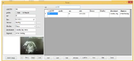
(a) (b)
Fig-5: Encrypting interface: (a) Loading image of Uterus with its information by the designed interface (b) Crypt the loaded image and save it in the server’s database
6. Transmission of single-slice
For high uploading transmission speed of big size images as Doppler or X-Ray images, especially through the Internet, users with our program can easily get one useful slice from the whole chosen image and save it directly wherever they need [15, 16, 17]. Figures below describe the slice transmission. The browser sends a request to the server with the parameters of the demanded region, and then the server sends the determined slice back to the browser. This transmission procedure includes four steps. Step 1: the user accesses the browser via wireless or wirednetwork to enter a search interface (using a special password) where he can determine the wanted department, then he can access the wanted image by its metadata.
© 2017, IRJET | Impact Factor value: 5.181 | ISO 9001:2008 Certified Journal
| Page 1575
Fig-6: The designed interface to get the wanted image by its Metadata
Fig-7: The decoded Doppler image inside the interface
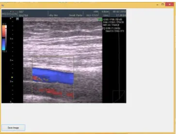
[image:4.595.37.290.94.216.2]Step 3: the user can view the wanted image in full size and then select the chosen slice. The browser then sends the request to the server to capture the region as shown in Fig. 8; information about the selected region include x, y axis coordinates and the slice’s width and height.
Fig-8: View the wanted image in full size and surround the useful region
[image:4.595.400.464.228.391.2]Step 4 is the slice position determination, via equations (1) and (2) where imgMaxW, imgMaxH are the maximum width and height of whole image; dispW, dispH represent width and height of display region; (xo, yo) are the coordinates of the upper-left corner of request region; SliceSize is the size of slice. The total number of slices contained in the display region is determined according to equations (1), (2) and (3) for the slice with index (a, b), its position in the whole image [2, 18].
) 1 (
) 1
(
disW sliceSize disH sliceSize
v (1) (1)
xo a sliceSize dispW
imgMaxW x
slice. ( 1) (2) (2)
yo b sliceSize dispH
imgMaxH y
slice. ( 1) (3) (3)
If the request slice is in cache, the server transmits the slice to the browser directly as shown in Fig. 9.
Fig-9: The selected slice transmitted by the server
7. Devices support for functionality evaluation
[image:4.595.51.273.261.373.2] [image:4.595.71.253.475.613.2]© 2017, IRJET | Impact Factor value: 5.181 | ISO 9001:2008 Certified Journal
| Page 1576
For evaluating the wireless connection functionality, we used a gigabit router [19, 20] to connect the server with the client laptop. Some simple changes were made on both laptops to enable sharing between them. After these configurations, the user can easily browse the database for the wanted department, download the useful images and select a single slice from the whole image.
We evaluated the response time (the time interval between the user sending the command to the server and the server producing the result) over the Wireless network in terms of image access and presentation. We evaluated the performance of our system for one and two users, by sending more than one image at a time and more than one slice. All concurrent requests are equally sent from two users. To evaluate the system under this condition, the minimum, average and maximum response time are collected for each request [21, 22].
8. Results and discussion
For our system we computed the following results: Response time
For one concurrent request, the minimum, average and maximum response time is 0.1 s, 0.2 s and 0.3 s, respectively. For two concurrent requests, the minimum, average and maximum response time is 0.1 s, 0.5 s and 1.6 s, respectively. The computed response time is for getting the browsers requests from one and two users to enter the system. The users can enter in the system by a special user password and user name given by the hospital, then to allow entering to the selected department by another password for each department (Sonar, Doppler, etc.) which are given by the server manager.
Practically, we found that the response time range for the wired network is more than the average value for two users and it is in average value with one user. While the practice response time for wireless network is around the average value for one user and more than average value for two users.
In case of two requests from wired and wireless network, the practical response time for the wired network is around the average value and the response time for the wireless network is more than the average value for two users. Amount of data delivered
For different images sizes, we computed the amount of data delivered for the whole selected image and compared it with the one of a single selected slice with a same size for all the images; the results are shown in Table 1.
During our test of the system by sending requests from more than one user at the same time and then sending the pictures to more than one user through the wired or wireless network, the response time is influenced only with maximum response time as shown on the results above. Also, image size is affecting the delivered amount of data,
which means that the sending of a single slice from the whole image will reduce the response time.
Table-1: Amount of data delivered for different image sizes
Image size (MB) For single slice (MB)
1.3 0.71
1.54 0.80
1.96 0.94
2.44 0.86
2.87 0.79
2.97 0.66
3.65 0.74
3.88 0.83
4.06 0.89
9. Conclusion
In this paper, we presented a telemedicine system for hospitals that is developed for medical images sharing through wired and wireless systems. Our implementation is based on designing a special database server for medical images which receives the medical images from the different scanning departments in the hospital. The received images of different types and sizes are encoded and decoded in this paper by a new method which helps high security and speed in the encryption and decryption process. Also, our software enables the user to select a wanted slice from the whole image forfast sending and low amount of saved data. In this paper the computed response time gives good results for wired and wireless system (the difference between them is almost not noticeable). The proposed system can be easily implemented in any hospital, with low cost and high security for the interred information.
REFERENCES
[1] M. Kaur, V. Wasson, “ROI Based Medical Image
Compression for Telemedicine Application”, in 4th International Conference on Eco-friendly Computing and Communication Systems, ICECCS, Procedia Computer Science, vol. 70, 2015, pp. 579-585.
[2] H. Shen, D. Ma, Y. Zhao, ”MIAPS: A web-based system for
© 2017, IRJET | Impact Factor value: 5.181 | ISO 9001:2008 Certified Journal
| Page 1577
[3] M. Kumar, S. Agrawal, “Color image encoding in DOST
domain using DWT and SVD”, in Optics & Laser Technology, vol. 75, 2015, pp. 138-145.
[4] E. X. Bator, J. M. Gleason, A. J. Lorenzo, “The burden of
attending a pediatric surgical clinic and family preferences toward telemedicine”, in Journal of Pediatric Surgery, vol. 50, 2015, pp. 1776-1782.
[5] D. Dragan, D. Iveti´c, “Request redirection paradigm in
medical image archive implementation”, in Computer Methods and Programs in Biomedicine, vol. 107, 2012, pp. 111-121.
[6] D. Taubman, R. Prandolini, ”Architecture philosophy
and performance of JPIP: Internet Protocol standard for JPEG2000”, in Int. Symp. Visual Comm. and Image Proc, vol. 5150, 2003, pp. 649-663.
[7] B. Brindha, G. Raghuraman, “Region based lossless
compression for digital images in telemedicine application”, in International Conference in Communications and Signal Processing (ICCSP), 2013, pp. 537-540.
[8] P. Wu, K. Xie, H. Yu, Y. Zheng and W. Mao Yu, “A New
Preprocessing Algorithm Used in Color Image Compression”, Advances in Future Computer and Control Systems, Springer Berlin Heidelberg, 2012, pp. 465-471.
[9] B. Kamsu-Foguem, and C. Foguem, “Telemedicine and
mobile health with integrative medicine in developing countries”, in Health Policy and Technology 3, vol. 4, 2014, pp. 264-271.
[10] W. Y. Hsu, “Segmentation-based compression: New
frontiers of telemedicine in telecommunication”, In Telemetric and Informatics, Vol. 32, 2015, pp. 475-485.
[11] [11]. L. Chen, D.Zhao, “Optical image encryption with
Hartley transforms”, Opt. Lett., vol. 31, 2006, pp. 3438-3440.
[12] Z. Liu, Y. Zhang, F. W. Liu, and S. L. Meng, “Optical color
image hiding scheme based on chaotic mapping and Hartley transform”, Opt. Lasers Eng., vol. 51, 2013, pp. 967-972.
[13] N. Singh, A.Sinha, and “Gyrator transform-based optical
image encryption, using chaos”, in Opt. Lasers Eng., vol. 47, 2009, pp. 539-546.
[14] M. R. Abuturab, “Color image security system using
double random-structured phase encoding gyrator transform domain”, in Appl. Opt., vol.51, 2012, pp. 3006-3016.
[15] P. Schelkens, A. Munteanu and J. Barbarien, “Wavelet
coding of volumetric medical dataset”, in Medical Imaging, IEEE Transactions on, 2003, vol. 22, pp. 441-458.
[16] R. Shah, P. Sharma, “Performance Analysis of Region of
Interest Based Compression Method for Medical Images”, in Fourth International Conference in Advanced Computing & Communication Technologies (ACCT) IEEE, 2014, pp. 53-58.
[17] V. Sanchez, R. Abugharbieh & P. Nasiopoulos, ”3-D
scalable medical image compression with optimized volume of interest coding”, in Medical Imaging, IEEE Transactions on, 2010, vol. 29, pp. 1808-1820.
[18] M. Firoozbakht, J. Dehmeshki and M. Martini,
”Compression of digital medical images based on multiple regions of interest”, in Fourth International
Conference in Digital Society, (ICDS'10), 2010, pp. 260-263.
[19] Z. R. Hesabi, M. Sardari and A. Beirami, ”A
memory-assisted lossless compression algorithm for medical images”, In IEEE International Conference In Acoustics, Speech and Signal Processing (ICASSP), 2014, pp. 2030-2034.
[20] V. K. Bairagi, and A. M. Sapkal, “ROI-based DICOM image
compression for telemedicine”, in Sadhana, 2013, vol. 38, pp. 123-131.
[21] S. Devi and K. Vidhya, “Development of medical image
compression techniques”, In IEEE Conference on Computational Intelligence and Multimedia Applications, (2007), vol. 3, pp. 97-101.
[22] L. Faggioni, E. Neri and F. Cerri, “Integrating image
processing in PACS”, in European Journal of Radiology, vol. 78, (2011), pp.210–224.