Ankl e m o ti o n i nfl u e n c e s t h e
e x t e r n a l k n e e a d d u c tio n m o m e n t
a n d m a y p r e d i c t w h o will
r e s p o n d t o l a t e r a l w e d g e i n s ol e s ?
: a n a n cill a r y a n a ly si s f r o m t h e
S ILK t ri al
C h a p m a n , GJ, P a r k e s , MJ, F o r s y t h e , L, F el s o n , DT a n d Jo n e s , RK
h t t p :// dx. d oi.o r g / 1 0 . 1 0 1 6 /j.jo c a . 2 0 1 5 . 0 2 . 1 6 4
T i t l e
An kl e m o tio n infl u e n c e s t h e e x t e r n al k n e e a d d u c tio n
m o m e n t a n d m a y p r e d i c t w h o will r e s p o n d t o l a t e r al
w e d g e in s ol e s ? : a n a n cill a r y a n aly si s f r o m t h e S ILK t ri al
A u t h o r s
C h a p m a n , GJ, P a r k e s , MJ, F o r s y t h e , L, F el s o n , DT a n d
Jo n e s , RK
Typ e
Ar ticl e
U RL
T hi s v e r si o n is a v ail a bl e a t :
h t t p :// u sir. s alfo r d . a c . u k /i d/ e p ri n t/ 3 4 9 2 8 /
P u b l i s h e d D a t e
2 0 1 5
U S IR is a d i gi t al c oll e c ti o n of t h e r e s e a r c h o u t p u t of t h e U n iv e r si ty of S alfo r d .
W h e r e c o p y ri g h t p e r m i t s , f ull t e x t m a t e r i al h el d i n t h e r e p o si t o r y is m a d e
f r e ely a v ail a bl e o nli n e a n d c a n b e r e a d , d o w nl o a d e d a n d c o pi e d fo r n o
n-c o m m e r n-ci al p r iv a t e s t u d y o r r e s e a r n-c h p u r p o s e s . Pl e a s e n-c h e n-c k t h e m a n u s n-c ri p t
fo r a n y f u r t h e r c o p y ri g h t r e s t r i c ti o n s .
Ankle motion in
fl
uences the external knee adduction moment and
may predict who will respond to lateral wedge insoles?: an ancillary
analysis from the SILK trial
G.J. Chapman
y
*, M.J. Parkes
z
, L. Forsythe
z
, D.T. Felson
z x k
, R.K. Jones
y z
ySchool of Health Sciences, University of Salford, Salford, UK
zArthritis Research UK Epidemiology Unit, Centre for Musculoskeletal Research, University of, Manchester, Manchester, UK
xNIHR Manchester Musculoskeletal Biomedical Research Unit (BRU), Manchester Academic Health Sciences Centre, Manchester, UK
kClinical Epidemiology Unit, Boston University School of Medicine, Boston, MA, USA
a r t i c l e i n f o
Article history: Received 13 May 2014 Accepted 23 February 2015
Keywords: Osteoarthritis Knee
Lateral wedge insoles Biomechanical response External knee adduction moment
s u m m a r y
Objective: Lateral wedge insoles are a potential simple treatment for medial knee osteoarthritis (OA) patients by reducing the external knee adduction moment (EKAM). However in some patients, an in-crease in their EKAM is seen. Understanding the role of the ankle joint complex in the response to lateral wedge insoles is critical in understanding and potentially identifying why some patients respond differently to lateral wedge insoles.
Method: Participants with medial tibiofemoral OA underwent gait analysis whilst walking in a control shoe and a lateral wedge insole. We evaluated if dynamic ankle joint complex coronal plane biome-chanical measures could explain and identify those participants that increased (biomebiome-chanical non-responder) or decreased (biomechanical non-responder) EKAM under lateral wedge conditions compared to the control shoe.
Results: Of the 70 participants studied (43 male), 33% increased their EKAM and 67% decreased their EKAM. Overall, lateral wedge insoles shifted the centre of foot pressure laterally, increased eversion of the ankle/subtalar joint complex (STJ) and the eversion moment compared to the control condition. Ankle angle at peak EKAM and peak eversion ankle/STJ complex angle in the control condition predicted if individuals were likely to decrease EKAM under lateral wedge conditions.
Conclusions: Coronal plane ankle/STJ complex biomechanical measures play a key role in reducing EKAM when wearing lateral wedge insoles. Thesefindings may assist in the identification of those individuals that could benefit more from wearing lateral wedge insoles.
©2015 The Authors. Published by Elsevier Ltd and Osteoarthritis Research Society International. This is an open access article under the CC BY license (http://creativecommons.org/licenses/by/4.0/).
Introduction
Knee osteoarthritis (OA) affects approximately 12.5% adults over the age of 65 years old1e3. This progressive condition is charac-terised by pain and stiffness in the joint resulting in difficulty in weight bearing activities including walking and stair climbing1,2,4,
resulting in adverse effects on loss of function, personal indepen-dence and ultimately a reduction in quality of life.
Knee OA commonly affects the medial tibiofemoral compart-ment of the joint presumably due to the high percentage (up to 60e80%) of the load being transmitted to this compartment of the knee compared to the lateral side5e8. During walking the ground reaction force (GRF) passes medial to the knee, creating an external knee adduction moment (EKAM) acting on the knee which adducts the tibia with respect to the femur. The EKAM is widely accepted as a surrogate measure of medial knee loading. The EKAM has been directly linked with lower limb malalignment, disease severity9, disease progression10and self-reported pain11,12. As focal mechani-cal loads play an important role in disease severity and progression, reduction in the EKAM can be used as an objective goal for load-modifying conservative treatments designed to reduce load, pain *Address correspondence and reprint requests to: G.J. Chapman, Leeds Institute
of Rheumatic and Musculoskeletal Medicine, University of Leeds and Leeds NIHR Biomedical Research Unit, 2nd Floor, Chapel Allerton Hospital, Harehills Lane, Leeds, LS7 4SA, UK. Tel: 44-113-3924926; Fax: 44-113-3924991.
E-mail addresses:[email protected](G.J. Chapman), matthew.parkes@
manchester.ac.uk (M.J. Parkes), [email protected] (L. Forsythe),
[email protected],[email protected](D.T. Felson),[email protected].
uk(R.K. Jones).
http://dx.doi.org/10.1016/j.joca.2015.02.164
1063-4584/©2015 The Authors. Published by Elsevier Ltd and Osteoarthritis Research Society International. This is an open access article under the CC BY license (http:// creativecommons.org/licenses/by/4.0/).
Osteoarthritis and Cartilage xxx (2015) 1e7
and potentially slow disease progression and thus improve physical function. One common conservative treatment for medial knee OA is the use of lateral wedge insoles which are placed inside patient's shoes in an attempt to redistribute the load on the knees and thus decrease the EKAM. Past research has demonstrated that lateral wedge insoles decrease EKAM in patients with medial knee OA13e22. Individuals' EKAM response to lateral wedge insoles is remark-ably variable, with up to 30% of treated patients demonstrating an increase (worsening) in EKAM15e22. Randomised controlled trials examining the effects of lateral wedge insoles on knee pain have failed to show significant effects on pain reduction13,14,23e25with a recent meta-analysis showing no significant change in pain when compared to neutral inserts26. The modest effect of EKAM reduc-tion and the disappointing effects of pain reducreduc-tion could be due to the variability in EKAM response to wearing lateral wedge insoles and/or that previous studies have grouped participants together, on the assumption that lateral wedge insoles uniformly reduce EKAM and thereby alleviate knee pain.
The majority of past research examining the effects of lateral wedge insoles on lower limb joints has concentrated on the knee joint. In one study, lateral wedge insoles had little effect on the hip27. At the foot, lateral wedge insoles shift the centre of force pressure (COFP) laterally, increasing the ankle eversion moment and shortening the knee-GRF lever arm, thus reducing the
EKAM14,17,20,21,27,28. Thesefindings suggest that the ankle/subtalar
joint complex (STJ) may play a key role in the mechanical mecha-nism(s) of lateral wedge insoles and medial knee loading. To our knowledge, no previous research has investigated to what extent lateral wedge insoles alter coronal plane foot and ankle biome-chanics and/or determine if changes in foot/ankle biomebiome-chanics are linked to changes in EKAM in a large cohort of medial knee OA patients. An aim of the present study was to conduct an analysis of ancillary measurements obtained as part of the SILK trial to gain greater insight into the effect of lateral wedge insoles on foot and ankle biomechanics and also to understand the relationship be-tween changes in foot and ankle biomechanics and change in EKAM. Furthermore, given the variability of biomechanical response to lateral wedge insoles, we also examined whether the effects of the lateral wedge insole on foot and ankle biomechanics would identify individuals with reductions in EKAM.
Overall, we hypothesised that by classifying participants by EKAM response, dynamic coronal plane ankle/STJ complex biomechanics can identify and help explain why some patients experience an increase, whereas others show a decrease in EKAM.
Participants and methods
Participants
Participants with knee pain were recruited from orthopaedic clinics, physiotherapy clinics, and through advertisements in the local media. Participants were included in the study if they re-ported at least mild knee pain during walking on aflat surface in the last week, assessed by the Knee Injury and OA Outcome Score pain subscale question P529(we required that a potential partici-pant scored their pain as either mild, moderate, severe or extreme), were aged between 40 and 85 years old and demonstrated medial tibiofemoral OA Kellgren Lawrence (KL, grade II or III) on radio-graph of the affected knee with greater medial than lateral joint space narrowing determined by an experienced academically-based musculoskeletal radiologist. Exclusion criteria included pa-tients experiencing more pain localised in the patellofemoral joint on examination than the medial joint, had tricompartmental knee OA or KL grade 1 or 4 tibiofemoral OA, the latter based on reports that those with KL 4 have not responded to lateral wedge insoles.
Other exclusion criteria included any lower limb realignment sur-gery; total knee replacement of the affected side; any foot or ankle problems that might negate the use of footwear modifying in-terventions; the use of a walking aid; knee surgery or injections in the previous 6 months or the use of foot orthotics in the past 6 months. The symptomatic knee was the only knee tested in this study. For those patients that had bilateral medial knee OA, the more painful knee was deemed the affected side. NHS Research Ethics approval was obtained for the study and all participants provided written informed consent.
Interventions
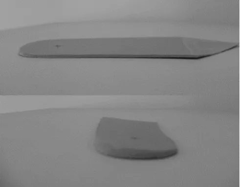
As an ancillary study to the SILK trial (ISRCTN: 83706683) which was a single visit randomised trial testing different lateral wedge insoles and shoes for their effect on the EKAM we performed an analysis on foot and ankle motion and how they related to knee moments in one of the conditions, a typical lateral wedge insole. This insole has previously been shown to reduce EKAM in medial knee OA patients22,30. The‘typical’lateral wedge was comprised of ethylene-vinyl acetate with a Shore A density of 60. This 5lateral wedge insole post from the heel to thefifth metatarsal head and did not have a medial arch support (see Fig. 1). The control shoe comprised of aflat, thin soled, leather shoe (Ecco Zen, UK). The lateral wedge was inserted into the control shoe bilaterally, with each participant having afive minute familiarisation period for each experimental condition. The order of control vs lateral wedge insole was randomised using computer-generated permutations, con-cealed in pre-sealed sequentially-numbered envelopes that were generated by the trial statistician, prior to participants' enrolment in the study, who was not present during recruitment or testing.
Protocol
[image:4.595.305.552.533.725.2]While wearing each condition, participants underwent 3D gait analysis. A 16 camera Qualisys (Qualisys, Sweden) OQUS3 motion analysis system (collected at 100 Hz) and four force plates (AMTI, USA) (collected at 200 Hz) embeddedflush in the ground were used to measure lower limb kinematics and kinetics. Each participant completed a minimum of three successful trials sequentially under each condition, at a self-selected walking speed. A successful trial was defined as a trial in which the participant walked naturally landing the whole foot of the affected limb on the force plate.
Fig. 1.Diagram illustrating the typical lateral wedge insole used in the present study. G.J. Chapman et al. / Osteoarthritis and Cartilage xxx (2015) 1e7
Participants were not informed about the force plates to minimise the participant from‘targeting’the force plates. The CAST marker set technique31was employed whereby rigid clusters of four non-orthogonal markers were positioned over the lateral shank, lateral thigh and sacrum to track the segmental kinematics in six degrees of freedom. Four retroreflective markers (positioned over thefirst andfifth metatarsal heads, the most posterior aspect of the calca-neus and the most anterior tip of the shoe) were glued securely to the control shoes with the foot being modelled as a rigid, single segment as reported previously28. A static calibration trial was collected, for each experimental condition, in which retroreflective markers were placed on bony landmarks to specify the location of the lower limb joints in relation the clusters and to approximate joint centres. Ankle and knee joint centres were calculated as midpoints between the malleoli and femoral epicondyles, respec-tively. The hip joint centre was calculated using the regression model based on the anterior and posterior superior iliac spine markers32. In Visual 3D (C-Motion, Rockville, Maryland), joint ki-nematics were calculated using an XeYeZ Euler rotation sequence equivalent to the joint coordinate system33and joint kinetic data were calculated using three-dimensional inverse dynamics. External knee adduction moments were normalised to participant's mass (Nm/kg) and knee adduction angular impulse (KAAI) nor-malised to participant's mass and time (Nm/kg*s). Additionally, coronal plane biomechanical measures relating to the ankle angle, external ankle eversion moments and the position of the COFP with respect to the foot were also calculated. Centre of force pressure measures were derived from the known location of the shoe (from the markers placed on the foot/ankle) on the force plate. Medio-lateral COFP was defined as the distance of the centre of force with respect to the midline of the foot (vector constructed by the heel and toe markers). A custom Matlab (Matlab, USA) programme was used to extract the peak EKAM during early stance (between 17% and 50% of stance34) and to calculate the KAAI. For each addi-tional parameter, data were extracted at the instant of peak EKAM in early stance and the peak value between heel strike and peak EKAM in early stance as this is where medial loading is greatest.
Data analysis and statistical analysis
In thefirst part of the analysis, we tested for effects of the lateral wedge insole on coronal ankle/STJ complex variables. Pairedt-tests were used to examine if each variable of interest changed during the lateral wedge condition, in comparison to the control condition. The second analysis investigated whether the change in coronal plane biomechanical measures was associated with the change in peak EKAM, when using the lateral wedge. We ran several uni-variate linear regression models, with the predictor variable for each model being the difference in one of the coronal plane vari-ables of interest, when wearing the lateral wedge. The outcome
variable in each model was the difference in EKAM between the lateral wedge and the control condition.
The third part of the analysis categorised participants based on whether their peak EKAM decreased or increased when wearing the lateral wedge insole. A participant with a reduction in peak EKAM in the lateralwedge condition was defined as a ‘ biome-chanical responder’, those with no change or an increase in peak EKAM were‘biomechanical non-responders’. We then used logistic regression to see which coronal ankle/STJ complex variable could predict response to EKAM (using this dichotomous classification). For this analysis, we used coronal ankle/STJ complex variables from thecontrol condition only, as this would test if we could effectively establish which (if any) coronal plane ankle/STJ complex variables were indicative of a response to lateral wedges, prior to an actual test of treatment.
Statistical analysis was performed using Stata version 13.1 (StataCorp, College Station, Texas, US) with the significance level set at P < 0.05. Model residuals were checked for normality using residual-versus-fitted plots. Due to the variables of interest all be-ing highly correlated with one another (i.e., highly collinear), all regression models tested (both linear and logistic) considered only one ankle outcome of interest, at any time. Models using several ankle outcomes as predictors simultaneously were avoided, since the highly collinear predictors would cause inappropriately large standard errors.
Results
Study sample characteristics
Seventy participants (43 male, 27 female) had medial knee OA (mean age 60.3 years (SD 9.6 yrs), height 1.69 m (0.09), weight 87.3 kg (18.5), BMI 30.5 kg/m2(4.9)). Of the 62 participants with KL data, the mean KL score was 2.63 and ranged from grade 2 to grade 3. We reviewed recent knee arthroscopy reports or MRIs for eight participants who did not have x-rays prior to the study to ensure that these participants also had medial>lateral cartilage loss and other features of OA. There was no difference in walking speed between conditions (mean gait speeds: control condition 1.163 m/ s; typical wedge 1.166 m/s).
Table Ioutlines the difference in coronal plane biomechanical variables when wearing the lateral wedge insole, compared to the control shoe. Notably, the lateral wedge produced an immediate, significant decrease in peak EKAM (overall change from control¼ 0.023 Nm/kg; 95% CI0.035 Nm/kg to0.011 Nm/kg) and KAAI (overall change from control ¼ 0.012 Nm/kg*s; 95% CI0.016 Nm/kg*s to0.009 Nm/kg*s). Expressed as a percentage change from the control shoe, these changes reflect a reduction in peak EKAM of 5.85%, and a reduction in KAAI of 7.95%. Please note
Table I
Effect of the typical lateral wedge insole on coronal plane biomechanical variables compared to the control shoe (N¼70). NB: the data on EKAM and KAAI has previously been reported35but is also indicated here for clarity
Variable Control shoe,
mean (SD)
Lateral wedge, mean (SD)
Difference, mean (95% CI),P
EKAM (Nm/kg) 0.394 (0.160) 0.371 (0.151) 0.023 (0.035 to0.011),<0.001 KAAI (Nm/kg*s) 0.156 (0.071) 0.144 (0.068) 0.012 (0.016 to0.009),<0.001 Ankle angle at peak EKAM () 3.457 (2.777) 4.399 (2.844) 0.942 (0.539 to 1.345),<0.001 Peak eversion ankle angle () 3.506 (2.770) 4.425 (2.843) 0.919 (0.518 to 1.321),<0.001 Centre of foot pressure at EKAM (mm)a 0.011 (0.006) 0.015 (0.006) 0.004 (0.005 to0.003),<0.001
Peak centre of foot pressure (mm)a 0.008 (0.007) 0.013 (0.006) 0.005 (0.006 to0.004),<0.001
Ankle eversion moment at EKAM (Nm/kg) 0.077 (0.064) 0.119 (0.062) 0.043 (0.050 to0.035),<0.001 Peak eversion ankle moment (Nm/kg) 0.091 (0.055) 0.126 (0.059) 0.035 (0.041 to0.029),<0.001 aNegative values indicate a lateral shift.
G.J. Chapman et al. / Osteoarthritis and Cartilage xxx (2015) 1e7 3
that the study sample demographics, peak EKAM and KAAI have been reported previously35.
Participants' response to wearing the lateral wedge insole varied considerably with 33% (n ¼ 23) of participants demonstrating
increased peak EKAM when wearing the lateral wedge insole (compared to the control shoe). The mean change in peak EKAM for these individuals was an increase of 0.028 Nm/kg (95% CI 0.018 Nm/kg to 0.037 Nm/kg), reflecting an increase of 8.15% compared to the control shoe. The remaining 67% of participants (n¼47) showed a decrease in peak EKAM when wearing the lateral wedge insole, compared to the control shoe (mean peak EKAM change0.048 Nm/kg; 95% CI0.059 to0.037 Nm/kg, reflecting a reduction of11.39% from the control shoe, in this subgroup).
The lateral wedge insole caused the ankle/STJ complex to be in a significantly more everted position (as shown by significantly greater peak ankle angle and ankle angle at peak EKAM) compared to the control condition (seeTable I). Overall, the lateral wedge insole caused a significant lateral shift in COFP at peak EKAM and a significantly greater peak COFP compared to the control shoe. Ankle eversion moment at peak EKAM and peak ankle eversion moment were significantly greater under the lateral wedge condition compared to control conditions.
Linear regression analysis revealed no significant relationships between change in coronal plane biomechanical variables under the lateral wedge condition and change in peak EKAM (seeTable II). Thefinal part of the analysis revealed participants with a higher peak ankle eversion angle (OR 1.248; 95% CI 1.025 to 1.519;
P¼0.027) or a higher ankle angle at peak EKAM (OR 1.241; 95% CI 1.021 to 1.509;P¼0.030) in the control condition, were more likely to classified as a biomechanical responder (i.e., EKAM more likely to decrease) to the lateral wedge insole (seeTable III), although no clear-cut threshold in ankle angle in the control condition between groups was found (see Supplementary Fig. 1). In additional ana-lyses, we found no difference infindings by gender.
Discussion
The present paper focuses on dynamic ankle/STJ complex biomechanics and investigated whether ankle biomechanics can assist in understanding and identifying why some individuals benefit (i.e., reduce peak EKAM) when wearing lateral wedge in-soles and others do not. Ourfindings suggest that coronal plane ankle/STJ complex biomechanical parameters play an important role in reducing peak EKAM and may predict those that are likely to experience a reduced peak EKAM when wearing lateral wedge insoles.
In the present study, those patients whose peak EKAM decreased in the lateral wedge condition compared to control (biomechanical responders) made up the majority the sample (67%) and experienced a decrease in their peak EKAM of 11.39% under the lateral wedge condition compared to control. In contrast,
the remaining third of participants whose peak EKAM increased in the lateral wedge condition (the biomechanical non-responders), increased their peak EKAM by a mean of 8.15% compared to the control condition. This variability in peak EKAM response to lateral wedge insoles is consistent with past research15e22and may be a factor why reported reductions in peak EKAM are
mod-est15,17e19,21,22,27,30,36. Similar reductions in peak EKAM and
vari-ability in response have been shown when wearing specialised footwear37e41. It is important to highlight that footwear may play a key role in peak EKAM response to interventions. However, more research is required to fully understand this issue. Thesefindings on peak EKAM suggest that grouping participants by peak EKAM response may demonstrate the true biomechanical effects of lateral wedge insoles on knee loading and be able to examine the clinical effects associated with this grouping. However, future research is required to confirm whether grouping participants by coronal ankle biomechanical variables assists in identifying those likely to benefit from wearing lateral wedge insoles and determine if there are clinical measurements that could be utilised instead of expen-sive 3D gait analysis.
The overall effect of the lateral wedge insoles resulted in a lateral shift in COFP, a more everted ankle/STJ complex and a greater eversion moment compared to the control condition which is consistent with those reported previously17,20,21,27. The increased eversion moment and everted ankle/STJ complex as well as the increased laterally displaced COFP suggests that these are potential key biomechanical responses to wearing lateral wedge insoles for reducing medial knee loading especially considering that Hinman
et al.17showed that shifting the COFP laterally caused the GRF to move towards the centre of the knee, reducing the knee-GRF dis-tance and EKAM. However, the present changes in coronal plane biomechanical variables did not significantly correlate with change in peak EKAM (see Table II), potentially due to the inclusion of participants that had an increase in the EKAM. Therefore, more research is required to determine the mechanism of action of lateral wedge insoles in individuals who have a positive EKAM response.
Predicting and/or identifying if an individual is likely to benefit from an intervention is an important issue. In the present study we attempted to determine if it is possible to predict those individuals that are likely to respond to lateral wedge insoles (i.e., decrease their peak EKAM; biomechanical responders) compared to those that will not respond (biomechanical non-responders), by using coronal plane ankle/STJ complex biomechanical variables within the control condition. Ourfindings demonstrated that those in-dividuals that have a greater everted ankle/STJ complex under the control condition were more likely to decrease their peak EKAM under lateral wedge conditions (Table III). This finding may be counter-intuitive given that those individuals that have a more inverted foot could potentially have greater range to rotate their foot into eversion. However, the presentfindings suggest that those patients with a more everted foot are more likely to be shifting the centre of foot pressure more laterally, thus potentially reducing
Table II
Relationship between change in variables of interest and change in peak EKAM when using the typical lateral wedge insole (N¼70)
Variable Association with change during typical lateral wedge use
b (95% CI) P
Ankle angle at peak EKAM () 0.002 (0.494 to0.005) 0.494 Peak eversion ankle angle () 0.002 (0.509 to0.005) 0.509 Centre of foot pressure at EKAM (mm)a 0.498 (0.754 to3.647) 0.754
Peak centre of foot pressure (mm)a 0.448 (0.737 to2.206) 0.737
Ankle eversion moment at EKAM (Nm/kg) 0.061 (0.749 to0.318) 0.749 Peak eversion ankle moment (Nm/kg) 0.055 (0.808 to0.394) 0.808 aNegative values indicate a lateral shift.
Table III
The association of gait parameters in the control condition with biomechanical response to the typical lateral wedge (N¼70)
Variable OR (95% CI) P
Ankle angle at peak EKAM () 1.241 (1.021 to 1.509) 0.030 Peak eversion ankle angle () 1.248 (1.025 to 1.519) 0.027 Centre of foot pressure at EKAM (mm)a* 1.047 (0.439 to 2.494) 0.918 Peak centre of foot pressure (mm)a* 1.031 (0.511 to 2.082) 0.931 Ankle eversion moment at EKAM (Nm/kg)* 0.996 (0.921 to 1.077) 0.922 Peak eversion ankle moment (Nm/kg)* 0.980 (0.893 to 1.075) 0.663
aNegative values indicate a lateral shift. For outcomes marked (*), odds ratios have
been rescaled to reflect a change of 0.01 units, rather than 1 unit. G.J. Chapman et al. / Osteoarthritis and Cartilage xxx (2015) 1e7
their EKAM compared to those with a more inverted foot, afinding that is consistent with other past biomechanical studies examining foot posture and knee loading42,43. Taken together, thesefindings suggest that those patients with a less everted ankle joint complex under control condition may have restricted frontal plane ankle range of motion, thus when a lateral wedge is inserted into their shoe, the restricted ankle range of motion may not allow the ankle joint complex to evert/pronate sufficiently to alter the load at the knee. However, due to the omission of clinical data, this is only a hypothesis and an area that future research needs to focus on. To our knowledge, this is the first study to demonstrate that it is possible to distinguish if an individual is likely to decrease their peak EKAM when wearing lateral wedge insoles. While this is an important finding, at present, sophisticated 3D motion analysis systems are required to highlight the small coronal plane kinematic differences and this may not be available in all hospitals and/or clinics. Therefore, future research needs to consider the use of a clinical assessment to determine the range of motion of lower limb joints and/or consider foot type to link the clinical assessment with the biomechanical analysis to determine if it is possible to simplify the identification of individuals that are likely to respond to wearing lateral wedge insoles. From a clinical point of view, our regression analyses has some potentially important implications, in that, being able to identify if someone is likely to respond to wearing lateral wedge insoles, the more beneficial the treatment is likely to be for those individuals. Conversely, categorising if someone is unlikely to respond to wearing lateral wedge insoles, other treatments should be prescribed which should be more beneficial (compared to lateral wedge insoles) for those patients. However, whilst these results are encouraging, future research is required to replicate thesefinding. There are limitations within the present study. Despite having a large sample of participants (n¼70), our decision to categorise participants based on biome-chanical response to lateral wedge insoles resulted in the sample size per group decreasing and therefore a potential reason for a number of close but insignificant findings. As the prediction of whether an individual is likely to respond to wearing lateral wedge insoles is based on ankle/STJ complex kinematics under the control condition, the type of footwear that was used in the study needs to be taken into account. It is plausible that different footwear char-acteristics/participants' own shoes may have differing effects on lower limb kinematics and kinetics. Therefore, future research is required to determine how footwear influences biomechanical response to wearing lateral wedge insoles and may also consider employing a more sophisticated foot model that allows for sepa-ration between hindfoot (i.e., four markers placed on the heel of the shoe) and forefoot kinematics during shod gait conditions. Another potential limitation of the current work is that we only measured the immediate effect of lateral wedge insoles on peak EKAM response and dichotomised participants based on their immediate peak EKAM response. It is plausible that peak EKAM response to lateral wedge insoles of some participants may have changed if worn longer. Past research has suggested that when specialised footwear is worn for six months, participants have reduced peak EKAM even when not wearing the prescribed specialised
foot-wear41,44, suggesting that knee OA patients show neuromuscular
adaptation to potential treatments for medial knee OA. Therefore, future research is required to understand if participants adapt to wearing lateral wedge insoles over time.
Conclusions
In conclusion, we have demonstrated that the ankle/STJ com-plex plays an important role in the reduction of peak EKAM when wearing lateral wedge insoles. Furthermore, our findings also
demonstrate that coronal plane ankle/STJ complex biomechanical measures under the control condition correlate with the likelihood of experiencing a reduction in peak EKAM when wearing lateral wedge insoles. These findings may provide future insights into determining who will respond to lateral wedge insoles.
Author contributions
DTF and RKJ conceived of the study idea and designed the study. GJC and LF collected and processed the data. GJC, MJP, DTF and RKJ analysed and interpreted the data. GJC drafted the manuscript. All authors revised the manuscript for intellectual content and approved of the final article prior to submission. RKJ (r.k.jones@ salford.ac.uk) and DTF ([email protected]) take full responsibility for the integrity of the work as a whole, from the inception to the
finished article.
Role of the funding source
Research in Osteoarthritis Manchester (ROAM) is supported by Arthritis Research UK special strategic award 18676. The Arthritis Research UK Centre of Excellence in Epidemiology is supported by grant number 20380. This study is an ancillary study carried out during the SILK trial (ISRCTN: 83706683). This report includes in-dependent research supported by the National Institute for Health Research Biomedical Research Unit Funding Scheme. The views expressed in this publication are those of the author(s) and not necessarily those of the NHS, the National Institute for Health Research or the Department of Health. The funding source had no role in the study design, collection, analysis and interpretation of the data; in the writing of the manuscript; or in the decision to submit the manuscript for publication.
Competing interests
RKJ may receive royalties from the lateral wedge insoles.
Acknowledgements
This study was supported by funding from Arthritis Research UK (Grant reference 18676). We wish to acknowledge the valuable help of the Research into Osteoarthritis in Manchester (ROAM) research team for aiding with the recruitment and screening of the individuals.
Supplementary data
Supplementary data related to this article can be found athttp:// dx.doi.org/10.1016/j.joca.2015.02.164.
References
1. Dillon CF, Rasch EK, Gu Q, Hirsch R. Prevalence of knee oste-oarthritis in the United States: arthritis data from the Third National Health and Nutrition Examination Survey 1991-94. J Rheumatol 2006;33:2271e9.
2. Felson DT. The epidemiology of knee osteoarthritis: results from the Framingham Osteoarthritis Study. Semin Arthritis Rheum 1990;20:42e50.
3. Peat G, McCarney R, Croft P. Knee pain and osteoarthritis in older adults: a review of community burden and current use of primary health care. Ann Rheum Dis 2001;60:91e7.
4. Guccione AA, Felson DT, Anderson JJ, Anthony JM, Zhang Y, Wilson PW,et al. The effects of specific medical conditions on the functional limitations of elders in the Framingham Study. Am J Public Health 1994;84:351e8.
G.J. Chapman et al. / Osteoarthritis and Cartilage xxx (2015) 1e7 5
5. Andriacchi TP, Mündermann A. The role of ambulatory me-chanics in the initiation and progression of knee osteoarthritis. Curr Opin Rheumatol 2006;18:514e8.
6. Johnson F, Leitl S, Waugh W. The distribution of load across the knee. J Bone Joint Surg Br 1980;62:346e9.
7. Prodromos CC, Andriacchi TP, Galante JO. A relationship be-tween gait and clinical changes following high tibial osteot-omy. J Bone Joint Surg 1985;67:1188e94.
8. Schipplein OD, Andriacchi TP. Interaction between active and passive knee stabilizers during level walking. J Orthop Res 1991;9:113e9.
9. Sharma L, Hurwitz DE, Thonar EJMA, Sum JA, Lenz ME,
Dunlop DD,et al. Knee adduction moment, serum hyaluronan
level, and disease severity in medial tibiofemoral osteoar-thritis. Arthritis Rheum 1998;41:1233e40.
10. Miyazaki T, Wada M, Kawahara H, Sato M, Baba H, Shimada S. Dynamic load at baseline can predict radiographic disease progression in medial compartment knee osteoarthritis. Ann Rheum Dis 2002;61:617e22.
11. Amin S, Luepongsak N, McGibbon CA, LaValley MP, Krebs DE,
Felson DT. Knee adduction moment and development of chronic knee pain in elders. Arthritis Care Res 2004;51:371e6. 12. Hurwitz DE, Sharma L, Andriacchi TP. Effect of knee pain on joint loading in patients with osteoarthritis. Curr Opin Rheu-matol 1999;11:422e6.
13. Baker K, Goggins J, Szumowski K, Xie H, LaValley M, Hunter DJ, et al. A randomized cross-over trial of a wedged insole for treatment of knee osteoarthritis. Arthritis Rheum 2005;52: S459e60.
14. Barrios JA, Crenshaw JR, Royer TD, Davis IS. Walking shoes and laterally wedged orthoses in the clinical management of medial tibiofemoral osteoarthritis: a one-year prospective controlled trial. Knee 2009;16:136e42.
15. Butler RJ, Marchesi S, Royer T, Davis IS. The effect of a subject-specific amount of lateral wedge on knee mechanics in pa-tients with medial knee osteoarthritis. J Orthop Res 2007;25: 1121e7.
16. Crenshaw SJ, Pollo FE, Calton EF. Effects of lateral-wedged insoles on kinetics at the knee. Clin Orthop Relat Res 2000: 185e92.
17. Hinman RS, Bowles KA, Metcalf BB, Wrigley TV, Bennell KL.
Lateral wedge insoles for medial knee osteoarthritis: effects on lower limb frontal plane biomechanics. Clin Biomech 2012;27: 27e33.
18. Hinman RS, Bowles KA, Payne C, Bennell KL. Effect of length on laterally-wedged insoles in knee osteoarthritis. Arthritis Rheum 2008;59:144e7.
19. Hinman RS, Payne C, Metcalf BR, Wrigley TV, Bennell KL.
Lateral wedges in knee osteoarthritis: what are their imme-diate clinical and biomechanical effects and can these predict a three-month clinical outcome? Arthritis Rheum 2008;59: 408e15.
20. Kakihana W, Akai M, Nakazawa K, Naito K, Torii S. Inconsistent knee varus moment reduction caused by a lateral wedge in knee osteoarthritis. Am J Phys Med Rehabil 2007;86:446e54.
21. Kakihana W, Akai M, Nakazawa K, Takashima T, Naito K,
Torii S. Effects of laterally wedged insoles on knee and subtalar joint moments. Arch Phys Med Rehabil 2005;86:1465e71.
22. Kerrigan DC, Lelas JL, Goggins J, Merriman GJ, Kaplan RJ,
Felson DT. Effectiveness of a lateral-wedge insole on knee varus torque in patients with knee osteoarthritis. Arch Phys Med Rehabil 2002;83:889e93.
23. Bennell KL, Bowles K-A, Payne C, Cicuttini F, Williamson E, Forbes A,et al. Lateral wedge insoles for medial knee osteo-arthritis: 12 month randomised controlled trial. BMJ 2011;342.
24. Maillefert JF, Hudry C, Baron G, Kieffert P, Bourgeois P,
Lechevalier D,et al. Laterally elevated wedged insoles in the treatment of medial knee osteoarthritis: a prospective ran-domized controlled study. Osteoarthritis Cartilage 2001;9: 738e45.
25. Pham T, Maillefert JF, Hudry C, Kieffert P, Bourgeois P,
Lechevalier D,et al. Laterally elevated wedged insoles in the treatment of medial knee osteoarthritis. A two-year prospec-tive randomized controlled study. Osteoarthritis Cartilage 2004;12:46e55.
26. Parkes MJ, Maricar N, Lunt M, LaValley MP, Jones RK, Segal NA, et al. Lateral wedge insoles as a conservative treatment for pain in patients with medial knee osteoarthritis a meta-anal-ysis. Jama-Journal Am Med Assoc 2013;310:722e30.
27. Butler RJ, Barrios JA, Royer T, Davis IS. Effect of laterally wedged foot orthoses on rearfoot and hip mechanics in pa-tients with medial knee osteoarthritis. Prosthet Orthot Int 2009;33:107e16.
28. Jones RK, Zhang M, Laxton P, Findlow AH, Liu A. The biome-chanical effects of a new design of lateral wedge insole on the knee and ankle during walking. Hum Mov Sci 2013;32:596e604. 29. Roos EM, Roos HP, Lohmander LS, Ekdahl C, Beynnon BD. Knee
Injury and Osteoarthritis Outcome Score (KOOS)e
develop-ment of a self-administered outcome measure. J Orthop Sports Phys Ther 1998;28:88e96.
30. Jones RK, Chapman GJ, Findlow AH, Forsythe L, Parkes MJ,
Sultan J,et al. A new approach to prevention of knee osteo-arthritis: reducing medial load in the contralateral knee. J Rheumatol 2013;40:309e15.
31. Cappozzo A, Catani F, Della Croce U, Leardini A. Position and orientation in space of bones during movement: anatomical frame definition and determination. Clin Biomech 1995;10: 171e8.
32. Bell AL, Brand RA, Pedersen DR. Prediction of hip joint centre location from external landmarks. Hum Mov Sci 1989;8:3e16. 33. Grood ES, Suntay WJ. A joint coordinate system for the clinical description of 3-dimensional motions e applications to the knee. J Biomed EngeTrans Asme 1983;105:136e44.
34. Thorp LE, Sumner DR, Block JA, Moisio KC, Shott S,
Wimmer MA. Knee joint loading differs in individuals with mild compared with moderate medial knee osteoarthritis. Arthritis Rheum 2006;54:3842e9.
35. Jones RK, Chapman GJ, Forsythe L, Parkes MJ, Felson DT. The relationship between reductions in knee loading and imme-diate pain response whilst wearing lateral wedged insoles in knee osteoarthritis. J Orthop Res 2014;32:1147e54.
36. Shimada S, Kobayashi S, Wada M, Uchida K, Sasaki S,
Kawahara H,et al. Effects of disease severity on response to lateral wedged shoe insole for medial compartment knee osteoarthritis. Arch Phys Med Rehabil 2006;87:1436e41. 37. Erhart JC, Muendermann A, Elspas B, Giori NJ, Andriacchi TP.
Variable-stiffness shoe lowers the knee adduction moment in subjects with symptoms of medial compartment knee osteo-arthritis. J Biomech 2008;41:2720e5.
38. Erhart JC, Muendermann A, Elspas B, Giori NJ, Andriacchi TP. Changes in knee adduction moment, pain, and functionality with a variable-stiffness walking shoe after 6 months. J Orthop Res 2010;28:873e9.
39. Jenkyn TR, Erhart JC, Andriacchi TP. An analysis of the mech-anisms for reducing the knee adduction moment during walking using a variable stiffness shoe in subjects with knee osteoarthritis. J Biomech 2011;44:1271e6.
40. Shakoor N, Lidtke RH, Sengupta M, Fogg LF, Block JA. Effects of specialized footwear on joint loads in osteoarthritis of the knee. Arthritis Rheum-Arthritis Care Res 2008;59:1214e20. G.J. Chapman et al. / Osteoarthritis and Cartilage xxx (2015) 1e7
41. Shakoor N, Lidtke RH, Wimmer MA, Mikolaitis RA, Foucher KC,
Thorp LE, et al. Improvement in knee loading after use
of specialized footwear for knee osteoarthritis: results of a six-month pilot investigation. Arthritis Rheum 2013;65: 1282e9.
42. Levinger P, Menz H, Fotoohabadi M, Feller J, Bartlett J,
Bergman N. Foot posture in people with medial compartment knee osteoarthritis. J Foot Ankle Res 2010;3:29.
43. Levinger P, Menz HB, Morrow AD, Bartlett JR, Feller JA,
Bergman NR. Relationship between foot function and medial knee joint loading in people with medial compartment knee osteoarthritis. J Foot Ankle Res 2013;6.
44. Haim A, Rubin G, Rozen N, Goryachev Y, Wolf A. Reduction in knee adduction moment via non-invasive biomechanical training: a longitudinal gait analysis study. J Biomech 2012;45: 41e5.
G.J. Chapman et al. / Osteoarthritis and Cartilage xxx (2015) 1e7 7