R E S E A R C H A R T I C L E
Open Access
Safety, tolerability, clinical, and joint
structural outcomes of a single
intra-articular injection of allogeneic
mesenchymal precursor cells in patients
following anterior cruciate ligament
reconstruction: a controlled double-blind
randomised trial
Yuanyuan Wang
1, Andrew Shimmin
2, Peter Ghosh
3, Paul Marks
4, James Linklater
5, David Connell
6, Stephen Hall
7,
Donna Skerrett
3, Silviu Itescu
3and Flavia M. Cicuttini
1*Abstract
Background:Few clinical trials have investigated the safety and efficacy of mesenchymal stem cells for the management of post-traumatic osteoarthritis. The objectives of this pilot study were to determine the safety and tolerability and to explore the efficacy of a single intra-articular injection of allogeneic human mesenchymal precursor cells (MPCs) to improve clinical symptoms and retard joint structural deterioration over 24 months in patients following anterior cruciate ligament (ACL) reconstruction.
Methods:In this phase Ib/IIa, double-blind, active comparator clinical study, 17 patients aged 18–40 years with unilateral ACL reconstruction were randomized (2:1) to receive either a single intra-articular injection of 75 million allogeneic MPCs suspended in hyaluronan (HA) (MPC + HA group) (n= 11) or HA alone (n= 6). Patients were monitored for adverse events. Immunogenicity was evaluated by anti-HLA panel reactive antibodies (PRA) against class I and II HLAs determined by flow cytometry. Pain, function, and quality of life were assessed using the Knee Injury and Osteoarthritis Outcome Score (KOOS) and SF-36v2 scores. Joint space width was measured from radiographs, and tibial cartilage volume and bone area assessed from magnetic resonance imaging (MRI). (Continued on next page)
* Correspondence:[email protected]
1Department of Epidemiology and Preventive Medicine, School of Public Health and Preventive Medicine, Monash University, Alfred Hospital, Melbourne, VIC 3004, Australia
Full list of author information is available at the end of the article
(Continued from previous page)
Results:Moderate arthralgia and swelling within 24 h following injection that subsided were observed in 4 out of 11 in the MPC + HA group and 0 out of 6 HA controls. No cell-related serious adverse effects were observed. Increases in class I PRA >10% were observed at week 4 in the MPC + HA group that decreased to baseline levels by week 104. Compared with the HA group, MPC + HA-treated patients showed greater improvements in KOOS pain, symptom, activities of daily living, and SF-36 bodily pain scores (p< 0.05). The MPC + HA group had reduced medial and lateral tibiofemoral joint space narrowing (p< 0.05), less tibial bone expansion (0.5% vs 4.0% over 26 weeks,p= 0.02), and a trend towards reduced tibial cartilage volume loss (0.7% vs–4.0% over 26 weeks,p= 0.10) than the HA controls.
Conclusions:Intra-articular administration of a single allogeneic MPC injection following ACL reconstruction was safe, well tolerated, and may improve symptoms and structural outcomes. These findings suggest that MPCs warrant further investigations as they may modulate some of the pathological processes responsible for the development of post-traumatic osteoarthritis following ACL reconstruction.
Trial registration:ClinicalTrials.gov (NCT01088191) registration date: March 11, 2010
Keywords:Mesenchymal precursor cells, Anterior cruciate ligament reconstruction, Pain, Function, Cartilage, Subchondral bone, Post-traumatic osteoarthritis
Background
Osteoarthritis (OA) is a widespread debilitating chronic condition that causes joint pain, functional disability, and impaired quality of life. It is a disease with a multifactorial aetiology, with obesity, ageing, occupational, hormonal, and genetic factors considered to be important contributors to its onset and progression [1]. Joint injury is also a well-established risk factor for the development of OA [2]. For approximately 12% of the OA population the condition is considered to arise secondary to a traumatic injury [3], leading to the classification of this group of patients as pre-senting with post-traumatic OA. Post-traumatic OA affects 5.6 million individuals in the United States with an annual direct medical cost of approximately $3 billion [3]. Anterior cruciate ligament (ACL) rupture is a relatively common in-jury, particularly during sporting activities. Individuals who sustained an ACL rupture account for an estimated 25% of the overall knee OA population [4, 5]. Approximately 80% of patients with traumatic rupture of the ACL show evi-dence of radiographic OA within 12–14 years following the injury, despite surgical repair [6, 7]. Moreover, there is no evidence that ACL reconstruction improves knee joint structural outcomes and the onset of OA [8].
Currently used modalities for the treatment of post-traumatic OA address the symptoms that arise subse-quent to the initiating injury but, apart from their anal-gesic and anti-inflammatory activities [9, 10], offer limited value in restoring the joint structural changes in-duced by the initial traumatic event or diminishing the progression to symptomatic OA. Adult mesenchymal stem cells (MSC) are an abundant source of self-renewing, multipotent, undifferentiated cells that can be readily isolated from bone marrow, adipose tissue, muscle, or synovium and then readily culture expanded
Stromal tissue within the perivascular niche of the bone marrow contains a heterogeneous population of MSC that may be isolated by ficoll gradient centrifuga-tion, followed by adherence in plastic culture flasks [20]. MSC preparations derived from adult human bone mar-row by this means contain low abundance of colony-forming units-fibroblast (CFU-F) and are contaminated with mature stromal cells, non-mesenchymal cell types, and a small population of immature mesenchymal pre-cursor cells (MPCs) [20, 21]. However, MPCs can be separated from the MSC and other stromal elements by magnetic-activated cell sorting using specific monoclonal antibodies that bind to antigens expressed on the surface of the MPCs [22, 23]. Examples of these antibodies in-clude: STRO-1, STRO-3, STRO-4 (HSP-90b), VCAM-1 (CD106), and CD146 [23–26]. The MPCs isolated using these antibodies for immuno-selection lack the pheno-typic characteristics of mature stromal elements and in-clude the majority of the CFU-F and plasticity present in the plastic-adherent bone marrow MSC population [23, 25, 27]. Previous studies utilising immune-selected STRO-1+or STRO-3+MPCs demonstrated that they ex-hibited superior clonogenicity, proliferative efficiency, and maintained their immunophenotype following cul-ture expansion compared to plastic-adherent MSC iso-lated from the same bone marrow aspirates [27–29].
Our previous preclinical studies using ovine models of post-traumatic OA [30] and collagen-induced arthritis [31, 32] have demonstrated the safety and efficacy of allogeneic STRO-3+-selected MPCs in preserving syn-ovial joint articular cartilage, downregulating pro-inflammatory cytokine activities, and preserving cartilage and endothelial cell function. These encouraging preclin-ical findings using allogeneic STRO-3+ MPCs provided the rationale for our hypothesis that these cells were safe and might provide long-term clinical and joint structural benefits when administered to patients after ACL recon-struction. Accordingly, the primary aims of the present study were to evaluate the safety and tolerability over 24 months of a single intra-articular injection of 75 million allogeneic MPCs suspended in HA compared to injec-tion of HA alone when administered to patients post-ACL rupture and surgical reconstruction. A secondary objective was to explore the efficacy of these agents in improving clinical outcomes, joint cartilage integrity, and subchondral bone remodelling over 24 months using radiographs and MRI.
Methods
Study design, setting, and participants
This study was a single centre, phase Ib/IIa randomised, double-blind, parallel group, active comparator clinical trial conducted in Melbourne, Australia, using criteria compliant with principles of good clinical practice
(cGCP) and in accordance with the declaration of Helsinki as mandated by the Therapeutic Goods Admin-istration (TGA) of Australia. The patient group recruited for the study during 2009–2012 consisted of 17 young adults aged 18–40 years who had sustained an initial, unilateral ACL injury and were subjected to an ACL re-construction within 6 months of the acute injury but had no visual evidence of joint articular cartilage lesions when viewed at the time of surgery. The details of inclu-sion and excluinclu-sion criteria are shown in Table 1. The trial was registered at ClinicalTrials.gov (NCT01088191) prior to recruitment. Ethics approval was obtained from the Cabrini Human Research Ethics Committee. All par-ticipants provided written informed consent before their participation.
Randomisation and intervention
Eligible participants were randomly assigned in a 2:1 fash-ion to receive intra-articular injectfash-ion of either 75 millfash-ion allogeneic MPCs suspended in HA (n= 11; the MPC + HA group) or HA alone (n= 6). Each consenting patient was assigned a three-digit patient identification number in con-secutive, ascending, and chronological order. The un-blinded designee not involved in study assessments consulted a central master randomisation list prepared by the project statistician, assigned a patient randomisation number, and allocated treatment in chronological ascend-ing order. The intra-articular injection was performed under ultrasound guidance (to ensure that the interventions were injected into the joint space) by a blinded radiologist with experience in intra-articular injections. After injection, the knee was gently flexed five times and the patients then remained in a supine position for 2 h. Vital signs and ad-verse events were recorded every 30 min. Concomitant medications were recorded along with the pain score on a visual analogue scale for the knee when resting, moving, and bending. After 2 h, if the patient experienced no unto-ward adverse events he/she could return home with written follow-up instructions. All patients followed a standard of care rehabilitation programme for ACL reconstruction.
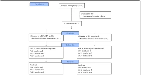
The study flowchart is shown in Fig. 1. Nine MPCs + HA and 5 HA-alone patients completed the visit week 26 post-injection; 7 MPCs + HA and 5 HA-alone pa-tients completed the visit week 52 post-injection; and 6 MPCs + HA and 4 HA-alone patients completed the final visit week 104 post-injection.
Investigational agents
expanded, and cryopreserved using the company’s propri-etary good manufacturing practice (GMP) procedures and product release criteria approved by the TGA. Allogeneic use of the MPCs for this study did not require donor re-cipient matching or rere-cipient immunosuppression. The MPCs (150 million cells in 4 mL cryopreservent) were supplied for the study in cryobags maintained in the vapour phase of liquid nitrogen at–140 to–196 degrees C. Immediately, prior to application, the cryobags and contents were thawed and 2 mL (75 million MPCs) drawn up into a syringe and mixed using a three-way tap, with 2 mL HA (sodium hyaluronate 10 mg/mL as Euflexxa®, Ferring Pharmaceuticals Inc., Parsippany, NJ, USA) from a pre-filled syringe.
Safety and tolerance
[image:4.595.54.291.97.763.2] [image:4.595.303.539.101.301.2]Adverse events were assessed via a telephone call on the day after the knee injection. Safety measures, including physical examination, vital signs, knee physical assessment, adverse events, laboratory parameters, and documentation of concomitant medications or therapies, were performed at 5 days, 28 days, and 8, 12, 26, 36, 52, 78, and 104 weeks following intra-articular injection. Laboratory blood haem-atological and biochemical tests included non-fasting chemistry (albumin, alkaline phosphatase, alanine amino-transferase, aspartate aminoamino-transferase, blood urea nitro-gen, creatinine, chloride, direct bilirubin, gamma-glutamyl transferase, glucose, lactic dehydrogenase, sodium, potas-sium, phosphorous, total bilirubin, total calcium, carbon dioxide, total cholesterol, total protein, and uric acid), erythrocyte sedimentation rate and C-reactive protein, complete blood count, HIV, and hepatitis B and hepatitis C viral testing. Because of the human origin of the MPCs and their culture expansion that included exposure to fetal bo-vine serum, the generation and persistence of anti-HLA
Table 1Inclusion and exclusion criteria Inclusion criteria
(1) Males or females aged 18–40 years;
(2) Anterior cruciate ligament (ACL) injury requiring reconstruction with bone bruising evident on pre-operative magnetic resonance im-aging (MRI) scan at screen or within 6 months of initial ACL injury;
(3) Have undergone unilateral ACL reconstruction surgery within 6 months of injury;
(4) Clinically stable knee after reconstruction—International Knee Documentation Committee clinical knee examination at time of surgery after reconstruction to be grade normal or nearly normal;
(5) Willing and able to undertake a standardized rehabilitation protocol as assessed by surgeon;
(6) ACL graft used was autograft hamstring;
(7) Willingness to participate in follow-up for 24 months from the time of initial treatment;
(8) Ability to understand and willingness to sign consent form; (9) If a female was of childbearing potential, then she must have confirmed negative urine and serum pregnancy test result at screening, and a negative urine pregnancy test prior to the administration of the study treatment, and agree to use a medically reliable method of preventing conception for the first 6 months after injection of the study treatment;
(10) Male patients with partners of childbearing potential must be willing to use a medically reliable method of preventing conception for the first 6 months after injection of the study treatment.
Exclusion criteria
(1) Women who are pregnant or breast feeding or planning to become pregnant during the first 6 months after injection of the study treatment;
(2) Known sensitivities to bovine (cow), murine (mouse), chicken products, and/or dimethyl sulphoxide;
(3) Known allergies to products from birds such as feathers, eggs, or poultry;
(4) Previous allergic reaction to hyaluronan (HA);
(5) Systemic or local infection at the screen visit or at the time of the study injection;
(6) History of any autoimmune disease, such as systemic lupus erythematosus, Addison’s disease, Crohn’s disease, or rheumatoid arthritis;
(7) Treatment with immunosuppression therapy within 6 months prior to screening;
(8) Chronic (at least 7 consecutive days) of systemic corticosteroids at a dose equivalent to >10 mg/day prednisolone within 14 days prior to screening;
(9) Acute or chronic infectious disease, including but not limited to human immunodeficiency virus;
(10) Treatment and/or uncompleted follow-up treatment of any inves-tigational therapy within 6 months before the procedure and/or intent to participate in any other investigational drug or cell therapy study dur-ing the 24-month follow-up period of this study;
(11) Recipient of prior allogeneic stem cell/progenitor cell therapy; (12) Undergoing a simultaneous procedure to the opposite knee; (13) Injury was work related and covered by workers compensation; (14) A medical condition, serious intercurrent illness, or extenuating circumstance that, in the opinion of the investigator, would preclude participation in the study or potentially decrease survival or interfere with ambulation or rehabilitation (e.g., histories of transient ischemic attack, stroke, uncontrolled diabetes, or liver disease);
(15) Presence of≥20% anti-human leukocyte antigen (HLA) antibody titres and/or having antibody specificities to donor HLAs;
(16) History or current evidence of alcohol or drug abuse or was a recreational user of illicit drugs or prescription medications;
(17) Significant damage to the collateral or posterior ligaments of the knee;
(18) Meniscal injury requiring more than a 1/3 resection or more than a single suture to reconstruction or a reconstruction that would alter the usual ACL rehabilitation;
(19) History of prior surgery to the study knee joint;
Table 1Inclusion and exclusion criteria(Continued)
(20) History of malignancy (excluding basal cell carcinoma that has been successfully excised);
(21) Chondral lesions noted at time of surgical reconstruction greater than grade 1a on any surfaces;
(22) Intra-articular steroid or corticosteroid or HA injections in preced-ing 3 months to the affected joint;
(23) Diffuse synovitis at time of surgery for the ACL reconstruction; (24) Indwelling metal of any description which precluded MRI examination such as, but not limited to, indwelling pacemaker, cerebral aneurysm clips, or electrical indwelling device such as bone stimulator or anything that would preclude patient from undergoing screening MRI;
(25) Not willing to return for required follow-up visits or there was a clear demonstration of likely poor compliance;
(26) Any other medical condition that, in the judgment of the Principal Investigator/Investigator, would prohibit the patient from participating in the study;
(27) Patient was legally or mentally incapacitated;
panel reactive antibodies (PRA), murine, and anti-bovine antibodies were tested. Immunogenicity was evalu-ated by anti-HLA PRA against class I and II HLAs mea-sured by flow cytometry.
Additional safety monitoring was implemented by es-tablishing an independent Safety Review Committee. The Safety Review Committee was an independent multidisciplinary group (independent of both the spon-sor and contract research organisation) consisting of one biostatistician and three physicians with orthopaedic/ rheumatologic expertise who collectively had experience in the management of patients and the conduct and monitoring of randomised clinical trials.
Knee pain, function, and quality of life
Pain, function, and quality of life were assessed using Knee Injury and OA Outcome Score (KOOS) [33] and SF-36v2 [34] at the time of screening, randomisation (baseline), and 6, 12, 18, and 24 months after intra-articular injection of the test substances. Patient re-sponses to intra-articular treatment using these clinical instruments were determined by calculating the change from their baseline score at 6, 12, 18, and 24 months. Thus, a positive value indicated an improvement and a negative value indicated a worsening.
Knee x-ray and joint space width measurement
Knee x-ray was performed using a standardised tech-nique, with views including weight-bearing anteroposter-ior and lateral, skyline and Rosenberg. Participants
underwent plain x-rays at screening (baseline) and 6, 12, 18, and 24 months after intra-articular injection and signs of OA were assessed using the Kellgren and Lawrence five-point grading system [35]. Joint space width was measured on the Rosenberg view [36] in millimetres using electronic callipers on a workstation (Inteleviewer, Intelerad, Montreal, Canada). Change in joint space width was calculated as a follow-up measure of change from the baseline, such that a positive value indicated an increase in joint space width and a negative value indicated joint space narrowing.
MRI acquisition and knee structure measurements
Knee structures were assessed from MRIs performed at screening (baseline), and 6, 12, and 24 months after in-jection. Knees were imaged in the sagittal plane on a 1.5-T whole body magnetic resonance unit using a com-mercial transmit-receive extremity coil (Signa HDxt; GE Medical Systems, USA). The following sequence param-eters were used: a T1-weighted fat suppressed 3D
gradi-ent recall acquisition in the steady state; flip angle 40 degrees; repetition time 23 ms; echo time 6.3 ms; field of view 16 cm; 64 partitions; 512 × 512 matrix; one acquisi-tion. Sagittal images were obtained at a partition thick-ness of 1.5 mm and an in-plane resolution of 0.31 × 0.31 mm. In addition, a coronal proton density fat-saturated acquisition, repetition time 5000 ms, echo time 57 ms, slice thickness 3.5 mm, 1 excitation, a field of view of 16 cm, and a matrix of 512 × 512 pixels was also obtained. Each MRI measurement was performed by a trained Assessed for eligibility (n=28)
Excluded (n=11)
- Not meeting inclusion criteria
Analysed At 6 months: n=9 At 12 months: n=7 At 24 months: n=6
Lost to follow-up (non-compliant) At 6 months: n=2
At 12 months: n=2 At 24 months: n=1 Allocated to MPC + HA (n=11)
- Received allocated intervention (n=11)
Lost to follow-up (non-compliant) At 6 months: n=1
At 12 months: n=0 At 24 months: n=1 Allocated to HA alone (n=6)
- Received allocated intervention (n=6)
Analysed At 6 months: n=5 At 12 months: n=5 At 24 months: n=4
Allocation
Analysis Follow-Up
Randomized (n=17)
Enrollment
[image:5.595.57.541.87.346.2]observer with an independent cross-check performed by a second trained observer, both blinded to the character-istics of participants, the group allocation of the partici-pants, and the sequence of MRIs. Knee joint articular cartilage pathology was graded using the Osteoarthritis Research Society International grading system [37].
Tibial cartilage volume was determined by image pro-cessing on an independent workstation using the soft-ware Osiris (Digital Imaging Unit, University Hospital of Geneva, Switzerland). The volumes of medial and lateral tibial cartilage plates were isolated from the total volume by manually drawing disarticulation contours around the cartilage boundaries on each section. The volume of the particular cartilage plate was determined by summing the pertinent voxels within the resultant binary volume. The coefficients of variation (CVs) for cartilage volume measures were 3.4% for medial tibia and 2.0% for lateral tibia [38]. Annual change in cartilage volume was calcu-lated as: (follow-up cartilage volume – baseline cartilage volume) divided by the period of time between MRI scans. Annual percentage change was obtained by dividing an-nual change by baseline cartilage volume, expressed as a percentage. Therefore, a positive value indicated an in-crease in cartilage volume and a negative value indicated cartilage volume loss.
Medial and lateral cross-sectional areas of the tibial plateau were determined by creating an isotropic volume from the input images which were reformatted in the axial plane, using the software program Osiris. Areas were directly measured from these axial images as previ-ously described [39]. CVs for the medial and lateral tibial plateau area were 2.3% and 2.4%, respectively [38]. An-nual change in bone area was calculated as: (follow-up bone area–baseline bone area) divided by the period of time between MRI scans. Annual percentage change was obtained by dividing annual change by baseline bone area, expressed as a percentage. Thus, a positive value indicated bone expansion and a negative value indicated reduced bone area.
Anthropometric measures
At screening, each participant had their height and weight measured and body mass index calculated.
Sample size calculation
The primary aim of this study was to assess the safety and tolerance, and the secondary aim was to explore the preliminary effect of the investigational agents on clin-ical and knee structural outcomes in patients following ACL reconstruction. The sample size of 24 in this ex-ploratory study was not based on attaining sufficient statistical power for tests of within- or between-group comparisons. There was no formal rule to stop the study based on the number of adverse events within a group.
It was possible to estimate the least chance of an adverse event in an individual subject so that one or more groups of 8 subjects were likely to have one or more events.
Statistical analyses
Descriptive statistics for characteristics of the study par-ticipants were tabulated. Continuous variables were assessed for normality. Independent samplesttests were used to compare means, and Chi-squared tests used to compare nominal characteristics between the two groups. Least squares analysis was used to compare the change in joint space width from baseline by time point between the two groups. There was no adjustment for multiple comparison performed in this study. A pvalue less than 0.05 (two-tailed) was regarded as statistically significant. All analyses were performed using IBM SPSS version 23.
Results
Baseline characteristics of study participants
The baseline characteristics of the study participants are shown in Table 2. There were no significant differences be-tween the two groups in terms of age, gender, body mass index, tibial cartilage volume, tibial bone area, joint space width, tibial cartilage volume, or bone area. Apart from the KOOS activities of daily living (ADL) scores for the HA injected individuals, both groups exhibited KOOS baseline scores that were below the cut-off scores for individuals with physical problems associated with injured knees [7]. However, participants in the MPC + HA group re-ported worse KOOS pain (p = 0.02), symptoms (p < 0.001), ADL score (p= 0.047), and SF-36v2 bodily pain (p = 0.01) score than the HA-alone group at baseline. No significant differences in baseline characteristics were observed between completers and non-completers at 6, 12, and 24 months of follow-up as shown in Fig. 1 (Additional files 1, 2 and 3: Tables S1–S3).
Adverse events
The incidence of adverse events was graded according to the National Cancer Institute Common Termin-ology Criteria for Adverse Events (NCICTCAE) and is summarised in Table 3. No participants experienced treatment-related or treatment-emergent serious ad-verse events that resulted in death, treatment discon-tinuation, or study termination over the course of the study.
maintained through to week 104. None of the study par-ticipants exhibited clinically significant abnormalities in haematology or blood chemistry laboratory results over the course of the study. No major changes in vital signs were reported following injection of the test substances, nor were there notable changes from baseline in the
physical examination data. No ectopic tissue formation was noted from the blinded MRI evaluations.
Serious adverse events
Two serious adverse events, fracture of the humerus and infective bursitis, were reported for patients in the MPC + HA group after week 52. These were not considered to be treatment related.
Effect of MPCs on knee pain, function, and quality of life over 24 months
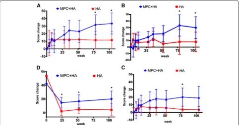
There was an improvement from baseline in both groups for all the KOOS dimensions over 24 months (Fig. 2, Additional file 4: Table S4). However, significant differ-ences between treatment groups were observed for KOOS symptoms and pain at 18 months (bothp= 0.03) and 24 months (p= 0.04 and 0.02, respectively), and for ADL at 18 months (p = 0.04), all in favour of patients who were injected with the MPC + HA preparation (Fig. 2, Additional file 4: Table S4). There was also an improvement over 24 months in both groups for all SF-36 physical components except for general health per-ception (Fig. 2, Additional file 5: Table S5). However, statistically significant differences between treatments were only observed for bodily pain at 6, 12, and 24 months (p= 0.02, 0.05, and 0.03, respectively), where injection with the MPH + HA preparation was found to be more benefi-cial than HA alone (Fig. 2, Additional file 5: Table S5).
Effect of MPCs on tibial cartilage volume change over 24 months
[image:7.595.304.539.99.298.2]There was no significant difference in annual change in medial or lateral tibial cartilage volume between the MPC
Table 2Baseline characteristics of study participants MPC + HA (n= 11)
HA alone (n= 6) p
*
Age, years 26.0 (3.6) 26.9
(10.3) 0.85
Females, number (%) 3 (27) 2 (33) 0.79
Body mass index, kg/m2 25.1 (3.1) 25.1 (4.6) 1.00 Interval between ACL injury and
reconstruction, days
76.5 (54.3) 61.7 (34.8)
0.56
Interval between ACL reconstruction and intra-articular injection, days
48.5 (14.0) 47.8 (8.3) 0.92
KOOS
Pain 69.1 (11.7) 84.2
(11.9) 0.02
Symptoms 59.7 (10.8) 83.9
(11.7)
<0.001
Activities of daily living 82.1 (11.0) 92.8 (6.4) 0.047 Sport and recreation function 29.7 (17.6) 40.0
(14.1) 0.47
Knee-related quality of life 40.9 (19.8) 50.0 (16.8)
0.36
SF-36 physical component score 41.3 (4.7) 45.9 (5.8) 0.11 Physical functioning 41.0 (6.2) 43.7 (1.7) 0.31 Role limitation: physical 40.0 (7.0) 42.6 (8.1) 0.51
Bodily pain 42.3 (8.6) 53.5 (5.4) 0.01
General health perception 55.2 (6.6) 55.4 (7.9) 0.95 Joint space width, mm
Medial tibiofemoral compartment
4.36 (0.55) 4.97 (0.63)
0.06
Lateral tibiofemoral compartment
5.38 (0.62) 5.67 (0.52)
0.36
Knee structure measured from MRI
Medial tibial cartilage volume, mm3
2456 (397) 2627 (476)
0.44
Lateral tibial cartilage
volume, mm3 3291 (401) 3548(847) 0.51
Medial tibial plateau bone area, mm2
2299 (337) 2187 (182)
0.46
Lateral tibial plateau
bone area, mm2 1472 (202) 1415(193) 0.58
Data are reported as mean (SD) or number (%)
*For difference between two groups using independent samplesttest or chi-squared test where appropriate
ACLanterior cruciate ligament,KOOSKnee Injury and Osteoarthritis Outcome Score,MRImagnetic resonance imaging
Table 3Incidence of adverse events
MPC + HA (n= 11)
HA alone (n= 6)
Total number of adverse events 94 39
Non-serious,n(%) 92 (97.9) 39
(100.0)
Serious,n(%) 2 (2.1)# 0 (0.0)
Adverse events by system organ class*,n(grade) Musculoskeletal and connective tissue disorders
11 (2) 6 (1)
General disorders and administration site conditions
3 (2) 1 (1)
Injury, poisoning, and procedural complications
2 (1) 2 (1)
Infections and infestations 1 (1) 1 (1)
Respiratory, thoracic, and mediastinal disorders 1 (1) 0
Immune system disorders 2 (2) 1 (2)
* Graded according to the National Cancer Institute Common Terminology Criteria for Adverse Events (NCICTCAE)
#
[image:7.595.56.289.101.585.2]+ HA and HA-alone groups over 6, 12, and 24 months (Table 4). However, the MPC + HA group tended to have a reduced rate of medial tibial cartilage volume loss com-pared with the HA-alone group over the first 6 months of the study (0.7% vs–4.0%,p= 0.10) (Table 4).
Effect of MPCs on tibial bone expansion over 24 months
The rate of total tibial bone expansion in the MPC + HA group showed a significantly decrease compared with the HA-alone group over the first 6 months post-treatment (0.5% vs 4.0%, p= 0.02) (Table 5). The trend
was maintained over the subsequent 12 and 24 months (bothp= 0.09).
Effect of MPCs on tibiofemoral joint space width over 24 months
Radiological assessments of the mean joint space width of patients when expressed as individual changes from baseline revealed a greater increase in the MPC + HA group than the HA-alone group at 12, 18, and 24 months post-administration (Fig. 3, Additional file 6: Table S6). In the medial compartment, injection with MPC + HA resulted in a greater joint space width in-crease compared with the HA-alone group at 18 months (p= 0.03) and 24 months (p= 0.07) (Fig. 3, Additional file 6: Table S6). In the lateral compartment, joint space width increased relative to baseline in the MPC + HA group but declined in the HA-alone group. This resulted in significant differences between treatment at 12 months (p= 0.01), 18 months (p= 0.03), and 24 months (p= 0.04) (Fig. 3, Additional file 6: Table S6).
Discussion
This study is the first to evaluate the therapeutic effects of a single intra-articular injection of allogeneic immuno-selected MPCs on the long-term (2 years) outcomes of knee joint symptoms, cartilage, and subchondral bone changes post-ACL reconstruction. The study showed that
[image:8.595.57.540.88.342.2]Fig. 2Change from baseline ina–cKnee Injury and Osteoarthritis Outcome Score (KOOS) (pain scores (a), symptom scores (b), and activities of daily living (ADL) scores (c)), anddSF-36 physical component scores over 24 months (mean and 95% confidence interval shown). *p< 0.05HA hyaluronan,MPCmesenchymal precursor cell
Table 4Annual percentage change from baseline in tibial cartilage volume over 24 months
MPC + HA HA alone p*
Medial tibia
6 months 0.7 (5.9) –4.0 (3.9) 0.10
12 months 0.3 (6.3) –2.4 (3.1) 0.36
24 months –1.4 (4.2) –3.3 (5.3) 0.54
Lateral tibia
6 months –1.4 (5.3) –2.7 (4.4) 0.65
12 months –4.7 (3.4) –2.6 (2.5) 0.25
24 months –3.7 (3.4) –0.8 (3.5) 0.22
Data are reported as mean (SD)
[image:8.595.56.291.581.707.2]intra-articular administration of allogeneic MPCs was safe and well tolerated over the 24-month study period follow-ing a sfollow-ingle injection. Moreover, usfollow-ing validated clinical instruments under blinded conditions, there was evidence for improvements in pain, function, and quality of daily life, as well as favourable effects on knee joint structural changes over 24 months, supporting further investigation of MPCs as potential disease-modifying agents in post-traumatic knee OA.
In terms of both safety and efficacy our findings were consistent with previous studies using culture-expanded autologous bone marrow-derived MSC administered via the intra-articular route for the management of OA and a variety of other joint problems [16, 19, 40–44]. Over our 2-year study no cell-related serious adverse events or ab-normalities in laboratory parameters or clinical signs were observed.
Although the patient group enrolled in our study were young adults with a recent ACL reconstruction who exhib-ited no radiographic signs of OA, they reported persistent joint pain at study entry with the MPC + HA group
exhibiting lower (worse) KOOS pain, symptom, and ADL scores than the HA group at baseline. Over the course of the study both MPC + HA- and HA-injected groups showed symptomatic improvement; however, the MPC + HA-treated group exhibited greater changes from baseline.
Although the primary aim of the study was to examine the safety and tolerability of the test substances, it also demonstrated favourable effects of MPCs on knee struc-tural outcomes relative to the HA-alone group. We found that tibial bone expansion was halted in the MPC + HA arm (0.5 ± 2.4%), while there was a significant bone expansion in the HA alone group (4.0 ± 2.3%) over 6 months, with a non-statistically significant trend served at 12 and 24 months. Although we did not ob-serve a statistically significant effect of MPCs on knee cartilage, there was a trend for slowing in cartilage vol-ume loss in the MPC + HA group (0.7 ± 5.9%) com-pared to the HA-only group (–4.0 ± 3.9%) over 6 months. This was supported by the finding of reduced joint space narrowing in the MPC + HA group com-pared to the HA-only group. Recent studies have shown that the earliest changes at the knee post-trauma are seen at the subchondral bone [45]. Hunter and col-leagues showed that both ACL injury and ACL recon-struction were associated with significant flattening of articulating bone curvature over 5 years [45]. We showed that there was tibial bone expansion at 2 and 4 years post-meniscectomy which predated changes in car-tilage volume [46]. Subchondral bone and carcar-tilage are intimately integrated, since the avascular cartilage relies on the integrity of vascularized subchondral bone to re-main functional. A greater tibial plateau bone area has been shown to be associated with the classical radio-graphic hallmarks of OA (osteophyte and joint space narrowing) [47] and cartilage defects [48]. Taken to-gether, these preliminary results suggest that the admin-istration of MPCs in post-ACL reconstruction could slow the rate of disease progression, thereby delaying the onset of post-traumatic OA in later years.
[image:9.595.56.292.110.292.2]The mechanisms of action responsible for the beneficial effect of MPCs on patient-reported clinical outcomes and
Table 5Annual percentage tibial bone expansion from baseline over 24 months
MPC + HA HA alone p*
Medial tibial plateau
6 months 0.8 (3.9) 4.2 (3.4) 0.13
12 months –1.9 (4.0) 1.5 (3.6) 0.17
24 months 0.1 (1.7) 0.8 (0.9) 0.43
Lateral tibial plateau
6 months 0.4 (4.2) 3.6 (3.6) 0.17
12 months –0.2 (4.0) 1.9 (2.8) 0.35
24 months –1.8 (2.6) 1.2 (3.5) 0.16
Total tibial plateau
6 months 0.5 (2.4) 4.0 (2.3) 0.02
12 months –1.2 (2.8) 1.7 (2.0) 0.09
24 months –0.7 (1.5) 1.0 (1.1) 0.09
Data are reported as mean (SD)
* For difference between two groups using independent samplesttest HAhyaluronan,MPCmesenchymal precursor cell
[image:9.595.56.539.591.703.2]radiograph- and MRI-derived structural outcomes are presently unresolved. However, MPCs are known to ex-hibit anti-inflammatory and immunomodulatory activities [11–14] which could suppress the injury-induced produc-tion of inflammatory mediators within joint tissues that are responsible for the clinical symptoms and breakdown of joint connective tissues [2]. The MPCs have surface re-ceptors for interleukin (IL)-6, tumour necrosis factor (TNF), IL-1, and IL-17, among others, that bind these cytokines when induced at sites of tissue injury and in-flammation. The receptor engagement results in secretion by the MPCs of prostaglandin E2 and indoleamine 2,3-dioxygenase which can polarise monocytes to an anti-inflammatory M2 phenotype and T helper 17 cells to FoxP3 T regulatory cells [14, 49]. This pathway results in increased IL-10 levels and reduction in the very inflamma-tory cytokines that initiated the response [30, 31, 50]. Moreover, co-culture of human synovial and cartilage ex-plants exposed to conditioned media from MSC pre-treated with TNF-alpha showed reduced expression of IL-beta and matrix metalloproteinases and upregulation of the cytokine signalling suppressor 1 gene while, in cartil-age, upregulation of IL-1 receptor antagonist and down-regulation of the proteoglycan degrading proteinase ADAMTS-5 was observed [51]. MPCs injected intraven-ously to sheep with collagen-induced joint inflammation was demonstrated to decrease cartilage erosions, synovial stromal cell activation, angiogenesis, and plasma levels of activin A and IL-17A, confirming that systemic adminis-tration of these cells can modulate both local joint and systemic inflammation [32].
The present study has limitations. It is a pilot phase Ib/IIa randomised controlled trial with the primary aim being to determine the safety and tolerability of a single intra-articular injection of allogeneic MPCs. The small sample size has limited the power of our study to show significant results for some structural and clinical out-comes such as cartilage volume loss and ADL score. However, it has demonstrated consistent findings be-tween clinical symptoms and knee structural changes. In terms of joint structural effects, we have observed a more pronounced effect of MPCs on subchondral bone remodelling accompanied by a preservative effect on ar-ticular cartilage which follows a biologically plausible se-quence of effect, particularly after ACL reconstruction. Although there was a higher rate of loss to follow-up (17.6% at 6 months, 29.4% at 12 months, and 41.2% at 24 months), there were no significant differences be-tween the completers and non-completers in terms of baseline characteristics of age, gender, body mass index, tibial cartilage volume, tibial bone area, or joint space width. The strengths of our study included the objective assessment of changes in cartilage volume and bone area over 24 months at four time points which were
measured from MRI using validated methods. Both MRI outcome measures have been shown to play a role in the pathogenesis of knee OA [48, 52]. Furthermore, each measurement was independently performed by a trained observer who was blinded to the participant characteris-tics, the group allocation of the participants, and the se-quence of MRIs, with high intra-observer reproducibility.
Conclusions
Intra-articular administration of a single injection of allogeneic immuno-selected MPCs following ACL re-construction was shown to be safe and well tolerated in this 24-month study. There was also preliminary evidence to support the efficacy of MPCs in improving both symp-toms and structural outcomes. These findings suggest that MPCs may modulate some of the pathological processes re-sponsible for the onset and progression of post-traumatic OA and warrant further investigation of their potential as a disease-modifying agent for the treatment of early joint injuries.
Additional files
Additional file 1:Table S1.Baseline characteristics of completers and non-completers at 6 months. (DOC 32 kb)
Additional file 2:Table S2.Baseline characteristics of completers and non-completers at 12 months. (DOC 32 kb)
Additional file 3:Table S3.Baseline characteristics of completers and non-completers at 24 months. (DOC 32 kb)
Additional file 4:Table S4.Change from baseline in KOOS scores over 24 months. (DOC 46 kb)
Additional file 5:Table S5.Change from baseline in SF-36 physical component scores over 24 months. (DOC 42 kb)
Additional file 6Table S6.Radiologically determined change in joint space width from baseline over 24 months. (DOC 36 kb)
Abbreviations
ACL:Anterior cruciate ligament; ADL: Activities of daily living; CFU-F: Colony-forming units-fibroblast; cGCP: Criteria compliant with principles of good clinical practice; CV: Coefficient of variation; GMP: Good manufacturing practice; HA: Hyaluronan; HLA: Human leukocyte antigen; IL: Interleukin; KOOS: Knee Injury and Osteoarthritis Outcome Score; MPC: Mesenchymal precursor cell; MRI: Magnetic resonance imaging; MSC: Mesenchymal stem cells; OA: Osteoarthritis; PRA: Panel reactive antibodies; TGA: Therapeutic Goods Administration; TNF: Tumour necrosis factor
Acknowledgements Not applicable.
Funding
This study was sponsored by Mesoblast Ltd. However, the sponsor had no role in the design of the study, collection, analysis, or interpretation of data, which was maintained under blinded conditions and finally analysed by an independent statistical CRO (Statistical Revelations Pty. Ltd., Melbourne, Australia), or preparation of the manuscript. YW is the recipient of National Health and Medical Research Council Career Development Fellowship (Clinical level 1, #1065464).
Availability of data and material
Authors’contributions
PG, SI, AS, SH, and FMC designed the study, and were involved in conducting the study. YW, AS, PM, JL, DC, SH, and FMC contributed to data collection. YW, DS, PG, SI, and FMC analysed and interpreted the data. YW, FMC, and PG drafted the manuscript. AS, PM, JL, DC, SH, and SI contributed to the interpretation of results. All authors reviewed the manuscript for important intellectual content and approved the final manuscript.
Ethics approval and consent to participate
Ethics approval was obtained from the Cabrini Human Research Ethics Committee. All participants gave written informed consent.
Consent for publication Not applicable.
Competing interests
YW and FMC received remuneration from Mesoblast for MRI measurements. PM, JL, and DC received remuneration from Mesoblast for setting up MRI sequences and parameters. AS and SH received remuneration from Mesoblast for the study. PG is a consultant to Mesoblast Ltd. but does not hold stock in the company. DS and SI are employees of Mesoblast, and SI holds stock in the company.
Publisher’s Note
Springer Nature remains neutral with regard to jurisdictional claims in published maps and institutional affiliations.
Author details
1Department of Epidemiology and Preventive Medicine, School of Public Health and Preventive Medicine, Monash University, Alfred Hospital, Melbourne, VIC 3004, Australia.2Melbourne Orthopaedic Group, 33 The Avenue, Windsor, VIC 3181, Australia.3Mesoblast Ltd., Level 38, 55 Collins Street, Melbourne, VIC 3000, Australia.4Imaging Associates Box Hill, Box Hill, VIC 3128, Australia.5Castlereagh Imaging, 60 Pacific Highway, St Leonards, NSW 2065, Australia.6Imaging @ Olympic Park, AAMI Park, 60 Olympic Boulevard, Melbourne, VIC 3000, Australia.7Emeritus Research, 291 Wattletree Road, Malvern East, VIC 3145, Australia.
Received: 20 December 2016 Accepted: 17 July 2017
References
1. Felson DT, Lawrence RC, Dieppe PA, Hirsch R, Helmick CG, Jordan JM, Kington RS, Lane NE, Nevitt MC, Zhang Y, et al. Osteoarthritis: new insights. Part 1: the disease and its risk factors. Ann Intern Med. 2000;133:635–46. 2. Lieberthal J, Sambamurthy N, Scanzello CR. Inflammation in joint injury and
post-traumatic osteoarthritis. Osteoarthritis Cartilage. 2015;23:1825–34. 3. Brown TD, Johnston RC, Saltzman CL, Marsh JL, Buckwalter JA.
Posttraumatic osteoarthritis: a first estimate of incidence, prevalence, and burden of disease. J Orthop Trauma. 2006;20:739–44.
4. Hill CL, Seo GS, Gale D, Totterman S, Gale ME, Felson DT. Cruciate ligament integrity in osteoarthritis of the knee. Arthritis Rheum. 2005;52:794–9. 5. Amin S, Guermazi A, Lavalley MP, Niu J, Clancy M, Hunter DJ, Grigoryan M,
Felson DT. Complete anterior cruciate ligament tear and the risk for cartilage loss and progression of symptoms in men and women with knee osteoarthritis. Osteoarthritis Cartilage. 2008;16:897–902.
6. von Porat A, Roos EM, Roos H. High prevalence of osteoarthritis 14 years after an anterior cruciate ligament tear in male soccer players: a study of radiographic and patient relevant outcomes. Ann Rheum Dis. 2004;63:269–73. 7. Lohmander LS, Ostenberg A, Englund M, Roos H. High prevalence of
knee osteoarthritis, pain, and functional limitations in female soccer players twelve years after anterior cruciate ligament injury. Arthritis Rheum. 2004;50:3145–52.
8. Wen C, Lohmander LS. Osteoarthritis: does post-injury ACL reconstruction prevent future OA? Nat Rev Rheumatol. 2014;10:577–8.
9. Jones P, Dalziel SR, Lamdin R, Miles-Chan JL, Frampton C: Oral non-steroidal anti-inflammatory drugs versus other oral analgesic agents for acute soft tissue injury. Cochrane Database Syst Rev. 2015;Issue 7. Art. No.:CD007789. 10. Lotz MK, Kraus VB. New developments in osteoarthritis. Posttraumatic
osteoarthritis: pathogenesis and pharmacological treatment options. Arthritis Res Ther. 2010;12:211.
11. Ullah I, Subbarao RB, Rho GJ. Human mesenchymal stem cells—current trends and future prospective. Biosci Rep. 2015;35:e00191.
12. Uccelli A, Moretta L, Pistoia V. Mesenchymal stem cells in health and disease. Nat Rev Immunol. 2008;8:726–36.
13. Ham O, Lee CY, Kim R, Lee J, Oh S, Lee MY, Kim J, Hwang KC, Maeng LS, Chang W. Therapeutic potential of differentiated mesenchymal stem cells for treatment of osteoarthritis. Int J Mol Sci. 2015;16:14961–78.
14. Iyer SS, Rojas M. Anti-inflammatory effects of mesenchymal stem cells: novel concept for future therapies. Expert Opin Biol Ther. 2008;8:569–81. 15. Wang Y, Yuan M, Guo QY, Lu SB, Peng J. Mesenchymal stem cells for
treating articular cartilage defects and osteoarthritis. Cell Transplant. 2015;24:1661–78.
16. Pers YM, Rackwitz L, Ferreira R, Pullig O, Delfour C, Barry F, Sensebe L, Casteilla L, Fleury S, Bourin P, et al. Adipose mesenchymal stromal cell-based therapy for severe osteoarthritis of the knee: a phase I dose-escalation trial. Stem Cells Transl Med. 2016;5:847–56.
17. Hirzinger C, Tauber M, Korntner S, Quirchmayr M, Bauer HC, Traweger A, Tempfer H. ACL injuries and stem cell therapy. Arch Orthop Trauma Surg. 2014;134:1573–8.
18. Dave LY, Nyland J, McKee PB, Caborn DN. Mesenchymal stem cell therapy in the sports knee: where are we in 2011? Sports Health. 2012;4:252–7. 19. Vangsness Jr CT, Farr 2nd J, Boyd J, Dellaero DT, Mills CR, LeRoux-Williams
M. Adult human mesenchymal stem cells delivered via intra-articular injection to the knee following partial medial meniscectomy: a randomized, double-blind, controlled study. J Bone Joint Surg Am. 2014;96:90–8. 20. Ramakrishnan A, Torok-Storb B, Pillai MM. Primary marrow-derived stromal
cells: isolation and manipulation. Methods Mol Biol. 2013;1035:75–101. 21. Nombela-Arrieta C, Ritz J, Silberstein LE. The elusive nature and function of
mesenchymal stem cells. Nat Rev Mol Cell Biol. 2011;12:126–31.
22. Malgieri A, Kantzari E, Patrizi MP, Gambardella S. Bone marrow and umbilical cord blood human mesenchymal stem cells: state of the art. Int J Clin Exp Med. 2010;3:248–69.
23. Gronthos S, Zannettino AC, Hay SJ, Shi S, Graves SE, Kortesidis A, Simmons PJ. Molecular and cellular characterisation of highly purified stromal stem cells derived from human bone marrow. J Cell Sci. 2003;116:1827–35. 24. Simmons PJ, Torok-Storb B. Identification of stromal cell precursors in human
bone marrow by a novel monoclonal antibody, STRO-1. Blood. 1991;78:55–62. 25. Gronthos S, Fitter S, Diamond P, Simmons PJ, Itescu S, Zannettino AC. A
novel monoclonal antibody (STRO-3) identifies an isoform of tissue nonspecific alkaline phosphatase expressed by multipotent bone marrow stromal stem cells. Stem Cells Dev. 2007;16:953–63.
26. Gronthos S, McCarty R, Mrozik K, Fitter S, Paton S, Menicanin D, Itescu S, Bartold PM, Xian C, Zannettino AC. Heat shock protein-90 beta is expressed at the surface of multipotential mesenchymal precursor cells: generation of a novel monoclonal antibody, STRO-4, with specificity for mesenchymal precursor cells from human and ovine tissues. Stem Cells Dev. 2009;18:1253–62.
27. See F, Seki T, Psaltis PJ, Sondermeijer HP, Gronthos S, Zannettino AC, Govaert KM, Schuster MD, Kurlansky PA, Kelly DJ, et al. Therapeutic effects of human STRO-3-selected mesenchymal precursor cells and their soluble factors in experimental myocardial ischemia. J Cell Mol Med. 2011;15:2117–29.
28. Nasef A, Zhang YZ, Mazurier C, Bouchet S, Bensidhoum M, Francois S, Gorin NC, Lopez M, Thierry D, Fouillard L, Chapel A. Selected Stro-1-enriched bone marrow stromal cells display a major suppressive effect on lymphocyte proliferation. Int J Lab Hematol. 2009;31:9–19.
29. Psaltis PJ, Paton S, See F, Arthur A, Martin S, Itescu S, Worthley SG, Gronthos S, Zannettino AC. Enrichment for STRO-1 expression enhances the cardiovascular paracrine activity of human bone marrow-derived mesenchymal cell populations. J Cell Physiol. 2010;223:530–40.
30. Read R, Cake M, Smith T, Little CB, Smith MM, Appleyard RC, Itescu S, Ghosh P. Dose dependent chondroprotective effects of allogeneic STRO-3+ mesenchymal precursor cells following direct intra-articular injection into joints of an ovine model of early osteoarthritis. Osteoarthritis Cartilage. 2008; 16 Suppl 4:S18–9.
31. Dooley LM, Abdalmula A, Washington EA, Kaufman C, Tudor EM, Ghosh P, Itescu S, Kimpton WG, Bailey SR. Effect of mesenchymal precursor cells on the systemic inflammatory response and endothelial dysfunction in an ovine model of collagen-induced arthritis. PLoS One. 2015;10:e0124144. 32. Abdalmula A, Dooley LM, Kaufman C, Washington EA, House JV, Blacklaws
mesenchymal precursor cells reduce inflammation and improve clinical outcomes in a large animal model of monoarthritis. Stem Cell Res Ther. 2017;8:22.
33. Roos EM, Roos HP, Lohmander LS, Ekdahl C, Beynnon BD. Knee Injury and Osteoarthritis Outcome Score (KOOS)—development of a self-administered outcome measure. J Orthop Sports Phys Ther. 1998;28:88–96.
34. Ware Jr JE, Sherbourne CD. The MOS 36-item short-form health survey (SF-36). I. Conceptual framework and item selection. Med Care. 1992;30:473–83. 35. Kellgren JH, Lawrence JS. Radiological assessment of osteo-arthrosis. Ann
Rheum Dis. 1957;16:494–502.
36. Dervin GF, Feibel RJ, Rody K, Grabowski J. 3-Foot standing AP versus 45 degrees PA radiograph for osteoarthritis of the knee. Clin J Sport Med. 2001; 11:10–6.
37. Eckstein F, Cicuttini F, Raynauld JP, Waterton JC, Peterfy C. Magnetic resonance imaging (MRI) of articular cartilage in knee osteoarthritis (OA): morphological assessmen. Osteoarthritis Cartilage. 2006;14(Suppl A):A46–75. 38. Wluka AE, Davis SR, Bailey M, Stuckey SL, Cicuttini FM. Users of oestrogen
replacement therapy have more knee cartilage than non-users. Ann Rheum Dis. 2001;60:332–6.
39. Wang Y, Wluka AE, Cicuttini FM. The determinants of change in tibial plateau bone area in osteoarthritic knees: a cohort study. Arthritis Res Ther. 2005;7:R687–693.
40. Peeters CM, Leijs MJ, Reijman M, van Osch GJ, Bos PK. Safety of intra-articular cell-therapy with culture-expanded stem cells in humans: a systematic literature review. Osteoarthritis Cartilage. 2013;21:1465–73. 41. Vega A, Martin-Ferrero MA, Del Canto F, Alberca M, Garcia V, Munar A,
Orozco L, Soler R, Fuertes JJ, Huguet M, et al. Treatment of knee osteoarthritis with allogeneic bone marrow mesenchymal stem cells: a randomized controlled trial. Transplantation. 2015;99:1681–90.
42. Orozco L, Munar A, Soler R, Alberca M, Soler F, Huguet M, Sentis J, Sanchez A, Garcia-Sancho J. Treatment of knee osteoarthritis with autologous mesenchymal stem cells: a pilot study. Transplantation. 2013;95:1535–41. 43. Orozco L, Munar A, Soler R, Alberca M, Soler F, Huguet M, Sentis J, Sanchez
A, Garcia-Sancho J. Treatment of knee osteoarthritis with autologous mesenchymal stem cells: two-year follow-up results. Transplantation. 2014; 97:e66–68.
44. Xia P, Wang X, Lin Q, Li X. Efficacy of mesenchymal stem cells injection for the management of knee osteoarthritis: a systematic review and meta-analysis. Int Orthop. 2015;39:2363–72.
45. Hunter DJ, Lohmander LS, Makovey J, Tamez-Pena J, Totterman S, Schreyer E, Frobell RB. The effect of anterior cruciate ligament injury on bone curvature: exploratory analysis in the KANON trial. Osteoarthritis Cartilage. 2014;22:959–68.
46. Wang Y, Dempsey AR, Lloyd DG, Mills PM, Wrigley T, Bennell KL, Metcalf B, Hanna F, Cicuttini FM. Patellofemoral and tibiofemoral articular cartilage and subchondral bone health following arthroscopic partial medial
meniscectomy. Knee Surg Sports Traumatol Arthrosc. 2012;20:970–8. 47. Wluka AE, Wang Y, Davis SR, Cicuttini FM. Tibial plateau size is related to
grade of joint space narrowing and osteophytes in healthy women and in women with osteoarthritis. Ann Rheum Dis. 2005;64:1033–7.
48. Ding C, Cicuttini F, Jones G. Tibial subchondral bone size and knee cartilage defects: relevance to knee osteoarthritis. Osteoarthritis Cartilage. 2007;15:479–86.
49. Aggarwal S, Pittenger MF. Human mesenchymal stem cells modulate allogeneic immune cell responses. Blood. 2005;105:1815–22. 50. Prockop DJ, Oh JY. Mesenchymal stem/stromal cells (MSCs): role as
guardians of inflammation. Mol Ther. 2012;20:14–20.
51. van Buul GM, Villafuertes E, Bos PK, Waarsing JH, Kops N, Narcisi R, Weinans H, Verhaar JA, Bernsen MR, van Osch GJ. Mesenchymal stem cells secrete factors that inhibit inflammatory processes in short-term osteoarthritic synovium and cartilage explant culture. Osteoarthritis Cartilage. 2012;20:1186–96.
52. Cicuttini FM, Wluka AE, Forbes A, Wolfe R. Comparison of tibial cartilage volume and radiologic grade of the tibiofemoral joint. Arthritis Rheum. 2003;48:682–8.
• We accept pre-submission inquiries
• Our selector tool helps you to find the most relevant journal
• We provide round the clock customer support
• Convenient online submission
• Thorough peer review
• Inclusion in PubMed and all major indexing services
• Maximum visibility for your research
Submit your manuscript at www.biomedcentral.com/submit