0095-1137/05/$08.00⫹0 doi:10.1128/JCM.43.8.3673–3680.2005
Copyright © 2005, American Society for Microbiology. All Rights Reserved.
Development of a New Oligonucleotide Array To Identify
Staphylococcal Strains at Species Level
Philippe Giammarinaro,
1Sabine Leroy,
1Jean-Paul Chacornac,
1Julien Delmas,
2and Regine Talon
1*
INRA—Centre de Clermont-Ferrand-Theix, UR 370, Microbiologie, 63122 Saint-Gene`s Champanelle,1 and Centre Hospitalo-Universitaire, Laboratoire de Bacte´riologie, 28 place Henri Dunant,
63001 Clermont-Ferrand,2France
Received 21 January 2005/Returned for modification 15 March 2005/Accepted 14 April 2005
The genus Staphylococcus is made up of 36 validated species which contain strains that are pathogenic, saprophytic, or used as starter cultures for the food industry. An oligonucleotide array targeting the manga-nese-dependent superoxide dismutase (sodA) gene was developed to overcome the drawbacks of the conven-tional methods of identification. Divergences of thesodAgene were used to design oligonucleotide probes, and we showed that each of the 36 species had a characteristic pattern of hybridization. To evaluate the array, we analyzed 38 clinical and 38 food or food plantStaphylococcusisolates identified by the phenotype-based system VITEK 2 (bioMe´rieux). This commercial kit failed to identify 8 (21%) of the clinical isolates and 32 (84%) of the food and food plant isolates. In contrast, the oligonucleotide array we designed provided an accurate and rapid method for the identification of staphylococcal strains, isolated from clinical, environmental, or food samples, at species level.
Staphylococci are widely spread in various niches such as clinical environments and food plants. Thirty-six validated de-scribed species, including 21 subspecies, belong to the Staphy-lococcusgenus according to theList of Bacterial Names with Standing in Nomenclature, updated 3 December 2004 (16). Some staphylococcal strains are used for their technological abilities, and others are associated with diseases in humans or animals.Staphylococcus xylosusandS. carnosusstrains are used as starter cultures in fermented meat products, because they contribute to their color and flavor (47). In these products, other staphylococci, such asS. simulans,S. succinus,S. equo-rum,S. warneri,S. epidermidis,S. saprophyticus, andS. aureus, may be found (8), but the last three are also known to be pathogens or opportunistic pathogens.S. saprophyticusis the predominant staphylococcal species involved in acute urinary tract infections of young adult women (32).S. epidermidis is involved in many infections such as bacteremia and prosthetic and natural valvular endocarditis (50).S. aureusis one of the leading causes of food-borne diseases and of nosocomial in-fections (28, 34).
Because of these yin/yang aspects, much effort has been expended in recent years to identify staphylococci. Several manual and automated methods based on phenotypic charac-teristics have been developed for identification of the Staphy-lococcusspecies that are most often isolated from clinical sam-ples (21, 25, 35, 37). Unfortunately, these systems have their limitations, mostly due to phenotypic differences between strains from the same species (33, 37–39). For this reason, methods based on molecular techniques have been developed. Genus- and species-specific primers have been designed for
the identification of bacteria belonging to the genus Staphylo-coccus and for the species-specific detection of S. aureus, S. epidermidis, S. saprophyticus, and S. xylosus (2, 18, 31, 33). Some authors associated several genus- and species-specific primer pairs in the same amplification reaction and were able to identify strains at genus level and up to four species (13). These PCR methods are quick and reliable, but they are lim-ited in the number of species that can be identified. Alternative approaches include denaturating gradient gel electrophoresis (8) and sequence determination of the 16S rRNA-encoding gene (rrs) (5, 45). However, closely related species may have nearly identical rrs sequences, impairing the discriminatory power of these techniques (46). To solve this problem, it is possible to use alternative target genes which exhibit more-divergent sequences thanrrs. So far thecpn60(19),gla (53),
femA(49),rpoB(14), andsodA(36) genes have been used. In previous studies, our laboratory reported the sequencing of thesodAgenes, encoding a manganese-dependent surper-oxide dismutase, ofS. xylosus and S. carnosus (3, 4). At the same time, Poyart et al. published the sequences of thesodA
genes of nearly all known species of staphylococci (36). Pair-wise comparison of these sequences revealed a mean identity (81.5%) lower than that calculated for the rrs sequences of staphylococci (98%). Therefore, thesodAgene will be a more discriminatory target sequence thanrrs for differentiation of closely related staphylococci. However, thesodAsequences of pairs of type strains of subspecies shared more than 99.3% identity and did not allow discrimination at the subspecies level, except for thesodAgenes ofStaphylococcus cohniisubsp.
cohniiandStaphylococcus cohniisubsp.urealyticus, which dis-play 4% sequence divergence. Furthermore, Poyart et al. dem-onstrated that thesodAsequences of strains of the same spe-cies isolated from food or clinical samples displayed less than 1.5% divergence from the sequence of the corresponding type strain (36). In conclusion, they proposed that the sequence
* Corresponding author. Mailing address: INRA, Centre de Cler-mont-Ferrand-Theix, UR 370, Microbiologie, 63122 Saint-Gene`s Champanelle, France. Phone: 33 (0) 473624170. Fax: 33 (0) 473624268. E-mail: [email protected].
3673
on May 15, 2020 by guest
http://jcm.asm.org/
polymorphism of thesodAgene could allow the development of assays based on DNA chip technologies.
The potential for microbial diagnostics of DNA microarrays, originally developed for whole-genome gene expression anal-yses (42, 48), is very high, since they allow simultaneous prod-uct interrogation with a large number of probe sequences (10, 11). Recent studies showed the accurateness of such tools at detecting and identifying a great number of bacteria at the genus or species level in a single assay (15, 51, 52). Despite their very interesting abilities, microarrays are not yet common in microbial diagnostic laboratories. Part of the reason is the considerable initial financial investment. A recent survey con-ducted by the Association of Biomolecular Resource Facilities (ABRF) Microarray Research Group estimated the mean cost for setting up a microarray facility at $286,000 (22). Similar but less expensive techniques can be used. Oligonucleotide probe sets spotted onto nylon or nitrocellulose membranes have been used for bacterial identification for 10 years. In 1994, Kaufhold et al. used allele-specific oligonucleotide probes fixed to a membrane to rapidly identify strains of group A streptococci (27). Since that preliminary work, other authors have used closely related techniques to identify bacteria at the genus or species level (6, 40, 41).
In this study, we demonstrated the accurateness of such a tool for identification of staphylococcal strains at species level. This system, which we called “Staph. Array,” couples PCR amplification of thesodAgene with an oligonucleotide-based array to efficiently discriminate all the 36 validated Staphylo-coccusspecies and the two subspecies ofS. cohnii.
MATERIALS AND METHODS
Bacterial strains and culture conditions.TheStaphylococcustype strains used are listed in Table 1. Strains were grown at 30°C in brain heart infusion broth or on brain heart infusion agar (Difco, Detroit, Mich.), with the notable exception ofS. saccharolyticus, which was grown anaerobically in a medium containing the following (in grams per liter): casein peptone, 10; meat peptone, 5; yeast extract, 5;L-cysteine HCl, 0.4; glucose, 10; NaCl, 5; thioglycolate, 2 (pH 7.2).
Oligonucleotide probe design.A database ofsodAgene sequences was con-structed, and local BLAST comparisons were done with tools embedded in BioEdit software (23). Alignments were done using the ClustalW (12) service at the public website of the European Bioinformatics Institute (http://www .ebi.ac.uk/clustalw/). To facilitate the probe design, the alignments were re-organized with the “Multialignment Cleaner” tool of the Annhyb package (http://bioinformatics.org/annhyb/). The hairpin and dimer formation abili-ties of oligonucleotides were tested with the “Oligo” tools of the same package. Melting temperatures of perfect-match duplexes and those of mismatched nu-cleotides were predicted by the nearest-neighbor method using MELTING (29). All oligonucleotide probes were synthesized with a 5⬘-terminal amino group by Operon Biotechnologies (Germany) to allow covalent coupling of probes to the membrane. Probes used in this study are reported in Table 2.
Array preparation.Procedures for covalent coupling of probes followed the protocol described previously, with a Cross-Blot dot blot apparatus (Sebia, France) used instead of a Miniblotter (26). Briefly, a charged nylon membrane (Biodyne C; Pall Biosupport, United Kingdom) was activated for 10 min with freshly prepared 16% (wt/vol) 1-(3-dimethylaminopropyl)-3-ethylcarbodiimide hydrochloride (Across Organics, France). The oligonucleotide probes were ap-plied to the membrane in parallel by using the grid with 34 vertical spacers. After 1 min at room temperature, the membrane was inactivated for 8 min with 100 mM NaOH and then washed with 2⫻SSPE (1⫻SSPE is 0.18 M NaCl, 10 mM NaH2PO4, and 1 mM EDTA, pH 7.7) (Promega, France) supplemented with 0.1% sodium dodecyl sulfate (SDS; Eurobio Biotechnology, France) for 5 min at 60°C.
Target preparation and hybridization procedures.Primers D1and D2, used to amplify the internal part of thesodAgene (sodAint), have been described previ-ously (36). D2was synthesized with a 5⬘-terminal digoxigenin group (DIG). Amplifications were done with a GenAmp PCR system 9700 PE thermal cycler
(Perkin-Elmer, France) and 25-l volumes containing 0.8M of each primer, 50
M of each deoxyribonucleoside triphosphate, 1.5 mM MgCl2, and 1 U ofTaq DNA polymerase in 1⫻buffer according to the manufacturer’s instructions (Promega, France). For efficient amplification from one colony picked up from the agar plate, the following conditions were used: 15 min at 4°C; 5 min at 95°C; 40 cycles of 30 s at 94°C, 1 min at 35°C, and 30 s at 72°C; and a final 2-min hold at 72°C. Relative quantification of the 480-bp amplified fragments was per-formed by comparison with SmartLadder (Eurogentec, France) after electro-phoresis through a 1.5% agarose gel and ethidium bromide staining.
The PCR products at a final concentration of 150 ng/ml in 0.5⫻SSPE–0.1% SDS were heat denatured and cooled on ice immediately. After 5 min of incu-bation at room temperature in 50 ml of 0.5⫻SSPE–0.1% SDS, the membrane was placed in the Cross-Blot dot blot apparatus. The 14 horizontal slots of the grid were filled with the denatured target, and hybridization occurred for 1 h at 50°C on a plane surface. The samples were removed carefully, and slots were filled with a prewarmed (60°C) 2⫻SSPE–0.5% SDS solution. After aspiration, the membrane was taken from the Cross-Blot dot blot apparatus and washed twice in 70 ml 2⫻SSPE–0.5% SDS for 15 min at 60°C in a rolling bottle. An additional wash with 70 ml 0.1⫻SSPE–0.5% SDS occurred for 7 min at room temperature. The hybridized targets were detected with the DIG color detection kit (Roche, France).
Other methods of identification.The ID-GPC card of the VITEK 2 system was used for biochemical identification as recommended by the manufacturer (bioMe´rieux). Analysis of the results was based on the report provided by the VITEK 2 (version 3.01) computer software. Results with low levels of confidence are indicated.
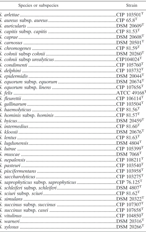
[image:2.585.303.539.77.451.2]Multiplex PCRs were performed to check the identification at the genus level TABLE 1. Type strains used in this study
Species or subspecies Strain
S. arlettae...CIP 103501T S. aureussubsp.aureus...CIP 65.8T S. auricularis...DSM 20609T S. capitissubsp.capitis...CIP 81.53T S. caprae...DSM 20608T S. carnosus...DSM 20501T S. chromogenes...CIP 81.59T S. cohniisubspcohnii...DSM 20260T S. cohniisubspurealyticus...CIP104024T S. condimenti...CIP 105760T S. delphini...CIP 103732T S. epidermidis...DSM 20044T S. equorumsubsp.equorum...DSM 20674T S. equorumsubsp.linens...CIP 107656T S. felis...ATCC 49168T S. fleurettii...CIP 106114T S. gallinarum...CIP 103504T S. haemolyticus...CIP 81.56T S. hominissubsp.hominis...CIP 81.57T S. hyicus...DSM 20459T S. intermedius...CIP 81.60T S. kloosii...DSM 20676T S. lentus...CIP 81.63T S. lugdunensis...DSM 4804T S. lutrae...CIP 105399T S. muscae...DSM 7068T S. nepalensis...CIP 108211T S. pasteuri...CIP 103540T S. piscifermentans...CIP 103958T S. saccharolyticus...CIP 103275T S. saprophyticussubsp.saprophyticus...CIP 76.125T S. schleiferisubsp.schleiferi...DSM 4807T S. sciurisubsp.sciuri...CIP 81.62T S. simulans...DSM 20322T S. succinussubsp.succinus...CIP 107307T S. succinussubsp.casei...CIP 107658T S. vitulinus...CIP 104850T S. warneri...DSM 20316T S. xylosus...DSM 20266T
on May 15, 2020 by guest
http://jcm.asm.org/
and the identification ofS. aureus,S. epidermidis,S. saprophyticus, andS. xylosus
strains (13).
The internal base compositions of the sodAgenes were determined using primers D1and D2 as previously described (36). Sequences were compared against a local database ofsodAintgene sequences. Identification to the species level was based onⱖ97% sequence identity with the type strain sequence and a ⱖ5% sequence difference from the next closest species.
Nucleotide sequence accession numbers.All partial staphylococcal sequences determined in this study were deposited in GenBank. The accession numbers of thesodAsequences ofStaphylococcus succinussubsp.succinus,Staphylococcus succinussubsp.casei,Staphylococcus equorumsubsp.linens,S. fleurettii, andS. nepalensisare AY845222, AY842858, AY878697, AY845223, and AY878698, respectively.
RESULTS
Determination of the sodA internal gene sequences from type strains of coagulase-negative staphylococci.ThesodAint
sequences of type strains ofS. equorumsubsp. linens,S. succi-nussubsp.succinus,S. succinussubsp.casei,S. fleurettii, andS. nepalensiswere amplified. These fragments were sequenced, and sequence comparisons were done using BLAST (1). The base composition ofS. equorumsubsp. linens sodAintwas
com-pletely identical to that previously published for theS. equorum
subsp. equorumtype strain. ThesodAintsequences ofS. succi-nussubsp. succinusandS. succinussubsp. caseidiffered in only one base pair. The highest sequence similarity values were (i) 93% forS. succinuscompared toS. gallinarum, (ii) 95% forS. fleurettiicompared toS. vitulinus, and (iii) 92% forS. nepalensis
compared toS. cohniisubsp. urealyticus.
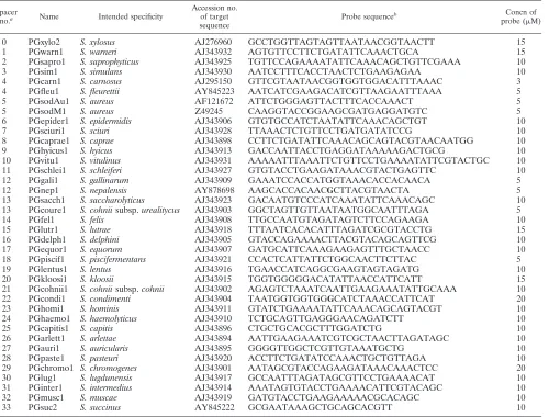
Design of probes. The available partial sequences ofsodA
[image:3.585.48.548.81.461.2]and those we determined were used to create characteristic probes for the 36 species. Oligonucleotides of 21 to 38 bases, with predicted melting temperatures from 61°C to 68°C, were chosen from dissimilar parts noted in alignedsodAsequences. We rejected sequences with predicted stable hairpins and dimers or with unsatisfactory specificities. A central mismatch was introduced into probes PGcondi1 and PGnep1. These G/T artificial mismatches were created to increase the specificity of these probes. Candidate probes were tested on the array under different conditions, and those that were adopted are reported in Table 2. Probe concentrations were empirically modified to allow better discrimination.
TABLE 2. Probes
Spacer
no.a Name Intended specificity
Accession no. of target sequence
Probe sequenceb Concn of
probe (M)
0 PGxylo2 S. xylosus AJ276960 GCCTGGTTAGTAGTTAATAACGGTAACTT 15
1 PGwarn1 S. warneri AJ343932 AGTGTTCCTTCTGATATTCAAACTGCA 15
2 PGsapro1 S. saprophyticus AJ343925 TGTTCCAGAAAATATTCAAACAGCTGTTCGAAA 10
3 PGsim1 S. simulans AJ343930 AATCCTTTCACCTAACTCTGAAGAGAA 10
4 PGcarn1 S. carnosus AJ295150 GTTCGTAATAACGGTGGTGGACATTTAAAC 3
4 PGfleu1 S. fleurettii AY845223 AATCATCGAAGACATCGTTAAGAATTTAAA 5
5 PGsodAu1 S. aureus AF121672 ATTCTGGGAGTTACTTTCACCAAACT 5
5 PGsodM1 S. aureus Z49245 CAAGGTACCGGAAGCGATGAGGATGTC 5
6 PGepider1 S. epidermidis AJ343906 GTGTGCCATCTAATATTCAAACAGCTGT 10
7 PGsciuri1 S. sciuri AJ343928 TTAAACTCTGTTCCTGATGATATCCG 10
8 PGcaprae1 S. caprae AJ343898 CCTTCTGATATTCAAACAGCAGTACGTAACAATGG 10
9 PGhyicus1 S. hyicus AJ343913 GACCAATTACCTGAGGATAAAAAGACTGCG 10
10 PGvitu1 S. vitulinus AJ343931 AAAAATTTAAATTCTGTTCCTGAAAATATTCGTACTGC 10
11 PGschlei1 S. schleiferi AJ343927 GTGTACCTGAAGATAAACGTACTGAGTTC 10
12 PGgali1 S. gallinarum AJ343909 GAAATCCACCATGGTAAACACCACAACA 5
12 PGnep1 S. nepalensis AY878698 AAGCACCACAACGCTTACGTAACTA 5
13 PGsacch1 S. saccharolyticus AJ343923 GACAATGTCCCATCAAATATTCAAACAGC 10
13 PGcoure1 S. cohniisubsp.urealitycus AJ343903 GGCTAGTTGTTAATAATGGCAATTTAGA 5
14 PGfel1 S. felis AJ343908 TTGCCAATGTAGATAGTCTTCCAGAAGA 10
15 PGlutr1 S. lutrae AJ343918 TTTAATCACACATTTAGATCGCGTACCTG 15
16 PGdelph1 S. delphini AJ343905 GTACCAGAAAACTTACGTACAGCAGTTCG 10
17 PGequor1 S. equorum AJ343907 GATGCATTCAAAGAAGAGTTTGCTAACC 10
18 PGpiscif1 S. piscifermentans AJ343921 CCACTCATTATTCTGGCAACTTCTTAC 5
19 PGlentus1 S. lentus AJ343916 TGAACCATCAGGCGAAGTAGTAGATG 10
20 PGkloosi1 S. kloosii AJ343915 TGGTGGGGGACATATTAACCATTCATT 15
21 PGcohnii1 S. cohniisubsp.cohnii AJ343902 AGAGTCTAAATCAATTGAAGAAATATTGCAAA 10
22 PGcondi1 S. condimenti AJ343904 TAATGGTGGTGGGCATCTAAACCATTCAT 20
23 PGhomi1 S. hominis AJ343911 GTATCTGAAAATATTCAAACAGCAGTACGT 10
24 PGhaemo1 S. haemolyticus AJ343910 TCTGCAGTTGAGGGAACAGATCTT 10
25 PGcapitis1 S. capitis AJ343896 CTGCTGCACGCTTTGGATCTG 10
26 PGarlett1 S. arlettae AJ343894 AATTGAAGAAATCGTCGCTAACTTAGATAGC 10
27 PGauri1 S. auricularis AJ343895 GGGGTTGGCTCGTTGTAAATGCTG 10
28 PGpaste1 S. pasteuri AJ343920 ACCTTCTGATATCCAAACTGCTGTTAGA 10
29 PGchromo1 S. chromogenes AJ343901 AATAGCGTACCAGAAGATAAACAAACTCC 20
30 PGlug1 S. lugdunensis AJ343917 GCCAATTTAGATAGCGTTCCTGAAAACAT 10
31 PGinter1 S. intermedius AJ343914 AAATAGTGTACCTGAAAACATTCGTACAGC 10
32 PGmusc1 S. muscae AJ343919 GATGTACCTGAAGAAAAACGCACAGC 10
33 PGsuc2 S. succinus AY845222 GCGAATAAAGCTGCAGCACGTT 10
aSpacer numbers 0 to 33 correspond to the spacer numbers in Table 3 and Fig. 1. bPositions of mismatches with target sequences are indicated by boldfaced characters.
on May 15, 2020 by guest
http://jcm.asm.org/
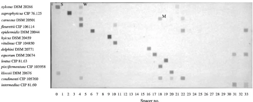
Validation with type strains.Our array was first tested with type strains of each validatedStaphylococcusspecies (Table 1). As expected, not only unique spots but unique patterns of spots were obtained (Fig. 1). This was due to the conditions of hybridization, which allowed some mismatched probe-target pairs to hybridize, i.e., some probes hybridized not only with the targets for which they were designed but also with targets from closely related species. However, comparisons of the pat-terns of hybridization showed that a unique pattern was found for each species (Table 3). The probes that we designed dis-criminated targets with differences in their base composition as low as 3%, since we distinguishedS. condimentifromS. car-nosusorS. piscifermentans and we obtained distinct patterns for the two subspecies of S. cohnii. We also discriminated species that are difficult to differentiate on the basis of theirrrs
sequences, such as theS. intermedius and S. delphinispecies and theS. nepalensisandS. cohniisubsp. urealyticusspecies (5, 44). We could not distinguish between the two subspecies ofS. succinus or the two subspecies of S. equorum because their
sodAintsequences were identical.
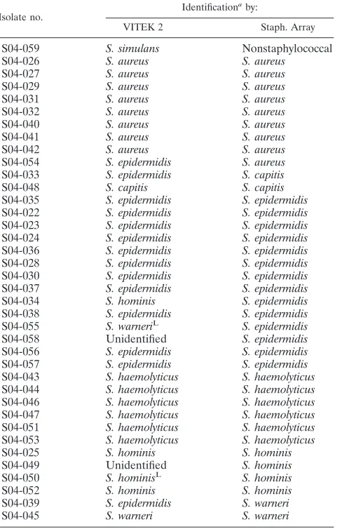
Application to strains isolated from clinical samples.A total of 38 strains (Table 4) from clinical samples were identified first by a phenotypic approach using the VITEK 2 system (bioMe´rieux), and these results were compared to the array identification. Results were identical for 30 strains ofS. aureus,
S. capitis, S. epidermidis, S. haemolyticus, S. hominis, and S. warneri. Six strains (16%) were misidentified by the VITEK 2. Three strains identified as S. epidermidis by VITEK 2 were identified as strains ofS. aureus,S. warneri, andS. capitisby the array. Two strains identified asS. epidermidisby the array were misidentified asS. warneriand S. hominisby VITEK 2. One strain identified asS. simulans by VITEK 2 did not give any hybridization result on the array. Multiplex PCR confirmed that this strain was not a staphylococcus. Two strains (5%) could not be identified by VITEK 2. They were identified asS. epidermidisandS. hominis.
Application to strains isolated from food or food plant sam-ples.A total of 38 strains (Table 5) from food or food plant samples were also identified by the VITEK 2 system, and the results of the identification were compared to the results from
the array. Some species commonly isolated from food or food plants, such asS. equorumandS. succinus(9), are not included in the VITEK card database; thus, the strains belonging to these species could not be identified by the VITEK 2 system. But even for species included in the ID-GPC database, some misidentification or lack of identification occurred. None of eightS. xylosus strains were correctly identified. Five strains were misidentified asS. saprophyticus, two were misidentified asS. cohniisubsp.urealyticus, and one was not identified. Only one of fourS. saprophyticusstrains was correctly identified; the others were either misidentified asS. chromogenesorS. auric-ularisor not identified. Of the two strains ofS. epidermidis, one was correctly identified, while the other was identified as Kocu-ria vaKocu-rians. TheS. warneri strain was also misidentified asK. varians. Strains of S. hominis, S. capitis, and S. sciuri were correctly identified. All the identifications done via the array were confirmed by multiplex PCR when suitable and by se-quencing of thesodAintfragments of these strains.
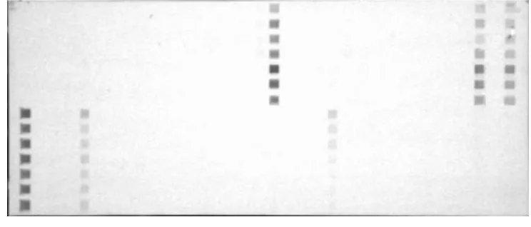
Stability of patterns obtained with wild-type strains. The stability of the patterns of hybridization obtained for wild-type strains was investigated. As an example, Fig. 2 shows the pat-terns obtained for strains ofS. xylosusandS. equorumisolated from food or food plant samples. Only slight variations in the intensity of hybridization spots occurred. The same stability of hybridization patterns was obtained whatever the origin of the strain (data not shown).
DISCUSSION
In this study, we present a new oligonucleotide array tool, called “Staph. Array,” for the identification of the 36 species of staphylococci described and validated and for the discrimina-tion of the twoS. cohniisubspecies. For this method, universal primers amplifying an internal part of the sodA gene were used, followed by hybridization of the denatured PCR prod-ucts onto an oligonucleotide array.
[image:4.585.77.512.66.242.2]Because a large amount ofrrssequence data is available in a public database, it is not surprising that this gene has been an obvious choice when molecular diagnostic tests based on DNA arrays have been developed. One important drawback of using
FIG. 1. Hybridization patterns obtained with reference type strains ofStaphylococcus. Lanes 0 to 33 correspond to the numbers of the spacers in which the probes described in Table 2 have been fixed. Levels of hybridization are indicated as follows: W, weak; M, medium; S, strong.
on May 15, 2020 by guest
http://jcm.asm.org/
rrsgenes is their conservative nature. Takahashi et al. pointed out that closely related species of staphylococci could have nearly identicalrrsbase composition (46), decreasing the dis-criminatory potential of that gene for staphylococci. To bypass this problem, some authors have used more-divergent genes to identify staphylococcal strains. ThefemA(24, 49),rpoB (14),
gla (53), cpn60 (19), and sodA (36) genes have been used. Array techniques have been used with thefemA and cpn60
genes. With femA a microarray was developed allowing dis-crimination of only fiveStaphylococcus species (S. aureus, S. epidermidis,S. haemolyticus,S. hominis, andS. saprophyticus) (24). The use of amplification products of the HSP60-encoding gene (cpn60) as probes produced better results; Goh et al. identified strains belonging to 30 species (20). However, their system failed to distinguish S. intermedius from S. delphini
strains and did not identify some other strains, probably be-cause they belonged to species not included in their 30-species panel. We choosesodAto develop our tool because sequences of that gene were available for 33 out of the 36 species of
staphylococci as opposed to 15 forfemA, 27 forrpoB, and 30 for cpn60. In the present work, the available sequence data were completed with thesodApartial base composition of the five type strains of S. equorum subsp. linens, S. fleurettii, S. nepalensis,S. succinussubsp. succinus, andS. succinussubsp. casei. This gene proved to be discriminatory at species level, since the sequences obtained were more than 5% divergent from the othersodAsequences present in GenBank. However, the sodAint sequences of the two subspecies of S. succinus
showed an identical base pair composition, like the sodAint
sequences of the two subspecies ofS. equorum. These results confirmed the lack of discriminatory power of thesodAint se-quences at the subspecies level (36).
[image:5.585.46.542.79.468.2]After initial database screening, oligonucleotide probes were selected on the basis of hybridization results obtained by using reference strains as templates. The conditions of hybrid-ization allowed some mismatched duplexes to form. Conse-quently, cross-hybridization of several probes with some tar-gets obtained from strains belonging to closely related species
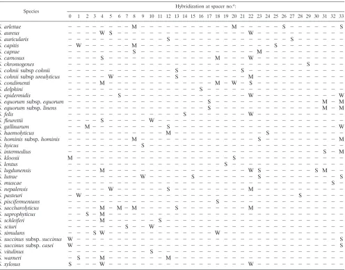
TABLE 3. Patterns obtained with type strains of the 36 species ofStaphylococcus
Species
Hybridization at spacer no.a:
0 1 2 3 4 5 6 7 8 9 10 11 12 13 14 15 16 17 18 19 20 21 22 23 24 25 26 27 28 29 30 31 32 33
S. arlettae ⫺ ⫺ ⫺ ⫺ ⫺ ⫺ ⫺ ⫺ M ⫺ ⫺ ⫺ ⫺ ⫺ ⫺ ⫺ ⫺ ⫺ ⫺ ⫺ M ⫺ ⫺ ⫺ ⫺ ⫺ S ⫺ ⫺ ⫺ ⫺ ⫺ ⫺ S
S. aureus ⫺ ⫺ ⫺ ⫺ W S ⫺ ⫺ ⫺ ⫺ ⫺ ⫺ ⫺ ⫺ ⫺ ⫺ ⫺ ⫺ ⫺ ⫺ ⫺ ⫺ W ⫺ ⫺ ⫺ ⫺ ⫺ ⫺ ⫺ ⫺ ⫺ ⫺ ⫺
S. auricularis ⫺ ⫺ ⫺ ⫺ ⫺ ⫺ ⫺ ⫺ ⫺ ⫺ ⫺ ⫺ S ⫺ ⫺ ⫺ ⫺ ⫺ ⫺ ⫺ ⫺ ⫺ ⫺ ⫺ ⫺ ⫺ ⫺ S ⫺ ⫺ ⫺ ⫺ ⫺ ⫺
S. capitis ⫺ W ⫺ ⫺ ⫺ ⫺ ⫺ ⫺ M ⫺ ⫺ ⫺ ⫺ ⫺ ⫺ ⫺ ⫺ ⫺ ⫺ ⫺ ⫺ ⫺ ⫺ ⫺ ⫺ S ⫺ ⫺ ⫺ ⫺ ⫺ ⫺ ⫺ ⫺
S. caprae ⫺ ⫺ ⫺ ⫺ ⫺ ⫺ ⫺ ⫺ S ⫺ ⫺ ⫺ ⫺ ⫺ ⫺ ⫺ ⫺ ⫺ ⫺ ⫺ ⫺ ⫺ ⫺ M ⫺ ⫺ ⫺ ⫺ ⫺ ⫺ ⫺ ⫺ ⫺ ⫺
S. carnosus ⫺ ⫺ ⫺ ⫺ S ⫺ ⫺ ⫺ ⫺ ⫺ ⫺ ⫺ ⫺ ⫺ ⫺ ⫺ ⫺ ⫺ M ⫺ ⫺ ⫺ W ⫺ ⫺ ⫺ ⫺ ⫺ ⫺ ⫺ ⫺ ⫺ ⫺ ⫺
S. chromogenes ⫺ ⫺ ⫺ ⫺ ⫺ ⫺ ⫺ ⫺ ⫺ ⫺ ⫺ ⫺ ⫺ ⫺ ⫺ ⫺ ⫺ ⫺ ⫺ ⫺ ⫺ ⫺ ⫺ ⫺ ⫺ ⫺ ⫺ ⫺ ⫺ S ⫺ ⫺ ⫺ ⫺
S. cohniisubspcohnii ⫺ ⫺ ⫺ ⫺ ⫺ ⫺ ⫺ ⫺ ⫺ ⫺ ⫺ ⫺ ⫺ S ⫺ ⫺ ⫺ ⫺ ⫺ ⫺ ⫺ S ⫺ ⫺ ⫺ ⫺ ⫺ ⫺ ⫺ ⫺ ⫺ ⫺ ⫺ ⫺
S. cohniisubspurealyticus ⫺ ⫺ ⫺ ⫺ ⫺ W ⫺ ⫺ ⫺ ⫺ ⫺ ⫺ ⫺ S ⫺ ⫺ ⫺ ⫺ ⫺ ⫺ ⫺ ⫺ M ⫺ ⫺ ⫺ ⫺ ⫺ ⫺ ⫺ ⫺ ⫺ ⫺ ⫺
S. condimenti ⫺ ⫺ ⫺ ⫺ M ⫺ ⫺ ⫺ ⫺ ⫺ ⫺ ⫺ ⫺ ⫺ ⫺ ⫺ ⫺ ⫺ M ⫺ W ⫺ S ⫺ ⫺ ⫺ ⫺ ⫺ ⫺ ⫺ ⫺ ⫺ ⫺ ⫺
S. delphini ⫺ ⫺ ⫺ ⫺ ⫺ ⫺ ⫺ ⫺ ⫺ ⫺ ⫺ ⫺ ⫺ ⫺ ⫺ ⫺ S ⫺ ⫺ ⫺ ⫺ ⫺ ⫺ ⫺ ⫺ ⫺ ⫺ ⫺ ⫺ ⫺ ⫺ ⫺ ⫺ ⫺
S. epidermidis ⫺ ⫺ ⫺ ⫺ ⫺ ⫺ S ⫺ ⫺ ⫺ ⫺ ⫺ ⫺ ⫺ ⫺ ⫺ ⫺ ⫺ ⫺ ⫺ ⫺ ⫺ W ⫺ ⫺ ⫺ ⫺ ⫺ ⫺ ⫺ ⫺ ⫺ ⫺ W
S. equorumsubsp.equorum ⫺ ⫺ ⫺ ⫺ ⫺ ⫺ ⫺ ⫺ ⫺ ⫺ ⫺ ⫺ ⫺ ⫺ ⫺ ⫺ ⫺ S ⫺ ⫺ ⫺ ⫺ ⫺ ⫺ ⫺ ⫺ ⫺ ⫺ ⫺ ⫺ ⫺ M ⫺ M
S. equorumsubsp.linens ⫺ ⫺ ⫺ ⫺ ⫺ ⫺ ⫺ ⫺ ⫺ ⫺ ⫺ ⫺ ⫺ ⫺ ⫺ ⫺ ⫺ S ⫺ ⫺ ⫺ ⫺ ⫺ ⫺ ⫺ ⫺ ⫺ ⫺ ⫺ ⫺ ⫺ M ⫺ M
S. felis ⫺ ⫺ ⫺ ⫺ ⫺ ⫺ ⫺ ⫺ ⫺ ⫺ ⫺ ⫺ ⫺ ⫺ S ⫺ ⫺ ⫺ ⫺ ⫺ ⫺ ⫺ W ⫺ ⫺ ⫺ ⫺ ⫺ ⫺ ⫺ ⫺ ⫺ ⫺ ⫺
S. fleurettii ⫺ ⫺ ⫺ ⫺ S ⫺ ⫺ ⫺ ⫺ ⫺ W ⫺ ⫺ ⫺ ⫺ ⫺ ⫺ ⫺ ⫺ ⫺ ⫺ ⫺ ⫺ ⫺ ⫺ ⫺ ⫺ ⫺ ⫺ ⫺ ⫺ ⫺ ⫺ ⫺
S. gallinarum ⫺ ⫺ M⫺ ⫺ ⫺ ⫺ ⫺ ⫺ ⫺ ⫺ ⫺ S ⫺ ⫺ ⫺ ⫺ ⫺ ⫺ ⫺ ⫺ ⫺ ⫺ ⫺ ⫺ ⫺ ⫺ ⫺ ⫺ ⫺ ⫺ ⫺ ⫺ W
S. haemolyticus ⫺ ⫺ ⫺ ⫺ ⫺ ⫺ ⫺ ⫺ ⫺ ⫺ ⫺ ⫺ M ⫺ ⫺ ⫺ ⫺ ⫺ ⫺ ⫺ ⫺ ⫺ ⫺ ⫺ S ⫺ ⫺ ⫺ ⫺ ⫺ ⫺ ⫺ ⫺ ⫺
S. hominissubsp.hominis ⫺ ⫺ ⫺ ⫺ ⫺ ⫺ ⫺ ⫺ M ⫺ ⫺ ⫺ ⫺ ⫺ ⫺ ⫺ ⫺ ⫺ ⫺ ⫺ ⫺ ⫺ ⫺ S ⫺ ⫺ ⫺ ⫺ ⫺ ⫺ ⫺ ⫺ ⫺ M
S. hyicus ⫺ ⫺ ⫺ ⫺ ⫺ ⫺ ⫺ ⫺ ⫺ S ⫺ ⫺ ⫺ ⫺ ⫺ ⫺ ⫺ ⫺ ⫺ ⫺ ⫺ ⫺ ⫺ ⫺ ⫺ ⫺ ⫺ ⫺ ⫺ ⫺ ⫺ ⫺ ⫺ ⫺
S. intermedius ⫺ ⫺ ⫺ ⫺ ⫺ ⫺ ⫺ ⫺ ⫺ ⫺ ⫺ ⫺ ⫺ ⫺ ⫺ ⫺ ⫺ ⫺ ⫺ ⫺ ⫺ ⫺ ⫺ ⫺ ⫺ ⫺ ⫺ ⫺ ⫺ ⫺ ⫺ S ⫺ M
S. kloosii M ⫺ ⫺ ⫺ ⫺ ⫺ ⫺ ⫺ ⫺ ⫺ ⫺ ⫺ ⫺ ⫺ ⫺ ⫺ ⫺ ⫺ ⫺ ⫺ S ⫺ ⫺ ⫺ ⫺ ⫺ ⫺ ⫺ ⫺ ⫺ ⫺ ⫺ ⫺ ⫺
S. lentus ⫺ ⫺ ⫺ ⫺ ⫺ ⫺ ⫺ ⫺ ⫺ ⫺ ⫺ ⫺ ⫺ ⫺ ⫺ ⫺ ⫺ ⫺ ⫺ S ⫺ ⫺ ⫺ ⫺ ⫺ ⫺ ⫺ ⫺ ⫺ ⫺ ⫺ ⫺ ⫺ ⫺
S. lugdunensis ⫺ ⫺ ⫺ ⫺ M ⫺ ⫺ ⫺ ⫺ ⫺ ⫺ ⫺ ⫺ ⫺ ⫺ ⫺ ⫺ ⫺ ⫺ ⫺ ⫺ ⫺ W S ⫺ ⫺ ⫺ ⫺ ⫺ ⫺ S M ⫺ ⫺
S. lutrae ⫺ ⫺ ⫺ ⫺ ⫺ ⫺ ⫺ ⫺ ⫺ W ⫺ ⫺ ⫺ ⫺ ⫺ S ⫺ ⫺ ⫺ ⫺ ⫺ ⫺ ⫺ S ⫺ ⫺ ⫺ ⫺ ⫺ ⫺ ⫺ ⫺ ⫺ S
S. muscae ⫺ ⫺ ⫺ ⫺ ⫺ ⫺ ⫺ ⫺ ⫺ ⫺ ⫺ ⫺ ⫺ ⫺ ⫺ ⫺ ⫺ ⫺ ⫺ ⫺ ⫺ ⫺ ⫺ ⫺ ⫺ ⫺ ⫺ ⫺ ⫺ ⫺ ⫺ ⫺ S ⫺
S. nepalensis ⫺ ⫺ ⫺ ⫺ ⫺ W ⫺ ⫺ ⫺ ⫺ ⫺ ⫺ S ⫺ ⫺ ⫺ ⫺ ⫺ ⫺ ⫺ ⫺ ⫺ M ⫺ ⫺ ⫺ ⫺ ⫺ ⫺ ⫺ ⫺ ⫺ ⫺ ⫺
S. pasteuri ⫺ W ⫺ ⫺ ⫺ ⫺ ⫺ ⫺ ⫺ ⫺ ⫺ ⫺ ⫺ ⫺ ⫺ ⫺ ⫺ ⫺ ⫺ ⫺ ⫺ ⫺ ⫺ ⫺ ⫺ ⫺ ⫺ ⫺ S ⫺ ⫺ ⫺ ⫺ ⫺
S. piscifermentans ⫺ ⫺ ⫺ ⫺ ⫺ ⫺ ⫺ ⫺ ⫺ ⫺ ⫺ ⫺ ⫺ ⫺ ⫺ ⫺ ⫺ ⫺ S ⫺ ⫺ ⫺ ⫺ ⫺ ⫺ ⫺ ⫺ ⫺ ⫺ ⫺ ⫺ ⫺ ⫺ ⫺
S. saccharolyticus ⫺ ⫺ ⫺ ⫺ M ⫺ M⫺ M ⫺ ⫺ ⫺ ⫺ S ⫺ ⫺ ⫺ ⫺ ⫺ ⫺ ⫺ ⫺ M ⫺ ⫺ ⫺ ⫺ ⫺ ⫺ ⫺ ⫺ ⫺ ⫺ ⫺
S. saprophyticus ⫺ ⫺ S ⫺ M ⫺ ⫺ ⫺ ⫺ ⫺ ⫺ ⫺ ⫺ ⫺ ⫺ ⫺ ⫺ ⫺ ⫺ ⫺ ⫺ ⫺ ⫺ ⫺ ⫺ ⫺ ⫺ ⫺ ⫺ ⫺ ⫺ ⫺ ⫺ ⫺
S. schleiferi ⫺ ⫺ ⫺ ⫺ M ⫺ ⫺ ⫺ ⫺ ⫺ ⫺ S ⫺ ⫺ ⫺ ⫺ ⫺ ⫺ ⫺ ⫺ ⫺ ⫺ ⫺ ⫺ ⫺ ⫺ ⫺ ⫺ ⫺ ⫺ ⫺ ⫺ ⫺ ⫺
S. sciuri ⫺ ⫺ ⫺ ⫺ ⫺ ⫺ ⫺ S ⫺ ⫺ W ⫺ ⫺ ⫺ ⫺ ⫺ ⫺ ⫺ ⫺ ⫺ ⫺ ⫺ ⫺ ⫺ ⫺ ⫺ ⫺ ⫺ ⫺ ⫺ ⫺ ⫺ ⫺ ⫺
S. simulans ⫺ ⫺ ⫺ S W ⫺ ⫺ ⫺ ⫺ ⫺ ⫺ ⫺ ⫺ ⫺ ⫺ ⫺ ⫺ ⫺ W ⫺ ⫺ ⫺ ⫺ ⫺ ⫺ ⫺ ⫺ ⫺ ⫺ ⫺ ⫺ ⫺ ⫺ ⫺
S. succinussubsp.succinus W ⫺ ⫺ ⫺ ⫺ ⫺ ⫺ ⫺ ⫺ ⫺ ⫺ ⫺ ⫺ ⫺ ⫺ ⫺ ⫺ ⫺ ⫺ ⫺ ⫺ ⫺ ⫺ ⫺ ⫺ ⫺ ⫺ ⫺ ⫺ ⫺ ⫺ ⫺ ⫺ S
S. succinussubsp.casei W ⫺ ⫺ ⫺ ⫺ ⫺ ⫺ ⫺ ⫺ ⫺ ⫺ ⫺ ⫺ ⫺ ⫺ ⫺ ⫺ ⫺ ⫺ ⫺ ⫺ ⫺ ⫺ ⫺ ⫺ ⫺ ⫺ ⫺ ⫺ ⫺ ⫺ ⫺ ⫺ S
S. vitulinus ⫺ ⫺ ⫺ ⫺ ⫺ ⫺ ⫺ ⫺ ⫺ ⫺ S ⫺ ⫺ ⫺ ⫺ ⫺ ⫺ ⫺ ⫺ ⫺ ⫺ ⫺ ⫺ ⫺ ⫺ ⫺ ⫺ ⫺ ⫺ ⫺ ⫺ ⫺ ⫺ ⫺
S. warneri ⫺ S ⫺ ⫺ M ⫺ ⫺ ⫺ ⫺ ⫺ ⫺ ⫺ M ⫺ ⫺ ⫺ ⫺ ⫺ ⫺ ⫺ ⫺ ⫺ ⫺ ⫺ ⫺ ⫺ ⫺ ⫺ ⫺ ⫺ ⫺ ⫺ ⫺ ⫺
S. xylosus S ⫺ ⫺ ⫺W ⫺ ⫺ ⫺ ⫺ ⫺ ⫺ ⫺ ⫺ ⫺ ⫺ ⫺ ⫺ ⫺ ⫺ ⫺ ⫺ ⫺ W ⫺ ⫺ ⫺ ⫺ ⫺ ⫺ ⫺ ⫺ ⫺ ⫺ ⫺
aSpacer numbers correspond to the numbers of the spacers in which the probes have been fixed, as reported in Table 2. W, weak; M, medium; S, strong;⫺, no
hybridization. See Fig. 1.
on May 15, 2020 by guest
http://jcm.asm.org/
was observed. However, a unique pattern of hybridization was obtained for each staphylococcal species, allowing us to iden-tify unknown strains. We used the “Staph. Array” system to identify 76 strains from clinical, food, or environmental food samples, and these identifications were compared with those obtained by the VITEK 2 system. VITEK 2 is one of the laboratories’ routine identification systems and has been shown to provide reliable results compared to other systems based on phenotypic identification (17, 30). Nineteen species commonly encountered in clinical isolates are included in the ID-GPC database of the VITEK 2 system. We showed that strains can be misidentified or not identified by the commercial system, even strains of species whose identification is covered by the VITEK 2 database; this is especially true for strains isolated from food or food plants. Strangely, identifications with good or higher levels of confidence were obtained when some strains belonging to species not included in the ID-GPC database were submitted to the VITEK 2 system (Table 5). Therefore, caution should be taken when VITEK 2 is used with unsuitable species. The problem of identification by the
VITEK 2 system could be explained by the intraspecies vari-ability of the phenotypic traits. In contrast to their phenotypic traits, the patterns obtained with “Staph. Array” were stable for strains belonging to the same species, whatever their ori-gins. This result confirmed the low intraspecies variation of the
sodAintsequences (36, 43).
Recent studies have shown that molecular methods give more-accurate results than kits based on biochemical assays (5, 43). Interestingly, one of these studies usedsodAsequencing to identify staphylococcal strains from clinical isolates (43). Iden-tification was based on 97% sequence identity with the type strain sequence and a 5% difference in sequence from the next closest species. However, these criteria did not allow discrim-ination of S. condimenti and S. carnosus (96.7% nucleotide identity) or of S. condimenti and S. piscifermentans (95.6% nucleotide identity). With “Staph. Array,” the hybridization profiles ofS. carnosus, S. piscifermentans, and S. condimenti
[image:6.585.45.289.92.469.2]were clearly distinct, so these three species can be identified (Fig. 1).
TABLE 4. Results of identification of clinical strains by the VITEK 2 and the Staph. Array system
Isolate no.
Identificationaby:
VITEK 2 Staph. Array
S04-059 S. simulans Nonstaphylococcal
S04-026 S. aureus S. aureus
S04-027 S. aureus S. aureus
S04-029 S. aureus S. aureus
S04-031 S. aureus S. aureus
S04-032 S. aureus S. aureus
S04-040 S. aureus S. aureus
S04-041 S. aureus S. aureus
S04-042 S. aureus S. aureus
S04-054 S. epidermidis S. aureus
S04-033 S. epidermidis S. capitis
S04-048 S. capitis S. capitis
S04-035 S. epidermidis S. epidermidis
S04-022 S. epidermidis S. epidermidis
S04-023 S. epidermidis S. epidermidis
S04-024 S. epidermidis S. epidermidis
S04-036 S. epidermidis S. epidermidis
S04-028 S. epidermidis S. epidermidis
S04-030 S. epidermidis S. epidermidis
S04-037 S. epidermidis S. epidermidis
S04-034 S. hominis S. epidermidis
S04-038 S. epidermidis S. epidermidis
S04-055 S. warneriL S. epidermidis
S04-058 Unidentified S. epidermidis
S04-056 S. epidermidis S. epidermidis
S04-057 S. epidermidis S. epidermidis
S04-043 S. haemolyticus S. haemolyticus
S04-044 S. haemolyticus S. haemolyticus
S04-046 S. haemolyticus S. haemolyticus
S04-047 S. haemolyticus S. haemolyticus
S04-051 S. haemolyticus S. haemolyticus
S04-053 S. haemolyticus S. haemolyticus
S04-025 S. hominis S. hominis
S04-049 Unidentified S. hominis
S04-050 S. hominisL S. hominis
S04-052 S. hominis S. hominis
S04-039 S. epidermidis S. warneri
S04-045 S. warneri S. warneri
[image:6.585.296.540.92.470.2]aL, identified at a low level of confidence.
TABLE 5. Results of identification of food or food plant strains by the VITEK 2 and the Staph. Array system
Isolate no.a Identification bby:
VITEK 2 Staph. Array
CIT S03-0458 Kocuria rosea S. arlettae*
CIT S03-0258 S. capitis S. capitis
CIT S03-0356 K. roseaL S. carnosus*
CIT S03-0354 K. roseaL S. carnosus*
CIT S03-0437 Kocuria varians S. epidermidis
CIT S03-0438 S. epidermidisL S. epidermidis
CIT S03-0631 Unidentified S. equorum* CIT S03-0632 S. cohniisubsp.urealyticus S. equorum* CIT S03-0203 S. cohniisubsp.urealyticusL S. equorum*
CIT S03-0204 S. cohniisubsp.urealyticus S. equorum* CIT S03-0205 S. cohniisubsp.urealyticus S. equorum* CIT S03-0215 S. cohniisubsp.urealyticusL S. equorum*
CIT S03-0214 K. variansL S. equorum*
CIT S03-0190 K. varians S. equorum*
CIT S03-0670 S. kloosii S. fleurettii* CIT S03-0417 S. hominisL S. hominis
CIT S03-0370 S. capitis S. pasteuri* CIT S03-0429 S. warneri S. pasteuri* CIT S03-0199 S. saprophyticus S. saprophyticus
CIT S03-0451 S. chromogenesL S. saprophyticus
CIT S03-0480 Unidentified S. saprophyticus
CIT S03-0481 S. auricularisL S. saprophyticus
CIT S03-0027 S. sciuri S. sciuri
CIT S03-0630 S. kloosiiL S. succinus*
CIT S03-0657 S. kloosiiL S. succinus*
CIT S03-0740 S. xylosusL S. succinus*
CIT S03-0749 S. kloosiiL S. succinus*
CIT S03-0579 S. cohniisubsp.cohniiL S. vitulinus*
CIT S03-0586 S. cohniisubsp.cohniiL S. vitulinus*
CIT S03-0406 K. variansL S. warneri
CIT S03-0519 S. saprophyticus S. xylosus
CIT S03-0520 S. cohniisubsp.urealyticusL S. xylosus
CIT S03-0523 S. saprophyticus S. xylosus
CIT S03-0525 S. saprophyticus S. xylosus
CIT S03-0526 S. saprophyticus S. xylosus
CIT S03-0527 Unidentified S. xylosus
CIT S03-0063 S. cohniisubsp.urealyticus S. xylosus
CIT S03-0179 S. saprophyticus S. xylosus
aCIT, Collection INRA de Theix.
bL, low level of confidence. *, species not included in the ID-GPC database of VITEK 2.
on May 15, 2020 by guest
http://jcm.asm.org/
The specificity of the probes we designed was ensured by comparison with the sequences available in GenBank (7). To date, screening of GenBank revealed that the sodA gene is present in about 30 bacterial genera, but most of the data covered strains ofStaphylococcus,Enterococcus,Streptococcus,
Pasteurella, andMycobacterium. We cannot exclude the possi-bility that yet unknown targets hybridize to our probes, but it is unlikely that targets from nonstaphylococcal strains produce patterns of hybridization that can be confused with staphylo-coccal patterns.
In conclusion, the tool “Staph. Array” allowed the rapid (less than 24 h) and accurate identification of staphylococcal strains at species level. It is the only tool described to date that distinguishes in one shot the 36 validated staphylococcal spe-cies and also discriminates the two subspespe-cies ofS. cohnii.
ACKNOWLEDGMENTS
We are grateful to Caroline Michaud, Christine Lamadon, and Lau-rent Lanore for excellent technical assistance.
This work was financially supported by the European project “Tra-disausage” QLK1-CT-2002-02240.
REFERENCES
1.Altschul, S. F., W. Gish, W. Miller, E. W. Myers, and D. J. Lipman.1990. Basic local alignment search tool. J. Mol. Biol.215:403–410.
2.Aymerich, T., B. Martin, M. Garriga, and M. Hugas.2003. Microbial quality and direct PCR identification of lactic acid bacteria and nonpathogenic staphylococci from artisanal low-acid sausages. Appl. Environ. Microbiol.
69:4583–4594.
3.Barriere, C., R. Bruckner, and R. Talon.2001. Characterization of the single superoxide dismutase ofStaphylococcus xylosus. Appl. Environ. Microbiol.
67:4096–4104.
4.Barriere, C., S. Leroy-Setrin, and R. Talon.2001. Characterization of cata-lase and superoxide dismutase inStaphylococcus carnosus833 strain. J. Appl. Microbiol.91:514–519.
5.Becker, K., D. Harmsen, A. Mellmann, C. Meier, P. Schumann, G. Peters, and C. von Eiff.2004. Development and evaluation of a quality-controlled ribosomal sequence database for 16S ribosomal DNA-based identification of
Staphylococcusspecies. J. Clin. Microbiol.42:4988–4995.
6.Behr, T., C. Koob, M. Schedl, A. Mehlen, H. Meier, D. Knopp, E. Frahm, U. Obst, K. Schleifer, R. Niessner, and W. Ludwig.2000. A nested array of rRNA targeted probes for the detection and identification of enterococci by reverse hybridization. Syst. Appl. Microbiol.23:563–572.
7.Benson, D. A., I. Karsch-Mizrachi, D. J. Lipman, J. Ostell, and D. L. Wheeler.2004. GenBank: update. Nucleic Acids Res.32(Database issue):
D23–D26.
8.Blaiotta, G., C. Pennacchia, D. Ercolini, G. Moschetti, and F. Villani.2003. Combining denaturing gradient gel electrophoresis of 16S rDNA V3 region and 16S–23S rDNA spacer region polymorphism analyses for the identifica-tion of staphylococci from Italian fermented sausages. Syst. Appl. Microbiol.
26:423–433.
9.Blaiotta, G., C. Pennacchia, F. Villani, A. Ricciardi, R. Tofalo, and E. Parente.2004. Diversity and dynamics of communities of coagulase-negative staphylococci in traditional fermented sausages. J. Appl. Microbiol.97:271– 284.
10.Bodrossy, L., and A. Sessitsch.2004. Oligonucleotide microarrays in micro-bial diagnostics. Curr. Opin. Microbiol.7:245–254.
11.Call, D. R., M. K. Borucki, and F. J. Loge.2003. Detection of bacterial pathogens in environmental samples using DNA microarrays. J. Microbiol. Methods53:235–243.
12.Chenna, R., H. Sugawara, T. Koike, R. Lopez, T. J. Gibson, D. G. Higgins, and J. D. Thompson.2003. Multiple sequence alignment with the Clustal series of programs. Nucleic Acids Res.31:3497–3500.
13.Corbie`re Morot-Bizot, S., R. Talon, and S. Leroy.2004. Development of a multiplex PCR for the identification ofStaphylococcusgenus and four staph-ylococcal species isolated from food. J. Appl. Microbiol.97:1087–1094. 14.Drancourt, M., and D. Raoult.2002.rpoBgene sequence-based
identifica-tion ofStaphylococcusspecies. J. Clin. Microbiol.40:1333–1338.
15.Dubois, J. W., S. Hill, L. S. England, T. Edge, L. Masson, J. T. Trevors, and R. Brousseau.2004. The development of a DNA microarray-based assay for the characterization of commercially formulated microbial products. J. Mi-crobiol. Methods58:251–262.
16.Euzeby, J. P.1997. List of Bacterial Names with Standing in Nomenclature: a folder available on the Internet. Int. J. Syst. Bacteriol.47:590–592. 17.Fahr, A. M., U. Eigner, M. Armbrust, A. Caganic, G. Dettori, C. Chezzi, L.
Bertoncini, M. Benecchi, and M. G. Menozzi.2003. Two-center collaborative evaluation of the performance of the BD Phoenix automated microbiology system for identification and antimicrobial susceptibility testing of Entero-coccusspp. andStaphylococcusspp. J. Clin. Microbiol.41:1135–1142. 18.Forsman, P., A. Tilsala-Timisjarvi, and T. Alatossava.1997. Identification of
staphylococcal and streptococcal causes of bovine mastitis using 16S–23S rRNA spacer regions. Microbiology143:3491–3500.
19.Goh, S. H., S. Potter, J. O. Wood, S. M. Hemmingsen, R. P. Reynolds, and A. W. Chow.1996. HSP60 gene sequences as universal targets for microbial species identification: studies with coagulase-negative staphylococci. J. Clin. Microbiol.34:818–823.
20.Goh, S. H., Z. Santucci, W. E. Kloos, M. Faltyn, C. G. George, D. Driedger, and S. M. Hemmingsen.1997. Identification ofStaphylococcusspecies and subspecies by the chaperonin 60 gene identification method and reverse checkerboard hybridization. J. Clin. Microbiol.35:3116–3121.
21.Grant, C. E., D. L. Sewell, M. Pfaller, R. V. Bumgardner, and J. A. Williams.
1994. Evaluation of two commercial systems for identification of coagulase-negative staphylococci to species level. Diagn. Microbiol. Infect. Dis.18:1–5. 22.Grills, G., C. Griffin, A. Massimi, K. Lilley, K. Knudtson, and J. VanEe.
2001. 2000/2001 ABRF Microarray Research Group study: a current profile of microarray laboratories. [Online.] http://abrf.org/ResearchGroups /Microarray/EPosters/MARG_poster_2001.pdf.
23.Hall, T. A.1999. BioEdit: a user-friendly biological sequence alignment
0 1 2 3 4 5 6 7 8 9 10 11 12 13 14 15 16 17 18 19 20 21 22 23 24 25 26 27 28 29 30 31 32 33
S. equorumCIT S03-0190
S. equorumCIT S03-0203
S. equorumCIT S03-0204
S. equorumCIT S03-0205
S. equorumCIT S03-0214
S. equorumCIT S03-0215
S. equorumCIT S03-0632
S. xylosusCIT S03-0519
S. xylosusCIT S03-0520
S. xylosusCIT S03-0523
S. xylosusCIT S03-0525
S. xylosusCIT S03-0526
S. xylosusCIT S03-0063
S. xylosusCIT S03-0527
[image:7.585.152.524.71.230.2]Spacer no.
FIG. 2. Hybridization patterns obtained with wild-type strains. Lanes 0 to 33 correspond to the numbers of the spacers in which the probes described in Table 2 have been fixed.
on May 15, 2020 by guest
http://jcm.asm.org/
editor and analysis program for Windows 95/98/NT. Nucleic Acids Symp. Ser.41:95–98.
24.Hamels, S., J. L. Gala, S. Dufour, P. Vannuffel, N. Zammatteo, and J. Remacle.2001. Consensus PCR and microarray for diagnosis of the genus
Staphylococcus, species, and methicillin resistance. BioTechniques31:1364– 1372.
25.Ieven, M., J. Verhoeven, S. R. Pattyn, and H. Goossens.1995. Rapid and economical method for species identification of clinically significant coagu-lase-negative staphylococci. J. Clin. Microbiol.33:1060–1063.
26.Kamerbeek, J., L. Schouls, A. Kolk, M. van Agterveld, D. van Soolingen, S. Kuijper, A. Bunschoten, H. Molhuizen, R. Shaw, M. Goyal, and J. van Embden.1997. Simultaneous detection and strain differentiation of Myco-bacterium tuberculosisfor diagnosis and epidemiology. J. Clin. Microbiol.
35:907–914.
27.Kaufhold, A., A. Podbielski, G. Baumgarten, M. Blokpoel, J. Top, and L. Schouls.1994. Rapid typing of group A streptococci by the use of DNA amplification and non-radioactive allele-specific oligonucleotide probes. FEMS Microbiol. Lett.119:19–25.
28.Le Loir, Y., F. Baron, and M. Gautier.2003.Staphylococcus aureusand food poisoning. Genet. Mol. Res.2:63–76.
29.Le Novere, N. 2001. MELTING, computing the melting temperature of nucleic acid duplex. Bioinformatics17:1226–1227.
30.Ligozzi, M., C. Bernini, M. G. Bonora, M. De Fatima, J. Zuliani, and R. Fontana.2002. Evaluation of the VITEK 2 system for identification and antimicrobial susceptibility testing of medically relevant gram-positive cocci. J. Clin. Microbiol.40:1681–1686.
31.Martineau, F., F. J. Picard, D. Ke, S. Paradis, P. H. Roy, M. Ouellette, and M. G. Bergeron.2001. Development of a PCR assay for identification of staphylococci at genus and species levels. J. Clin. Microbiol.39:2541–2547. 32.Martineau, F., F. J. Picard, C. Menard, P. H. Roy, M. Ouellette, and M. G. Bergeron.2000. Development of a rapid PCR assay specific for Staphylococ-cus saprophytiStaphylococ-cusand application to direct detection from urine samples. J. Clin. Microbiol.38:3280–3284.
33.Morot-Bizot, S., R. Talon, and S. Leroy-Setrin.2003. Development of spe-cific PCR primers for a rapid and accurate identification ofStaphylococcus xylosus, a species used in food fermentation. J. Microbiol. Methods55:279– 286.
34.Palavecino, E.2004. Community-acquired methicillin-resistant Staphylococ-cus aureusinfections. Clin. Lab. Med.24:403–418.
35.Perl, T. M., P. R. Rhomberg, M. J. Bale, P. C. Fuchs, R. N. Jones, F. P. Koontz, and M. A. Pfaller.1994. Comparison of identification systems for
Staphylococcus epidermidisand other coagulase-negativeStaphylococcus spe-cies. Diagn. Microbiol. Infect. Dis.18:151–155.
36.Poyart, C., G. Quesne, C. Boumaila, and P. Trieu-Cuot.2001. Rapid and accurate species-level identification of coagulase-negative staphylococci by using thesodAgene as a target. J. Clin. Microbiol.39:4296–4301. 37.Renneberg, J., K. Rieneck, and E. Gutschik.1995. Evaluation of Staph ID 32
system and Staph-Zym system for identification of coagulase-negative staph-ylococci. J. Clin. Microbiol.33:1150–1153.
38.Rhoden, D. L., and J. M. Miller.1995. Four-year prospective study of STAPH-IDENT system and conventional method for reference identifica-tion ofStaphylococcus,Stomatococcus, andMicrococcusspp. J. Clin. Micro-biol.33:96–98.
39.Rossi, F., R. Tofalo, S. Torriani, and G. Suzzi.2001. Identification by 16S–
23S rDNA intergenic region amplification, genotypic and phenotypic clus-tering ofStaphylococcus xylosusstrains from dry sausages. J. Appl. Microbiol.
90:365–371.
40.Rudi, K., S. L. Flateland, J. F. Hanssen, G. Bengtsson, and H. Nissen.2002. Development and evaluation of a 16S ribosomal DNA array-based approach for describing complex microbial communities in ready-to-eat vegetable sal-ads packed in a modified atmosphere. Appl. Environ. Microbiol.68:1146– 1156.
41.Rudi, K., O. M. Skulberg, R. Skulberg, and K. S. Jakobsen.2000. Applica-tion of sequence-specific labeled 16S rRNA gene oligonucleotide probes for genetic profiling of cyanobacterial abundance and diversity by array hybrid-ization. Appl. Environ. Microbiol.66:4004–4011.
42.Schena, M., D. Shalon, R. Heller, A. Chai, P. O. Brown, and R. W. Davis.
1996. Parallel human genome analysis: microarray-based expression moni-toring of 1000 genes. Proc. Natl. Acad. Sci. USA93:10614–10619. 43.Sivadon, V., M. Rottman, J. C. Quincampoix, V. Avettand, S. Chaverot, P. de
Mazancourt, P. Trieu-Cuot, and J. L. Gaillard.2004. Use ofsodA sequenc-ing for the identification of clinical isolates of coagulase-negative staphylo-cocci. Clin. Microbiol. Infect.10:939–942.
44.Spergser, J., M. Wieser, M. Taubel, R. A. Rossello-Mora, R. Rosengarten, and H. J. Busse.2003.Staphylococcus nepalensissp. nov., isolated from goats of the Himalayan region. Int. J. Syst. Evol. Microbiol.53:2007–2011. 45.Takahashi, T., M. Kaneko, Y. Mori, M. Tsuji, N. Kikuchi, and T. Hiramune.
1997. Phylogenetic analyses ofStaphylococcusbased on the 16S rDNA se-quence and assignment of clinical isolates from animals. J. Vet. Med. Sci.
59:775–783.
46.Takahashi, T., I. Satoh, and N. Kikuchi.1999. Phylogenetic relationships of 38 taxa of the genusStaphylococcus based on 16S rRNA gene sequence analysis. Int. J. Syst. Bacteriol.49:725–728.
47.Talon, R., S. Leroy-Se´trin, and S. Fadda.2002. Bacterial starters involved in the quality of fermented meat products, p. 175–191.InF. Toldra´ (ed.), Research advances in quality of meat and meat products. Research Signpost, Trivandrum, Kerala, India.
48.Tao, H., C. Bausch, C. Richmond, F. R. Blattner, and T. Conway.1999. Functional genomics: expression analysis of Escherichia coligrowing on minimal and rich media. J. Bacteriol.181:6425–6440.
49.Vannuffel, P., M. Heusterspreute, M. Bouyer, B. Vandercam, M. Philippe, and J. L. Gala.1999. Molecular characterization offemAfrom Staphylococ-cus hominisandStaphylococcus saprophyticus, andfemA-based discrimina-tion of staphylococcal species. Res. Microbiol.150:129–141.
50.von Eiff, C., G. Peters, and C. Heilmann.2002. Pathogenesis of infections due to coagulase-negative staphylococci. Lancet Infect. Dis.2:677–685. 51.Wang, R. F., M. L. Beggs, B. D. Erickson, and C. E. Cerniglia.2004. DNA
microarray analysis of predominant human intestinal bacteria in fecal sam-ples. Mol. Cell. Probes18:223–234.
52.Warsen, A. E., M. J. Krug, S. LaFrentz, D. R. Stanek, F. J. Loge, and D. R. Call.2004. Simultaneous discrimination between 15 fish pathogens by using 16S ribosomal DNA PCR and DNA microarrays. Appl. Environ. Microbiol.
70:4216–4221.
53.Yugueros, J., A. Temprano, B. Berzal, M. Sanchez, C. Hernanz, J. M. Luengo, and G. Naharro. 2000. Glyceraldehyde-3-phosphate dehydroge-nase-encoding gene as a useful taxonomic tool for Staphylococcus spp. J. Clin. Microbiol.38:4351–4355.