PCR Detection of
Yersinia pestis
in Fleas: Comparison
with Mouse Inoculation
DAVID M. ENGELTHALER, KENNETH L. GAGE,* JOHN A. MONTENIERI, MAY CHU,ANDLEON G. CARTER
Division of Vector-Borne Infectious Diseases, Centers for Disease Control and Prevention, Public Health Service, U.S. Department of Health and Human Services, Fort Collins, Colorado 80522
Received 3 November 1998/Returned for modification 25 January 1999/Accepted 4 March 1999
The “gold standard” for identifyingYersinia pestis-infected fleas has been inoculation of mice with pooled flea material. Inoculated mice are monitored for 21 days, and those that die are further analyzed forY. pestis
infection by fluorescent-antibody assay and/or culture. PCR may provide a more rapid and sensitive alternative for identifyingY. pestisin fleas. To compare these assays, samples were prepared from 381 field-collected fleas. Each flea was analyzed individually by both PCR and mouse inoculation. Sixty of the 381 flea samples were positive forY. pestisby PCR; 48 of these PCR-positive samples caused death in mice (80.0% agreement). None of the 321 PCR-negative samples caused death. Among the 12 mice that survived inoculation with PCR-positive samples, 10 were later demonstrated by serology or culture to have been infected withY. pestis. This suggests that death of inoculated mice is less reliable than PCR as an indicator of the presence ofY. pestis in flea samples. Mouse inoculation assays produce results that are comparable to PCR only when surviving as well as dead mice are analyzed for infection. The rapidity and sensitivity (10 to 100 CFU ofY. pestis) of PCR suggest that it could serve as a useful alternative to mouse inoculation for routine plague surveillance and outbreak investigations.
Yersinia pestis, the etiological agent of plague, is typically transmitted between rodent hosts and other mammals by the bites of infectious fleas. The risks of human infection are high-est during periods of epizootic activity in local rodent popula-tions (9). These risks are reduced by public education and the implementation of measures to limit the spread of plague, including the use of insecticides and rodent management tech-niques. Timely application of preventative measures is made possible by effective surveillance programs that are capable of rapidly identifying epizootics. For surveillance and outbreak investigation purposes, fleas are often easier and safer to col-lect and handle than animals potentially infected withY. pestis or other pathogens (5). Data on which host and flea species are involved in a given epizootic also can provide valuable infor-mation for designing locally appropriate control programs.
The “gold standard” for testing fleas forY. pestisinfection has been the inoculation of mice with ground flea suspensions (14, 15). Typically, inoculated mice are monitored for 21 days, and tissues from mice that die are tested by fluorescent-anti-body analysis for evidence ofY. pestisinfection (8). Surviving mice are not routinely tested because mice are extremely sus-ceptible to most wild-type strains ofY. pestis(50% lethal dose, 1 to 100 organisms) (14), and considerable time and personnel are required to process these additional samples. Mouse assays usually require at least 3 days, and often more, until infected mice succumb toY. pestisinfection; this creates a considerable delay in obtaining results fromY. pestis-infected flea samples. Also, false-negative results can occur when death is used as the assay endpoint, because some mice occasionally survive infec-tion withY. pestis(4).
Recently, molecular techniques have been proposed as a
means of more rapidly identifying plague bacteria in fleas. DNA hybridization probes were developed but were unable to reliably detect fewer than 105plague bacteria (13, 17). More
recently, several PCR assays have been developed for plague diagnosis (1, 7, 11). These assays provide much greater sensi-tivity than the above DNA probe techniques. Identification of Y. pestisin fleas by PCR has been described by Hinnebusch and Schwan (7). Although this assay decreases the time necessary to detect bacteria in fleas, it has not been compared to the standard mouse inoculation assay. We report here a modifica-tion of the PCR assay described by Hinnebusch and Schwan (7) and a comparison of this assay with mouse inoculation.
MATERIALS AND METHODS
Y. pestisstrains.To test the sensitivity of the PCR assay, the virulent strain CO96-3188 was used. The avirulent A1122 strain was used as a positive control for all PCR assays involving field-collected fleas.Y. pestisstrains were grown in brain heart infusion (BHI) broth at 28°C and harvested during log-phase growth. Serial 10-fold dilutions were then spread on blood-agar plates to determine the number of CFU, according to standard methods.
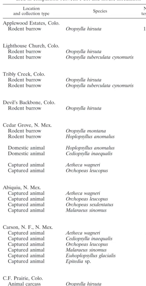
Flea samples.For comparison of mouse inoculation and PCR assays, 381 fleas of 12 different species were collected at various locations in Colorado and New Mexico from rodent burrows, domestic animals, captured animals, or animal carcasses (Table 1). Following collection, fleas were stored individually at270°C or in pools in 2% saline held at 4°C. Epizootic plague activity was identified or suspected at each of the locations where fleas were collected. Fleas for the sensitivity assays were from a colony ofOropsylla montanawhich was derived from fleas originally collected in Bernalillo County, New Mexico, in 1992.
Preparation of fleas for PCR and mouse inoculation.Fleas for comparison of PCR and mouse inoculation assays were prepared by triturating individual fleas in 100ml of BHI broth in 1.5-ml microcentrifuge tubes with sterile sea sand and disposable pestles. Fleas pooled and stored in saline at the time of collection were individually washed in sterile saline before trituration. Fifty microliters of the triturated flea-BHI infusion was pipetted into a separate 1.5-ml microcen-trifuge tube to which 350ml of sterile 0.85% (physiological) saline was added for mouse inoculation. The remaining 50 ml of the ground flea suspension was heated to 95°C for 10 min and then immediately centrifuged for 10 s at maximum speed (15,6003g) to pellet flea tissue and sand. The supernatants were assayed byplaPCR within 10 min. Leftover supernatants were stored at270°C.
Fleas used for testing the sensitivity of the PCR assay were triturated in six separate tubes containing 100ml of BHI broth spiked with 106, 105, 104, 103, 102,
and 101CFU ofY. pestisCO96-3188. The triturated flea material was then * Corresponding author. Mailing address: DVBID/CDC, PHS, U.S.
Dept. of Health & Human Services, P. O. Box 2087, Fort Collins, CO 80522. Phone: (970) 221-6450. Fax: (970) 221-6476. E-mail: klg0@cdc .gov.
1980
on May 15, 2020 by guest
http://jcm.asm.org/
heated and pelleted as described above. Pools of 0, 1, 5, 10, 20, and 25 fleas were used per sample.
PCR amplification targets.Primer sequences were the same as those de-scribed by Hinnebusch and Schwan (7) and were derived from the published sequence data for the plasminogen activator gene (pla) (16). These primers, Yp1 (59-ATCTTACTTTCCGTGAGAA-39) and Yp2 (59-CTTGGATGTTGAGCTT CCTA-39), correspond to nucleotides 971 to 990 and 1431 to 1450, respectively, and produce a 478-bp amplification product. All PCR tests with theplaprimers will hereafter be referred to asplaPCR assays. A confirmatory PCR assay used primers targeting thecaf1 gene ofY. pestis, as described by Chu et al. (2). These primers correspond to nucleotides 1 to 18 and 496 to 513 of thecaf1 gene, which encodes the structural region of the F1 antigen (6), and produce a 513-bp amplification product. All PCR tests with thecaf1 primers will hereafter be referred to ascaf1 PCR assays.
PCR.TheplaPCR protocol used in this study is a modification of the protocol described by Hinnebusch and Schwan (7). Briefly, for each PCR assay, 2.5ml of the flea-BHI preparation was combined with 0.25ml of each primer (Yp1 and Yp2; 30 pmol of primer/ml), 50 mM MgCl2, and 21.75ml of deionized, distilled
water in a 0.65-ml tube containing a Ready-To-Go PCR bead (Amersham Phar-macia Biotech, Piscataway, N.J.). The PCR bead contained 1.5 U ofTaqDNA
polymerase, 10 mM Tris HCl, 1.5 mM MgCl2, 200mM (each) deoxynucleoside
triphosphate (dNTP) and stabilizers, including bovine serum albumin. Positive controls were prepared by adding 2.5ml ofY. pestisA1122 to the above reagents, and negative controls were prepared by adding 2.5ml of sterile BHI to the above reagents. Two drops of sterile mineral oil were then added to the tubes before they were sealed and placed in a thermocycler (Minicycler; MJ Research, Wa-tertown, Mass.) with the following amplification program: initial denaturation at 95°C for 5 min, followed by 35 cycles of denaturing at 95°C for 1 min, annealing at 51°C for 1 min, and primer extension at 72°C for 2 min. After the last cycle, primer extension was continued at 72°C for 10 min. The preparation of thecaf1 PCR samples for the confirmatory PCR, withcaf1 primers, was the same as described above, but the denaturation, annealing, and extension steps of the thermocycler program were done according to Chu et al. (2). Samples were analyzed by electrophoresis on 2% agarose gels, according to standard methods (12).
Mouse inoculation and sample collection.Three hundred microliters of each 400-ml flea-saline suspension was inoculated subcutaneously in the lower abdo-men of individual white laboratory (Swiss Webster) mice (one mouse/flea sus-pension). Following inoculation, mice were labeled and held in cages and sup-plied with food and water ad libitum. Mice were checked twice daily for signs of morbidity or mortality. Mice that died within 21 days after inoculation were stored at220°C prior to a postmortem examination, which included macroscopic observations of the appearance of the lymph nodes, liver, and spleen, along with collection of portions of these organs or nodes for bacterial examination. Sur-viving mice were held for 21 days before tests were canceled. Postmortem tissue samples were taken from all mice that were inoculated withplaPCR-positive fleas and survived to day 21. Blood samples also were collected from these mice and from a random sampling of 30 mice inoculated with PCR-negative fleas. All experiments with mice were done according to a protocol approved by the Division of Vector Borne Infectious Disease’s Animal Care and Use Committee (AUC no. 97-09-008-AM).
PHA.The passive hemagglutination assay (PHA) was used to examine mouse serum samples for anti-F1 antibodies. Samples with titers greater than 1:10 were considered to be positive. This methodology, which is described elsewhere (18), was selected because it is the standard serological assay used by the World Health Organization Collaborating Center on Plague at the Centers for Disease Control and Prevention.
FA testing.Impression smears of mouse tissues were analyzed by fluorescent-antibody (FA) assays as a presumptive test for the presence ofY. pestiscells. The FA tests were conducted as described elsewhere (3).
Bacterial culture.To obtain bacterial isolates from mouse tissue, small pieces of spleen, liver, and lymph node tissues were streaked on sheep blood (6%) agar plates. To obtain bacterial isolates from flea suspensions, 50ml of the flea-saline suspension was also streaked on blood agar plates. All plates were incubated at 37°C for at least 48 h and examined for characteristic colonial morphology consistent withY. pestisgrowth. Confirmation of identity of the isolate was by a positive FA test, biochemical profiles, and by evidence of lysis withY. pestis -specific bacteriophage at both 25°C and 37°C (3). Bacteriophage lysis assays were done at both temperatures to eliminate false-positive reactions with Yersinia pseudotuberculosis, which can occur at 37°C but not at 25°C.
RESULTS
Sensitivity and pooling.Our modification of the plaPCR assay appeared to have sensitivity comparable to that of the original assay described by Hinnebusch and Schwan (7) and was able to detect as few as 10 CFU per sample, as determined by testing 10-fold serial dilutions ofY. pestissuspended in BHI broth containing ground, uninfected flea material (data not shown). The sensitivity of this assay did not noticeably change when up to 20 fleas were suspended in each serial dilution. Consistent results were not obtained, however, when 25 fleas per pool were used.
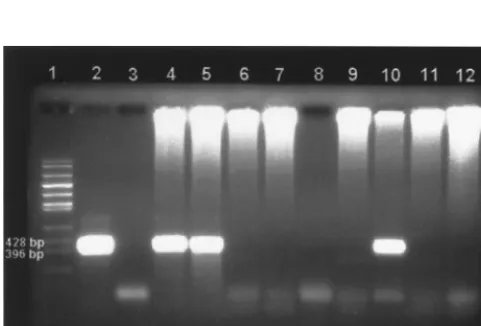
PCR-mouse inoculation comparison. Of the 381 field-col-lected flea samples tested bypla PCR, 60 were positive (i.e., contained a predicted 478-bp fragment that appeared identical in size to the one observed in the positive controls) (Table 2). The plaPCR-positive specimens included 28 of 208 (13.5%) Oropsylla hirsutaspecimens and 32 of 58 (55.2%)O. tubercu-lata cynomurisspecimens tested. None of the other flea species in our study wereplaPCR positive. Figure 1 shows an example of the pla PCR assay results for flea samples collected in northern Colorado.
[image:2.612.51.293.90.564.2]Of the 60plaPCR-positive flea suspensions, 48 caused death in mice; all dead mice were positive by FA forY. pestis infec-tion. This resulted in 80% agreement between PCR and mouse TABLE 1. Location, collection type, species, and number of fleas
used in comparison between PCR and mouse inoculation
Location
and collection type Species testedNo.
Applewood Estates, Colo.
Rodent burrow Oropsylla hirsuta 113
Lighthouse Church, Colo.
Rodent burrow Oropsylla hirsuta 27
Rodent burrow Oropsylla tuberculata cynomuris 21
Tribly Creek, Colo.
Rodent burrow Oropsylla hirsuta 29
Rodent burrow Oropsylla tuberculata cynomuris 7
Devil’s Backbone, Colo.
Rodent burrow Oropsylla hirsuta 43
Cedar Grove, N. Mex.
Rodent burrow Oropsylla montana 24
Rodent burrow Hoplopsyllus anomalus 6
Domestic animal Hoplopsyllus anomalus 1 Domestic animal Cediopsylla inaequalis 4
Captured animal Aetheca wagneri 1
Captured animal Orchopeas leucopus 1
Abiquiu, N. Mex.
Captured animal Aetheca wagneri 2
Captured animal Orchopeas leucopus 2
Captured animal Orchopeas sexdentatus 2
Captured animal Malaraeus sinomus 1
Carson, N. F., N. Mex.
Captured animal Aetheca wagneri 51
Captured animal Cediopsylla inaequalis 4
Captured animal Orchopeas leucopus 3
Captured animal Malaraeus sinomus 1
Captured animal Euhoplopsyllus glacialis 1
Captured animal Epitediasp. 1
C.F. Prairie, Colo.
Animal carcass Oropsylla hirsuta 6
Animal carcass Oropsylla tuberculata cynomuris 30
on May 15, 2020 by guest
http://jcm.asm.org/
inoculation when death was used as an endpoint for the assay. Of the 12 mice that survived inoculation withplaPCR-positive flea suspensions, 10 had positive PHA serology titers (range, 1:32 to 1:8,192) at the end of the 21-day test period. When killed on day 21, 5 of these 10 mice also were determined by FA assay to haveY. pestisin the liver, spleen, or lymph nodes. Additionally,Y. pestiswas isolated from the tissues of four of these five FA-positive mice. Overall, inoculation of mice with 58 of the 60 pla PCR-positive flea suspensions produced a positive FA assay, PHA serology, or Y. pestis isolate from mouse tissues (96.7% agreement). Both of thepla PCR-posi-tive flea suspensions that did not produce a detectable infec-tion in mice were positive when retested bycaf1 PCR.Y. pestis also was isolated directly from one of these two flea suspen-sions. All PCR-negative flea suspensions were negative by mouse inoculation (100% agreement) when death of mouse was used as the endpoint. Additionally, each of 30 randomly selected serum samples from mice inoculated withpla PCR-negative flea suspensions was PCR-negative by PHA.
DISCUSSION
We have shown that our modification of theplaPCR assay has many advantages over mouse inoculation for identifyingY. pestisin fleas, including its sensitivity and rapidity. Hinnebusch and Schwan (7) demonstrated that theplaprimers used in our study were suitable for amplifying target DNA fromY. pestis strains collected in Asia, Africa, and the Americas, suggesting
that PCR assays based on these primers should have wide applicability for surveillance purposes and epidemiological in-vestigations. These authors also indicated that theplaprimers did not produce false positives when tested with samples ofY. pseudotuberculosis,Yersinia enterocolitica, andRickettsia typhi. The translated coding region of theplagene has some degree of homology with theompTgene ofEscherichia coli(16), but the pla primers used in this study target the 39 noncoding region of the plasminogen activator gene. A comparison of the plaprimer sequences used in this study showed no homology with the ompT gene sequence (GenBank accession no. X06903).
The sensitivity of theplaPCR assay (10 to 100 CFU ofY. pestis) did not appear to diminish noticeably when assays were done with pools of as many as 20 fleas, which is only slightly less than the maximum of 25 fleas per pool usually recom-mended for the mouse inoculation assay (15). Testing to date has indicated that ourplaPCR results are less reliable when the pool size is increased to 25 fleas. Although positives were occasionally obtained with samples that contained 25 fleas and were spiked with as few as 10 CFU, the 100ml of BHI ap-peared to be insufficient to suspend that amount of flea mate-rial. This difficulty in suspending flea material may partially explain the inconsistent results. Consistent results may be ob-tainable by further modifying the procedures used to prepare the suspensions of flea material, but this type of modification was not evaluated during our study.
Our results suggest that the mouse inoculation assay can produce false-negative results when death is used as an end-point, as indicated by the fact that only 80% of the mice that were inoculated withpla PCR-positive flea suspensions actu-ally died. Among the 12 mice that survived inoculation withpla PCR-positive flea suspensions, 10 were later demonstrated by serology or bacterial isolation to have been infected withY. pestis. These 10 flea suspensions might have contained insuf-ficientY. pestisto produce a fatal infection in mice or might have contained bacteria of lower virulence than typical wild-type strains. Regardless of why some mice failed to die when inoculated withY. pestis-infected flea suspensions, pla PCR reliably detectedY. pestisin these samples. The results ofpla PCR and mouse inoculation, however, agreed closely (96.7%) when all mice, including those that survived to 21 days, were analyzed for evidence ofY. pestisinfection. It is important that all the PCR-positive fleas were collected either from prairie dog burrows in areas of suspected plague activity or from a dead prairie dog known to have died from plague. AlthoughY. pestiscan be isolated occasionally from fleas collected from apparently healthy animals, all such fleas examined during our study were PCR negative.
[image:3.612.55.552.84.174.2]Additional testing of the twoplaPCR-positive mouse inoc-ulation-negative flea suspensions clearly demonstrated or strongly suggested that these suspensions did indeed containY. pestis. Both flea suspensions were also positive bycaf1 PCR.
[image:3.612.54.295.499.662.2]FIG. 1. Ethidium bromide-stained agarose gel showingplaPCR results for samples of individualO. hirsutacollected from Applewood Estates, Colo. Large bands represent a 478-bp fragment amplified from theY. pestis plagene. Lane 1, molecular size marker; lane 2, positive control (A1122); lanes 3 and 8, negative controls; lanes 4 to 7 and 9 to 12, flea samples (4, 5, and 10 are positive; 6, 7, 9, 11, and 12 are negative). Note: The high-molecular-weight DNA at the top of the gel is probably flea DNA in that it appears only in the lanes containing PCR samples from flea suspensions.
TABLE 2. Collection type, test results, and percent agreement between PCR and mouse inoculation assays
Collection type No. offleas tested
No. of PCR-positive
fleas (%)
No. of PCR-positive fleas that caused death in mice
(% agreement)
No. of PCR-positive fleas that caused infection in mice
(% agreement)
Rodent burrow 270 24 (8.8) 20 (83.3) 24 (100.0)
Domestic animal 5 0 0 0
Captured animal 70 0 0 0
Animal carcass 36 36 (100.0) 28 (77.8) 34 (94.4)
Total 381 60 (15.7) 48 (80.0) 58 (96.7)
on May 15, 2020 by guest
http://jcm.asm.org/
Although culturingY. pestisdirectly from flea material is often ineffective because of contamination (15), we were able to isolateY. pestis from one of the pla and caf1 PCR-positive, mouse inoculation-negative flea suspensions, providing clear proof that this flea was infected withY. pestis. We were unable to isolate Y. pestis from the remaining pla and caf1 PCR-positive flea suspension. Although, as with all PCR assays, the positive plaand caf1 PCR results obtained from the culture-negative sample could have resulted from an external source of contamination, it is possible that this flea suspension contained dead bacteria with sufficient amounts of intactY. pestisDNA to be amplified by PCR. However, it is unlikely that the positive result for this last flea suspension was due to amplicon con-tamination, because we obtained positive results with two dif-ferent primer sets (pla and caf1), including one (caf1) not previously used in our laboratory.
In addition to standard precautions taken to minimize pos-sible sources of contamination in PCR assays (10), we took further steps to ensure sample quality. For example, field-collected fleas are often stored under various conditions, in-cluding on dry ice (270°C), in 2% saline with Tween 80 de-tergent, or in 70% ethyl alcohol. Prior to this study, we found that the 2% saline-Tween 80 solutions used to store infected fleas occasionally tested positive for Y. pestis by PCR and mouse inoculation (data not shown). Contamination of the 2% saline, therefore, is likely to result in the contamination of other fleas stored in the same container. These findings led us to wash fleas stored in saline solutions in fresh, sterile, 2% saline before triturating these samples for PCR, which ap-peared to eliminate the false-positive results occasionally ob-tained with unwashed flea material. Other methods, however, may prove superior for removing foreign DNA from the out-side of fleas stored under this condition. Obviously, fleas stored in 70% alcohol cannot be tested by mouse inoculation but are suitable for PCR. It seems reasonable that fleas stored in alcohol also should be rinsed thoroughly prior to analysis by PCR to minimize cross-contamination.
Storage of fleas in alcohol is likely to be advantageous under certain circumstances, including those situations in which fleas are to be held without freezing for long periods (more than a few days) prior to analysis by PCR. We obtained good results when fleas were held dry on dry ice and at 270°C in an ultra-low freezer. Immediate freezing of samples reduces han-dling of the specimens, thereby reducing the risk of contami-nation. Our modified PCR protocol provided a simpler assay that required fewer steps to complete than the one described initially by Hinnebusch and Schwan (7). The PCR beads used with the mineral oil appeared to result in an assay whose sensitivity was comparable to that of these authors’ “hot start” method. When commercially prepared PCR beads are used, the likelihood of PCR reagent contamination is minimized, and the potential for contamination is further reduced because the number of times the tubes need to be opened is minimized. Sample preparation times also were decreased, by decreasing the number of preparation steps, without a noticeable change in performance.
On average, the sample preparation, PCR amplification, and electrophoresis of reaction products take about 4 to 5 h to complete. This is much faster than the mouse inoculation as-say, which usually requires a minimum of 3 days to identify a positive sample and 21 days to confirm a negative one, not including the additional time required to perform serological or cultural assays on serum samples or tissues, respectively. We have not calculated a cost comparison, but the price of PCR reagents is likely to be offset by the additional person-hours needed to care for the mice and to conduct the bacteriological
and serological testing involved with mouse inoculation assays. More importantly, the rapid turnaround time for samples en-sures that surveillance data and results from epidemiological investigations will be available within hours after samples are received. This information can be invaluable in determining what control and prevention methods should be implemented to reduce risks posed by individual rodent and flea species found within the affected area.
Although this PCR assay should be adequate for most sur-veillance purposes requiring rapid identification ofY. pestisin fleas, it cannot substitute for bacterial culture methods when additional data are needed, such as those pertaining to viru-lence, plasmid profile analysis, or antibiotic susceptibility. Be-cause the PCR assay is unable to distinguish betweenY. pestis strains with various degrees of virulence or antibiotic suscep-tibility, we recommend that representative samples of the trit-urated flea material be kept at270°C for future analysis.
Our results show that the death of inoculated mice is a less reliable indicator than PCR of the presence ofY. pestisin flea samples. The results of the modified pla PCR assay and the mouse inoculation assay were similar only when all surviving mice were tested for evidence ofY. pestisinfection at the end of the 21-day assay period. This is important, because under the current protocols, surviving mice are rarely tested after 21 days because of time and personnel constraints. It should be noted, however, that even follow-up testing of the surviving mice in our study failed to identify twoY. pestis-infected flea pools. In conclusion, the rapidity of theplaPCR test (;4 h), along with its sensitivity, makes it a suitable alternative to mouse inoculation assay for identifying infected fleas during surveillance activities and outbreak investigations.
ACKNOWLEDGMENTS
We thank Pam Reynolds and Ted Brown of the New Mexico Divi-sion of Health and Richard Grossman of the Larimer County Health Department (Colo.) for their assistance in collecting flea samples. We also thank Brook M. Yockey, Zenda L. Berrada, and Todd S. Deppe for their assistance in performing diagnostic serology tests and culture identification and William Black and three anonymous reviewers for their comments on the manuscript.
REFERENCES
1.Campbell, J., J. Lowe, S. Walz, and J. Ezzel. 1993. Rapid and specific identification ofYersinia pestisby using a nested polymerase chain reaction procedure. J. Clin. Microbiol.31:758–759.
2.Chu, M. C., J. E. Bracher, K. J. Moss, and B. M. Yockey.1998. Unpublished data.
3.Chu, M. C., B. A. Wilmoth, L. G. Carter, and T. A. Quan.Laboratory manual of plague diagnostic tests, in press. World Health Organization, Geneva, Switzerland.
4.Englesberg, E., T. H. Chen, J. B. Levy, L. E. Foster, and K. F. Meyer.1954. Virulence inPasteurella pestis. Science119:413–414.
5.Gage, K. L.1998. Plague, p. 885–903.InL. Collier (ed.), Topley and Wilson’s microbiology and microbial infections, 9th ed. Oxford University Press, Inc., New York, N.Y.
6.Galyov, E. E., O. Y. Smirnov, A. V. Karlishev, K. I. Volkovoy, A. I. Denesyuk, I. V. Nazimov, K. S. Rubtsov, V. M. Abramov, S. M. Dalvadyanz, and V. P. Zavyalov.1990. Nucleotide sequence of theYersinia pestisgene encoding F1 antigen and the primary structure of the protein. FEBS Lett.277:230–232. 7.Hinnebusch, J., and T. G. Schwan.1993. New method for plague surveillance
using polymerase chain reaction to detectYersinia pestisin fleas. J. Clin. Microbiol.31:1511–1514.
8.Hudson, B. W., L. Kartman, and F. M. Prince.1966.Pasteurella pestis detection in fleas by fluorescent antibody staining. Bull. W. H. O.34:709– 714.
9.Kartman, L., F. M. Prince, S. F. Quan, and H. E. Stark.1958. New knowl-edge on the ecology of sylvatic plague. Ann. N. Y. Acad. Sci.70:668–711. 10. Kwok, S.1990. Procedures to minimize PCR-product carry-over, p. 142–145.
InM. A. Innis, D. G. Gelfand, J. J. Sninsky, and T. J. White (ed.), PCR protocols: a guide to methods and applications. Academic Press, Inc., San Diego, Calif.
11. Leal, N. C., F. G. C. Abath, L. C. Alves, and A. M. P. de Almedia.1996. A
on May 15, 2020 by guest
http://jcm.asm.org/
simple PCR-based procedure for plague diagnosis. Rev. Inst. Med. Trop. Sao Paulo38:371–373.
12.Maniatis, T., E. F. Fritsch, and J. Sambrook.1982. Molecular cloning: a laboratory manual. Cold Spring Harbor Laboratory, Cold Spring Harbor, N.Y.
13.McDonough, K. A., T. G. Schwan, R. E. Thomas, and S. Falkow.1988. Identification of aYersinia pestis-specific DNA probe with potential for use in plague surveillance. J. Clin. Microbiol.25:2515–2519.
14.Poland, J. D., and A. M. Barnes.1979. Plague, p. 515–597.InJ. H. Steele (ed.), CRC handbook series in zoonoses. CRC Press, Inc., Boca Raton, Fla. 15.Quan, T. J., A. M. Barnes, and J. D. Polland.1981. Yersiniosis, p. 723–745.
InA. Barlows and W. J. Hausler, Jr. (ed.), Diagnostic procedures for bac-terial, mycotic and parasitic infections. 6th ed. APHA, Inc., Washington, D.C. 16.Sodeinde, O. A., and J. D. Goguen.1989. Nucleotide sequence of the plas-minogen activator gene ofYersinia pestis: relationship toompTofEscherichia coliand geneEofSalmonella typhimurium. Infect. Immun.57:1517–1523. 17.Thomas, R. E., K. A. McDonough, and T. G. Schwan.1989. Use of DNA
hybridization probes for detection of the plague bacillus (Yersinia pestis) in fleas (Siphonoptera: Pulicidae and Ceratophyillidae). J. Med. Entomol.26:
342–348.
18. World Health Organization.1970. Passive hemagglutination test. WHO Tech. Rep. Ser.447:23–25.