Copyright © 2002, American Society for Microbiology. All Rights Reserved.
Development of a Multilocus Sequence Typing Scheme for the Pig
Pathogen
Streptococcus suis
: Identification of Virulent Clones
and Potential Capsular Serotype Exchange
Samantha J. King,
1James A. Leigh,
2Peter J. Heath,
3Inmaculada Luque,
4Carmen Tarradas,
4Christopher G. Dowson,
1and Adrian M. Whatmore
1*
Infectious Disease Research Group, Department of Biological Sciences, University of Warwick, Coventry CV4 7AL,1
Institute for Animal Health, Compton, Newbury RG20 7NN,2and Veterinary Laboratories Agency, Rougham
Hill, Bury Saint Edmunds IP33 2RX,3United Kingdom, and Departamento Patolgía Infecciosa,
Campus Universitario de Rabanales, 14071 Co´rdoba, Spain4
Received 14 January 2002/Returned for modification 9 June 2002/Accepted 23 July 2002
Streptococcus suisis an important pathogen of pigs and occasionally causes serious human disease. However, little is known about theS. suispopulation structure, the clonal relationships between strains, the potential of particular clones to cause disease, and the relevance of serotype as a marker for epidemiology. Here we describe a multilocus sequence typing (MLST) scheme forS. suisdeveloped in order to begin to address these issues. Seven housekeeping gene fragments from each of 294S. suisisolates obtained from variousS. suisdiseases and from asymptomatic carriage representing 28 serotypes and nine distinct countries of origin were sequenced. Between 32 and 46 alleles per locus were identified, giving the ability to distinguish >1.6ⴛ1011sequence types
(STs). However only 92 STs were identified in this study. Of the 92 STs 18 contained multiple isolates, the most common of which, ST1, was identified on 141 occasions from six countries. Assignment of the STs to lineages resulted in 37 being identified as unique and unrelated STs while the remaining 55 were assigned to 10 complexes. ST complexes ST1, ST27, and ST87 dominate the population; while the ST1 complex was strongly associated with isolates from septicemia, meningitis, and arthritis, the ST87 and ST27 complexes were found to contain significantly higher numbers of lung isolates. In agreement with the observed distribution of disease-causing isolates of S. suis, most isolates previously characterized as of high virulence in porcine infection models belong to ST1, while isolates belonging to other STs appear to be less virulent in general. Finally nine STs were found to contain isolates of multiple serotypes, and many isolates belonging to the same serotypes were found to have very disparate genetic backgrounds. As well as highlighting that the serotype can often be a poor indicator of genetic relatedness betweenS. suisisolates, these findings suggest that capsular genes may be moving horizontally through theS. suispopulation.
Streptococcus suisis an important pathogen associated with
a range of diseases in pigs, including pneumonia, meningitis, septicemia, and arthritis, although the organism is also fre-quently carried asymptomatically.S. suishas substantial impli-cations for the swine industry both in terms of animal welfare concerns and economic considerations and can cause serious zoonotic infection of humans, where it has been associated with septicemia, meningitis, and endocarditis (2, 46). The pre-vention and control ofS. suisinfection of swine by vaccine and treatment with antimicrobials are hampered by increasing an-tibiotic resistance and poor understanding of the biology, cru-cial virulence factors, and protective antigens of the organism (46).
There are currently 35 serotypes ofS. suisrecognized based on the immunogenicity of capsular antigens (18, 19, 20, 24, 35). Although the majority of disease is caused by a small number of serotypes, it is recognized that the capsular serotype is a poor marker of virulence. Virulence can vary substantially both within and among serotypes, and not all isolates of the same
serotype cause the same disease (46). Serotype 2 is considered the most virulent and the most frequently isolated from dis-ease. However, the importance of particular serotypes can vary geographically. For example, some 70% of isolates from por-cine disease in France belong to serotype 2 (4), and this sero-type was recently reported to be the most frequently isolated in countries such as Italy and Spain (49, 54). In contrast serotype 9 is reportedly particularly common in Belgium and Holland (54) and previously Australia (16), whereas serotypes 1 and 14, in addition to 2, are common in the United Kingdom (22, 54). The prevalence of serotypes also varies temporally. In Canada over the last 7 years the percentage of serotype 2 strains iso-lated from diseased animals decreased from 22 to 15% (23). Similarly, the importance of serotype 14, which was a major invasive pathogen in the United Kingdom, though apparently not elsewhere, in the middle-to-late 1990s (22), has declined recently.
Previous studies have indicated thatS. suisis a genetically diverse species (3, 21, 31, 34, 42) although many involved small numbers of isolates, focused on isolates representing a single serotype or geographical location, and used a variety of tradi-tional approaches such that it is difficult to compare results among studies. In spite of acknowledged difficulties in the categorization ofS. suisisolates as virulent or avirulent (see * Corresponding author. Present address: Department of Bacterial
Diseases, Veterinary Laboratories Agency, Addlestone KT15 3NB, United Kingdom. Phone: 44 1932 357311. Fax: 44 1932 357423. E-mail: [email protected].
3671
on May 15, 2020 by guest
http://jcm.asm.org/
reference 17 for a discussion), there is considerable evidence that virulent isolates are genetically distinct from avirulent isolates, suggesting a clonal association with virulence in S. suis. Staats et al. (47) found that most serotype 2 isolates from septicemia represented a single ribotype, whereas less-virulent isolates were genetically heterogeneous. Similarly Smith et al. (42) found that virulent isolates of serotypes 1 and 2 had distinct ribotype profiles, while Rasmussen et al. (37) demon-strated that particular ribotype profiles were clearly associated with clinical-pathology observations. A recent study of 99 strains by macrorestriction of DNA revealed four major clusters, one of which was strongly associated with invasive disease, and indicated that isolates from pigs with meningitis and septicemia showed a significantly higher degree of genetic homogeneity than isolates from pigs with pneumonia and healthy pigs (1).
Many basic questions about the population biology ofS. suis
and the nature, origin, and spread of virulent clones could be answered if isolates of this species could be characterized un-ambiguously. AsS. suisis responsible for a range of diseases and can be carried asymptomatically, a study of the population structure of this organism could reveal clones or clonal groups with an apparently increased capacity to cause disease or an increased association with particular clinical manifestations of
S. suis. As well as helping understand the epidemiology ofS.
suisinfection and the biological relevance of the current sero-typing approach, such work could facilitate the rapid identifi-cation of potentially virulent strains within herds, which could then be treated prophylactically. Multilocus sequence typing (MLST) is a highly discriminatory and unambiguous method of characterizing bacterial isolates that has now been successfully employed in the characterization of several species (10, 11, 12, 13, 29). MLST is based on the nucleotide sequences of internal fragments of housekeeping genes, in which mutations are as-sumed to be largely neutral (40). For each gene fragment the different nucleotide sequences are assigned allele numbers and the sequence type (ST) of each isolate is defined by the alleles present at each of seven distinct loci. Isolates that share the same ST are assumed to be members of the same clone, that is, they have a recent common ancestor. Due to the high numbers of alleles at each of the seven loci it is highly unlikely that isolates will have the same profile by chance. An important advantage of MLST is that sequence data are portable and can be readily compared among laboratories. In addition, the data obtained can be used to address questions about the evolution-ary and population biology of bacterial species (14, 45).
Here we describe the development of an MLST scheme for
S. suis. The scheme is based on the nucleotide sequences of
seven housekeeping gene fragments and was used to evaluate 294 isolates from diverse geographical backgrounds. The iso-lates were derived from both asymptomatic carriage and from a range of disease states and represent some 28 different se-rotypes. The relationship of ST with serotype, currently the standard epidemiological marker forS. suis, is described, and additionally significant differences in the distributions of strains isolated from different disease states among clonal com-plexes are discussed.
MATERIALS AND METHODS
Bacterial isolates. A total of 301S. suisisolates were used in this study. Reference strains of serotypes 1 and 2 through 34 were supplied by L. A. Devriese (Faculty of Veterinary Medicine, University of Ghent, Ghent,
Bel-gium), M. Gottschalk (Faculte´ de Me´dicine Ve´te´rinaire, Universite´ de Montre´al, Montreal, Canada), and P. Heath (Veterinary Laboratories Agency, Bury Saint Edmunds, United Kingdom) (7). Twenty-one well-characterized serotype 2 iso-lates, including many isolates characterized in previous virulence studies, and 11 serotype 14 isolates from cases of meningitis were supplied by P. Norton (Insti-tute for Animal Health, Newbury, United Kingdom). One hundred thirty-six field isolates, obtained by the Veterinary Laboratories Agency from diverse geographical sources across England, were included in this study. These were selected to represent isolates from diverse serotypes, sites of isolation, and clinical backgrounds, including invasive-disease isolates (meningitis, septicemia, and arthritis) and lung isolates from cases of pneumonia or other respiratory problems. One further isolate from the Warwick strain collection obtained from a case of porcine septicemia in the United Kingdom was included. A similar sample of 81 Spanish field isolates obtained by C. Tarradas and I. Luque was included in this study. The sample consisted of 38 “carried” isolates from the tonsils of healthy pigs and 43 clinical isolates, again with diverse clinical back-grounds (27). Two porcine isolates, one previously described as atypical (26), provided by C. Lammler, Institut fu¨r Tierarztliche, Giessen, Germany, were included. Fifteen isolates obtained fromS. suisdisease of humans were included in addition to the serotype 14 type strain, which was also originally a human isolate. These isolates were obtained from Augustine Cheng (Department of Microbiology, Faculty of Medicine, The Chinese University of Hong Kong, Hong Kong; six isolates), M. Gottschalk (three isolates), G. Grise (Centre Hosptialier d’Elbeuf, Elbeuf, France; three isolates), C. Lammler (one isolate), P. Heath (one isolate), and B. Francois, Hospital Universitaire Dupuytren, Limoges, France (one isolate; B. Francois, V. Gissot, M. C. Ploy, and P. Vignon, Letter, J. Clin. Microbiol.36:2395, 1998).
Preparation of chromosomal DNA.Chromosomal DNA was prepared from all isolates as described previously (52).
Identification of housekeeping loci used for MLST.Seven housekeeping gene loci were used in this study. The sequences ofcpn60, encoding a 60-kDa chap-eronin (6),dpr, a putative peroxide resistance protein (32), andrecA, which encodes homologous recombination factor (48), were obtained from sequence databases. The sequences ofaroA(encoding 5-enolpyruvylshikimate 3-phos-phate synthase) andthrA(encoding aspartokinase/homoserine dehydrogenase) were kindly provided by A. Allen (University of Cambridge, Cambridge, United Kingdom). Finally the sequences forgki(encoding glucose kinase) andmutS
(encoding a DNA mismatch repair enzyme) were obtained by us following PCR amplification and sequencing using primers designed to amplify the correspond-ing genes from other streptococci.
Amplification and nucleotide sequence determination.PCR was performed under standard conditions with 30 cycles of 95°C for 1 min,X°C for 1 min, and 72°C for 1 min per kilobase of predicted product size, whereX°C represents an annealing temperature appropriate for the particular primer set used. Products were visualized by agarose gel electrophoresis on 1.0% agarose in the presence of 1g of ethidium bromide ml⫺1. Details of all oligonucleotides used in this study are given in Table 1. PCR products were purified by passage through QiaQuick PCR product purification columns (Qiagen) and directly sequenced from both ends with the Beckman CEQ2000 system according to the manufac-turer’s instructions. Sequences were analyzed with the DNASTAR software, and in-frame internal fragments of the genes were selected for use in the MLST scheme.
Allele and ST assignment.For each locus distinct allele sequences were as-signed arbitrary allele numbers with no weighting given to the degree of se-quence divergence among alleles. For each isolate, the alleles at each of the seven loci defined the allelic profile or ST. The STs were assigned arbitrary numbers in order of description. STs were grouped into lineages or clonal complexes with the program BURST written and developed by E. Feil and located in the START, version 1.05, package of programs developed by K. Jolley (http://outbreak.ceid.ox.ac.uk/software.htm). The members of a lineage were defined as groups of two or more independent isolates that shared identical alleles at five or more loci. Each lineage was named after the ST identified as a putative ancestral type by BURST followed by “complex.” If no ancestral type was identified, the lineage was named after the STs contained within the clonal complex, for example, the ST-2/55 complex.
Confirmation of serotype.The vast majority of isolates had been serotyped prior to receipt. However, in cases where an ST contained multiple serotypes, the serotypes were confirmed, where possible, by a serotype-specific PCR assay developed by Smith et al. (43, 44), which allows identification of the most common serotypes, 1 (plus 14), 2 (plus 1/2), 7, and 9.
Computational analyses.The degree of clonality within the data set was estimated by calculating the index of association (IA) and its significance for all STs and for a subset of STs representative of each clonal complex by using a
on May 15, 2020 by guest
http://jcm.asm.org/
program written by Keith Jolley (http://www.mlst.net/indexassoc/indexass-oc.htm). The determination of the number of nucleotide polymorphic sites, the calculation ofdN/dS(wheredNrepresents nonsynonymous base substitutions and
dSrepresents synonymous base substitutions), and the construction of the
den-drogram by using UPGMA (unweighted pair group method with arithmetic mean) were performed by using START (http://outbreak.ceid.ox.ac.uk/soft-ware.htm). The number of amino acid alterations, the maximum percent nucle-otide divergence, and the average percent nuclenucle-otide divergence among alleles at a given locus were calculated by using the MEGA package, version 1.0 (S. Kumar, K. Tamura, and M. Nei, MEGA—molecular evolutionary genetics anal-ysis 1.01, Pennsylvania State University, University Park, Pa., 1993). The test of Sawyer (39) was applied to the synonymous polymorphic sites within the alleles at each locus, and the significance of any clustering of polymorphic sites was evaluated by using 10,000 resamplings of the data.
RESULTS
Development of an MLST scheme forS. suis.Chromosomal DNA was obtained from the 301 isolates, and the seven house-keeping gene loci from 294 isolates including the reference strains of 27 serotypes (serotypes 1 to 19, 23 to 25, and 27 through 31) were amplified. One or more housekeeping genes from the type strains of seven serotypes (20 through 22, 26, and 32 through 34) could not be amplified, and thus these serotypes were not examined further in this study. For all the remaining 294 isolates the sequences of the seven loci were determined and allelic profiles were assigned. The alleles defined for the MLST scheme were based on sequences of between 318
(cpn60) and 354 (recA) nucleotides, and between 32 (thrA) and
46 (gki) alleles per locus were present. The proportion of variable nucleotide sites present in the selected housekeeping genes ranged from 11.0 (thrA) to 29.0% (gki) (Table 2), while the number of polymorphic amino acid sites ranged from 3.4
(recA) to 17.9% (dpr). The proportions of nucleotide alter-ations that changed the amino acid sequence (dN) and the
proportions of silent changes (dS) were calculated for each
gene. From these data thedN/dSratios for all seven loci were
calculated, and all were substantially less than 1 (Table 2). For the 294S. suisisolates, the mean number of alleles per locus was 40, providing the theoretical potential to distinguish⬎1.6
⫻1011different genotypes.
Relatedness ofS. suisisolates.Figure 1 shows a dendrogram constructed from the matrix of pairwise allelic differences be-tween the STs of all 294 isolates. The isolates resolved into 92 STs, 74 of which (80.4%) were identified only once. The most common ST, ST1, was identified 141 times in the data set, while 17 other STs contained between 2 and 13 members. Assignment of STs to lineages with BURST revealed that 37 were both unique and unrelated to any others, while the re-maining 55 were assigned to 10 lineages (Table 3). The ST1 complex was the largest and contained 165 isolates represent-ing 14 STs. The ST27 complex included 49 members, the ST87 complex comprised 19 isolates, and the ST61 complex con-tained 4 isolates. The remaining six lineages concon-tained two member STs (Table 3).
Evidence of recombination. The extent of recombination within theS. suispopulation was assessed by determining the IA(30). The IAfor the complete data set was 4.874, but a value
[image:3.587.44.549.84.183.2]of 2.09 was obtained on reduction of the data set to a single representative of each ST. When randomized data sets (1,000 trials) were used, the latter value was significantly greater than zero, which would be the expected value for a population at linkage equilibrium (i.e., freely recombining). However, in TABLE 1. Primers used for amplification and sequencing of the seven loci included in theS. suisMLST scheme
Locus Sequence
Forward (5⬘–3⬘) Reverse (5⬘–3⬘)
dpr CGTCTTTCAGCCCGCGTCCA GACCAAGTTCTGCCTGCAGC
thrAa GATTCAGAACGTCGCTTTGT AAGTTTTCATAGAGGTCAGC
cpn60 TTGAAAAACGTRACKGCAGGTGC ACGTTGAAIGTACCACGAATC
recA TATGATGAGTCAGGCCATG CGCTTAGCATTTTCAGAACC
gki GGAGCCTATAACCTCAACTGG AAGAACGATGTAGGCAGGATT
aroA TTCCATGTGCTTGAGTCGCTA ACGTGACCTACCTCCGTTGAC
mutS CGCAGAGCAGATGGAAGATCC CCCATAGCTGTTTTGGTTTCATC
aThe primer 5⬘-AAGAATGGATCATCAACCGT-3⬘was used for the forwardthrAsequencing reaction.
TABLE 2. Characteristics of housekeeping gene loci included in theS. suisMLST scheme
Locus Putative function ofgene product sequencedSize of fragment (bp)
No. of alleles identified
No. of polymorphic
nucleotide sites (%)
No. of polymorphic
amino acid sites (%)
Nucleotide divergence between pairs of
alleles (%) dN/dS GenBank accession No.
Maximum Avg
aroA EPSP synthase 366 39 54 (14.8) 20 (16.4) 16 (4.4) 7.5 0.0812 AJ491619–AJ491657
cpn60 60-kDa chaperonin 318 43 79 (24.8) 8 (7.6) 39 (12.2) 21.1 0.0063 AJ491576–AJ491618
dpr Peroxide resistance 336 37 61 (18.2) 20 (17.9) 24 (7.1) 9.6 0.0957 AJ491539–AJ491575
gki Glucose kinase 321 46 93 (29.0) 9 (8.4) 54 (16.8) 20.4 0.0174 AJ491493–AJ491538
mutS DNA mismatch repair enzyme 339 42 89 (26.3) 12 (10.6) 41 (12.1) 18.5 0.0153 AJ491451–AJ491492
recA Homologous recombination
factor 354 41 53 (15.0) 4 (3.4) 32 (9.0) 10.9 0.0147 AJ491373–AJ491413
thrA Aspartokinase/homoserine
dehydrogenase 336 32 37 (11.0) 11 (9.8) 10 (3.0) 4.4 0.0715 AJ491341–AJ491372
on May 15, 2020 by guest
http://jcm.asm.org/
[image:3.587.43.547.587.730.2]populations in which recombination is sufficient to randomize alleles at different loci over a long time period, the recent expansion of clones can result in the appearance of multiple isolates with similar genotypes. Such a sampling bias is a par-ticular problem in a population such as that described here, which is likely to be dominated by virulent clones which have risen to high frequency. To address this issue, the IA was
recalculated by using one member of each lineage (47 STs). For this sample the IAwas reduced to 0.230 and no significant
linkage disequilibrium among alleles was observed. In support of the suggestion of the importance of recombination in the long-term evolution ofS. suis, there was evidence of recombi-nation within thecpn60,gki, andmutSgenes, with the test of Sawyer (39) showing a highly nonrandom distribution of syn-onymous polymorphic sites (P聿0.0001 [sum of the squares of the condensed fragment lengths statistic]). There was no sig-nificant clustering of polymorphisms within the other gene fragments.
Relationship between STs and serotypes.Of the 18 STs that contain more than one isolate 9 contained multiple serotypes (Table 3); the dendrogram (Fig. 2), which is reduced to show-ing only one member of each ST, highlights these 9 STs. When possible the serotypes of selected isolates were confirmed by PCR using specific primers (43, 44). Where large numbers of strains representing each different serotype were present in an individual ST (and thus there was little chance of the observed distribution reflecting serotyping errors), three representatives were selected for confirmatory PCR. Three of these STs con-tain serotypes which may, on the basis of the occurrence of some cross-reactivity in diagnostic serotyping, be closely re-lated such as 2 and 1/2 or 1 and 14 (17). Thus these potential serotype exchanges should be treated with caution until more is known about the genetic relationships between the capsular loci of these serotypes. However, the remaining six STs contain serotypes with unambiguous serological responses which are not thought to be closely related to any other serotype.
[image:4.587.75.253.72.390.2]In the converse situation many serotypes were found to be associated with multiple STs. Thus, as illustrated in Fig. 2, serotype 2 isolates are represented in 16 of the 92 STs, sero-type 9 isolates are represented in 7 distinct STs, and serosero-type 7 isolates are represented in 5 distinct STs. Again amplification with serotype-specific primers was used to confirm the wide distribution of serotypes 1 (plus 14), 2 (plus 1/2), 7, and 9. Hence, although many isolates of the same serotypes are closely related, these data clearly highlight that the serotype can be a poor marker of genetic relatedness ofS. suisisolates. Relationships among ST, disease state, and country of iso-lation.The most frequently isolated ST, ST1, was identified in six countries and hence appears to be globally distributed (Ta-ble 3). Of the other 17 STs that include multiple isolates 6 were also found to contain strains from more than one country. Isolates belonging to the five STs containing five or more isolates originated from a diversity of sources. Strains isolated from different clinical states are widely distributed throughout the dendrogram; however, there is some evidence that isolates from different clone complexes have differing propensities to cause particular disease states. Thus an examination of Fig. 1 shows that the ST1 complex contains a substantially higher proportion of isolates from meningitis, septicemia, and arthri-tis than the ST27 and ST87 complexes. Chi-square tests com-paring the numbers of invasive isolates with the numbers of other isolates in particular complexes indicate that the ST1 complex contains a significantly higher number of isolates from invasive disease than the ST87 (P⬍0.01) and ST27 (P⬍0.01) complexes. Both the ST87 (P⬍0.01) and ST27 (P⬍0.0001) complexes contain a significantly higher number of isolates from lung disease than the ST1 complex. The correlation be-tween lung isolates and two individual STs appears even stron-ger. Five of the 11 ST29 isolates were isolated from the lung in cases of porcine pneumonia (an additional 2 of the 11 were also lung isolates, one from a case of septicemia and one from a sudden death, while the remaining 4 isolates include 1 from healthy carriage, 1 brain isolate, 1 from an unknown disease, and 1 unknown). Similarly all three ST65 isolates were from the lung. Carried isolates are widely distributed throughout the dendrogram and are represented in 22 of 92 STs. Sixteen isolates from human invasive disease were included in the study and were found to represent four STs, although the FIG. 1. Dendrogram of 294 isolates ofS. suisisolates constructed
by UPMGA cluster analysis. Ninety-two STs were identified and three major clonal complexes were defined by using the program BURST. The members of a lineage were defined as groups of two or more independent isolates that had identical alleles at five or more loci. Percentages of isolates from the three major complexes isolated from invasive disease (meningitis, septicemia, and arthritis), pneumonia and other respiratory disease, porcine carriage, human disease, and other states (including reference strains previously used in virulence studies and isolates of unknown origin and those from other disease states) are indicated.
on May 15, 2020 by guest
http://jcm.asm.org/
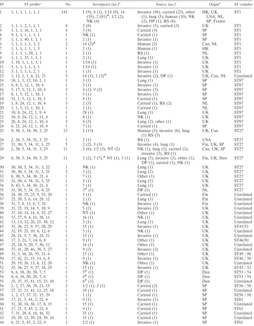
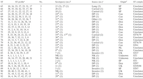
TABLE 3. Characteristics of isolates belonging to the 92 STs identified in this study
ST ST profilea No. Serotype(s) (no.)b Source (no.)c Origind ST complex
1 1, 1, 1, 1, 1, 1, 1 141 1 (9), 8 (1), 1/14 (9), 14 (35), 2 (81)#, 1/2 (2), NK (4)
Invasive (94), carried (22), other (1), lung (5), human (10), NK (2), DP (1), RS (6)
HK, UK, USA, NL, SP, France
ST1
2 1, 1, 1, 2, 1, 1, 1 8 2 (8) Invasive (5), carried (3) UK ST1
3 1, 1, 1, 16, 1, 1, 1 4 3 (4) Carried (4) SP ST1
4 9, 1, 1, 1, 1, 1, 1 1 NK (1) Carried (1) SP ST1
5 1, 1, 1, 40, 1, 1, 1 1 2 (1) Invasive (1) SP ST1
6 1, 1, 1, 1, 1, 1, 2 2 14 (2)# Human (2) Can, NL ST1
7 1, 1, 1, 1, 1, 1, 3 1 2 (1) Human (1) HK ST1
8 1, 1, 1, 1, 28, 1, 1 1 2 (1) RS (1) NL ST1
9 1, 1, 1, 35, 1, 1, 1 1 2 (1) Lung (1) UK ST1
10 1, 10, 1, 1, 1, 1, 1 1 1/14 (1) Invasive (1) UK ST1
11 3, 1, 1, 1, 1, 1, 1 1 1/14 (1) Invasive (1) UK ST1
12 3, 1, 1, 1, 1, 2, 1 1 1 (1) Invasive (1) UK ST1
13 1, 12, 1, 1, 6, 21, 21 3 14 (1), 1 (2)# Invasive (2), DP (1) UK, Can, NL Unrelated
14 18, 1, 5, 12, 10, 1, 1 1 3 (1) Lung (1) SP ST87
15 8, 8, 5, 12, 1, 10, 4 1 3 (1) Invasive (1) SP ST87
16 5, 17, 5, 12, 1, 10, 4 3 4 (1), 9 (2) Invasive (3) SP ST87
17 8, 1, 5, 12, 1, 10, 1 1 5 (1) Invasive (1) SP ST87
18 31, 1, 5, 12, 1, 10, 1 1 8 (1) Carried (1) SP ST87
19 1, 8, 24, 12, 1, 10, 4 3 2 (3) Carried (1), RS (2) NL ST87
20 1, 1, 5, 12, 1, 10, 1 1 2 (1) Carried (1) NL ST87
21 18, 8, 24, 12, 1, 9, 4 1 28 (1) Lung (1) UK ST87
22 18, 8, 24, 12, 1, 11, 4 1 8 (1) NK (1) UK ST87
23 26, 8, 24, 12, 1, 10, 4 3 4 (3) Lung (2), other (1) UK ST87
24 6, 21, 24, 12, 1, 10, 4 1 7 (1) Carried (1) SP ST87
25 9, 30, 5, 34, 30, 3, 25 13 2 (13) Human (3), invasive (6), lung
(1) RS (3) UK, Can ST27
26 2, 26, 5, 34, 31, 3, 25 1 2 (1) RS (1) USA ST27
27 31, 30, 5, 34, 31, 3, 25 5 2 (2), 3 (3) Invasive (4), lung (1) Fin, UK, SP ST27 28 2, 30, 5, 34, 31, 3, 25 11 2 (6), 1/2 (3), NT (2) NK (1), lung (2), carried (2),
invasive (5), RS (1) Can, UK, SP ST27 29 8, 30, 5, 34, 30, 3, 25 11 2 (2), 7 (7),#NT (1), 3 (1) Lung (5), invasive (2), other (1),
DP (1), carried (1), NK (1) Fin, UK, Den ST27
30 30, 30, 5, 34, 31, 3, 22 1 NK (1) Lung (1) UK ST27
31 30, 30, 5, 34, 31, 3, 25 2 3 (2) Lung (2) UK ST27
32 8, 30, 5, 34, 30, 21, 4 1 7 (1) Other (1) UK ST27
33 31, 30, 4, 34, 31, 3, 25 1 3 (1) Lung (1) UK ST27
34 8, 43, 5, 34, 30, 21, 4 1 7 (1) Lung (1) UK ST27
35 31, 30, 5, 34, 31, 4, 25 1 3#(1) DP (1) NL ST27
36 24, 30, 35, 25, 9, 33, 20 1 3 (1) Carried (1) Fin Unrelated
37 25, 39, 5, 6, 14, 29, 12 1 2 (1) Lung (1) Fin Unrelated
38 31, 7, 8, 13, 6, 7, 24 1 NK (1) Invasive (1) Fin Unrelated
39 21, 25, 19, 14, 5, 10, 9 2 5 (2) Invasive (2) UK Unrelated
40 37, 10, 14, 14, 8, 32, 27 1 NT (1) Other (1) UK Unrelated
41 15, 27, 9, 4, 16, 26, 11 1 16 (1) NK (1) UK Unrelated
42 13, 13, 32, 28, 25, 38, 12 1 3 (1) Lung (1) UK Unrelated
43 35, 36, 27, 9, 37, 28, 29 1 15 (1) Invasive (1) UK ST43/52
44 32, 19, 25, 10, 6, 12, 6 1 5 (1) NK (1) UK Unrelated
45 28, 31, 5, 7, 36, 24, 31 1 15 (1) Invasive (1) UK Unrelated
46 17, 3, 21, 7, 14, 6, 8 1 9 (1) Other (1) UK ST46/50
47 25, 24, 9, 29, 7, 30, 12 1 16 (1) Other (1) UK Unrelated
48 37, 41, 20, 46, 38, 5, 27 2 9 (2) Invasive (2) UK Unrelated
49 31, 3, 16, 26, 35, 21, 6 1 15 (1) Other (1) UK ST49 / 88
50 17, 42, 21, 15, 14, 6, 8 1 9 (1) Invasive (1) UK ST46 / 50
51 29, 19, 26, 13, 6, 12, 20 1 NK (1) Other (1) UK Unrelated
52 35, 36, 27, 9, 37, 18, 29 1 15 (1) Invasive (1) UK ST43 / 52
53 8, 6, 18, 20, 28, 7, 5 1 5#(1) DP (1) Den ST53 / 54
54 8, 6, 18, 20, 28, 7, 4 1 4#(1) DP (1) Den ST53 / 54
55 25, 37, 15, 41, 15, 23, 10 1 6#(1) DP (1) Den Unrelated
56 1, 2, 17, 36, 29, 21, 15 2 1/2 (1), 2 (1) Carried (2) SP ST56 / 58
57 27, 22, 11, 43, 12, 25, 18 1 10 (1) Carried (1) SP Unrelated
58 1, 2, 17, 37, 21, 15, 29 1 1 (1) Carried (1) SP ST56 / 58
59 17, 21, 5, 44, 2, 22, 4 1 9 (1) Invasive (1) SP ST61
60 31, 30, 18, 38, 17, 8, 15 1 15 (1) Carried (1) SP Unrelated
61 17, 21, 5, 45, 2, 22, 4 1 9 (1) Carried (1) SP ST61
62 7, 31, 28, 8, 18, 16, 32 1 21 (1) Carried (1) SP Unrelated
63 19, 29, 12, 39, 24, 39, 16 1 15 (1) Carried (1) SP Unrelated
64 8, 21, 5, 45, 2, 22, 4 1 1/2 (1) Invasive (1) SP ST61
Continued on following page
on May 15, 2020 by guest
http://jcm.asm.org/
majority belong to ST1. Two of these four STs, ST1 and ST25, also contain a large number of isolates from various porcine disease states.
MLST findings in relation to isolates previously character-ized in virulence studies.Fourteen reference isolates included in this study had previously been included in studies ofS. suis
virulence using porcine infection models (Table 4) (33). The virulence of these strains had been determined by one of three different methodologies, either intravenously (i.v.) (33, 36) or by using the method of Vecht et al. (50) or the method of Galina et al. (15). The methods of both Vecht et al. (50) and Galina et al. (15) use intranasal inoculation ofS. suisfollowing inoculation withBordetella bronchisepticaor porcine reproduc-tive and respiratory syndrome virus, respecreproduc-tively. Six of the 14 isolates were representatives of ST1, and all 6 were defined as highly virulent in whichever model they were tested including both i.v. (3 isolates) and intranasal challenge (3 isolates). A further five isolates were members of the ST27 complex. Three ST25 isolates included isolates defined as of high, intermedi-ate, or low virulence by i.v. inoculation, while two other mem-bers of the ST27 complex, an ST26 isolate and an ST28 isolate, were described as of low virulence in intranasal and i.v. chal-lenges, respectively. ST19, represented by two isolates and a member of the ST87 complex, was associated only with strains of intermediate and low virulence as determined by intranasal challenge. Thus correlation of ST and observed virulence il-lustrates a trend suggesting that ST1 isolates are highly virulent
and that isolates of other common STs, such as ST19 and ST25, may be somewhat less capable of causing invasive disease.
DISCUSSION
The primary objective of this work was to increase under-standing of the population structure ofS. suisby developing an unambiguous typing scheme which could then be expanded by other investigators and in turn to use this as a framework to help understand the differential virulence ofS. suis isolates. Such a framework can be used as a basis on which the distri-bution and alleles of virulence genes ofS. suiscan be super-imposed. Some of these genes, such as the suilysin gene, are already known to be absent from manyS. suisisolates (25), and studies may identify genes (or alleles of genes) of particular importance inS. suispathogenesis, which will in turn aid stud-ies aimed at developing more-effective vaccines and therapeu-tics againstS. suis. Previous studies ofS. suisgenetic diversity have largely included strains of only one serotype or isolates from a single country and have generally applied typing meth-ods of low resolution that pose problems of reproducibility among laboratories. This study, however, uses two large col-lections of strains from the United Kingdom and Spain and includes smaller numbers of isolates from seven other coun-tries. In addition this study utilizes MLST, which affords high discrimination, reproducibility, and potential accessibility of data over the Internet. MLST has already been used success-TABLE 3—Continued
ST ST profilea No. Serotype(s) (no.)b Source (no.)c Origind ST complex
65 10, 16, 23, 27, 23, 31, 17 3 15 (2), 27 (1) Lung (3) SP Unrelated
66 38, 35, 10, 42, 33, 17, 30 1 28 (1) Carried (1) SP Unrelated
67 27, 15, 11, 22, 12, 27, 11 1 10 (1) Carried (1) SP Unrelated
68 14, 20, 31, 27, 27, 34, 18 1 24#(1) DP (1) Can Unrelated
69 23, 33, 22, 30, 3, 15, 12 1 25#(1) DP (1) Can Unrelated
70 38, 28, 30, 15, 32, 26, 7 1 31#(1) Other (1) Can Unrelated
71 11, 5, 13, 11, 26, 36, 14 1 13#(1) DP (1) Den Unrelated
72 12, 9, 33, 3, 19, 35, 19 1 27#(1) DP (1) Can Unrelated
73 33, 37, 2, 19, 13, 5, 12 1 16#(1) DP (1) Den Unrelated
74 31, 2, 6, 31, 10, 14, 6 1 12#(1) DP (1) Den Unrelated
75 25, 32, 9, 15, 9, 13, 8 1 28#(1) DP (1) Can Unrelated
76 8, 23, 36, 24, 21, 22, 12 2 19#(1), 17#(1) Carried (2) Can ST76/79
77 39, 38, 7, 32, 11, 3, 8 1 30#(1) DP (1) Can Unrelated
78 23, 11, 5, 7, 22, 8, 20 1 10#(1) DP (1) Den Unrelated
79 8, 21, 36, 45, 21, 12, 22 1 18#(1) Carried (1) Can ST76/79
80 8, 21, 3, 45, 2, 22, 23 1 23#(1) DP (1) Can ST61
81 22, 1, 20, 21, 4, 21, 26 1 15#(1) DP (1) NL Unrelated
82 25, 34, 29, 5, 34, 19, 28 1 9#(1) DP (1) Den Unrelated
83 8, 30, 5, 34, 39, 3, 25 1 7 (1) Invasive (1) UK ST27
84 4, 1, 1, 1, 1, 1, 1 1 NK (1) NK (1) Unknown ST1
85 34, 14, 37, 33, 20, 40, 21 1 NK (1) NK (1) Unknown Unrelated
86 1, 1, 1, 1, 1, 1, 25 1 2 (1) NK (1) SP ST1
87 18, 8, 24, 12, 1, 10, 4 1 8#(1) DP (1) Den ST87
88 36, 3, 16, 18, 35, 21, 6 1 11 (1) DP (1) UK ST49/88
89 8, 8, 24, 12, 1, 10, 4 1 3 (1) Invasive (1) SP ST87
90 20, 40, 5, 23, 40, 41, 1 1 NK (1) Invasive (1) UK Unrelated
91 31, 18, 5, 12, 41, 10, 18 1 11#(1) DP (1) Den Unrelated
92 16, 4, 34, 17, 42, 37, 13 1 29#(1) DP (1) Can Unrelated
aAllele numbers for each gene, presented in the following order:aroA, cpn60, dpr, gki, mutS, recA, thrA. b#, reference strain of a particular serotype. (7), represented by this ST. NK, not known; NT, not typeable.
cInvasive isolates were obtained from cases of septicemia, meningitis, or arthritis. Lung isolates were obtained from cases of bronchopneumonia or other
uncomplicated respiratory disease. NK, not known; DP, isolated from a diseased pig with no further details known; RS, reference isolate (14, which have been characterized previously in porcine models of infection, were included; see Table 4).
dSP, Spain; HK, Hong Kong; UK, United Kingdom; Can, Canada; NL, The Netherlands; Fin, Finland; Den, Denmark; USA, United States.
on May 15, 2020 by guest
http://jcm.asm.org/
[image:6.587.41.539.85.359.2]fully in the characterization of other bacteria and has been validated against other molecular typing methodologies (10, 11, 12, 13, 29).
To facilitate this study, seven loci that could be amplified and sequenced from a wide range of S. suis isolates were chosen. The seven loci were not subject to positive selection, as demonstrated by the dN/dS ratio for each locus, which was
substantially less than 1 (adN/dSratio⬎1 implies selection for
amino acid change). For the 294 strains included in this study the number of alleles identified per locus was on average 40, which is consistent with previous phenotypic and genotypic analyses suggesting thatS. suis represents a diverse species. The difficulty in amplifying all seven loci from some of the type
strains is likely a reflection of this diversity. Type strains of seven serotypes (20 to 22, 26, and 32 to 34) were excluded from the study due to unsuccessful amplification of at least one housekeeping gene in each case. Six of these seven type strains have previously been shown to be more distantly related sero-types on the basis of 16S rDNA andcpn60sequencing (6, 7). This study supports the argument that at least some of these serotypes may represent separate species (6). PCR amplifica-tion from the remaining type strain (serotype 21) was unsuc-cessful for a single locus, presumably due to divergence within this allele The IA, a measure of the extent of recombination for
the complete data set, was 4.874, but this was reduced to 0.230 if one representative of each lineage was included. This value was not significantly different from the IAof zero expected for
a population at linkage equilibrium. This implies that, although the observed population structure is clearly biased by the re-peated recovery of recently arisen highly successful clones, in the longer term there is little clonal framework within theS. suis population. In support of this there was evidence of a history of horizontal gene transfer identified incpn60, mutS, andgkiby Sawyer’s test (39).
In spite of the observed genetic diversity three major clonal groups dominate theS. suispopulation examined in this study. Most striking was the ST1 complex, containing 165 isolates, within which 141 isolates were found to represent ST1 itself. Isolates belonging to ST1 originated from six countries includ-ing several European nations, the United States, and Hong Kong, and a high proportion were associated with the classicS. suis invasive diseases, septicemia, meningitis, and arthritis. Such a high occurrence of one ST could suggest that the MLST scheme described here does not have a high power of discrim-ination. However, the mean number of alleles identified per locus was 40, providing the potential to distinguish⬎1.6⫻1011
[image:7.587.49.282.73.507.2]different genotypes. Hence a more likely explanation is that ST1 isolates represent a highly successful clone which arose relatively recently and which has rapidly spread worldwide. Repeated recovery of such indistinguishable isolates from in-vasive disease in different countries clearly implies that STs, such as ST1, define strains with an increased capacity to cause FIG. 2. Distribution of serotypes relative to ST. The dendrogram
shown in Fig. 1 is reduced to show only one member of each ST. STs representing isolates of multiple serotypes are boxed (see Table 3).F, distribution of serotype 9;⽧, distribution of serotype 7; arrows, dis-tribution of serotype 2. STs belonging to the three major clonal com-plexes are bracketed.
TABLE 4. MLST analysis of isolates previously examined in porcine models of infection
Strain Virulence ST (ST complex)
H11/1 Highc 1 (1)
D282 Higha 1 (1)
P1/7 Highc 1 (1)
3881 Higha 1 (1)
87555 Highb 1 (1)
B831 Highc 1 (1)
1591 Highd 25 (27)
B554 Intermediatec 25 (27)
DH5 Lowb 26 (27)
TD-10 Lowc, d 25 (27)
0891 Lowd 28 (27)
3921 Intermediatea 19 (87)
3898 Lowa 19 (87)
3912 Lowa 8 (1)
aVirulence determined by the intranasal method of Vecht et al. (50). bVirulence determined by the intranasal method of Galina et al. (15). cVirulence determined by i.v. inoculation (33).
dVirulence determined by i.v. inoculation (36).
on May 15, 2020 by guest
http://jcm.asm.org/
[image:7.587.296.542.92.243.2]disease. This might reflect a variety of factors such as increased fitness, the possession of certain virulence factors or allelic variants thereof, and the selection of clones possessing partic-ular antibiotic resistance profiles by therapeutic or prophylac-tic use. Two other major complexes were apparent, the ST27 and ST87 complexes, although no individual ST was as domi-nant as ST1 within these complexes. Although isolates from septicemia and meningitis fall within these complexes, there appears to be a significantly higher proportion of lung isolates in the ST27 and ST87 complexes than in the ST1 complex. This observation could suggest that someS. suisstrains may have a propensity to cause pneumonia and others, such as members of ST1, may be better equipped to cause the classicS. suis inva-sive diseases, septicemia, meningitis, and arthritis. However, as
S. suisis often isolated from the lungs of pigs with pneumonia
along with other agents potentially involved in respiratory dis-ease (such asPasteurella multocida,Haemophilus parasuis,
Ac-tinobacillus pleuropneumoniae, swine influenza virus,B.
bron-chiseptica, andMycoplasmaspp. [22, 28, 38]), the role ofS. suis
in pneumonia remains somewhat controversial. Although S. suisis isolated in pure culture from pneumonia with reasonable frequency (1, 22) further epidemiological studies are required to address the etiological role ofS. suisin porcine pneumonia and the particular clones involved.
To help understand the relationships between clonal groups and virulence, 14S. suis isolates which had previously been included in virulence studies using porcine models were in-cluded in this study (Table 4). Six of these isolates were found to belong to ST1, and, in agreement with the suggestion above that ST1 isolates are of high virulence potential, all were highly virulent in the models irrespective of whether infection was via the intranasal or the i.v. route. Five of the isolates were found to belong to the ST27 complex, members of which, on the basis of the epidemiological observations already discussed, may have a lower potential to cause classic invasive diseases such as septicemia and meningitis than members of the ST1 complex. The five ST27 complex isolates have been reported to have various virulence potentials (Table 4), with three members classified as of low virulence while two others were classified as of intermediate or high virulence. However the two studies that produced these results used i.v. inoculation, which by-passes the normal route of colonization and invasion via the nasopharyngeal membranes and the palatine tonsils (8, 53) and thus may not be an accurate indication of the potential to cause disease in the field. Two other isolates belonged to ST19, a member of the ST87 complex that again appears to contain isolates less strongly associated with invasive disease and that were described as being of low or intermediate virulence in an intranasal model of infection. Note that several discrepancies in the literature with regard to the virulence of individual isolates have previously been identified and that there have been conflicting findings in experimental infection models. Thus, for example, strain DH5 has been variously reported to be of high or low virulence (for a discussion see reference 25 and the letter from M. Gottschalk, R. Higgins, and S. Quessy [J. Clin. Microbiol.37:4202-4203, 1999]). Conflicting data may reflect the age and immunological status of the animals used and/or the inoculum and the system used to score clinical symptoms and highlight the need for international agreement on a virulence model and virulent and avirulent control strains
(17). However, in spite of these caveats, there appears to be some correlation between the observed virulence in experi-mental infection models and the observed distribution of dis-ease-associated strains in this MLST study.
Previous studies using multilocus enzyme electrophoresis, DNA restriction endonuclease analysis, and ribotyping have demonstrated similarities between strains ofS. suisfrom hu-mans and pigs (3, 21, 31). In this study 16 human isolates were included; 10 belong to ST1, which, as already discussed, con-tains a large number of isolates from various porcine disease states. Of the remaining six, three are representatives of two STs containing only human isolates, which form part of the ST1 complex, while the remaining three belong to ST25, which is a member of the ST27 complex. The three human ST25 isolates were from Canada; this may reflect the prevailing clonal groups present there although further investigation of porcine isolates from Canada is required to confirm this. Sim-ilarly it is likely that the isolation of ST1 from humans, at least in Europe, reflects the prevalence of strains belonging to this ST in porcine invasive disease. There is therefore no convinc-ing evidence forS. suisclones with an increased propensity to infect humans, and it seems that strains causing human infec-tion may reflect the dominant clones in the local pig popula-tion.
Isolates of many of the most common serotypes were dis-tributed widely across the dendrogram. Thus, for example, serotype 2 was found to be present in 16 of the 92 STs, and in the converse situation a number of STs which contain isolates of multiple serotypes were identified. Considerable genetic diversity between strains of the same serotype has previously been reported, and these observations make the use of sero-typing as a means of strain identification for epidemiological studies unreliable. While many of these STs are closely related and therefore most likely descended from a recent common ancestor, the same serotype is seen in isolates which differ at all seven loci. One implication of these observations is that cap-sular genes, which specify serotype, may be spread horizontally through the population. Horizontal spread of capsular genes has been demonstrated in Streptococcus pneumoniae (9), where, as in S. suis, capsular genes which are conserved be-tween serotypes flank variable serotype-specific loci encoding antigenic specificity (43) and recombination between the con-served regions flanking the serotype-specific genes results in serotype exchange. ForS. pneumoniae the proposed mecha-nism involves natural transformation although, asS. suisis not known to be naturally transformable, the mechanism here re-mains unknown. However, similar horizontal movement of sur-face markers such as the M protein is known to occur in other non-naturally transformable streptococci such asStreptococcus
pyogenes(51). Isolates of serotype 2 are the most widely
dis-tributed across the dendrogram. This may simply reflect the higher number of serotype 2 isolates included in the study. Alternatively, as any serotype exchange presumably occurs during the colonization of the host with multiple serotypes as demonstrated previously (5, 41) and as serotype 2 is among the most frequently isolated serotypes (1, 54), it may be that these strains simply come into contact with strains of other serotypes more frequently. It is interesting to speculate that the recent epidemiology of S. suis in the United Kingdom may reflect such a serotype exchange event. As mentioned previously
on May 15, 2020 by guest
http://jcm.asm.org/
rotype 14 became increasingly associated with the types of invasive disease normally associated with serotype 2 S. suis
during the 1990s. Virtually all serotype 14 isolates character-ized in this study belonged to ST1, which, as already described, contains a large number of virulent isolates, the majority of which belong to serotype 2. Thus it is possible that selective pressures imposed by increasing immunity to the dominant serotype 2 may have resulted in the emergence of a virulent variant which has retained the highly successful ST1 genotype but which has acquired a type 14 capsular locus.
In conclusion, we have devised the first unambiguous typing system forS. suis.Three major complexes were identified. The dominant ST1 complex represented 165 isolates and was strongly associated with invasive disease (septicemia, meningi-tis, and arthritis), while members of the smaller ST27 and ST87 complexes appear less strongly associated with these diseases but may be associated with respiratory disease. The wide dis-tribution of serotypes throughout the dendrogram and the identification of nine STs containing multiple serotypes suggest that the capsular genes may be moving horizontally through the population. It is hoped that this MLST framework will now be expanded to include isolates from other countries. It will be of particular interest to examine North American isolates, as it is hypothesized that serotype 2 isolates there have virulence potentials and virulence factors different from those of Euro-pean isolates (17). In addition future studies will aim to further the understanding of S. suis pathogenesis by examining the distribution and allelic diversity of potential virulence deter-minants ofS. suisrelative to the MLST framework.
ACKNOWLEDGMENTS
We gratefully acknowledge all of the colleagues listed in Materials and Methods, who provided isolates for inclusion in this study and sequence data prior to publication.
This work was supported by a project grant from the BBSRC (grant reference 88/S11598), and A.M.W. was supported by a Wellcome Trust Research Fellowship in Biodiversity (grant reference 053589).
REFERENCES
1.Allgaier, A., R. Goethe, H. J. Wisselink, H. Smith, and P. Valentin-Weigand.
2001. Relatedness ofStreptococcus suisof various serotypes and clinical backgrounds as evaluated by macrorestriction analysis and expression of potential virulence traits. J. Clin. Microbiol.39:445–453.
2.Arends, J. P., and H. C. Zanen.1988. Meningitis caused byStreptococcus suis
in humans. Rev. Infect. Dis.10:131–137.
3.Beaudoin, M., J. Harel, R. Higgins, M. Gottschalk, M. Frenette, and J. I. MacInnes.1992. Molecular analysis of isolates ofStreptococcus suiscapsular type 2 by restriction-endonuclease-digested DNA separated on SDS-PAGE and by hybridisation with an rDNA probe. J. Gen. Microbiol.138:2639–2645. 4.Berthelot-Herault, F., H. Morvan, A. Keribin, M. Gottschalk, and M. Ko-bisch.2000. Production of muraminidase-released protein (MRP), extracel-lular factor (EF) and suilysin by field isolates ofStreptococcus suiscapsular types 2, 1/2, 9,7 and 3 isolated from swine in France. Vet. Res.31:473–479. 5.Brisebois, L. M., R. Charlebois, R. Higgins, and M. Nadeau.1989. Preva-lence ofStreptococcus suisin four to eight week old clinically healthy piglets. Can. J. Vet. Res.54:174–177.
6.Brousseau, R., J. E. Hill, G. Prefontaine, S. Goh, J. Harel, and S. Hemming-sen.2001.Streptococcus suisserotypes characterized by analysis of chapero-nin 60 gene sequences. Appl. Env. Microbiol.67:4828–4833.
7.Chatellier, S., M. Gottschalk, R. Higgins, R. Brousseau, and J. Harel.1999. Relatedness ofStreptococcus suisserotype 2 isolates from different geo-graphical origins as evaluated by molecular fingerprinting. J. Clin. Microbiol.
37:362–366.
8.Clifton-Hadley, F. A., and T. J. L. Alexander.1980. The carrier site and carrier rate ofStreptococcus suistype 2 in pigs. Vet. Rec.107:40–41. 9.Coffey, T. J., M. C. Enright, M. Daniels, J. K. Morona, R. Morona, W.
Hryniewicz, J. C. Paton, and B. G. Spratt.1998. Recombinational exchange at the capsular polysaccharide biosynthetic locus leads to frequent serotype changes among natural isolates ofStreptococcus pneumoniae. Mol. Micro-biol.27:73–83.
10.Dingle, K. E., F. M. Colles, D. R. A. Wareing, R. Ure, A. J. Fox, F. E. Bolton, H. J. Bootsma, R. J. L. Willems, R. Urwin, and M. C. J. Maiden.2001. Multilocus sequence typing system forCampylobacter jejuni. J. Clin. Micro-biol.39:14–23.
11.Enright, M., and B. G. Spratt.1998. A multilocus sequence typing scheme forStreptococcus pneumoniae: identification of clones associated with serious invasive disease. Microbiology144:3049–3060.
12.Enright, M. C., N. P. J. Day, C. E. Davies, S. J. Peacock, and B. G. Spratt.
2000. Multilocus sequence typing for characterization of methicillin-suscep-tible clones ofStaphylococcus aureus. J. Clin. Microbiol.38:1008–1015. 13.Enright, M. C., B. G. Spratt, A. Kalia, J. H. Cross, and D. E. Bessen.2001.
Multilocus sequence typing ofStreptococcus pyogenesand the relationships betweenemmtype and clone. Infect. Immun.69:2416–2427.
14.Feil, E. J., M. C. J. Maiden, M. Achtman, and B. G. Spratt.1999. The relative contributions of recombination and mutation to the divergence of clones ofNeisseria meningitidis. Mol. Biol. Evol.16:1496–1502.
15.Galina, L., C. Pijoan, M. Sitjar, W. T. Christianson, K. Rossow, and J. E. Collins.1994. Interaction betweenStreptococcus suisserotype 2 and porcine reproductive and respiratory syndrome virus in specific-pathogen-free pig-lets. Vet. Rec.134:60–64.
16.Gogolewski, R. P., R. W. Cook, and C. J. O’Connell.1990.Streptococcus suis
serotypes associated with disease in weaned pigs. Aust. Vet. J.67:202–204. 17.Gottschalk, M., and M. Segura.2000. The pathogenesis of the meningitis caused byStreptococcus suis: the unresolved questions. Vet. Microbiol.76:
259–272.
18.Gottschalk, M., R. Higgins, M. Jacques, K. R. Mittal, and J. Henrichsen.
1989. Description of 14 new capsular types ofStreptococcus suis. J. Clin. Microbiol.27:2633–2636.
19.Gottschalk, M., R. Higgins, M. Jacques, M. Beaudoin, and J. Henrichsen.
1991. Isolation and characterization ofStreptococcus suis capsular types 9–22. J. Vet. Diagn. Investig.3:60–65.
20.Gottschalk, M., R. Higgins, M. Jacques, M. Beaudoin, and J. Henrichsen.
1991. Characterization of six new capsular types (23 through 28) of Strepto-coccus suis. J. Clin. Microbiol.29:2590–2594.
21.Hampson, D. J., D. J. Trott, I. L. Clarke, C. G. Mwaniki, and I. D. Robert-son.1993. Population structure of Australian isolates ofStreptococcus suis. J. Clin. Microbiol.31:2895–2900.
22.Heath, P. J., and B. W. Hunt.2001.Streptococcus suisserotypes 3 to 28 associated with disease in pigs. Vet. Rec.148:207–208.
23.Higgins, R., and M. Gottschalk.2000. Distribution of theStreptococcus suis
capsular types in 1999. Can. Vet. J.41:414.
24.Higgins, R., M. Gottschalk, M. Boudreau, A. Lebrun, and J. Henrichsen.
1995. Description of six new capsular types (29–34) ofStreptococcus suis. J. Vet. Diagn. Investig.7:405–406.
25.King, S. J., P. J. Heath, I. Luque, C. Tarradas, C. G. Dowson, and A. M. Whatmore.2001. Distribution and genetic diversity of suilysin in Streptococ-cus suisisolated from different diseases of pigs and characterization of the genetic basis of suilysin absence. Infect. Immun.69:7572–7582.
26.Lammler, C., and R. Weiss.1997. Characterisation of an unusual Strepto-coccus suisisolated from an aborted fetus of a pig. Med. Sci. Res.25:263– 264.
27.Luque, I., C. Tarradas, R. Astorga, A. Perea, H. J. Wisselink, and U. Vecht.
1999. The presence of muramidase released protein and extracellular factor protein in various serotypes ofStreptococcus suisisolated from diseased and healthy pigs in Spain. Res. Vet. Sci.66:69–72.
28.Macinnes, J. I., and R. Desrosiers.1999. Agents of the “suis-ide diseases” of swineActinobacillus suis,Haemophilus suisandStreptococcus suis. Can. J. Vet. Res.63:83–89.
29.Maiden, M. C. J., J. A. Bygraves, E. Feil, G. Morelli, J. E. Russell, R. Urwin, Q. Zhang, J. Zhou, K. Zurth, D. A. Caugant, I. M. Feavers, M. Achtman, and B. G. Spratt.1998. Multilocus sequence typing: a portable approach to the identification of clones within populations of pathogenic microorganisms. Proc. Natl. Acad. Sci. USA95:3140–3145.
30.Maynard-Smith, J., N. H. Smith, M. O’Rouke, and B. G. Spratt.1993. How clonal are bacteria? Proc. Natl. Acad. Sci. USA90:4384–4388.
31.Mogollon, J. D., C. Pijoan, M. P. Murtaugh, E. L. Kaplan, J. E. Collins, and P. P. Cleary.1990. Characterization of prototype and clinically defined strains ofStreptococcus suisby genomic fingerprinting. J. Clin. Microbiol.
28:2462–2466.
32.Niven, D. F., and A. Ekins.2001. Iron content ofStreptococcus suisand evidence for adprhomologue. Can. J. Microbiol.47:412–416.
33.Norton, P. M., C. Rolph, P. N. Ward, R. W. Bentley, and J. A. Leigh.1999. Epithelial invasion and cell lysis by virulent strains ofStreptococcus suisis enhanced by the presence of suilysin. FEMS Immunol. Med. Microbiol.
26:25–35.
34.Okwumabua, O., J. Staats, and M. M. Chengappa.1995. Detection of heterogeneity in Streptococcus suisisolates by DNA restriction fragment length polymorphisms of rRNA genes (ribotyping). J. Clin. Microbiol.33:
968–972.
35.Perch, B., K. B. Pedersen, and J. Henrichsen.1983. Serology of capsulated streptococci pathogenic for pigs: six new serotypes ofStreptococcus suis. J. Clin. Microbiol.17:993–996.
on May 15, 2020 by guest
http://jcm.asm.org/
36.Quessy, S., J. D. Dubreuil, M. Caya, and R. Higgins.1995. Discrimination of virulent and avirulentStreptococcus suiscapsular type 2 isolates from differ-ent geographical origins. Infect. Immun.63:1975–1979.
37.Rasmussen, S. R., F. M. Aarestrup, N. E. Jensen, and S. E. Jorsal.1999. Associations ofStreptococcus suisserotype 2 ribotype profiles with clinical disease and antimicrobial resistance. J. Clin. Microbiol.37:404–408. 38.Reams, R. Y., D. D. Harrington, L. T. Glickman, H. L. Thacker, and T. L.
Bowersock.1996. Multiple serotypes and strains ofStreptococcus suisin naturally infected swine herds. J. Vet. Diagn. Investig.8:119–121. 39.Sawyer, S.1989. Statistical tests for detecting gene conversion. Mol. Biol.
Evol.6:526–538.
40.Selander, R. K., D. A. Caugant, H. Ochman, J. M. Musser, M. N. Gilmour, and T. S. Whittam.1986. Methods of multilocus enzyme electrophoresis for bacterial population genetics and systematics. Appl. Environ. Microbiol.
51:873–884.
41.Sihvonen, L., D. N. Kurl, and J. Salmela.1986. Infection withStreptococcus suisserotypes 1 and 2 in the same diseased pig. Acta Vet. Scand.27:626–628. 42.Smith, H. E., M. Rijnsburger, N. Stockhofe-Zurwieden, H. J. Wisselink, U. Vecht, and M. A. Smits.1997. Virulent strains ofStreptococcus suisserotype 2 and highly virulent strains ofStreptococcus suisserotype 1 can be recog-nized by a unique ribotype profile. J. Clin. Microbiol.35:1049–1053. 43.Smith, H. E., L. van Bruijnsvoort, H. Buijs, H. J. Wisselink, and M. A. Smits.
1999. Rapid PCR test forStreptococcus suisserotype 7. FEMS Microbiol. Lett.178:265–270.
44.Smith, H. E., V. Veenbergen, J. van der Velde, M. Damman, H. J. Wisselink, and M. A. Smits.1999. Thecpsgenes ofStreptococcus suisserotypes 1, 2, and 9: development of rapid serotype-specific PCR assays. J. Clin. Microbiol.
37:3146–3152.
45.Spratt, B. G.1999. Multilocus sequence typing: molecular typing of bacterial pathogens in an era of rapid DNA sequencing and the Internet. Curr. Opin. Microbiol.2:312–316.
46.Staats, J. J., I. Feder, O. Okwumabua, and M. M. Chengappa.1997. Strep-tococcus suis: past and present. Vet. Res. Commun.21:381–407. 47.Staats, J. J., B. L. Plattner, J. Nietfeld, S. Dritz, and M. M. Chengappa.1998.
Use of ribotyping and hemolysin activity to identify highly virulent Strepto-coccus suistype 2 isolates. J. Clin. Microbiol.36:15–19.
48.Takamatsu, D., M. Osaki, and T. Sekizaki.2001. Construction and charac-terization ofStreptococcus suis-Escherichia colishuttle cloning vectors. Plas-mid45:101–113.
49.Tarradas, C., C. Borge, A. Arenas, A. Maldonado, R. Astorga, A. Miranda, and I. Luque.2001. Suilysin production byStreptococcus suisisolated from diseased and healthy carrier pigs in Spain. Vet. Rec.148:183–184. 50.Vecht, U., J. P. Arends, E. J. van der Molen, and L. A. van Leengoed.1989.
Differences in virulence between two strains ofStreptococcus suistype II after experimentally induced infection of newborn germ-free pigs. Am. J. Vet. Res.50:1037–1043.
51.Whatmore, A. M., V. Kapur, D. J. Sullivan, J. M. Musser, and M. A. Kehoe.
1994. Non-congruent relationships between variation in emm gene se-quences and the population genetic structure of group A streptococci. Mol. Microbiol.14:619–631.
52.Whatmore, A. M., V. A. Barcus, and C. G. Dowson.1999. Genetic diversity of the streptococcal competence (com) gene locus. J. Bacteriol.181:3144– 3154.
53.Williams, D. M., G. H. Lawson, and A. C. Rowland.1973. Streptococcal infection in piglets: the palatine tonsils as portals of entry forStreptococcus suis. Res. Vet. Sci.15:352–362.
54.Wisselink, H. J., H. E. Smith, N. Stockhofe-Zurwieden, K. Peperkamp, and U. Vecht.2000. Distribution of capsular types and production of murami-dase-released protein (MRP) and extracellular factor (EF) ofStreptococcus suisstrains isolated from diseased pigs in seven European countries. Vet. Microbiol.74:237–248.