Total Surface Area Is Useful for Differentiating between Aggressive
and Favorable Multifocal Papillary Thyroid Carcinomas
Jung-Soo Pyo,
1Jin Hee Sohn,
1Guhyun Kang,
2Dong-Hoon Kim,
1and Jisup Yun
3Departments of 1Pathology and 3Surgery, Kangbuk Samsung Hospital, Sungkyunkwan University School of Medicine, Seoul; 2Department of Pathology, Sanggye Paik Hospital, Inje University College of Medicine, Seoul, Korea.
Received: April 11, 2014 Revised: May 22, 2014 Accepted: May 29, 2014
Corresponding author: Dr. Jin Hee Sohn, Department of Pathology,
Kangbuk Samsung Hospital,
Sungkyunkwan University School of Medicine, 29 Saemunan-ro, Jongno-gu,
Seoul 110-746, Korea.
Tel: 82-2-2001-2391, Fax: 82-2-2001-2398 E-mail: [email protected]
∙ The authors have no financial conflicts of interest.
© Copyright:
Yonsei University College of Medicine 2015
This is an Open Access article distributed under the terms of the Creative Commons Attribution Non-Commercial License (http://creativecommons.org/ licenses/by-nc/3.0) which permits unrestricted non-commercial use, distribution, and reproduction in any medium, provided the original work is properly cited.
Purpose: The purpose of the present study was to identify more useful parameters for predicting behaviors of multifocal papillary thyroid carcinoma (PTC). Materials and Methods: We investigated dominant tumor diameter (TD), total tumor diame-ter (TTD), and total surface area (TSA) in tumors from 588 patients and evaluated their usefulness as parameters for assessment of tumor behaviors in multifocal PTCs. Results: In 588 PTCs, tumor multifocality was found in 179 PTCs (30.4%). Multifocal tumors were significantly associated with extrathyroidal extension, lymph node metastasis, and higher tumor stage grouping (p<0.001, p<0.001, and p<0.001, respectively). The rates of nodal metastasis increased with greater TSA and TTD in PTCs. Multifocal papillary thyroid microcarcinomas (mPMCs) with TSA >3.14 cm2 had higher rates of nodal metastasis than mPMCs with TSA ≤3.14 cm2
(p=0.038); however, there was no significant difference between mPMCs with TTD >1.0 cm and with TTD ≤1.0 cm (p=0.325). In addition, nodal metastasis was more frequent in mPMCs with TSA >3.14 cm2 than in unifocal papillary thyroid
micro-carcinomas (uPMCs) (TD ≤1.0 cm) (p=0.002), but not overt unifocal PTCs (TD >1.0 cm) (p=0.244). Conclusion: Our results suggest that mPMCs with TSA >3.14 cm2 show more aggressive behavior than uPMCs and mPMCs with TSA ≤3.14 cm2.
TSA could be useful in distinguishing aggressive mPMCs from favorable cases.
Key Words: Papillary thyroid carcinoma, multifocal, total surface area, lymph node metastasis
INTRODUCTION
Papillary thyroid carcinoma (PTC) is the most common malignant tumor of the endocrine organs. Papillary thyroid microcarcinoma (PMC) of the thyroid is de-fined as a PTC of 1 cm or less in greatest diameter, according to the World Health
Organization classification system.1 PMCs are more common than overt PTCs
(larger than 1 cm) and have a more favorable prognosis. Nevertheless, for PMCs, unfavorable factors affecting aggressive behavior and prognosis are unclear.
Multifocal tumors may represent either multiple independent primary tumors or intraglandular dissemination from a primary tumor,2 and tumor multifocality has
examination and representative sections with normal-looking parenchyma from each lobe were histologically examined.
The multifocal tumors may represent either multiple in-dependent primary tumors or intraglandular dissemination from a primary tumor.12 In this study, regardless of origin,
multifocal tumors were defined as tumors with a distance between each tumor greater than 0.5 cm.
Definition and evaluation of TD, TTD, and TSA
In the present study, the dominant tumor was defined as the largest single tumor for multifocal PTC or the primary for unifocal PTC. In multifocal tumors, TTD and TSA were defined as the total combined diameter and the surface area of each tumor, respectively.
The shape of PTCs was mostly ovoid to round, with di-mensions taller than they were wide. We estimated tumor surface area by the following formula:13
Surface area=0.5·π·z· z+c·(arcsin (e) e
z= a+b,e= c2+z2 2 2
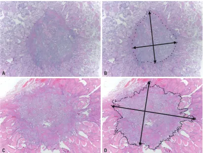
where a, b, and c are the perpendicular diameter of each tumor focus with a>b>c. In the present study, three-dimen-sional diameters were measured on gross and microscopic examination as shown in Fig. 1. PMC (TD ≤1.0 cm) entails a more favorable prognosis than overt PTC (TD >1.0 cm). In addition, previous studies have reported that PMC with TD >0.5 cm show more aggressive behaviors than PMC with TD ≤0.5 cm.14-17 Thus, in consideration of tumor
be-havior, we divided subgroups into PTCs with TD ≤0.5 cm, PTCs with 0.5 cm<TD≤1.0 cm, and PTCs with TD >1.0 cm, respectively. We assigned TD 0.5 cm to TSA 0.785 cm2
and TD 1.0 cm to TSA 3.14 cm2.
Statistical analysis
All statistical analyses were performed using SPSS soft-ware version 18.0 (SPSS Inc., Chicago, IL, USA). Correla-tion between tumor multifocality and clinicopathological parameters was determined by either the χ2 test or the
Fish-er exact test (two-sided). In addition, using TD, TTD, and TSA, the correlation between tumor multifocality and lymph node metastasis was investigated. Multivariate logistic re-gression analysis was performed to identify the most influ-ential variables associated with lymph node metastasis. The results are reported as p-values and odds ratios (ORs) with 95% confidence intervals (CIs). All results with p<0.05 were considered statistically significant.
higher rates in multifocal PMCs (mPMCs) than in unifocal PMCs (uPMCs).8,9 Based on the American Thyroid
Associa-tion management guidelines,10 hemithyroidectomy or
subto-tal thyroidectomy is generally recommended as adequate treatment for uPMCs; however, in multifocal PMCs, surgical treatment and postoperative management are controversial.
In the present study, we investigated dominant tumor di-ameter (TD), total tumor didi-ameter (TTD), and total surface area (TSA) in multifocal PTCs. We evaluated the clinico-pathological significance of tumor multifocality and the role of TSA as a new parameter for assessing tumor behav-ior in PTC.
MATERIALS AND METHODS
Patients
Five hundred and eighty-eight consecutively resected classi-cal variants of PTC at the Department of Pathology, Kang-buk Samsung Hospital, Sungkyunkwan University School of Medicine (Seoul, Korea) from January 1, 2010 to De-cember 31, 2011 were analyzed. The correlation between tumor multifocality and clinicopathological characteristics was evaluated by reviewing medical charts, pathological re-cords, and glass slides, following the 7th edition of American Joint Committee on Cancer (AJCC) tumor node metastasis (TNM) classification.11 The pathological features were
re-viewed by two pathologists. The patients’ mean age was 45.5±11.4 years (range 19‒81 years); mean tumor size was 0.98±0.61 cm; and patients had undergone either total thy-roidectomy (n=431) or lobectomy (n=157) with lymph node dissection. Central node dissection was performed in all pa-tients, and modified radical neck dissection was selectively performed in patients confirmed as having metastasis in the lateral neck lymph nodes via preoperative fine-needle aspi-ration biopsy or frozen section (n=32). In terms of patholog-ic TNM stage grouping, there were 381 cases in stage I, one in stage II, 195 in stage III, and 11 in stage IV. All 298 pa-tients under 45 years old in the present study were classified as stage I. This protocol was reviewed and approved by the Institutional Review Board of Kangbuk Samsung Hospital (approval No. KBC13145).
Definition and pathologic assessment of tumor multifocality
In our institution, sections were cut to a thickness of 0.2 cm for pathologic examination. All tumors detected during gross
(
42.3%, and 61.6% of each TSA group, respectively. Based on TTD groups, the nodal metastasis rates were 19.4%, 43.7%, and 64.9%, respectively. Nodal metastasis was found in a higher rate in the larger TSA and TTD groups than in the smaller groups (p<0.001 and p<0.001, respec-tively) (Table 2). In multivariate analysis, TSA and TTD were independent predictors of lymph node metastasis (p< 0.001, OR=1.157, 95% CI 1.089‒1.230 and p<0.001, OR= 3.185, 95% CI 1.851‒5.481, respectively).
Next, we evaluated the usefulness of TSA and TTD in dis-tinguishing aggressive mPMCs from favorable mPMCs. The groups of TSA and TTD were subdivided based on criteria of overt PTCs (TD >1.0 cm), which represented TSA >3.14 cm2 and TTD >1.0 cm, respectively. Tumors with TSA >3.14
cm2 comprised 13 of 104 mPMCs (12.5%) and those with
TTD >1.0 cm consisted of 47 of 104 mPMCs (45.2%). Lymph node metastasis was observed in 42 (46.2%) of 91
mPMCs with TSA ≤3.14 cm2 and 10 (76.9%) of 13 mPMCs
with TSA >3.14 cm2. Multifocal PMCs with TSA >3.14 cm2
had higher rates of nodal metastasis than mPMCs with TSA ≤3.14 cm2 (p=0.038); however, there was no significant
dif-ference between mPMCs with TTD >1.0 cm and those with TTD ≤1.0 cm (p=0.325) (Table 3).
RESULTS
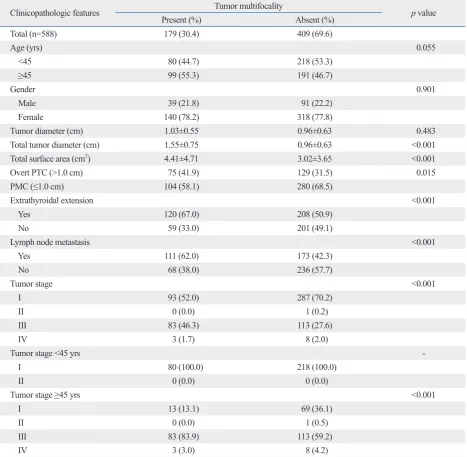
First, we investigated the clinicopathological significances of tumor multifocality in 588 classical PTCs. Tumor multi-focality was noted in 179 of 588 PTCs (30.4%), in which overt PTCs showed a higher rate of tumor multifocality than PMCs (p=0.015). Tumor multifocality was significantly cor-related with extrathyroidal extension (p<0.001), lymph node metastasis (p<0.001), and higher tumor stage (p<0.001) but not with age (p=0.055) or gender (p=0.901). There were dif-ferences between multifocal and unifocal PTCs in TSA (p <0.001) and TTD (p<0.001); however, there was no differ-ence in TD (p=0.483) (Table 1). In multivariate analysis, multifocality was an independent predictor of lymph node metastasis (p<0.001, OR=2.227, 95% CI 1.554‒3.192).
All PTCs were classified based on TSA and TTD, which represented PTCs with TD ≤0.5 cm, PTCs with 0.5 cm< TD≤1.0 cm and PTCs with TD >1.0 cm, respectively. TSA
and TTD groups were subdivided as TSA ≤0.785 cm2,
0.785 cm2<TS≤3.14 cm2, and TSA >3.14 cm2, as well as
[image:3.595.99.511.66.375.2]TTD ≤0.5 cm, 0.5 cm<TTD≤1.0 cm, and TTD >1.0 cm, re-spectively. Lymph node metastasis was found in 36.4%,
Fig. 1. Examples of measurements of tumor diameter. (A and B) Well-demarcated papillary thyroid carcinoma. (C and D) Papillary thyroid carcinoma with infiltrative border (×20).
A
C
B
Table 1. Correlation between Tumor Multifocality and Clinicopathological Features of PTCs
Clinicopathologic features Tumor multifocality p value
Present (%) Absent (%)
Total (n=588) 179 (30.4) 409 (69.6)
Age (yrs) 0.055
<45 80 (44.7) 218 (53.3)
≥45 99 (55.3) 191 (46.7)
Gender 0.901
Male 39 (21.8) 91 (22.2)
Female 140 (78.2) 318 (77.8)
Tumor diameter (cm) 1.03±0.55 0.96±0.63 0.483
Total tumor diameter (cm) 1.55±0.75 0.96±0.63 <0.001
Total surface area (cm2) 4.41±4.71 3.02±3.65 <0.001
Overt PTC (>1.0 cm) 75 (41.9) 129 (31.5) 0.015
PMC (≤1.0 cm) 104 (58.1) 280 (68.5)
Extrathyroidal extension <0.001
Yes 120 (67.0) 208 (50.9)
No 59 (33.0) 201 (49.1)
Lymph node metastasis <0.001
Yes 111 (62.0) 173 (42.3)
No 68 (38.0) 236 (57.7)
Tumor stage <0.001
I 93 (52.0) 287 (70.2)
II 0 (0.0) 1 (0.2)
III 83 (46.3) 113 (27.6)
IV 3 (1.7) 8 (2.0)
Tumor stage <45 yrs
I 80 (100.0) 218 (100.0)
II 0 (0.0) 0 (0.0)
Tumor stage ≥45 yrs <0.001
I 13 (13.1) 69 (36.1)
II 0 (0.0) 1 (0.5)
III 83 (83.9) 113 (59.2)
IV 3 (3.0) 8 (4.2)
PTC, papillary thyroid carcinoma; PMC, papillary thyroid microcarcinoma.
Table 2. Correlation between Total Surface Area and Total Tumor Diameter and Lymph Node Metastasis in Papillary Thyroid Carcinomas
Total surface area (cm2) ≤0.785 0.785–3.14 >3.14 p value
All tumors (n=588) 66 319 203
Lymph node metastasis (%) <0.001
Yes 24 (36.4) 135 (42.3) 125 (61.6)
No 42 (63.6) 184 (57.7) 78 (38.4)
Total tumor diameter (cm) ≤0.5 0.5–1.0 >1.0 p value
All tumors (n=588) 108 229 251
Lymph node metastasis (%) <0.001
Yes 21 (19.4) 100 (43.7) 163 (64.9)
[image:4.595.55.530.592.732.2]controversial.19-21 Lin, et al.20 and Mazeh, et al.22 reported
that tumor multifocality is significantly correlated with tu-mor recurrence. In mPMCs, a significant correlation be-tween tumor multifocality and lymph node metastasis has also been reported.8 According to previous reports and our
results, tumor multifocality may be a high risk factor. How-ever, tumor multifocality is not included in any staging sys-tem, including AGES (Age, Grade, Extent, Size), AMES (Age, Distant metastasis, Extent, Size), MACIS (Distant metastasis, Age, Completeness of resection, Local invasion, Size), GAMES (Grade, Age, Distant metastasis, Extent, Size), or AJCC TNM classification.11,23,24 Despite the
signif-icant correlation between tumor multifocality and aggres-sive behavior, little is known about the reason for its exclu-sion as a risk factor.
The prognosis for PMC is favorable, even in the presence of regional lymph node metastases and local invasion.25 The
rates of locoregional and regional lymph node recurrence have been reported to be between 2% and 5.7% in PMCs.26-28
According to the American Thyroid Association manage-ment guidelines,10 lobectomy and/or central lymph node
dis-section is considered an adequate treatment for uPMCs without other higher risk features. Total thyroidectomy with lymph node dissection is recommended for overt PTCs with multifocality,10,22 however, optimal treatment guidelines for
multifocal PMCs remain controversial. A subset of PMCs with aggressive behavior requires surgical treatment and postoperative management similar to overt PTCs.29,30 Thus,
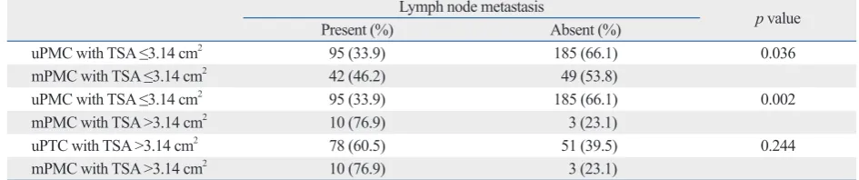
As shown in Table 4, uPMCs exhibited lymph node metas-tasis in 95 of 280 cases (33.9%). Nodal metasmetas-tasis was found in higher rates in mPMCs with TSA ≤3.14 cm2 (p=0.036), as
well as mPMCs with TSA >3.14 cm2 (p=0.002), than in
uPMCs; however, there was no difference between mPMCs with TSA >3.14 cm2 and unifocal overt PTCs (p=0.370).
DISCUSSION
Tumor multifocality of PTCs has been shown to be corre-lated with aggressive behavior, such as extrathyroidal ex-tension and lymph node metastasis; nevertheless, the man-agement guidelines of mPMCs remain controversial. The present study is the first, to the best of our knowledge, to show that assessment using TSA could be useful in differ-entiating between aggressive and favorable mPMCs and in deciding postoperative management.
In our study, tumor multifocality was observed in 30.4% of PTCs and was significantly correlated with extrathyroi-dal extension, lymph node metastasis, and tumor stage grouping (Table 1). Although Katoh, et al.18 reported that
multifocal tumors were found in 78.1% of all PTCs, tumor multifocality recently has been reported in only 20‒40% of cases.3-8 Other studies have shown that tumor multifocality
is significantly associated with lymph node metastasis, which is consistent with our results.19-21 The correlation
[image:5.595.69.541.84.159.2]be-tween tumor multifocality and extrathyroidal extension is
Table 3. Correlation between Total Surface Area and Lymph Node Metastasis of Multifocal Papillary Thyroid Microcarcinomas
mPMCs Lymph node metastasis p value
Present (%) Absent (%)
TSA ≤3.14 cm2 42 (46.2) 49 (53.8) 0.038
TSA >3.14 cm2 10 (76.9) 3 (23.1)
TTD ≤1.0 cm 26 (45.6) 31 (54.4) 0.325
TTD >1.0 cm 26 (55.3) 21 (44.7)
mPMC, multifocal papillary thyroid microcarcinoma; TSA, total surface area; TTD, total tumor diameter.
Table 4. Correlation between Total Surface Area and Lymph Node Metastasis of Multifocal Papillary Thyroid Microcarcinomas
Lymph node metastasis p
value
Present (%) Absent (%)
uPMC with TSA ≤3.14 cm2 95 (33.9) 185 (66.1) 0.036
mPMC with TSA ≤3.14 cm2 42 (46.2) 49 (53.8)
uPMC with TSA ≤3.14 cm2 95 (33.9) 185 (66.1) 0.002
mPMC with TSA >3.14 cm2 10 (76.9) 3 (23.1)
uPTC with TSA >3.14 cm2 78 (60.5) 51 (39.5) 0.244
mPMC with TSA >3.14 cm2 10 (76.9) 3 (23.1)
[image:5.595.67.540.200.299.2]of multifocal tumors. Accordingly, prediction of lymph node metastasis or tumor recurrence via tumor number has limitations.
In the present study, we evaluated the usefulness of a new parameter, TSA, in predicting nodal metastasis of PTCs. The criteria for TSA analysis were 0.785 cm2 and 3.14 cm2,
which are equivalent to TD 0.5 cm and TD 1.0 cm, respec-tively. Regardless of tumor multifocality, the larger TSA group showed significantly higher rates of nodal metastasis than the smaller TSA group (Table 2). Although overt PTCs showed higher rates of lymph node metastasis than PMCs (p<0.001), there was no significant difference in nodal me-tastasis between mPMCs with TSA >3.14 cm2 and overt
PTCs (p=0.465) (data not shown). In addition, as shown in Table 4, mPMCs with TSA >3.14 cm2 showed higher rates
of lymph node metastasis than mPMCs with TSA ≤3.14 cm2 and uPMCs. Andea, et al.13,33 previously reported that
the assessment using TSA in multifocal breast carcinomas and TSA rather than TTD was useful for the prediction of lymph node metastasis. According to our results, PTCs with TSA >3.14 cm2 showed higher rates of extrathyroidal
extension than PTCs with TSA ≤3.14 cm2 (p<0.001).
How-ever, there was no significant difference in extrathyroidal ex-tension between mPMCs with TSA >3.14 cm2 and mPMCs
with TSA ≤3.14 cm2 (p=0.194). In the present study,
three-dimensional diameters were measured on gross and micro-scopic examination. If a tumor has marked infiltrative bor-der, the estimated surface area may be smaller than the real surface area, which would warrant consideration in prac-tice. Notwithstanding, according to our results and previous reports, TSA rather than TTD could be useful for differenti-ating between higher and lower risk groups of nodal metas-tasis in multifocal tumors.
In conclusion, the present study showed that multifocal PMCs can be evaluated via TSA, and patients with mPMCs with TSA >3.14 cm2 may be managed in a different manner
than patients with uPMCs or mPMCs with TSA ≤3.14 cm2.
As a new parameter, TSA could be a useful parameter for deciding on preoperative, surgical, and postoperative man-agement of mPTCs.
REFERENCES
1. DeLellis RA, Lloyd RV, Heitz PU, Eng C. World Health Organi-zation Classification of Tumours. Pathology and genetics of tu-mours of endocrine organs. Lyon: IARC Press; 2004. p.57-66.
it is important to differentiate multifocal PMCs requiring more aggressive surgical treatment and postoperative man-agement. In addition, more detailed assessment of different surgical treatment and postoperative management options for multifocal tumors are needed.
In the present study, tumor multifocality was observed at a higher rate in overt PTCs than it was in PMCs. Based on the 7th edition of the AJCC TNM classification systems,11
primary tumor (T) stage is to be evaluated via tumor size and extrathyroidal extension; however, in multifocal PTCs, tumor size is evaluated by the largest tumor size, regardless of tumor number. The evaluation of extrathyroidal exten-sion is performed for all tumors, including the largest tumor and other smaller tumors. Although PTCs show higher rates of tumor multifocality than other tumors,14 little is known
about the effects of tumor multifocality on aggressive be-havior. Whether the second tumor of multifocal PTCs af-fects aggressive behavior remains a subject for further in-vestigation.
The assessment parameters for surgical treatment and postoperative management of multifocal PMCs are not ful-ly understood. In the assessment for tumor multifocality of PTCs, new trials have attempted to assess the use of total tumor diameter or tumor number. Zhao, et al.8 introduced
TTD as an assessment tool; however, these results were not included in the comparison of lymph node metastasis be-tween mPMCs with TTD >1 cm and with TTD <1 cm. Our results revealed no significant difference in lymph node me-tastasis rates between mPMCs with TTD >1 cm and with TTD ≤1 cm (Table 3). In addition, the shape of PTCs was mainly ovoid to round with dimensions taller than they were wide.31 Therefore, tumor diameter cannot accurately
repre-sent true tumor bulk, as tumor volume or total surface area can. Tumor surfaces are proportional to the second and third power of the diameter. By adding the diameters of multifo-cal tumors, TTD could overestimate total surface area. These results suggest that TTD cannot distinguish aggres-sive mPMCs from more favorable mPMCs and uPMCs. Lin, et al.20 reported that tumor number was significantly
associated with tumor recurrence. The detection of multifo-cal tumors is dependent on surgimultifo-cal extent, degree of in-spection, and microscopic examination;32 however, it is
dif-ficult to microscopically examine the entire thyroid gland, and tumor multifocality may be missed. In addition, if bilat-eral lobes are not examined through total thyroidectomy, tumors in the contralateral lobe may be missed.32 These
Thyroid 2014;24:245-53.
18. Katoh R, Sasaki J, Kurihara H, Suzuki K, Iida Y, Kawaoi A. Mul-tiple thyroid involvement (intraglandular metastasis) in papillary thyroid carcinoma. A clinicopathologic study of 105 consecutive patients. Cancer 1992;70:1585-90.
19. Park SY, Park YJ, Lee YJ, Lee HS, Choi SH, Choe G, et al. Anal-ysis of differential BRAF(V600E) mutational status in multifocal papillary thyroid carcinoma: evidence of independent clonal ori-gin in distinct tumor foci. Cancer 2006;107:1831-8.
20. Lin JD, Chao TC, Hsueh C, Kuo SF. High recurrent rate of multi-centric papillary thyroid carcinoma. Ann Surg Oncol 2009;16: 2609-16.
21. Kim HJ, Kim NK, Choi JH, Kim SW, Jin SM, Suh S, et al. Radio-active iodine ablation does not prevent recurrences in patients with papillary thyroid microcarcinoma. Clin Endocrinol (Oxf) 2013; 78:614-20.
22. Mazeh H, Samet Y, Hochstein D, Mizrahi I, Ariel I, Eid A, et al. Multifocality in well-differentiated thyroid carcinomas calls for total thyroidectomy. Am J Surg 2011;201:770-5.
23. Rodríguez-Cuevas S, Labastida-Almendaro S, Cortés-Arroyo H, López-Garza J, Barroso-Bravo S. Multifactorial analysis of surviv-al and recurrences in differentiated thyroid cancer. Comparative evaluation of usefulness of AGES, MACIS, and risk group scores in Mexican population. J Exp Clin Cancer Res 2002;21:79-86. 24. Voutilainen PE, Siironen P, Franssila KO, Sivula A, Haapiainen
RK, Haglund CH. AMES, MACIS and TNM prognostic classifi-cations in papillary thyroid carcinoma. Anticancer Res 2003;23: 4283-8.
25. Appetecchia M, Scarcello G, Pucci E, Procaccini A. Outcome af-ter treatment of papillary thyroid microcarcinoma. J Exp Clin Cancer Res 2002;21:159-64.
26. Hay ID, Hutchinson ME, Gonzalez-Losada T, McIver B, Reinalda ME, Grant CS, et al. Papillary thyroid microcarcinoma: a study of 900 cases observed in a 60-year period. Surgery 2008;144:980-7. 27. Besic N, Pilko G, Petric R, Hocevar M, Zgajnar J. Papillary
thy-roid microcarcinoma: prognostic factors and treatment. J Surg Oncol 2008;97:221-5.
28. Pelizzo MR, Boschin IM, Toniato A, Pagetta C, Piotto A, Ber-nante P, et al. Natural history, diagnosis, treatment and outcome of papillary thyroid microcarcinoma (PTMC): a mono-institutional 12-year experience. Nucl Med Commun 2004;25:547-52. 29. Lee J, Rhee Y, Lee S, Ahn CW, Cha BS, Kim KR, et al. Frequent,
aggressive behaviors of thyroid microcarcinomas in Korean pa-tients. Endocr J 2006;53:627-32.
30. Page C, Biet A, Boute P, Cuvelier P, Strunski V. ‘Aggressive pap-illary’ thyroid microcarcinoma. Eur Arch Otorhinolaryngol 2009; 266:1959-63.
31. Moon WJ, Jung SL, Lee JH, Na DG, Baek JH, Lee YH, et al. Be-nign and malignant thyroid nodules: US differentiation--multi-center retrospective study. Radiology 2008;247:762-70. 32. Haymart MR, Cayo M, Chen H. Papillary thyroid
microcarcino-mas: big decisions for a small tumor. Ann Surg Oncol 2009;16: 3132-9.
33. Andea AA, Wallis T, Newman LA, Bouwman D, Dey J, Visscher DW. Pathologic analysis of tumor size and lymph node status in multifocal/multicentric breast carcinoma. Cancer 2002;94:1383-90. 2. Shattuck TM, Westra WH, Ladenson PW, Arnold A. Independent
clonal origins of distinct tumor foci in multifocal papillary thyroid carcinoma. N Engl J Med 2005;352:2406-12.
3. Siassakos D, Gourgiotis S, Moustafellos P, Dimopoulos N, Hadji-yannakis E. Thyroid microcarcinoma during thyroidectomy. Sin-gapore Med J 2008;49:23-5.
4. Mercante G, Frasoldati A, Pedroni C, Formisano D, Renna L, Pi-ana S, et al. Prognostic factors affecting neck lymph node recur-rence and distant metastasis in papillary microcarcinoma of the thyroid: results of a study in 445 patients. Thyroid 2009;19:707-16. 5. Ogilvie JB, Patel KN, Heller KS. Impact of the 2009 American
Thyroid Association guidelines on the choice of operation for well-differentiated thyroid microcarcinomas. Surgery 2010;148: 1222-6.
6. Ciuffreda L, De Martino D, Bonfitto N, Scaramuzzi R. [Our expe-rience on surgical treatment of papillary thyroid microcarcinoma]. G Chir 2011;32:41-4.
7. Connor MP, Wells D, Schmalbach CE. Variables predictive of bi-lateral occult papillary microcarcinoma following total thyroidec-tomy. Otolaryngol Head Neck Surg 2011;144:210-5.
8. Zhao Q, Ming J, Liu C, Shi L, Xu X, Nie X, et al. Multifocality and total tumor diameter predict central neck lymph node metasta-ses in papillary thyroid microcarcinoma. Ann Surg Oncol 2013;20: 746-52.
9. Lim YC, Choi EC, Yoon YH, Kim EH, Koo BS. Central lymph node metastases in unilateral papillary thyroid microcarcinoma. Br J Surg 2009;96:253-7.
10. American Thyroid Association (ATA) Guidelines Taskforce on Thyroid Nodules and Differentiated Thyroid Cancer, Cooper DS, Doherty GM, Haugen BR, Kloos RT, Lee SL, et al. Revised American Thyroid Association management guidelines for pa-tients with thyroid nodules and differentiated thyroid cancer. Thy-roid 2009;19:1167-214.
11. Edge SB, Byrd DR, Compton CC, Fritz AG, Greene FL, Trotti A. AJCC Cancer Staging Manual. 7th ed. New York: Springer-Ver-lag; 2009. p.87-100.
12. Pyo JS, Kang G, Kim DH, Park C, Kim JH, Sohn JH. The prog-nostic relevance of psammoma bodies and ultrasonographic intra-tumoral calcifications in papillary thyroid carcinoma. World J Surg 2013;37:2330-5.
13. Andea AA, Bouwman D, Wallis T, Visscher DW. Correlation of tumor volume and surface area with lymph node status in patients with multifocal/multicentric breast carcinoma. Cancer 2004;100: 20-7.
14. Chow SM, Law SC, Chan JK, Au SK, Yau S, Lau WH. Papillary microcarcinoma of the thyroid-Prognostic significance of lymph node metastasis and multifocality. Cancer 2003;98:31-40. 15. Machens A, Holzhausen HJ, Dralle H. The prognostic value of
primary tumor size in papillary and follicular thyroid carcinoma. Cancer 2005;103:2269-73.
16. Pelizzo MR, Boschin IM, Toniato A, Piotto A, Bernante P, Pagetta C, et al. Papillary thyroid microcarcinoma (PTMC): prognostic factors, management and outcome in 403 patients. Eur J Surg On-col 2006;32:1144-8.