0095-1137/09/$08.00⫹0 doi:10.1128/JCM.00197-09
Copyright © 2009, American Society for Microbiology. All Rights Reserved.
Fine-Needle Aspiration, an Efficient Sampling Technique for
Bacteriological Diagnosis of Nonulcerative
Buruli Ulcer
䌤
Miriam Eddyani,
1* Alexandra G. Fraga,
2Fernando Schmitt,
3Ce
´cile Uwizeye,
1Krista Fissette,
1Christian Johnson,
4Julia Aguiar,
5Ghislain Sopoh,
6Yves Barogui,
7Wayne M. Meyers,
8Jorge Pedrosa,
2and Franc¸oise Portaels
1Mycobacteriology Unit, Department of Microbiology, Institute of Tropical Medicine, Antwerp, Belgium1; Life and
Health Sciences Research Institute, School of Health Sciences, University of Minho, Braga, Portugal2;
Institute of Molecular Pathology and Immunology and Medical Faculty, Porto University, Porto, Portugal3; Programme National de Lutte contre l’Ulce`re de Buruli, Cotonou, Benin4;
Centre Sanitaire et Nutritionnel Gbemoten, Zagnanado, Benin5; Centre de De´pistage et de
Traitement de l’Ulce`re de Buruli, Allada, Benin6; Centre de De´pistage et de
Traitement de l’Ulce`re de Buruli, Lalo, Benin7; and Armed Forces Institute of
Pathology, Washington, DC 203068
Received 30 January 2009/Returned for modification 20 March 2009/Accepted 9 April 2009
Invasive punch or incisional skin biopsy specimens are currently employed for the bacteriological confir-mation of the clinical diagnosis of Buruli ulcer (BU), a cutaneous infectious disease caused byMycobacterium ulcerans. The efficacy of fine-needle aspirates (FNA) using fine-gauge needles (23G by 25 mm) for the laboratory confirmation of BU was compared with that of skin tissue fragments obtained in parallel by excision or punch biopsy. In three BU treatment centers in Benin, both types of diagnostic material were obtained from 33 clinically suspected cases of BU and subjected to the same laboratory analyses: i.e., direct smear examination, IS2404PCR, and in vitro culture. Twenty-three patients, demonstrating 17 ulcerative and 6 nonulcerative lesions, were positive by at least two tests and were therefore confirmed to have active BU. A total of 68 aspirates and 68 parallel tissue specimens were available from these confirmed patients. When comparing the sensitivities of the three confirmation tests between FNA and tissue specimens, the latter yielded more positive results, but only for PCR was this significant. When only nonulcerative BU lesions were considered, however, the sensitivities of the confirmation tests using FNA and tissue specimens were not significantly different. Our results show that the minimally invasive FNA technique offers enough sensitivity to be used for the diagnosis of BU in nonulcerative lesions.
Mycobacterium ulceransdisease, or Buruli ulcer (BU), a dev-astating skin disease that may affect bone, is the third most common mycobacteriosis after tuberculosis and leprosy. The disease is focally endemic in Africa, the Americas, Australia, and Asia, where rural populations are the most affected (14, 28, 32).
For many years, the standard treatment of BU was surgical excision of affected tissue (1, 29). Now, however, specific an-tibiotic therapy for 8 weeks without surgery has been increas-ingly implemented, especially in patients with small lesions, whether closed or ulcerative (6, 9, 31). This recent develop-ment increases the importance of confirming the clinical diag-nosis of BU by laboratory techniques to avoid therapy with
antimycobacterial drugs when the lesion is not caused byM.
ulcerans (30). However, with the decreasing importance of surgery as a therapeutic intervention, tissues for microbiolog-ical diagnosis are often not available. Alternatively, punch
bi-opsy specimens, requiring local anesthesia, have been em-ployed for confirmation of the clinical diagnosis of BU (23).
In fact, while microbiological tests on ulcerative forms of BU can be performed on multiple swabs of exudates taken from the undermined edges of ulcers, for nonulcerative forms the microbiological diagnosis must be carried out on tissue frag-ments, i.e., collected by incisional or punch biopsy (30). As a minimal invasive technique, fine-needle aspiration (FNA), commonly used for the cytologic diagnosis of tumors (10, 11), lymphadenopathies caused by trypanosomiasis (17) and tuber-culosis (8, 15, 27), leprous osteitis (20), or leprotic neuritis (16), would seem to offer an advantageous alternative.
In addition, FNA could be valuable for the follow-up of the infection, during and after treatment with antimycobacterial drugs. Compared to punch biopsy specimens, FNA includes less risk of ulceration or possible subsequent reactivation of healing lesions since it is a minimally invasive procedure. Very recently, Phillips et al. (24) also reported the use of FNA for the diagnosis of BU.
In three BU treatment centers in Benin (Centres de De
´p-istage et de Traitement de l’Ulce`re de Buruli [CDTUB] at
Zagnanado, Allada, and Lalo), FNA was applied to clinically suspected cases of BU. The efficacy of this technique, as a source of diagnostic material, was compared in parallel with
* Corresponding author. Mailing address: Mycobacteriology Unit, Department of Microbiology, Institute of Tropical Medicine, Nation-alestraat 155, B-2000 Antwerp, Belgium. Phone: 32(0)32476336. Fax: 32(0)32476333. E-mail: [email protected].
䌤Published ahead of print on 22 April 2009.
1700
on May 16, 2020 by guest
http://jcm.asm.org/
that of tissue fragments obtained by excision or punch biopsy, concerning the laboratory confirmation of both ulcerative and nonulcerative BU lesions.
MATERIALS AND METHODS
Collection of specimens in Benin.Sampling was carried out in November 2006. During anesthesia and before surgery, 84 FNA samples were taken from lesions of 33 consecutively enrolled patients with a clinical diagnosis of BU. Sampling was carried out by the same study physician (Y.B.) in all BU treatment centers. Patients were under treatment for various periods of time (see Table 3) with streptomycin and rifampin combined with surgery (31). Depending on the size of the lesion, up to six aspirates were taken from each lesion using fine-gauge needles (23G by 25 mm) attached to 10-ml syringes. An optimal aspiration technique is very important to obtain sufficient material (12). The needle is advanced through the skin without applying suction. For nonulcerative forms, the middle of the lesion is sampled; for ulcerative forms, the undermined edges are sampled (30). Suction is applied while moving the needle back and forth repeatedly through the lesion in multiple directions. Aspiration is terminated when fluid first appears at the needle hub. With cessation of aspiration and with the needle still inside the skin, negative pressure within the syringe returns the syringe piston to the base of the syringe barrel automatically. The reason for releasing suction before the needle is withdrawn is to leave the maximum amount of aspirated material inside the needle. This is so because if the needle is withdrawn from the lesion under negative pressure in the syringe, the aspirated specimen is drawn into the syringe and is dispersed into numerous small droplets. These droplets would stick to the walls of the syringe barrel and would be virtually impossible to recover (10). After withdrawal from the lesion, the needle is detached from the syringe, and the syringe is allowed to fill with air. The needle is then placed back onto the syringe, uncovered, and the specimen is dispersed into the transport medium.
Aspirates were stored at 4°C in a liquid transport medium (LTM) containing Middlebrook 7H9 broth supplemented with polymyxin B, amphotericin B, nali-dixic acid, trimethoprim, and azlocillin (PANTA, Becton Dickinson, Sparks, MD) and OADC (oleic acid, albumin, dextrose, and catalase) (Difco Laborato-ries, Detroit, MI). The needle was detached from the syringe, and the content of the needle hub was smeared on a slide for direct smear examination (DSE).
During surgery, parallel tissues were removed at the same sites from which the aspirates were taken. In the CDTUB of Zagnanado, fragments of⫾1 g from surgically excised tissues were taken, while in the CDTUB of Allada and Lalo, 4-mm punch biopsy specimens were used. Tissue specimens were stored at 4°C in a sterile vial with 1 ml of a semisolid transport medium with the same components as LTM plus 0.5% agar. This medium is efficient for storage of clinical specimens from patients with BU (7).
All specimens were transported to Belgium at ambient temperature and pro-cessed within 2 weeks.
Microbiological analysis of specimens in Belgium.Smears prepared in Benin were stained with auramine and read using fluorescence microscopy according to the American Thoracic Society scale (4). The LTM containing the aspirate was processed for IS2404PCR (13) and in vitro culture after decontamination using the reversed Petroff technique (22).
Tissue fragments were cut into small pieces, ground aseptically with a mortar and pestle, and suspended in 2 ml phosphate-buffered saline as previously de-scribed (30). One drop of this suspension was dried on a slide, stained with auramine, and read using fluorescence microscopy. IS2404PCR and in vitro culture were performed on the suspensions.
When culture tubes of specimens with positive results by PCR and DSE were contaminated by other microorganisms, the specimens were processed again for in vitro culture after decontamination with oxalic acid as previously described by Portaels et al. (25) and Yeboah-Manu et al. (33). Briefly, 0.5 ml of 0.2% mala-chite green, 0.5 ml NaOH, and 0.1 ml of 0.8% actidione were added to 0.5 ml of the specimen suspended in phosphate-buffered saline. This mixture was left for 30 min at room temperature and then centrifuged for 15 min at 3,500⫻g. The supernatant was discarded, and 1 ml of 5% oxalic acid was added to the sediment and left at room temperature for 20 min. The suspension was then centrifuged again for 15 min at 3,500⫻g, after which 0.1 ml of the pellet, suspended in 1 ml distilled water, was inoculated onto Lo¨wenstein-Jensen medium, incubated at 30°C, and examined weekly for growth. Positive cultures were identified to the species level based on phenotypic characteristics (18).
Statistical analysis.Data were analyzed statistically with Epi Info v. 3.3.2 and SPSS v. 16.0. Pearson’s chi-square test was used to compare proportions, and Student’sttest was applied to compare means of symmetric distributions.
Sen-sitivities were calculated as the ratio of positives for a certain test to the total number of specimens from confirmed patients.
Ethical provisions.This study had the agreement of the Ministry of Health of Benin. All patients (or their parents or guardians, for children younger than 15 years) gave their oral informed consent to participate in the study.
RESULTS
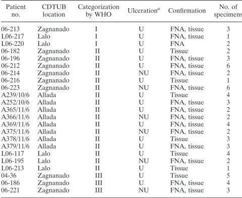
Among the 33 patients, 23 were confirmed to have active BU according to WHO guidelines (30): at least two positive tests among the DSE, PCR, and culture methods (Table 1). Six patients did not have BU, and four were found to have inactive BU (only one positive test by either DSE or PCR). Among the 23 confirmed BU patients, 17 had ulcers and 6 had nonulcer-ative lesions: plaques (3), edema (2), or osteomyelitis (1). When classified according to WHO guidelines (31), 3 patients had early lesions (category I), 17 had large lesions (category II), and 3 had disseminated lesions (category III) (Table 1). Altogether 68 aspirates and 68 parallel tissue specimens were available from these confirmed patients.
DSE.Sensitivity of DSE by FNA was 32.4% (22/68) (Table
2). This is not significantly lower than the sensitivity on tissue
specimens, which was 38.2% (26/68) (P⫽0.59). A comparison
of the sensitivities of DSE by FNA of ulcerative and nonulcer-ative lesions revealed that nonulcernonulcer-ative lesions were signifi-cantly more often positive for acid-fast bacilli (AFB) than
ulcerative lesions (64.7% versus 21.6%;P⫽0.003). In tissue
specimens, the sensitivity of DSE was also higher for
nonul-cerative lesions, but this was not significant (P ⫽0.08). The
sensitivity of DSE was higher by FNA than on tissue specimens from nonulcerative lesions, but this was not statistically
signif-icant (64.7% versus 58.8%;P⫽0.70).
IS2404PCR.In general, the sensitivity of PCR for the total
number of FNA specimens was 48.5% (33/68) (Table 2). This is significantly lower than the sensitivity for tissue specimens,
which was 85.3% (58/68) (P⬍0.0001). When comparing the
[image:2.585.300.540.99.295.2]sensitivities of PCR on FNA of ulcerative and nonulcerative
TABLE 1. Treating CDTUB, categorization according to WHO, clinical presentation, and type of specimen with at least
one positive result for confirmed BU patients
Patient no.
CDTUB location
Categorization
by WHO Ulceration
a Confirmation No. of
specimens
06-213 Zagnanado I U FNA, tissue 3
L06-217 Lalo I U FNA, tissue 1
L06-220 Lalo I U FNA 2
06-182 Zagnanado II U Tissue 2
06-196 Zagnanado II U FNA, tissue 3
06-212 Zagnanado II U FNA, tissue 6
06-214 Zagnanado II NU FNA, tissue 2
06-216 Zagnanado II U Tissue 1
06-223 Zagnanado II NU FNA, tissue 6
A239/10/6 Allada II U Tissue 4
A252/10/6 Allada II U FNA, tissue 3
A365/11/6 Allada II U FNA, tissue 2
A366/11/6 Allada II NU FNA, tissue 2
A369/11/6 Allada II U FNA, tissue 4
A375/11/6 Allada II NU FNA, tissue 2
A378/11/6 Allada II U Tissue 3
A379/11/6 Allada II U FNA, tissue 3
L06-117 Lalo II U Tissue 4
L06-195 Lalo II NU FNA, tissue 2
L06-213 Lalo II U Tissue 1
04-36 Zagnanado III U Tissue 5
06-186 Zagnanado III U FNA, tissue 4
06-221 Zagnanado III NU FNA, tissue 3
aU, ulcerative lesion; NU, nonulcerative lesion.
on May 16, 2020 by guest
http://jcm.asm.org/
lesions, significantly more PCR-positive samples were found in
nonulcerative lesions (88.2% versus 35.3%; P ⬍ 0.001). In
tissue specimens, the sensitivities of PCR for ulcerative and
nonulcerative lesions were not significantly different (P ⫽
1.00). For nonulcerated lesions alone, the sensitivities of PCR on FNA and tissue specimens were identical (88.2%).
In vitro culture.The sensitivity of in vitro culture with FNA specimens was 17.6% (12/68) (Table 2). This is not significantly lower than the sensitivity on tissue specimens, which was 25.0%
(17/68) (P⫽0.40). When comparing the sensitivities of in vitro
culture of FNA of ulcerative and nonulcerative lesions, the latter were significantly more often positive (9.8% versus
41.2%; P ⫽ 0.007). Also in tissue specimens, nonulcerative
lesions yielded more positive cultures (P⬍0.001). The
sensi-tivities of in vitro culture in nonulcerative lesions with FNA and tissue specimens were not significantly different (41.2%
versus 58.8%;P⫽0.30).
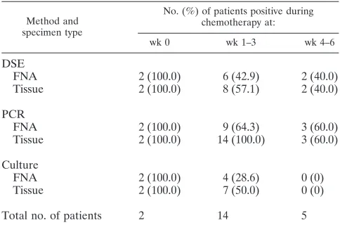
Influence of chemotherapy on positivity of bacteriological tests.In specimens from patients under treatment, DSE and PCR remained positive, although a decrease in positivity was observed both for FNA and tissue specimens (Table 3). In contrast, FNA and tissue specimens from patients under treat-ment for 4 weeks or more did not yield any positive cultures.
DISCUSSION
The first attempt to confirm BU diagnosis by puncturing a lesion with a needle was made in 1957 in the Democratic Republic of Congo on an extensive edematous lesion of the arm. A smear of the exudate stained by the Ziehl-Neelsen method revealed AFB (14).
In a study by Singh et al. (27), the sensitivity of laboratory tests for the confirmation of tuberculous lymphadenitis was higher with biopsy specimens than with FNA material. In our study, when the sensitivities of laboratory tests obtained for the total number of FNA and tissue specimens (regardless of clin-ical form) were compared, tissue specimens yielded more pos-itive results—but only by PCR, the most senspos-itive test. Impor-tantly, when considering only nonulcerative BU lesions, the sensitivities obtained from FNA and tissue specimens were not significantly different by PCR or by any of the other confirma-tory tests (Table 2). These results emphasize the potential of FNA for confirmation of the diagnosis of nonulcerative forms of BU since ulcers can easily be confirmed on swabs. With nonulcerative forms of BU, however, an invasive sampling technique is currently necessary, and FNA is a much less trau-matic approach.
In the present study, three bacteriological confirmation tests were performed and PCR was found to be the most sensitive technique, for both FNA and tissue specimens, for ulcerative as well as for nonulcerative lesions (Table 2). The second most sensitive confirmation test was DSE, followed by in vitro cul-ture. Comparable observations have been reported in other studies (5, 7, 23). These findings are analogous to those re-ported for the use of FNA for the diagnosis of tuberculous lymphadenitis (2, 8, 15, 27). Also, a recent study by Phillips et al. (24) reports PCR as the most sensitive technique for
con-firming the presence ofM. ulceransin FNA of nonulcerative
BU lesions.
[image:3.585.42.541.81.183.2]In the present study, FNA samples from nonulcerative le-sions had significantly higher positivity rates than FNA samples from ulcerative lesions for all of the tested laboratory methods. This may be due to the sampling technique, since in ulcerative lesions it is probably more difficult to find the most productive sampling site. In the ulcerative lesions that have not been debrided, the necrotic slough in the base of the ulcer contains the majority of AFB. After debridement, the largest numbers of AFB are seen at the base of the ulcer in the undermined
TABLE 2. Results from DSE, PCR, and in vitro culture of FNA and tissue specimens of ulcerative and nonulcerative BU lesions
Parameter
No. (%) of specimensa
Tissue FNA
Ulcerative (17 patients)
Nonulcerative
(6 patients) Total (23 patients)
Ulcerative (17 patients)
Nonulcerative
(6 patients) Total (23 patients)
DSE positive 16 (31.4) 10 (58.8) 26 (38.2) 11 (21.6)* 11 (64.7)* 22 (32.4)
PCR positive 43 (84.3) 15 (88.2) 58 (85.3)† 18 (35.3)† 15 (88.2)† 33 (48.5)†
Culture positive 7 (13.7)§ 10 (58.8)§ 17 (25.0) 5 (9.8)¶ 7 (41.2)¶ 12 (17.6)
Total no. of specimens 51 17 68 51 17 68
aStatistically significant differences between the results of the analyses as evaluated by Pearson chi-square test are indicated as follows for the values compared: *,
P⫽0.003; †,P⬍0.0001; †,P⫽0.0005; §,P⫽0.0006; and ¶,P⫽0.007.
TABLE 3. Results of DSE, PCR, and in vitro culture of FNA and tissue specimens among patients with different durations
of chemotherapy
Method and specimen type
No. (%) of patients positive during chemotherapy at:
wk 0 wk 1–3 wk 4–6
DSE
FNA 2 (100.0) 6 (42.9) 2 (40.0)
Tissue 2 (100.0) 8 (57.1) 2 (40.0)
PCR
FNA 2 (100.0) 9 (64.3) 3 (60.0)
Tissue 2 (100.0) 14 (100.0) 3 (60.0)
Culture
FNA 2 (100.0) 4 (28.6) 0 (0)
Tissue 2 (100.0) 7 (50.0) 0 (0)
Total no. of patients 2 14 5
on May 16, 2020 by guest
http://jcm.asm.org/
[image:3.585.42.284.563.723.2]edges. Using tissue fragments in a larger study, we have also observed a higher positivity rate in the confirmation tests for nonulcerative lesions (plaques and edemas) than for ulcerative lesions (F. Portaels et al., unpublished data). Positivity of mi-crobiological confirmation tests also varies in tuberculous lymphadenitis, depending on the type of lesion (19, 21).
Recent unpublished studies by our group of more than 1,000 BU patients show that at least two tissue specimens from the same lesion must be analyzed to achieve acceptable sensitivity for the bacteriological confirmation of BU (F. Portaels, unpub-lished data). The taking of multiple FNA aspirations from different sites of the lesion may thus be justified to increase the sensitivity of the laboratory tests. In the present study, patients were anesthetized before the sampling procedures because these preceded surgery. FNA is routinely used for cytologic diagnoses without anesthesia (12). The pain when taking five to seven FNA in breast lesions of 79 patients has been reported to be 3.19 on a scale of 0 (no pain) to 10 (worst possible pain) (34). It is therefore expected that for sampling of diagnostic FNA specimens from BU lesions, anesthesia would also not be necessary.
In both FNA and tissue specimens, DSE and PCR remained positive during treatment (Table 3). This type of phenomenon has been observed in patients with tuberculosis, whose sputum smears may remain positive for AFB after up to 36 weeks of treatment while cultures became negative much earlier (3). Moreover, the overall sensitivities of in vitro culture with FNA as well as tissue specimens were lower in this study than those in previous studies (7, 23). The reason for this is probably that most of the patients included in this study were undergoing treatment when the specimens were collected, therefore reduc-ing the number of viable bacilli in the lesion. Thus, this study also demonstrates the importance of FNA for follow-up of chemotherapy when the use of more invasive sampling tech-niques could be deemed unethical. Indeed, according to our observations, the outcome of in vitro culture from nonulcer-ative BU lesions could be best assessed in FNA specimens.
We have evaluated several small collections of FNA speci-mens taken by other teams of investigators who used 19G needles and found them much less productive in the confirma-tion of BU (data not shown). The types of lesions sampled and the techniques employed were not uniform; however, direct comparisons of samples taken by 19G and 23G needles seem warranted.
Siegmund et al. (26) found a great variation in positivity rates of bacteriological tests between BU treatment centers, suggesting that the quality of the clinical diagnosis and of sampling specimens differed between centers. We therefore recommend the development of training sessions on the dif-ferent sampling techniques for the confirmation of BU: tissue or swab specimens from ulcerative lesions and FNA from non-ulcerative lesions.
In conclusion, our results support the use of FNA, a mini-mally invasive technique, as a sensitive sampling method for the diagnosis and follow-up of BU in nonulcerative lesions. The facts that the available sampling techniques for confirma-tion of BU diagnosis cause high levels of discomfort and that BU affects primarily children reinforce the need to develop less invasive sampling methods as proposed here. Additionally, due to the present sampling difficulties, antibiotic treatment is
of-ten performed without bacteriological confirmation of BU, implying the risk of submitting patients to long
chemothera-peutic protocols in skin lesions not caused by M. ulcerans.
Moreover, in these cases, confirmation of BU diagnosis may prevent potentially harmful surgery. Our findings have there-fore a potential impact in benefit of patients with nonulcerative lesions. However, studies in larger case series of patients with nonulcerative lesions are warranted before making any recom-mendations to replace the more invasive techniques currently used for the diagnosis of BU. In addition, further studies are needed to optimize the sensitivity of BU confirmation tests on FNA taken with needles of different sizes and on the number of FNA aspirates that would be ideal to obtain an optimal sensitivity for the laboratory confirmation of BU diagnosis.
ACKNOWLEDGMENTS
This work was partly supported by the Damian Foundation (Brus-sels, Belgium), the Directorate-General for Development Cooper-ation (Brussels, Belgium), FWO-Flanders (Brussels, Belgium; grant K.1.197.07.N.00), the Health Services of Fundac¸a˜o Calouste Gul-benkian (Lisbon, Portugal), the European Commission, project no. INCO-CT-2005-051476-BURULICO, and the Stop Buruli initiative funded by the UBS Optimus Foundation.
We thank Martine Debacker for help with the statistical analyses and all health staff and examined patients of the CDTUB of Zagna-nado, Allada, and Lalo for cooperation.
REFERENCES
1.Aguiar, J., and C. Steunou.1997. Les ulce`res de Buruli en zone rurale au Be´nin: prise en charge de 635 cas. Med. Trop.57:83–90.
2.Aljafari, A. S., E. A. G. Khalil, K. E. Elsiddig, I. A. El Hag, M. E. Ibrahim, M. E. M. O. Elsafi, A. M. Hussein, I. M. Elkhidir, G. S. Sulaiman, and A. M. Elhassan.2004. Diagnosis of tuberculous lymphadenitis by FNAC, microbi-ological methods and PCR: a comparative study. Cytopathology15:44–48. 3.Al-Moamary, M. S., W. Black, E. Bessuille, R. K. Elwood, and S. Vedal.1999.
The significance of the persistent presence of acid-fast bacilli in sputum smears in pulmonary tuberculosis. Chest116:726–731.
4.American Thoracic Society.1981. Diagnostic standards and classification of tuberculosis and other mycobacterial diseases (14th edition). Am. Rev. Re-spir. Dis.123:343–358.
5.Bretzel, G., V. Siegmund, J. Nietsche, K. H. Herbinger, W. Thompson, E. Klutse, K. Crofts, W. Massavon, S. Etuaful, R. Thompson, K. Asamoah-Opare, P. Racz, F. Vloten, C. van Berberich, T. Kruppa, E. Ampadu, B. Fleischer, and O. Adjei.2007. A stepwise approach to the laboratory diag-nosis of Buruli ulcer disease. Trop. Med. Int. Health12:89–96.
6.Chauty, A., M.-F. Ardant, A. Adeye, H. Euverte, A. Gue´de´non, C. Johnson, J. Aubry, E. Nuermberger, and J. Grosset.2007. Promising clinical efficacy of streptomycin-rifampin combination for treatment of Buruli ulcer ( Mycobac-terium ulceransdisease). Antimicrob. Agents Chemother.51:4029–4035. 7.Eddyani, M., M. Debacker, A. Martin, J. Aguiar, C. R. Johnson, C. Uwizeye,
K. Fissette, and F. Portaels.2008. Primary culture ofMycobacterium ulcerans
from human tissue specimens after storage in a semisolid transport medium. J. Clin. Microbiol.46:69–72.
8.Erso¨z, C., A. Polat, M. S. Serin, L. Soylu, and O. Demircan.1998. Fine needle aspiration (FNA) cytology in tuberculous lymphadenitis. Cytopathol-ogy9:201–207.
9.Etuaful, S., B. Carbonnelle, J. Grosset, S. Lucas, C. Horsfield, R. Phillips, M. Evans, D. Ofori-Adjei, E. Klustse, J. Owusu-Boateng, G. K. Amedofu, P. Awuah, E. Ampadu, G. Amofah, K. Asiedu, and M. Wansbrough-Jones.
2005. Efficacy of the combination rifampin-streptomycin in preventing growth ofMycobacterium ulceransin early lesions of Buruli ulcer in humans. Antimicrob. Agents Chemother.49:3182–3186.
10.Furnival, C. M., M. A. Hocking, H. E. Hughes, M. M. Reid, and L. H. Blumgart.1975. Aspiration cytology in breast cancer: its relevance to diag-nosis. Lancet2:446–448.
11.Gamborino, E., C. Carrilho, J. Ferro, M. S. Khan, C. Garcia, M. C. Suarez, H. Yokoyama, and F. C. Schmitt.2000. Fine-needle aspiration diagnosis of Kaposi’s sarcoma in a developing country. Diagn. Cytopathol.23:322–325. 12.Geisinger, K. R., M. W. Stanley, S. S. Raab, J. F. Silverman, and A. Abati.
2003. Modern cytopathology, p.9–34. Churchill Livingstone, New York, NY. 13.Guimaraes-Peres, A., F. Portaels, P. de Rijk, K. Fissette, S. R. Pattyn, J.-P. van Vooren, and P.-A. Fonteyne.1999. Comparison of two PCRs for detec-tion ofMycobacterium ulcerans. J. Clin. Microbiol.37:206–208.
14.Janssens, P. G., S. R. Pattyn, W. M. Meyers, and F. Portaels.2005. Buruli
on May 16, 2020 by guest
http://jcm.asm.org/
ulcer: an historical overview with updating to 2005. Bull. Se´anc. Acad. R. Sci. Outre-Mer51:165–199.
15.Kim, S.-S., S.-M. Chung, J.-N. Kim, M. A. Lee, and E.-H. Ha.1996. Appli-cation of PCR from the fine needle aspirates for the diagnosis of cervical tuberculous lymphadenitis. J. Korean Med. Sci.11:127–132.
16.Kumar, B., I. Kaur, and M. S. Kumaran.2004. Pure neuritic leprosy in India: an appraisal. Int. J. Lepr.72:284–290.
17.Lejon, V., D. Legros, L. Rosengren, M. Gastellu Etchegorry, and P. Bu¨scher.
2001. Biological data and clinical symptoms as predictors of astrogiosis and neurodegeneration in patients with second-stageTrypanosoma brucei gam-biensesleeping sickness. Am. J. Trop. Med. Hyg.65:931–935.
18.Le´vy-Fre´bault, V. V., and F. Portaels.1992. Proposed minimal standards for the genusMycobacteriumand for description of new slowly growing Myco-bacteriumspecies. Int. J. Syst. Bacteriol.42:315–323.
19.Nataraj, G., S. Kurup, A. Pandit, and P. Mehta.2002. Correlation of fine needle aspiration cytology, smear and culture in tuberculous lymphadenitis: a prospective study. J. Postgrad. Med.48:113–116.
20.Nori, S. D. A.V., D. M. Thappa, and N. Siddarayu.2004. Leprous osteitis presenting as bone cyst and erosions. Dermatol. Online J.10:17. 21.Pahwa, R., S. Hedau, S. Jain, N. Jain, V. M. Arora, N. Kumar, and B. C. Das.
2005. Assessment of possible tuberculous lymphadenopathy by PCR com-pared to non-molecular methods. J. Med. Microbiol.54:873–878. 22.Palomino, J. C., and F. Portaels.1998. Effects of decontamination methods
and culture conditions on viability of Mycobacterium ulcerans in the BACTEC system. J. Clin. Microbiol.36:402–408.
23.Phillips, R., C. Horsfield, S. Kuijper, A. Lartey, I. Tettey, S. Etuaful, B. Nyamekye, P. Awuah, K. M. Nyarko, F. Osei-Sarpong, S. Lucas, A. H. J. Kolk, and M. Wansbrough-Jones.2005. Sensitivity of PCR targeting the IS2404insertion sequence ofMycobacterium ulceransin an assay using punch biopsy specimens for diagnosis of Buruli ulcer. J. Clin. Microbiol.43:3650– 3656.
24.Phillips, R. O., F. S. Sarfo, F. Osei-Sarpong, A. Boateng, I. Tetteh, A. Lartey, E. Adentwe, W. Opare, K. B. Asiedu, and M. Wansbrough-Jones.9 February 2009. Sensitivity of PCR forMycobacterium ulceranson fine-needle aspirates for diagnosis of Buruli ulcer. J. Clin. Microbiol. [Epub ahead of print.] doi:10.1128/JCM.01842-08.
25.Portaels, F., A. De Muynck, and M. P. Sylla.1988. Selective isolation of mycobacteria from soil: a statistical analysis approach. J. Gen. Microbiol.
134:849–855.
26.Siegmund, V., O. Adjei, J. Nitscke, W. Thompson, E. Klutse, K. H. Herbin-ger, R. Thompson, F. van Vloten, P. Racz, B. Fleischer, T. Loescher, and G. Bretzel.2007. Dry reagent-based polymerase chain reaction compared with other laboratory methods available for the diagnosis of Buruli ulcer disease. Clin. Infect. Dis.45:68–75.
27.Singh, K. K., M. Muralidhar, A. Kumar, T. K. Chattopadhyaya, K. Kapila, M. K. Singh, S. K. Sharma, N. K. Jain, and J. S. Tyagi.2000. Comparison of in house polymerase chain reaction with conventional techniques for the detection ofMycobacterium tuberculosisDNA in granulomatous lymphade-nopathy. J. Clin. Pathol.53:355–361.
28.Sizaire, V., F. Nackers, E. Comte, and F. Portaels.2006.Mycobacterium ulceransinfection: control, diagnosis, and treatment. Lancet Infect. Dis.
6:288–296.
29.World Health Organization.2000. Buruli ulcer,Mycobacterium ulcerans in-fection. WHO/CDS/CPE/GBUI/2000.1. K. Asiedu, R. Scherpbier, and M. Raviglione (ed.). World Health Organization, Geneva, Switzerland. 30.World Health Organization.2001. Buruli ulcer. Diagnosis ofMycobacterium
ulcerans disease. A manual for health care providers. WHO/CDS/GPE/ GBUI/2001.4. F. Portaels, P. Johnson, and W. M. Meyers (ed.). World Health Organization, Geneva, Switzerland.
31.World Health Organization.2004. Provisional guidance on the role of spe-cific antibiotics in the management of Mycobacterium ulcerans disease (Buruli ulcer), WHO/CDS/GPE/GBUI/2004.10. World Health Organiza-tion, Geneva, Switzerland.
32.World Health Organization.2008. Buruli ulcer: progress report, 2004–2008. Wkly. Epidemiol. Rec.83:145–156.
33.Yeboah-Manu, D., T. Bodmer, E. Mensah-Quainoo, S. Owusu, D. Ofori-Adjei, and G. Pluschke.2004. Evaluation of decontamination methods and growth media for primary isolation ofMycobacterium ulceransfrom surgical specimens. J. Clin. Microbiol.42:5875–5876.
34.Zografos, G. C., F. Zagouri, and T. N. Sergentanis.2008. Fine-needle aspi-ration breast biopsy: analysis of pain. Diagn. Cytopathol.37:74–75.