Matrix-Assisted Laser Desorption
Ionization–Time of Flight Mass
Spectrometry for Use with Positive
Blood Cultures: Methodology,
Performance, and Optimization
Matthew L. Faron,a Blake W. Buchan,a,bNathan A. Ledeboera,b
Medical College of Wisconsin, Milwaukee, Wisconsin, USAa; Wisconsin Diagnostic Laboratories, Milwaukee, Wisconsin, USAb
ABSTRACT Early initiation of effective antibiotics for septic patients is essential for
patient survival. Matrix-assisted desorption ionization–time of flight mass spectrome-try (MALDI-TOF MS) has revolutionized clinical microbiology for isolate identification and has the possibility to impact how blood culture testing is performed. This re-view discusses the various uses of MALDI-TOF MS for the identification and suscepti-bility testing of positive blood cultures, the performance of these methods, and the outcomes involved with its implementation.
KEYWORDS antimicrobial susceptibilities, MALDI-TOF MS, bacterial identification,
blood culture, diagnostics
B
loodstream infections pose a serious health risk to patients, as mortality rates can range from 25 to 80% depending upon various comorbidities and microbial factors causing disease (1, 2). Essential to patient survival is the administration of effective antibiotics, as data demonstrate that each hour a patient is on inappropriate therapy, their chance of survival decreases by 7.6% (3). Empirical treatment with broad-spectrum antibiotics is started in patients suspected of bacteremia; however, diagnostics are necessary, as recent studies have observed that 25 to 33% of patients are inappropri-ately treated within the first 24 h due to lack of coverage from organisms resistant to broad-spectrum antibiotics (4, 5). To improve patient care, rapid bacterial identification and susceptibility results for blood cultures are needed.Many molecular assays have been developed to improve the turnaround time (TAT) of blood cultures using PCR and microarray technologies. These assays are FDA cleared, employ a simple workflow, significantly reduce the TAT, may give genotypic resistance information, and are highly sensitive, leading to their adoption by many clinical laboratories. However, there are limitations to these assays. The cost is often high for clinical laboratories, as an initial investment is needed for the instruments (usually 2 to 4 instruments depending on specimen burden), and the cost per test ranges from approximately $15 to 300 per specimen tested. In addition, the list of targets that the assay can detect is limited by the assay’s chemistry. These targets cover the common causes of bacteremia/candidemia but are usually limited to 1 to 16 organisms (6–8). An alternative approach that can overcome these limitations is the use of matrix-assisted desorption ionization–time of flight mass spectrometry (MALDI-TOF MS) from positive blood cultures.
The use of MALDI-TOF MS for bacterial identification has revolutionized clinical microbiology because of its rapid TAT, comprehensive list of organisms that the technology can identify, and the low cost per isolate (after the initial cost of the
Accepted manuscript posted online30 August 2017
CitationFaron ML, Buchan BW, Ledeboer NA. 2017. Matrix-assisted laser desorption ionization–time of flight mass spectrometry for use with positive blood cultures: methodology, performance, and optimization. J Clin Microbiol
55:3328 –3338.https://doi.org/10.1128/JCM
.00868-17.
EditorColleen Suzanne Kraft, Emory University
Copyright© 2017 American Society for
Microbiology.All Rights Reserved.
Address correspondence to Nathan A. Ledeboer, [email protected].
crossm
on May 16, 2020 by guest
http://jcm.asm.org/
instrument itself). The way in which MALDI-TOF MS identifies bacteria has been well published, and several reviews can be found on the subject (9, 10). The concept of incorporating MALDI-TOF MS identification from positive blood cultures is similar, with the exception of a purification step(s) to remove nonbacterial proteins found in the blood bottle matrix. In this minireview, we discuss the methodology of performing MALDI-TOF MS from positive blood culture, evaluate the performance of different preparations, discuss ways to perform antimicrobial susceptibility testing (AST) from positive blood cultures, and review the clinical impact of the methodology.
METHODOLOGY
A limitation of MALDI-TOF MS is the need for pure isolates, which requires most testing to be performed from direct colonies; however, positive blood cultures are an ideal specimen for direct testing, as the majority (89 to 95%) of blood cultures are monomicrobial (11). The risk of testing polymicrobial cultures can be partially mitigated by performing a Gram stain prior to processing. Furthermore, on average, a positive blood culture bottle contains approximately 108CFU/ml, which is well above the limit
of detection for MALDI-TOF MS identification (104to 105CFU) (12, 13). However, as MALDI-TOF MS only
discriminates protein content based on size, proteins from the blood culture are measured, reducing the spectrum match and lowering the identification/confidence score. Purification of the bacterial cells is simple, and several methods consisting of both laboratory-developed tests (LDTs) and commercial products have been published. Hands-on time can differ between each of these methods, and the reported times to identification are summarized in Table 1.
Blood cultures are primary patient specimens with the potential to harbor several serious pathogens. Technologists preparing the bacterial pellet should wear proper personal protective equipment (PPE) and work in a biosafety cabinet at all times. Ideally, centrifugation buckets would be loaded and closed within the hood to reduce any chance of aerosolization with highly infectious agents, such as
Burkhold-eriaorFrancisella. Working within the hood is advised throughout the extraction process. Extraction
methods with formic acid and the matrix alone have been shown to kill several vegetative biothreat agents, includingBrucella melitensisandFrancisella tularensis(14). Endospores ofBacillus anthracisare not effectively inactivated by these common procedures and may require pretreatment of trifluoroacetic acid (15). Once appropriately inactivated, MALDI target plates can be safely handled outside a biosafety cabinet.
LDT protocols.Procedures for purification of bacterial pellets differ significantly between published
protocols but generally consist of a lysis step followed by centrifugation and washing to remove the matrix proteins. A general workflow is shown in Fig. 1. Studies differ on starting amounts, but between 1 and 6 ml of positive blood is aliquoted for processing. Low-speed centrifugation is used to remove the erythrocytes from the bacterial cells, often followed by an additional lysis procedure to further remove any remnant blood cells. Common reagents for lysis include 5% saponin, ammonium chloride, trifluo-roacetic acid (TFA), 10⫻Triton, and sterile water (16–22). Lysed samples are then centrifuged to isolate a bacterial pellet and washed with either water, ethanol, or a saline solution. Bacterial pellets are then loaded onto the MALDI-TOF MS target plate either by a direct smear or following common extraction methods for bacterial isolates from colonies (22, 23). An alternative method to remove serum proteins is the use of serum separator tubes (17). These tubes allow the removal of blood cells, leaving a pellet of bacteria on top of the gel where the serum remains. Protocols using this method then involve resuspension of the bacterial pellet and transfer to a new tube. Additional wash steps may be added for optimal purification. A summary of several LDT methodologies is shown in Table 1.
Commercial purification kits. The lack of standardization and the difficulty for laboratories of
validating LDT assays have led to the development of commercial products that can be bought and used by the laboratory. Currently, two systems are available: the Sepsityper kit (Bruker Daltonics, Billerica, MA) and the Vitek MS blood culture kit (bioMérieux, Inc., Durham, NC). These kits significantly differ from each other in the methodology for purification. Both kits are used for purification of a bacterial pellet and are capable of being used on any MALDI-TOF MS platform. It should be noted that currently, both kits are labeled as research use only (RUO), and in accordance with the College of American Pathologist guidelines, these kits cannot be used for patient care.
The Sepsityper (RUO) kit follows a workflow similar to those described for LDT methods. From positive blood culture, 1 ml of blood is transferred to an Eppendorf tube; 200l of lysis buffer is added, mixed, and centrifuged; and the supernatant is discarded. A simple wash step is added to purify the bacterial pellet, which can then be added to the target plate by either direct inoculation or further extraction processes. The benefits of this commercial kit are that the laboratory uses standardized reagents produced under good manufacturing practices, removing the variability resulting from stock solutions made by laboratory personnel.
The Vitek MS blood culture RUO kit is another commercially available kit but uses a different approach from centrifugation and instead relies on a vacuum filter for purification. The system was previously described in detail by Fothergill et al. (24). Briefly, blood samples are lysed using the kit’s lysis buffer [0.6% polyoxyethylene (10) oleyl ether in 0.4 M 3-cyclohexylamino-1-propanesulfonic acid] and then placed on a 0.45-m-pore-size filter. The lysate is then washed three times with 20 mM Na phosphate (0.05% Brij 97 and 0.45% NaCl buffer). The bacterial film remaining on the filter is scraped off and added to a target plate. Blood samples can be batched with up to 3 samples in a single run, and the process takes approximately 20 min.
on May 16, 2020 by guest
http://jcm.asm.org/
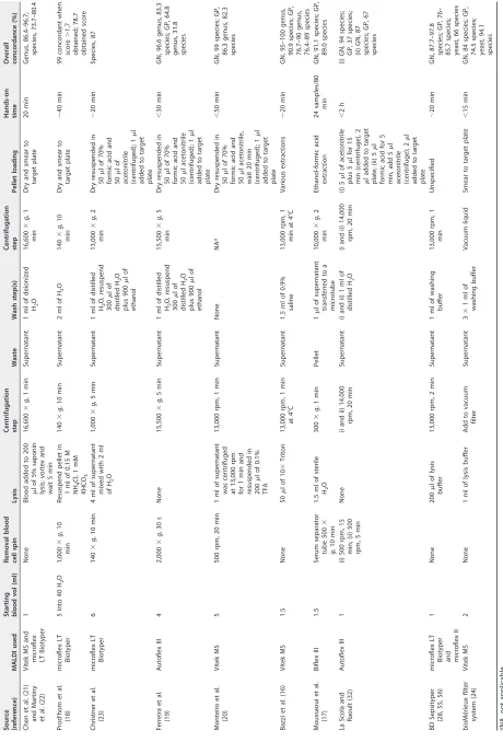
TABLE 1 Summary of MALDI-TOF MS blood culturing methods Source (reference) MALDI used Starting blood vol (ml) Removal blood cell spin Lysis Centrifugation step Waste Wash step(s) Centrifugation step Pellet loading Hands-on time Overall concordance (%) Chen et al. (21 ) and Martiny et al. (22 ) Vitek MS and microflex LT Biotyper 1 None Blood added to 200 l of 5% saponin lysis; vortex and wait 5 min 16,600 ⫻ g , 1 min Supernatant 1 ml of deionized H2 O 16,600 ⫻ g ,1 min Dry and smear to target plate 20 min Genus, 86.4–96.7; species, 73.7–80.4 Prod’hom et al. (18 ) microflex LT Biotyper 5 into 40 H2 O 1,000 ⫻ g ,1 0 min Resuspend pellet in 1 ml of 0.15 M NH 4 C l,1m M KHCO 3 140 ⫻ g , 10 min Supernatant 2 ml of H2 O 140 ⫻ g ,1 0 min Dry and smear to target plate ⬃ 40 min 99 concordant when score ⬎ 1.7 obtained; 78.7 obtained score Christner et al. (23 ) microflex LT Biotyper 6 140 ⫻ g , 10 min 4 ml of supernatant mixed with 2 ml of H2 O 1,000 ⫻ g , 5 min Supernatant 1 ml of distilled H2 O, resuspend 300 lo f distilled H2 O plus 900 lo f ethanol 13,000 ⫻ g ,2 min Dry resuspended in 50 lo f7 0 % formic acid and 50 lo f acetonitrile (centrifuged); 1 l added to target plate ⬃ 20 min Species, 87 Ferreira et al. (19 ) Autoflex III 4 2,000 ⫻ g , 30 s None 15,500 ⫻ g , 5 min Supernatant 1 ml of distilled H2 O, resuspend 300 lo f distilled H2 O plus 900 lo f ethanol 15,500 ⫻ g ,5 min Dry resuspended in 50 lo f7 0 % formic acid and 50 l acetonitrile (centrifuged); 1 l added to target plate ⬍ 30 min GN, 96.6 genus, 83.3 species; GP, 64.8 genus, 31.8 species Monteiro et al. (20 ) Vitek MS 5 500 rpm, 20 min 1 ml of supernatant was centrifuged at 13,000 rpm for 1 min and resuspended in 200 l of 0.1% TFA 13,000 rpm, 1 min Supernatant None NA a Dry resuspended in 50 lo f7 0 % formic acid and 50 l acetonitrile, wait 20 min (centrifuged); 1 l added to target plate ⬍ 50 min GN, 99 species; GP, 86.3 genus, 82.3 species Bazzi et al. (16 ) Vitek MS 1.5 None 50 lo f1 0 ⫻ Triton 13,000 rpm, 1 min at 4°C Supernatant 1.5 ml of 0.9% saline 13,000 rpm, 1 min at 4°C Various extractions ⬃ 20 min GN, 95–100 genus, 90.9 species; GP, 76.7–90 genus, 76.4–89 species Moussaoui et al. (17 ) Biflex III 1.5 Serum separator tube 500 ⫻ g ,1 0m in 1.5 ml of sterile H2 O 300 ⫻ g , 1 min Pellet 1 l of supernatant transferred to a microtube 10,000 ⫻ g ,2 min Ethanol-formic acid extraction 24 samples/80 min GN, 91.1 species; GP, 89.0 species La Scola and Raoult (32 ) Autoflex III 1 (i) 500 rpm, 15 min; (ii) 500 rpm, 5 min None (i and ii) 14,000 rpm, 20 min Supernatant (i and ii) 1 ml of distilled H2 O (i and ii) 14,000 rpm, 20 min (i) 5 l of acetonitrile plus 5 l for 15 min (centrifuge), 2 l added to target plate; (ii) 5 l formic acid for 5 min, add 5 l acetonitrile (centrifuge); 2 l added to target plate ⬍ 2 h (i) GN, 94 species; GP, 37 species; (ii) GN, 87 species; GP, 67 species BD
Sepsityper (28,
55 , 56 ) microflex LT
Biotyper and microflex
II 1 None 200 l of lysis buffer 13,000 rpm, 2 min Supernatant 1 ml of washing buffer 13,000 rpm, 1 min Unspecified ⬃ 20 min GN, 87.7–97.8 species; GP, 76– 85.7 species; yeast, 66 species bioMérieux filter system (24 ) Vitek MS 2 None 1 ml of lysis buffer Add to vacuum filter Supernatant 3 ⫻ 1m lo f washing buffer Vacuum liquid Smear to target plate ⬍ 15 min GN, 84 species; GP, 74.5 species; yeast, 94.1 species aNA, not applicable.
on May 16, 2020 by guest
http://jcm.asm.org/
[image:3.585.53.513.71.749.2]Microcolonies.A final method to identify organisms growing in blood culture bottles is to perform testing on short-incubation colonies or microcolonies (25). Blood culture is directly plated to tryptic soy agar (TSA) with 5% sheep blood agar and incubated at 35°C⫾2°C for a shortened period, such as 5 h, to allow for growth of microcolonies. These colonies are then treated as normal colony growth with respect to performing MALDI-TOF MS identification. Testing of microcolonies increases TAT compared to other methods, but it removes the need to perform labor-intensive purification. Slow-growing organisms, such as anaerobes and yeast (Candidaspecies), require extended periods of growth to obtain enough organism for testing (26). The short incubation period does not allow differentiation of various colony morphologies, so the inability of MALDI-TOF MS to distinguish polymicrobial specimens is still applicable to microcolony testing.
ASSAY PERFORMANCE AND OPTIMIZATION
With the lack of standardization, the overall performance of these methods varies greatly, with data ranging from 60 to 99% concordance to a species level (16, 19, 24). Due to the variety of methods used, direct comparisons between studies are difficult, as various extraction methods could have large effects on performance. In addition, many common pitfalls are observed in isolate identification with MALDI-TOF MS, such as reduced efficiency for identification of Gram positives (GP) and yeast, as well as limitations in closely related species that can affect testing from positive blood cultures. Table 1 summarizes several of these studies, highlighting the method of purification used along with average accuracy for organism testing.
Performance of Gram-negative bacterial identification. The composition of Gram-negative (GN) cell walls is such that extraction of proteins through common MALDI-TOF MS protocols is reliable and highly accurate. Purification methods from positive blood culture have results similar to those of direct colony testing, with concordance ranging from 84 to 99% to a species-level identification, with most studies observing ⬎90% concordance (19, 20). In one study, the authors found reliable identification to the genus (but not species) level forEnterobacter cloacaeand to the species level forProteus vulgaris(n⫽5 for each organism) (16). The use of microcolo-nies works well for GN bacteria, but anaerobic organisms fail to grow if the incubation is not extended (16, 26). Due to the genetic similarities between Escherichia coliand
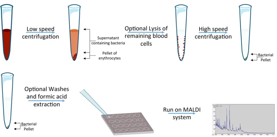
FIG 1Procedures between studies to perform identification directly from positive blood cultures using MALDI-TOF MS can differ greatly but usually
follow a similar workflow. A sample of blood culture is drawn from the bottle and is centrifuged to pellet the erythrocytes. In some studies, after the supernatant is collected, an optional lysis of remaining blood culture cells is performed to further clean the sample to produce less background. High-speed centrifugation is performed to pellet the remaining bacteria. This pellet can be washed and centrifuged a few times to improve the purification of bacterial pellets. Once purified, either an extraction can be performed or the bacteria can be added to a target plate and tested on a MALDI system for identification.
on May 16, 2020 by guest
http://jcm.asm.org/
[image:4.585.41.525.66.306.2]Shigella, testing of these organisms cannot be differentiated by MALDI-TOF MS, a limitation found in both isolate and blood culture testing.
Performance of Gram-positive bacterial identification. Testing GP organisms directly from positive blood cultures reduces the accuracy of MALDI-TOF MS. The percent agreement to the species level for GP organisms ranges from 65 to 96%, with most publications observing an overall GP identification rate around 80% (21, 27, 28). Using the Bruker system, viridians group streptococci are more difficult to identify and are often misidentified as Streptococcus pneumoniae (16, 17). Misidentification is a known limitation, as this occurs using direct colony testing, suggesting that it is more of a limitation of the library and unlikely to be the effects from testing blood culture. This issue is not observed using Vitek MS (bioMérieux, Inc., Durham, NC) due to the binning method utilized when assigning weight to the presence or absence of indi-vidual peaks (29). Scores directly from colonies can be improved using the Biotyper by the addition of spectra and a novel algorithm. Harju et al. reported an algorithm that uses organism list scores combined with the expanded MALDI Biotyper database (additional 1,000 strains added) (30). The new algorithm improved the identification of mitis species group streptococci from 66 misidentifications to just 1 when results were compared to those with 16S rRNA gene sequencing. This algorithm was performed only on isolates but would likely improve identifications directly from positive blood bottles, as similar studies expanding libraries for the identification ofBurkholderiaspecies have improved blood culture testing (31).
Data on the accuracy of MALDI-TOF MS directly from blood for staphylococcal species demonstrate mixed results. In several studies, the identification of Staphylococ-cus aureus was accurate in ⬎95% of the isolates tested, but the accuracy rate for coagulase-negative staphylococci (CoNS) fell below 90% (21, 28). However, another study observed only 40% correct identifications toS. aureusand 32% toStaphylococcus epidermidiscompared to MALDI-TOF MS from a colony (32). The use of formic acid may account for some of these differences, as extraction with formic acid increased the correct identification ofS. aureusfrom 40% to 58% and ofS. epidermidisfrom 32% to 80%, and it increased the mean score from 1.46 to 2.0 (32). In addition, the studies that found⬎95% agreement forS. aureusalso used formic acid for the extraction. Testing with microcolonies worked well for the identification of S. aureus, as two studies observed high accuracy, obtaining 87.5% and 95.5% correct identification rates (25, 33). Identifications of CoNS were different between the two studies. The study that reported 95.5% agreement forS. aureusalso had a better percent agreement for CoNS, at 78% compared to 21.7%, respectively. The study that had better performance used formic acid for extraction, whereas that with lower performance used trifluoroacetic acid (TFA), which may have affected the performance. Alternatively, these differences may be due to the different bottle types used, as previous data suggest performance differences when using different Bactec bottles, with Bactec Plus (Becton Dickinson, Sparks, MD) performing better than BacT/Alert SA (bioMérieux, Nürtingen, Germany) and BacT/Alert FA (bioMérieux, Nürtingen, Germany), the latter of which (Bactec Plus) was used by a study reporting poorer performance (34). These poor-performing studies used bottles with charcoal, which has been shown to decrease the accuracy of MALDI-TOF MS performed directly from blood culture (35, 36). Finally, one study modified the spectral analysis parameters by removing the interpretations of any peaks with an m/zratio below 4,000, which increased GP identification from 80% to 89% and specifically improved the detection ofS. epidermidis(28). These low peaks likely represent residual blood components, and overall removal increased confidence scores in 68% of all specimens tested (28).
Performance of yeast identification.The cell walls of yeast are difficult to disrupt using common MALDI-TOF MS methods, which affects the performance of MALDI-TOF MS identification for both colony and positive blood culture testing. Without additional modifications to the extraction methods, studies that have tested yeast direct from blood observe identification rates ranging from 0 to 70% compared to standard
on May 16, 2020 by guest
http://jcm.asm.org/
identification methods (depending on the publication, the standard of care consists of biochemical analysis or MALDI-TOF MS from direct colonies) (27, 28). MALDI-TOF MS of microcolonies also fails to identify yeast, with one study failing to identify any of the 36 yeast cultures after a 5-h subculture, likely due to insufficient growth (25).
Optimized protocols for the detection of yeast have been published. In a study by Yan et al., 42Candidaspecies were tested, and all were correctly identified compared to phenotypic methods (37). Unique to this study was the addition of 2 water lysis steps prior to performing the Sepsityper kit to increase the removal of blood cells from the sample. This reduces the background spectral noise from the blood matrix and helps isolate the yeast cells.
Polymicrobial specimens.Testing of polymicrobial blood specimens is unreliable due to the inherent difficulty of MALDI-TOF MS matching mixed spectra to an instru-ment’s library. In one study, direct blood testing found 22 polymicrobial bottles. In 18 specimens, one of the organisms was detected, 2 reported no reliable identification, and the remaining two resulted in an incorrect identification (32). This poor perfor-mance has been observed in other studies as well, with one study identifying a single organism in 64% (9/14) of the specimens tested and another observing single detection in 76% (16/21) of polymicrobial specimens (21, 28). Organism abundance likely explains why only one organism can often be detected in polymicrobial specimens. MALDI-TOF MS peaks are composed of not only the size but the amount of detected material, suggesting that high-abundance organisms may have enough defined peaks to allow the software to distinguish the spectra over the background of the second organism. Interestingly, by reducing the cutoff to scores of ⬎1.6 with the Biotyper system and using top-10-matched pattern choices, Chen et al. (21) could detect both organisms in 24% (5/21) of polymicrobial specimens. Testing of micro-colonies also fails to reliably report both organisms from polymicrobial specimens, with data demonstrating similar results at 83% (24/29) identification of the preva-lent organism growing.
Modification of cutoff values. Several studies have evaluated their data using various cutoff scores to see if different thresholds increase accuracy while resulting in no incorrect identifications. The Vitek MS system generates a confidence score in terms of percentage, and few studies have been performed to determine the agreement of direct from positive blood culture identification with low scores. One study did com-ment that 4/5 specimens that had a match rate of less than 75% had either a correct species or genus identification, suggesting that lowering the acceptable percent con-fidence score for reporting may increase identification (24). Similarly, lowering accept-able scores for identification for the Bruker system may improve overall detection. Instead of a percent confidence score, the Bruker system reports scores in one of three categories (cutoffs): unreliable identification (⬍1.7), genus identification (1.7 to 1.99), or reliable species identification (⬎2.0). Reducing the cutoffs to report species identifica-tion can improve identificaidentifica-tion directly from blood culture. In one study, the authors found that reanalysis of 304 blood cultures demonstrated that cutoffs could be dropped toⱖ1.6 without causing any misidentifications and raise species identification rates from 75 to 92% (23). The reduction in score was due to suboptimal specimen conditions where high background and low analyte concentrations created poor database matching. However, the authors did caution that species that are highly similar in spectra, such as the viridans streptococci, could be at risk for incorrect identifications using lower cutoffs. Another study looking at 566 blood cultures con-sisting of both GN and GP organisms and found similar results, allowing correct identification of an additional 5.2% samples when using scores ofⱖ1.4 that contained the same organism in the top 4 matches (17). Differentiating between poor specimen preparation and closely related species can be improved by reporting only species results when the score of the top match to the first different species isⱖ0.3 (22). One study specifically looking at the identification of yeast found that scores of⬎1.2 were
on May 16, 2020 by guest
http://jcm.asm.org/
reliable if samples were tested in quadruplicate and all 4 top identifications matched (38).
ANTIMICROBIAL SUSCEPTIBILITY TESTING
In addition to rapid identification, administration of appropriate antibiotics is es-sential to improve patient care. Organism identification can guide antibiotic therapy; however, many organisms have unpredictable resistance patterns and need full AST to determine optimal treatment. MALDI-TOF MS can be used to detect resistance mech-anisms, but currently, these methods are labor-intensive. Three major methods have been described, which include: detection of enzymatic activity, direct analysis of bacterial extracts, and comparison of protein concentrations in organisms growing with or without the presence of antibiotics.
Detection of enzymatic activity.One mechanism of resistance to antibiotics is the enzymatic modification of either the antibiotic or the antibiotic’s target substrate. Examples of these are the hydrolysis of the-lactam ring by-lactamases and meth-ylation of rRNA conferring resistance to aminoglycosides (39). Detection of-lactamase activity with MALDI-TOF MS was first reported in 2011 (40). In this study, carbapen-emase and non-carbapencarbapen-emase-producing strains were inoculated with and without ertapenem for 2.5 h. A centrifuged supernatant was then added to a MALDI-TOF MS target plate and visualized for peaks below 1,000 Da. Hydrolysis of ertapenem was detected by a shift in peaks of unhydrolyzed ertapenem at 476, 798, and 521 Da (based on sodium binding) to hydrolyzed ertapenem at 450 Da (40). Similar testing has been performed to test positive blood cultures. One study evaluated 55 blood cultures containingKlebsiella pneumoniaeand measured the hydrolyzation of ertapenem de-fined by a peak at 472.5 Da (41). Bacterial pellets isolated using the Sepsityper kit were resuspended in 30l of an ertapenem solution at 1 mg/ml and tested every 30 min over 3 h. All 55 isolates correlated with Vitek 2 results tested from subcultured isolates. A similar study looking at-lactam resistance inEnterobacteriaceaewas also performed from positive blood cultures using cefotaxime (0.5 mg/ml), and the results were compared to those with Etest and Check-MDR CT 101 and 102 microarray kits (Check-Points, Wageningen, The Netherlands) (42). Resistance was determined using a com-parison of the peak intensities of hydrolyzed and nonhydrolyzed products. One hun-dred consecutive blood cultures were tested, 94 of which matched the standard-of-care result. Of the 6 major errors, 2Klebsiella oxytocaisolates had the same MALDI-TOF MS result from both direct positive blood culture and isolate, but these did not match the Etest results. The remaining 4 only generated major errors from the blood culture. No very major errors were detected. Of note, common matrices used for MALDI-TOF MS have peaks below 1,000 Da with higher intensity than the antibiotic products, meaning that matrix composition must be considered before testing isolates. Furthermore, high-quality bacterial purification is needed, as many blood proteins are⬍1,000 Da and could affect identification (43). Anything below this range also requires calibration, as these instruments are not originally set up to accurately measure these m/z. Finally, methylation of rRNA is performed in a manner similar to that in-lactamase studies, but instead of antibiotic products, a 16-Da peak shift of rRNA is measured as methyl-ation increases the mass of the molecule (44). Methods that detect enzymatic modifi-cation are limited, as a lack of enzymatic activity does not rule out resistance by other mechanisms, such as porin alterations, efflux pumps, or alterations in penicillin-binding proteins.
Direct analysis of bacterial pellets. An ideal methodology from a workflow standpoint would be to differentiate resistant and sensitive organisms from differences in protein peaks. Several studies have been performed to identify unique peaks to differentiate methicillin-susceptibleS. aureus(MSSA) and methicillin-resistantS. aureus
(MRSA), vancomycin-resistant Enterococcus from vancomycin-sensitive Enterococcus, and-lactamases from each other (45–48). These studies have shown some success; however, in the case of differentiating MSSA and MRSA, there is contracting evidence, with one study able to find 14 MRSA- and 2 MSSA-specific peaks, whereas the other
on May 16, 2020 by guest
http://jcm.asm.org/
study could not differentiate the two types (45, 46). Differences in extraction protocols might be attributed to these differences, but further studies are needed to determine the utility of this method. Another limitation to this method is that inducible resistance may not have substantial peaks for detection.
Comparison of protein concentrations.A final method to use MALDI-TOF MS for the detection of resistance is by comparing peak intensities of isolates growing with and without antibiotic. In this method, a single dilution of an isolate is created and used to inoculate 2 nutrient-rich broths: one broth with the antibiotic of interest and another without. The bacteria are then incubated at 35°C for 1 to 4 h, pelleted, and then tested using the MALDI-TOF MS for spectrum analysis. The broth without antibiotic acts as a reference for peak intensity as optimal growth and is used to compare to the peaks with antibiotics. Resistant organisms will continue to replicate and have protein con-centrations similar to those of the control tube, but susceptible isolates will fail to grow and contain fewer proteins, creating a reduction in peak intensity. One study evaluated 99 patient-derived positive blood cultures to determine the effectiveness of this method for cefotaxime, piperacillin-tazobactam, and ciprofloxacin (49). Purified pellets using both the Sepsityper kit and serum separator tubes were diluted in Mueller-Hinton containing either no antibiotic or concentrations of antibiotic one dilution higher than the EUCAST breakpoints forEnterobacteriaceae. The final concentration of bacteria was diluted to 5⫻106CFU/ml. After a 2.5-h incubation at 37°C, each dilution was tested by MALDI-TOF MS, and an automated software evaluated the relative growth. The assay had accuracy similar to that of current gold standard MIC testing, with only 1 cipro-floxacin mismatch and 5 piperacillin-tazobactam mismatches, 3 of which resulted in very major errors. Retesting from subculture gave the correct value. Finally, the authors commented that for piperacillin-tazobactam testing, 3 h was needed to obtain suffi-cient growth separation between susceptible and nonsusceptible organisms. In theory, these data are promising for rapid resistance detection, especially as any drug-bug combination could be tested. However, reproducibility may be difficult, as differences in starting materials and transfer to a target plate might skew results.
IMPACT
Assessment of clinical impact is essential to understand the value of new assays and technologies developed in the clinical microbiology field. Knowledge of impact is not only important to establish best practices but also aids in justifying the workflow and cost of any new system. To date, there have been a few publications evaluating the effects of rapid identification of blood cultures using MALDI-TOF MS protocols. Unsur-prisingly, these studies have found that rapid identification from blood culture bottles with MALDI-TOF MS reduces the TAT, ranging from 18.1 to 34.3 h, compared to that of conventional culture, which ranges from 48 to 72 h (50, 51). No evaluations have been made with other molecular assays, but the TAT would be comparable, with a slightly shorter TAT for MALDI-TOF MS, as most purification methods take less than 30 min. Multiple studies also demonstrated that the time to optimal treatment was also improved, with results ranging from 12 to 36 h quicker than the hospitals’ previous workflow (50, 52). The study that observed a greater time to optimal treatment also introduced an antimicrobial stewardship program, which might explain the difference in the time to optimal treatment. These data are also supported by the work of French et al. that retrospectively assessed how the use of MALDI-TOF MS from positive blood cultures would affect patient care (53). The authors found that out of 115 cases, 28 would have had clear clinical benefit, and 5 of these patients would have received appropriate antibiotics 24 h earlier.
The combination of rapid identification (ID) with AST may have an even larger effect on patient outcome. One study combined the use of MALDI-TOF MS and the BD Phoenix (BD Diagnostic Systems, Sparks, MD) for AST from positive blood cultures (54). Using a pre-/postcomparison study of workflows, the authors found that therapy could be adjusted by an average of 75 h (⫾48 h), reduced length of stay from 11.0 days to 9.3, reduced the mortality rate from 10.7 to 5.6%, and reduced the average cost per
on May 16, 2020 by guest
http://jcm.asm.org/
survivor by an average of $19,547.00 when testing from positive blood cultures was implemented. This testing was only performed with bottles containing GN organisms, which may have a larger impact on patient care than GP infections due to the impact that genotypic data (MRSA and vancomycin-resistantEnterococcus[VRE]) have on patient care. The costs of instrumentation and testing also play a considerable role in defining what methods a laboratory implements. Unlike molecular assays that can only be used on various specimen types, investment in MALDI-TOF MS will reduce identification costs for testing from most colonies and positive blood cultures. The addition of blood culture testing is an added benefit to increase the rate of investment. The cost of implementation can vary due to several factors, such as the success rate of MALDI-TOF MS, the use of commercialized kits, such as the Sepsityper, for purification, and the current method for identification. One study suggests that laboratories switching from MALDI-TOF MS to direct colony testing to a Sepsityper kit purification methodology may see an increase in cost up to $3.64, which included the cost of the 14.8% of cultures that could not be identified from direct blood culture along with the cost difference in the Sepsityper kit (51). The additional cost of the technologist’s time was not included in this analysis. The authors do comment that the cost would be marginal if switching laboratory testing from a commercial molec-ular panel instead of MALDI-TOF MS for identification.
SUMMARY
Each laboratory has different resources and capabilities, and the needs for their patient population vary, which means laboratory directors need to weigh the benefits and challenges when considering the use of MALDI-TOF MS for identification directly from blood cultures. Currently, all methods require extensive laboratory validation, and no FDA-cleared assays exist. Furthermore, the accuracy of identification, especially in GP organisms, is higher in molecular assays. However, the benefits may outweigh these limitations. Overall, MALDI-TOF MS is more cost-effective than molecular identification, is not limited to the panel generated by the molecular assay, and is quicker than current molecular assays. Molecular assays are only useful for blood cultures, so laboratories with limited budgets will need to purchase equipment specific to blood culture identification instead of purchasing a MALDI-TOF MS instrument that can also test isolates, creating more utility for the cost of instrumentation. Laboratories capable of having both MALDI-TOF MS and molecular identification could optimize patient care by splitting the workflow based on Gram stain. A possible ideal workflow may be to only run GN organisms for MALDI-TOF MS identification. MALDI-TOF MS accuracy for GN matches that of molecular assays, and although MALDI-TOF MS lacks genotypic resis-tance markers, the variety of resisresis-tance in GN infections often requires full AST for optimal patient management.
In any workflow where a laboratory chooses to incorporate MALDI-TOF MS for blood culture, it will be important for the laboratories to define which protocol is appropriate. The literature contains a variety of methodologies to perform purification of bacterial pellets. Unfortunately, the number of studies and the low sample size in studies that compare methods with the same specimen make it difficult to determine optimal testing practices. However, similarities, such as the use of an erythrocyte lysis step along with sterile water washes and formic acid extraction, appear to be consistent steps in publications demonstrating improved performance. Current commercialized purifica-tion kits may help standardize reagents but do not demonstrate superior performance compared to LDT methods, and they add to the overall cost per sample tested.
In the current state of the field, AST directly from blood culture with MALDI-TOF MS is out of reach for most laboratories. In addition, data are lacking to determine how comparable these results will be to gold standard broth microdilutions. Advancements in standardization of methods are needed for AST from MALDI-TOF MS to be useful, which is why automation and microfluidics may play an essential role in this transition. Instrumentation could be developed where blood culture is placed into a cartridge, purified through lysis and filtration, and loaded into wells with and without panels of antibiotics, which are then incubated for a period before automated spotting to a
on May 16, 2020 by guest
http://jcm.asm.org/
MALDI-TOF MS target. Technologists would then just run the plates and software comparing growth through peak intensity and could report sensitive/resistant results, with the potential for MICs to be reported as well. To get to this point, more research is needed, but the potential applications for MALDI-TOF MS in all aspects of blood culture testing are promising.
ACKNOWLEDGMENT
We thank Arjun A. S. Ahluwalia for his artistic input in designing Fig. 1.
REFERENCESS
1. Angus DC, Wax RS. 2001. Epidemiology of sepsis: an update. Crit Care Med 29:S109 –116.https://doi.org/10.1097/00003246-200107001-00035. 2. Levy MM, Rhodes A, Phillips GS, Townsend SR, Schorr CA, Beale R, Osborn T, Lemeshow S, Chiche JD, Artigas A, Dellinger RP. 2014. Surviv-ing Sepsis Campaign: association between performance metrics and outcomes in a 7.5-year study. Intensive Care Med 40:1623–1633.https:// doi.org/10.1007/s00134-014-3496-0.
3. Kumar A, Roberts D, Wood KE, Light B, Parrillo JE, Sharma S, Suppes R, Feinstein D, Zanotti S, Taiberg L, Gurka D, Kumar A, Cheang M. 2006. Duration of hypotension before initiation of effective antimicrobial ther-apy is the critical determinant of survival in human septic shock. Crit Care Med 34:1589 –1596. https://doi.org/10.1097/01.CCM.0000217961 .75225.E9.
4. Ibrahim EH, Sherman G, Ward S, Fraser VJ, Kollef MH. 2000. The influence of inadequate antimicrobial treatment of bloodstream infections on patient outcomes in the ICU setting. Chest 118:146 –155.https://doi.org/ 10.1378/chest.118.1.146.
5. Shorr AF, Micek ST, Welch EC, Doherty JA, Reichley RM, Kollef MH. 2011. Inappropriate antibiotic therapy in Gram-negative sepsis increases hos-pital length of stay. Crit Care Med 39:46 –51.https://doi.org/10.1097/ CCM.0b013e3181fa41a7.
6. Ledeboer NA, Lopansri BK, Dhiman N, Cavagnolo R, Carroll KC, Granato P, Thomson R, Jr, Butler-Wu SM, Berger H, Samuel L, Pancholi P, Swyers L, Hansen GT, Tran NK, Polage CR, Thomson KS, Hanson ND, Winegar R, Buchan BW. 2015. Identification of Gram-negative bacteria and genetic resistance determinants from positive blood culture broths by use of the Verigene Gram-negative blood culture multiplex microarray-based mo-lecular assay. J Clin Microbiol 53:2460 –2472.https://doi.org/10.1128/ JCM.00581-15.
7. Salimnia H, Fairfax MR, Lephart PR, Schreckenberger P, DesJarlais SM, Johnson JK, Robinson G, Carroll KC, Greer A, Morgan M, Chan R, Loef-felholz M, Valencia-Shelton F, Jenkins S, Schuetz AN, Daly JA, Barney T, Hemmert A, Kanack KJ. 2016. Evaluation of the FilmArray blood culture identification panel: results of a multicenter controlled trial. J Clin Mi-crobiol 54:687– 698.https://doi.org/10.1128/JCM.01679-15.
8. Wolk DM, Struelens MJ, Pancholi P, Davis T, Della-Latta P, Fuller D, Picton E, Dickenson R, Denis O, Johnson D, Chapin K. 2009. Rapid detection of Staphylococcus aureus and methicillin-resistant S. aureus (MRSA) in wound specimens and blood cultures: multicenter preclinical evaluation of the Cepheid Xpert MRSA/SA skin and soft tissue and blood culture assays. J Clin Microbiol 47:823– 826.https://doi.org/10.1128/JCM.01884-08.
9. Angeletti S. 2016. Matrix assisted laser desorption time of flight mass spectrometry (MALDI-TOF MS) in clinical microbiology. J Microbiol Meth-ods 138:20 –29.https://doi.org/10.1016/j.mimet.2016.09.003.
10. Clark AE, Kaleta EJ, Arora A, Wolk DM. 2013. Matrix-assisted laser de-sorption ionization–time of flight mass spectrometry: a fundamental shift in the routine practice of clinical microbiology. Clin Microbiol Rev 26:547– 603.https://doi.org/10.1128/CMR.00072-12.
11. Buetti N, Marschall J, Atkinson A, Kronenberg A. 2016. National blood-stream infection surveillance in Switzerland 2008 –2014: different pat-terns and trends for university and community hospitals. Infect Control Hosp Epidemiol 2:1– 8.
12. Ferreira L, Sanchez-Juanes F, Gonzalez-Avila M, Cembrero-Fucinos D, Herrero-Hernandez A, Gonzalez-Buitrago JM, Munoz-Bellido JL. 2010. Direct identification of urinary tract pathogens from urine samples by matrix-assisted laser desorption ionization-time of flight mass spectrometry. J Clin Microbiol 48:2110 –2115.https://doi.org/10.1128/JCM.02215-09.
13. Hsieh SY, Tseng CL, Lee YS, Kuo AJ, Sun CF, Lin YH, Chen JK. 2008. Highly efficient classification and identification of human pathogenic bacteria
by MALDI-TOF MS. Mol Cell Proteomics 7:448 – 456.https://doi.org/10 .1074/mcp.M700339-MCP200.
14. Cunningham SA, Patel R. 2015. Standard matrix-assisted laser desorption ionization–time of flight mass spectrometry reagents may inactivate potentially hazardous bacteria. J Clin Microbiol 53:2788 –2789.https:// doi.org/10.1128/JCM.00957-15.
15. Lasch P, Nattermann H, Erhard M, Stammler M, Grunow R, Bannert N, Appel B, Naumann D. 2008. MALDI-TOF mass spectrometry compatible inactivation method for highly pathogenic microbial cells and spores. Anal Chem 80:2026 –2034.https://doi.org/10.1021/ac701822j. 16. Bazzi AM, Rabaan AA, El Edaily Z, John S, Fawarah MM, Al-Tawfiq JA.
2016. Comparison among four proposed direct blood culture microbial identification methods using MALDI-TOF MS. J Infect Public Health 10:308 –315.https://doi.org/10.1016/j.jiph.2016.05.011.
17. Moussaoui W, Jaulhac B, Hoffmann AM, Ludes B, Kostrzewa M, Riegel P, Prevost G. 2010. Matrix-assisted laser desorption ionization time-of-flight mass spectrometry identifies 90% of bacteria directly from blood culture vials. Clin Microbiol Infect 16:1631–1638.https://doi.org/10.1111/j.1469 -0691.2010.03356.x.
18. Prod’hom G, Bizzini A, Durussel C, Bille J, Greub G. 2010. Matrix-assisted laser desorption ionization–time of flight mass spectrometry for direct bacterial identification from positive blood culture pellets. J Clin Micro-biol 48:1481–1483.https://doi.org/10.1128/JCM.01780-09.
19. Ferreira L, Sanchez-Juanes F, Porras-Guerra I, Garcia MI, Garcia-Sanchez JE, Gonzalez-Buitrago JM, Munoz-Bellido JL. 2011. Microorgan-isms direct identification from blood culture by matrix-assisted laser desorption/ionization time-of-flight mass spectrometry. Clin Microbiol Infect 17:546 –551.https://doi.org/10.1111/j.1469-0691.2010.03257.x. 20. Monteiro J, Inoue FM, Lobo AP, Sugawara EK, Boaretti FM, Tufik S. 2015.
Fast and reliable bacterial identification direct from positive blood cul-ture using a new TFA sample preparation protocol and the Vitek MS system. J Microbiol Methods 109:157–159. https://doi.org/10.1016/j .mimet.2014.12.009.
21. Chen JH, Ho PL, Kwan GS, She KK, Siu GK, Cheng VC, Yuen KY, Yam WC. 2013. Direct bacterial identification in positive blood cultures by use of two commercial matrix-assisted laser desorption ionization–time of flight mass spectrometry systems. J Clin Microbiol 51:1733–1739.https:// doi.org/10.1128/JCM.03259-12.
22. Martiny D, Dediste A, Vandenberg O. 2012. Comparison of an in-house method and the commercial Sepsityper kit for bacterial identification di-rectly from positive blood culture broths by matrix-assisted laser desorption-ionisation time-of-flight mass spectrometry. Eur J Clin Microbiol Infect Dis 31:2269 –2281.https://doi.org/10.1007/s10096-012-1566-1. 23. Christner M, Rohde H, Wolters M, Sobottka I, Wegscheider K,
Aep-felbacher M. 2010. Rapid identification of bacteria from positive blood culture bottles by use of matrix-assisted laser desorption–ionization time of flight mass spectrometry fingerprinting. J Clin Microbiol 48: 1584 –1591.https://doi.org/10.1128/JCM.01831-09.
24. Fothergill A, Kasinathan V, Hyman J, Walsh J, Drake T, Wang YF. 2013. Rapid identification of bacteria and yeasts from positive-blood-culture bottles by using a lysis-filtration method and matrix-assisted laser de-sorption ionization–time of flight mass spectrum analysis with the SARA-MIS database. J Clin Microbiol 51:805– 809.https://doi.org/10.1128/JCM .02326-12.
25. Verroken A, Defourny L, Lechgar L, Magnette A, Delmee M, Glupczynski Y. 2015. Reducing time to identification of positive blood cultures with MALDI-TOF MS analysis after a 5-h subculture. Eur J Clin Microbiol Infect Dis 34:405– 413.https://doi.org/10.1007/s10096-014-2242-4.
26. Gonzalez MD, Weber CJ, Burnham CA. 2016. Rapid identification of microorganisms from positive blood cultures by testing early growth on
on May 16, 2020 by guest
http://jcm.asm.org/
solid media using matrix-assisted laser desorption ionization–time of flight mass spectrometry. Diagn Microbiol Infect Dis 85:133–135.https:// doi.org/10.1016/j.diagmicrobio.2016.02.018.
27. Morgenthaler NG, Kostrzewa M. 2015. Rapid identification of pathogens in positive blood culture of patients with sepsis: review and meta-analysis of the performance of the Sepsityper kit. Int J Microbiol 2015: 827416.https://doi.org/10.1155/2015/827416.
28. Buchan BW, Riebe KM, Ledeboer NA. 2012. Comparison of the MALDI Biotyper system using Sepsityper specimen processing to routine mi-crobiological methods for identification of bacteria from positive blood culture bottles. J Clin Microbiol 50:346 –352.https://doi.org/10.1128/JCM .05021-11.
29. Rychert J, Burnham CA, Bythrow M, Garner OB, Ginocchio CC, Jennemann R, Lewinski MA, Manji R, Mochon AB, Procop GW, Richter SS, Sercia L, Westblade LF, Ferraro MJ, Branda JA. 2013. Multicenter evaluation of the Vitek MS matrix-assisted laser desorption ionization–time of flight mass spectrometry system for identification of Gram-positive aerobic bacteria. J Clin Microbiol 51:2225–2231.https://doi.org/10.1128/JCM.00682-13. 30. Harju I, Lange C, Kostrzewa M, Maier T, Rantakokko-Jalava K, Haanpera M.
2017. Improved differentiation of Streptococcus pneumoniae and other S. mitis group streptococci by MALDI Biotyper using an improved MALDI Biotyper database content and a novel result interpretation algorithm. J Clin Microbiol 55:914 –922.https://doi.org/10.1128/JCM.01990-16.
31. Inglis TJ, Healy PE, Fremlin LJ, Golledge CL. 2012. Use of matrix-assisted laser desorption/ionization time-of-flight mass spectrometry analysis for rapid confirmation of Burkholderia pseudomallei in septicemic melioid-osis. Am J Trop Med Hyg 86:1039 –1042.https://doi.org/10.4269/ajtmh .2012.11-0454.
32. La Scola B, Raoult D. 2009. Direct identification of bacteria in positive blood culture bottles by matrix-assisted laser desorption ionisation time-of-flight mass spectrometry. PLoS One 4:e8041.https://doi.org/10.1371/ journal.pone.0008041.
33. Kohlmann R, Hoffmann A, Geis G, Gatermann S. 2015. MALDI-TOF mass spectrometry following short incubation on a solid medium is a valuable tool for rapid pathogen identification from positive blood cultures. Int J Med Microbiol 305:469 – 479.https://doi.org/10.1016/j.ijmm.2015.04.004. 34. Schmidt V, Jarosch A, Marz P, Sander C, Vacata V, Kalka-Moll W. 2012.
Rapid identification of bacteria in positive blood culture by matrix-assisted laser desorption ionization time-of-flight mass spectrometry. Eur J Clin Microbiol Infect Dis 31:311–317. https://doi.org/10.1007/ s10096-011-1312-0.
35. Wüppenhorst N, Consoir C, Lorch D, Schneider C. 2012. Direct identifi-cation of bacteria from charcoal-containing blood culture bottles using matrix-assisted laser desorption/ionisation time-of-flight mass spec-trometry. Eur J Clin Microbiol Infect Dis 31:2843–2850.https://doi.org/ 10.1007/s10096-012-1638-2.
36. Szabados F, Michels M, Kaase M, Gatermann S. 2011. The sensitivity of direct identification from positive BacT/ALERT (bioMérieux) blood cul-ture bottles by matrix-assisted laser desorption ionization time-of-flight mass spectrometry is low. Clin Microbiol Infect 17:192–195.https://doi .org/10.1111/j.1469-0691.2010.03229.x.
37. Yan Y, He Y, Maier T, Quinn C, Shi G, Li H, Stratton CW, Kostrzewa M, Tang YW. 2011. Improved identification of yeast species directly from positive blood culture media by combining Sepsityper specimen pro-cessing and Microflex analysis with the matrix-assisted laser desorption ionization Biotyper system. J Clin Microbiol 49:2528 –2532.https://doi .org/10.1128/JCM.00339-11.
38. Spanu T, Posteraro B, Fiori B, D’Inzeo T, Campoli S, Ruggeri A, Tumbarello M, Canu G, Trecarichi EM, Parisi G, Tronci M, Sanguinetti M, Fadda G. 2012. Direct MALDI-TOF mass spectrometry assay of blood culture broths for rapid identification of Candida species causing bloodstream infections: an observational study in two large microbiology laborato-ries. J Clin Microbiol 50:176 –179.https://doi.org/10.1128/JCM.05742-11. 39. Smith CA, Baker EN. 2002. Aminoglycoside antibiotic resistance by en-zymatic deactivation. Curr Drug Targets Infect Disord 2:143–160.https:// doi.org/10.2174/1568005023342533.
40. Burckhardt I, Zimmermann S. 2011. Using matrix-assisted laser desorp-tion ionizadesorp-tion–time of flight mass spectrometry to detect carbapenem resistance within 1 to 2.5 hours. J Clin Microbiol 49:3321–3324.https:// doi.org/10.1128/JCM.00287-11.
41. Sakarikou C, Ciotti M, Dolfa C, Angeletti S, Favalli C. 2017. Rapid detec-tion of carbapenemase-producing Klebsiella pneumoniae strains
de-rived from blood cultures by matrix-assisted laser desorption ionization–time of flight mass spectrometry (MALDI-TOF MS). BMC Mi-crobiol 17:54.https://doi.org/10.1186/s12866-017-0952-3.
42. Jung JS, Popp C, Sparbier K, Lange C, Kostrzewa M, Schubert S. 2014. Evaluation of matrix-assisted laser desorption ionization–time of flight mass spectrometry for rapid detection of beta-lactam resistance in Enterobacteriaceae derived from blood cultures. J Clin Microbiol 52: 924 –930.https://doi.org/10.1128/JCM.02691-13.
43. Dwivedi P, Schultz AJ, Hill HH. 2010. Metabolic profiling of human blood by high-resolution ion mobility mass spectrometry (IM-MS). Int J Mass Spectrom 298:78 –90.https://doi.org/10.1016/j.ijms.2010.02.007. 44. Kirpekar F, Douthwaite S, Roepstorff P. 2000. Mapping
posttranscrip-tional modifications in 5S ribosomal RNA by MALDI mass spectrometry. RNA 6:296 –306.https://doi.org/10.1017/S1355838200992148. 45. Edwards-Jones V, Claydon MA, Evason DJ, Walker J, Fox AJ, Gordon DB.
2000. Rapid discrimination between sensitive and methicillin-resistant Staphylococcus aureus by intact cell mass spectrometry. J Med Microbiol 49:295–300.https://doi.org/10.1099/0022-1317-49-3-295. 46. Bernardo K, Pakulat N, Macht M, Krut O, Seifert H, Fleer S, Hunger F,
Kronke M. 2002. Identification and discrimination of Staphylococcus aureus strains using matrix-assisted laser desorption/ionization-time of flight mass spectrometry. Proteomics 2:747–753. https://doi.org/10 .1002/1615-9861(200206)2:6⬍747::AID-PROT747⬎3.0.CO;2-V.
47. Camara JE, Hays FA. 2007. Discrimination between wild-type and ampicillin-resistant Escherichia coli by matrix-assisted laser desorption/ ionization time-of-flight mass spectrometry. Anal Bioanal Chem 389: 1633–1638.https://doi.org/10.1007/s00216-007-1558-7.
48. Griffin PM, Price GR, Schooneveldt JM, Schlebusch S, Tilse MH, Urbanski T, Hamilton B, Venter D. 2012. Use of matrix-assisted laser desorption ionization–time of flight mass spectrometry to identify vancomycin-resistant enterococci and investigate the epidemiology of an outbreak. J Clin Microbiol 50:2918 –2931.https://doi.org/10.1128/JCM.01000-12. 49. Jung JS, Hamacher C, Gross B, Sparbier K, Lange C, Kostrzewa M,
Schubert S. 2016. Evaluation of a semiquantitative matrix-assisted laser desorption ionization–time of flight mass spectrometry method for rapid antimicrobial susceptibility testing of positive blood cultures. J Clin Microbiol 54:2820 –2824.https://doi.org/10.1128/JCM.01131-16. 50. Verroken A, Defourny L, le Polain de Waroux O, Belkhir L, Laterre PF,
Delmee M, Glupczynski Y. 2016. Clinical impact of MALDI-TOF MS iden-tification and rapid susceptibility testing on adequate antimicrobial treatment in sepsis with positive blood cultures. PLoS One 11:e0156299.
https://doi.org/10.1371/journal.pone.0156299.
51. Lagacé-Wiens PR, Adam HJ, Karlowsky JA, Nichol KA, Pang PF, Guenther J, Webb AA, Miller C, Alfa MJ. 2012. Identification of blood culture isolates directly from positive blood cultures by use of matrix-assisted laser desorp-tion ionizadesorp-tion–time of flight mass spectrometry and a commercial extrac-tion system: analysis of performance, cost, and turnaround time. J Clin Microbiol 50:3324 –3328.https://doi.org/10.1128/JCM.01479-12.
52. Beganovic M, Costello M, Wieczorkiewicz SM. 2017. Effect of matrix-assisted laser desorption ionization–time of flight mass spectrometry (MALDI-TOF MS) alone versus MALDI-TOF MS combined with real-time antimicrobial stewardship interventions on time to optimal antimicro-bial therapy in patients with positive blood cultures. J Clin Microbiol 55:1437–1445.https://doi.org/10.1128/JCM.02245-16.
53. French K, Evans J, Tanner H, Gossain S, Hussain A. 2016. The clinical impact of rapid, direct MALDI-TOF identification of bacteria from posi-tive blood cultures. PLoS One 11:e0169332. https://doi.org/10.1371/ journal.pone.0169332.
54. Perez KK, Olsen RJ, Musick WL, Cernoch PL, Davis JR, Land GA, Peterson LE, Musser JM. 2013. Integrating rapid pathogen identification and antimicro-bial stewardship significantly decreases hospital costs. Arch Pathol Lab Med 137:1247–1254.https://doi.org/10.5858/arpa.2012-0651-OA.
55. Yonetani S, Ohnishi H, Ohkusu K, Matsumoto T, Watanabe T. 2016. Direct identification of microorganisms from positive blood cultures by MALDI-TOF MS using an in-house saponin method. Int J Infect Dis 52:37– 42.
https://doi.org/10.1016/j.ijid.2016.09.014.
56. Juiz PM, Almela M, Melcion C, Campo I, Esteban C, Pitart C, Marco F, Vila J. 2012. A comparative study of two different methods of sample preparation for positive blood cultures for the rapid identification of bacteria using MALDI-TOF MS. Eur J Clin Microbiol Infect Dis 31: 1353–1358.https://doi.org/10.1007/s10096-011-1449-x.