Abstract: The process and knowledge of image processing is applied to diagnose diabetic retinopathy from images of retina.The proposed work uses wavelet transform to decompose the given image. Colour and texture features are been extracted from the frequency domain co-efficients. Classification of the image is performed using Euclidean distance and support vector machine (SVM).
Keywords: Diabetic Retinopathy, Euclidean distance, Feature extraction, Support vector machine (SVM), Wavelet Transform
I. INTRODUCTION
Diabetic retinopathy is the most common diabetic eye disease and a leading cause of blindness in adults. It is caused by changes in the blood vessels of the retina. In some people with diabetic retinopathy, blood vessels may swell and leak fluid. In other people, abnormal new blood vessels grow on the surface of the retina. The retina is the light-sensitive tissue at the back of the eye. A healthy retina is necessary for good vision. If you have diabetic retinopathy, at first you may not notice changes to your vision. But over time, diabetic retinopathy can get worse and cause vision loss. Diabetic retinopathy usually affects both eyes. In the proposed system an attempt is made to classify the given image based on their severity into mild, moderate and severe diabetic retinopathy. In the process of classification wavelet transform is been used to decompose the image for extracting colour and texture features. Applying Euclidean distance & SVM classifier on these features, the proposed system classifies the image to mild, moderate and severe diabetic retinopathy images. The proposed system can be used in telemedicine [5] [6] where it helps to eliminate distance barriers and can improve access to medical services that would often not be consistently available in distant rural communities.
II. LITERATURESURVEY
ManjushaS et, al [1] proposed that the visual features are extracted using wavelet transform, colour, texture and edge descriptors and the user feedback is done by Interactive Genetic algorithm. The visual contents of the images in the database are extracted and described by multidimensional feature vectors. Wavelet based features provide valid information required for classification of the retinal images [2]. Jisha. K. P, A. Vasuki et, al [3] discussed gray level co-occurrence matrix for texture feature extraction. Wavelet transform can be implemented by using wavelet sub-bands of R, G and B components of images [4]. Chakrasali.S et, al has proposed the use of wavelet transform on medical images in telemedicine to eliminate the distance barriers and provide medical support to rural areas [5] [6]. The literature survey shows limited work in the detection of diabetic retinopathy using wavelet transform. It has highlighted the need of the proposed system in the field of Telemedicine.
III.WAVELETTRANSFORM
Technology (IJRASET)
. Fig.1 Wavelet Decomposition
Fig.2 Two level decomposition
IV.IMPLEMENTATION
Fundus image is an RGB color image, and this feature will be investigated in the study for localization of the blood vessels. The blue channel is characterized by low contrast and does not contain much information, the red channel usually contains too much noise or it is simply saturated, since most of the features emit a signal in the red channel, while the green component of the color retina image gives the best result in the contrast of blood vessels (darker blood vessels on a bright background). Hence, the green channel of the image is used in the proposed system. The Fig 3 clearly shows that green channel is best for analysis when compared to other two channels.
[image:3.612.153.460.464.588.2]
(a) (b) (c) Fig 3: Colour Channels of Retinal Image :(a)Green (b)Blue (c)Red
Once the green channel is extracted, it will be processed for image enhancement where adaptive histogram equalization, denoising and morphological open operations are performed in order to increase the contrast of an image, remove noise, remove optic disk and finally extracting the blood vessels. Further, this will be transformed into a binary image and level two wavelet decomposition is applied on it since it gives the valid information required ,whereas in level one decomposition the blood vessels are still narrowed and in case of level three decomposition valid information is been lost. The features will be extracted from LL band of the decomposed image. Using Euclidean distance method, classification between a normal and affected image is performed. In order to classify within the affected images SVM classifier is been used.
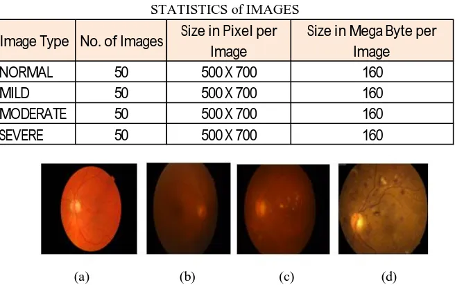
STATISTICS of IMAGES
Image Type No. of Images Size in Pixel per Image
Size in Mega Byte per Image
NORMAL 50 500 X 700 160
MILD 50 500 X 700 160
MODERATE 50 500 X 700 160
SEVERE 50 500 X 700 160
(a) (b) (c) (d) Fig 4: Example of Retinal Image :(a)Normal (b)Mild (c)Moderate (d)Severe
B. Experimental Results
[image:4.612.143.471.167.373.2]The Fig 5 shows the input fundus image and the blood vessels after image enhancemet, which is then fed to wavelet transform for decomposition.
Fig 5 Wavelet Decomposition LL band
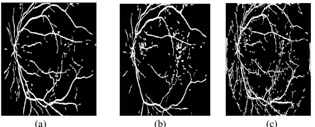
The Fig 6 & Fig 7 shows the wavelet decomposition of normal and affected eye fundus image respectively. The decomposition is performed till second level and its respective LL band is also shown.
[image:4.612.181.429.451.525.2][image:4.612.174.443.585.698.2]
Technology (IJRASET)
Fig 7 Wavelet Decomposition for Affected eye fundus and its LL band
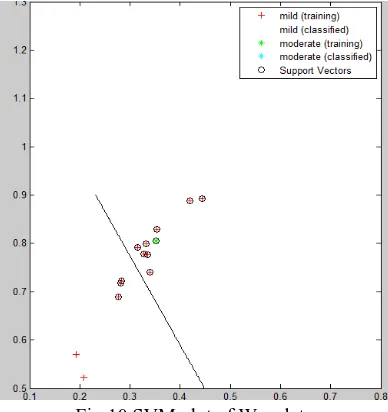
[image:5.612.172.437.83.220.2]The Fig 8 & Fig 9 shows the result of Euclidean distance for both normal and affected eye respectively. Given an image using Euclidean distance method it’s easier to identify whether a given image is normal or affected. Fig 10 Shows that to further classify the affected images SVM classifier has been used.
[image:5.612.210.408.288.369.2]Fig 8 Euclidean Distance result of Normal Eye .
Fig 9 Euclidean Distance result of Affected Eye
[image:5.612.210.404.509.715.2]
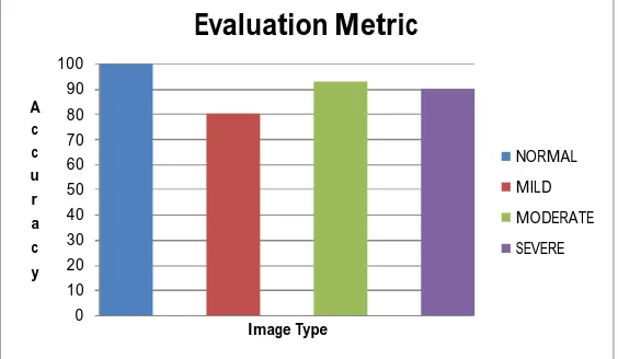
0 10 20 30 40 50 60 c u r a c y Image Type NORMAL MILD MODERATE SEVERE
Fig 11 Evaluation Metric of Wavelet Transform
V. CONCLUSIONS
The proposed system is used for an early detection of Diabetic Retinopathy which will aid the physicians for easy detection of retinopathy. The system helps in classifying whether a given image is normal or affected, if it is affected the system also specifies at which stage (mild, moderate, severe) it is currently. The proposed system has shown that the green channel is the best for blood vessels extraction and also found that wavelet decomposition of level two gives the valid information required. It can be concluded that Euclidean distance method gives the better result for the classification between normal and affected image. Whereas, for further classification of affected images SVM shows the better results. This work can be extended to other frequency domain transform to compare the performances.
VI.ACKNOWLEDGMENT
I would like to thank my college BNMIT for providing me with the facilities for aiding me in completing my project. I am thankful to Dr. Saritha Chakrasali, my guide for her constant support, advice and motivation. I owe my sincere gratitude to Dr.Hemanth Murthy from Retina Institute of Karnataka who provided me with the data set and information required for my work.
REFERENCES
[1] ManjushaS, Nelwin Raj ,Content Based Image Retrieval Using Wavelet Transform and Feedback Algorithm, International Journal of Innovative Research in
Science, Engineering and Technology, July 2014
[2] Yusra .T. Mshari, Hameed A. Younis ,Content Based Image Retrieval using Haar Wavelet to Extracted Colour Histogram and Texture Features ,International
Journal of Computer Science and Mobile Computing August 2015.
[3] isha. K. P, Thusnavis Bella Mary. I, Dr. A. Vasuki, An Image Retrieve Al Technique Based On Texture Features Using Semantic Properties, International Conference on Signal Processing, Image Processing and Pattern Recognition [ICSIPR], 2013.
[4] Sheetal Jagannath Dalavi, Mahadev S. Patil, Sanjay R. Patil, Content Based Image Retrieval by Using Daubechies Wavelet Transform, International Journal of
Innovative Research in Science, Engineering and Technology, March 2014
[5] Ramachandran Murugesan ,Saritha Chakrasali, Mahadevan Sumathi, Raman Bhaskaran, Secure Dual Watermarking in the Wavelet Domain for Teleradiology, International Conference on Advances in Information and Communication Technologies (ICICOT07), ISBN 0230634370 Macmillan Publisher, 2007. [6] Ramachandran Murugesan, Saritha Chakrasali, Raman Bhaskaran, Wavelet Based Image Adaptive Dual watermarking for Teleradiology, 52nd Congress of