Review
1
Does irisin link physical exercise with Alzheimer’s
2
disease?
3
Dewan Md. Sumsuzzman1,2,3,#, Yunho Jin1,2,3,#, Jeonghyun Choi1,2,3, Sang-Rae Lee4,*,
4
Yonggeun Hong1,2,3,5,*
5
1 Department of Rehabilitation Science, Graduate School of Inje University, Gimhae, Korea;
6
[email protected] (D.Md.S); [email protected] (Y.J.)
7
2 Biohealth Products Research Center (BPRC), Inje University, Gimhae, Korea
8
3 Ubiquitous Healthcare & Anti-aging Research Center (u-HARC), Inje University, Gimhae, Korea
9
4 National Primate Research Center (NPRC), Korea Research Institute of Bioscience and Biotechnology
10
(KRIBB), Ochang, Korea.
11
5 Department of Physical Therapy, College of Healthcare Medical Science & Engineering, Gimhae, Korea
12
* Correspondence: [email protected] (Y.H.); [email protected] (S.-R.L); Tel.: 55-320-3681 (Y.H.);
+82-13
43-240-6322 (S.-R.L); Fax: +82-55-329-1678 (Y.H.); +82-43-240-6309 (S.-R.L)
14
# These authors contributed equally to this work.
15
16
Abstract: Irisin, a skeletal muscle-secreted myokine, produced in response to physical exercise, has
17
protective functions in both the central and the peripheral nervous systems, including the regulation
18
of brain-derived neurotrophic factors and modification of telomere length. Such beneficial effects
19
may inhibit or delay the emergence of neurodegenerative diseases, including Alzheimer’s disease
20
(AD). This review is based on the hypothesis that irisin produced by physical exercise helps control
21
AD progression. Herein, we describe the physiology of irisin and its potential role in delaying or
22
preventing AD. Although current and ongoing studies on irisin show promising results, further
23
research is required to clarify its potential as a meaningful therapeutic target for treating human
24
diseases.
25
Keywords: Physical Exercise, Irisin, Neurodegeneration, Aging, Alzheimer's disease
26
27
1. Introduction
28
Alzheimer’s disease (AD) is a devastating age-associated neurodegenerative disorder
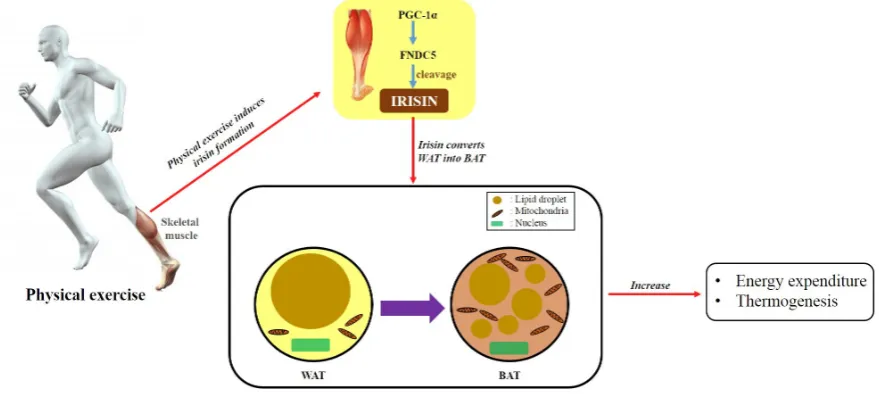
29
characterized by progressive cognitive and functional decline. Extracellular amyloid-β (Aβ)
30
aggregation and intracellular neurofibrillary tangles are considered the pathological hallmarks of
31
AD. Notwithstanding several previous studies, the etiology of AD is largely unknown. However, a
32
series of neurodegenerative events in the hippocampus, as well as microglial activation,
33
neuroinflammation, oxidative stress, metabolic energy failure, and consequent neuronal apoptosis
34
are believed to be closely correlated with the pathogenesis of AD [1–6]. Physicalexerciseameliorates
35
various neurodegenerative events and reduces the consequent production of harmful factors [7].
36
Indeed, aerobic exercise reverses hippocampal volume loss, causing a 2% increase followed by
37
improved memory function [8]. Physical exercise slows the neurodegeneration-induced decline of
38
executive functioning [9], and many studies have highlighted the effects of exercise in various organs,
39
such as the liver, brain, adipose tissue, and heart. Unlike other organs, skeletal muscles are directly
40
affected by exercise [10]. Skeletal muscle is a secretary organ that produces and releases cytokines
41
and other peptides that function in manner similar to hormones [11]. These secretions may underlie
42
the beneficial effects of exercise. Hundreds of secretome components of skeletal muscle are involved
43
in muscle communication with other organs [10]. Among these components, irisin has attracted great
44
attention, as it has recently been identified as a muscle-derived myokine released from skeletal
45
muscle immediately after exercise. This review discusses the beneficial role of irisin and its potential
46
protective effects against AD.
47
48
2. Irisin, the exercise-induced myokine, originated from the PGC-1α/FNDC5 pathway
49
The transcriptional coactivator, peroxisome proliferator-activated receptor gamma coactivator
50
1-alpha (PGC-1α), regulates many biological processes involved in energy metabolism [12], and it
51
modulates the factors secreted from skeletal muscle [12]. Fibronectin type III domain-containing
52
protein 5 (FNDC5) is one of numerous muscle gene products affected by PGC-1α. FNDC5
53
proteolytically cleaved to form the hormone irisin [12]; after cleavage of its extracellular portion, irisin
54
is secreted into the blood [12, 13]. Irisin is also synthesized in various tissues of different species [14].
55
Irisin upregulates UCP1 and transforms white adipose tissue (WAT) into brown adipose tissue
56
(BAT), thereby increasing thermogenesis and the energy consumption of adipose tissue [15].
57
Additionally, it ameliorates insulin resistance, lowers blood glucose, and promotes weight loss.
58
Furthermore, irisin further encourages cell proliferation and inhibits cell apoptosis. Previous studies
59
have also indicated that irisin sustains the levels, and increases the proliferation, of human umbilical
60
vein endothelial cells [16]. Irisin was also shown to increase the proliferation of H19-7 mouse
61
hippocampal neurons [17]. Meanwhile, irisin suppresses the high-glucose-induced apoptosis of
62
vascular endothelial cells and improves their function via the extracellular signal-regulated kinase
63
(ERK) and the 5’-adenosine monophosphate-activated protein kinase (AMPK)-PI3K-protein kinase B
64
(Akt)-eNOS signaling pathways [16, 18, 19]. Furthermore, by interfering with oxidative stress and
65
inflammation, irisin protects against palmitic acid-induced apoptosis in liver cells [20].
66
67
3. Neuroprotective implications of irisin via the Akt/ERK signaling pathway
68
Irisin is expressed not only in the skeletal muscle and the heart but also in the brain [21]. It largely
69
inhibits brain infarct volume and reduces neuroinflammation and post-ischemic oxidative stress. One
70
group of scientists demonstrated that irisin activates the Akt and ERK1/2 signaling pathways in brain
71
tissue [22]. Previous studies have also shown that irisin stimulates ERK1/2 signaling in adipocytes
72
[23], endothelial cells [24], and bone marrow stromal cells [25], and activates Akt signaling in
73
hepatocytes [26]. These results indicate that the activation of both Akt and ERK1/2 may be important
74
for the neuroprotective effects of irisin because specific chemical inhibitors of the Akt and ERK1/2
75
pathways abolished the neuroprotection conferred by irisin. The same group also proved that mouse
76
plasma irisin levels are negatively correlated with plasma tumor necrosis factor-alpha (TNF-α) and
77
Interleukin-6 levels [22]. Finally, they demonstrated that the novel exercise-induced hormone irisin
78
protects against neuronal injury via activation of the Akt and ERK1/2 signaling pathways [22]. These
79
results suggest that irisin contributes to the neuroprotective effects of physical exercise in cerebral
80
ischemia and is a promising agent for the prevention and treatment of ischemic stroke. Recent
81
research has disclosed a role for chronic neuroinflammation in the pathophysiology of
82
neurodegenerative diseases such as AD, and attention has focused the use of anti-TNF and
TNF-83
modulating agents for prevention and treatment [27]. The brains of treated animals exhibited a
84
significant reduction in pro-inflammatory TNF-α, and a diminished burden of neurofibrillary
85
tangles, amyloid precursor protein, and Aβ plaques. The brief discussion above allows a clearer
86
mechanistic understanding of the role of proinflammatory mediators such as TNF-α in AD, and
87
suggests that irisin could be a novel target to reduce proinflammatory mediators for the prevention
88
or treatment of AD.
4. Irisin protects the nervous system
93
Physical activity has many positive effects, including lowering the risk of developing heart
94
disease, stroke, and diabetes. Exercise, particularly endurance exercise, has salutary effects on brain
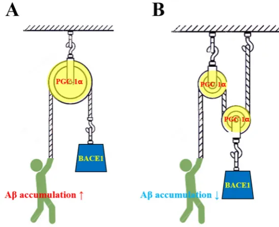
95
health and cognitive functioning [28-30]. The improvement in cognitive functioning following
96
exercise may be prominent in older adults [31]. Exercise ameliorates negative outcomes in
97
neurological diseases, such as depression, epilepsy, stroke, AD, and Parkinson’s disease [32–37]. The
98
beneficial effects of exercise on the brain are most discernible in the hippocampus and its dentate
99
gyrus, a region of the brain associated with learning and memory. Several studies have shown that
100
exercise has markedly favorable effects on the brain including increased size, blood vessel growth of
101
the human hippocampus, synaptic plasticity, and, importantly, de novo neurogenesis in the dentate
102
gyrus in various animal models [28, 29]. These results are intriguing as the hippocampus is the region
103
of the brain that is most affected by AD [38, 39]. As physical exercise has diverse benefits, the
104
discovery of the exercise hormone irisin has attracted a great deal of attention [12]. Human studies
105
have demonstrated that 10 weeks of physical training increases plasma levels of irisin [12].
106
Subsequent studies substantiated acute exercise-altered irisin levels [40, 41]. Irisin expression is
107
induced by exercise, and this myokine converts WAT into BAT, leading to increased caloric
108
expenditure [42]. Of the two types of adipose tissues, WAT stores energy as a form of fat, whereas
109
BAT burns energy [43]. With the brown appearance derived from abundant mitochondria and small
110
lipid droplets, BAT expresses UCP1, which is responsible for heat production via the uncoupling of
111
respiration from ATP synthesis [43] (Figure 1). This type of adipose tissue is rich in metabolically
112
active adults [44].
113
114
115
116
Figure 1. The general role of irisin. Physical exercise induces irisin. During exercise, the
117
transcriptional coactivator PGC-1α modulates several factors secreted from skeletal muscle. Among
118
the factors, FNDC5 is proteolytically cleaved to form irisin. This exercise-induced myokine converts
119
WAT into BAT, thereby increasing thermogenesis and energy consumption. However, irisin has a
120
range of functions beyond its role in adipose conversion.
121
122
The contribution of irisin is not confined to physical fitness and fat browning; the central nervous
123
system may be another beneficiary. The beneficial roles of exercise described above are likely to be
124
associated with irisin. Irisin administration increased the proliferation of hippocampal cells in vitro
125
[45], and expression of FNDC5 resulted in elevated irisin concentrations and brain-derived
126
neurotropic factor (BDNF) gene expression in culture [46]. These findings suggest that irisin could be
127
a therapeutic target in neurodegenerative disorders [15, 47, 48]. PGC-1α, which functions upstream
128
of the irisin precursor, FNDC5, has been reported to benefit tissues that have no primary metabolic
129
functions, such as the brain [15]. PGC-1α-null mice show adverse neuropathological behaviors, such
130
as stimulus-induced myoclonus, excessive startle responses, dystonic posture, and limb clasping [49].
Additionally, it has been suggested that PGC-1α is a key controller of energy metabolism in the early
132
stages of neurological disorders [50]. The irisin precursor, FNDC5, is increased by endurance exercise
133
in the mouse hippocampus, and forced expression of FNDC5 in primary cortical neurons induces
134
augmented BDNF expression [51]. Peripheral delivery of FNDC5 to the liver induces the expression
135
of BDNF and other protective genes and elevates levels of blood irisin [51]. As BDNF is a critical
136
regulator of neural plasticity, irisin may act as a key regulator of neuronal survival following
137
neurodegenerative diseases, such as AD. BDNF is responsible for regulating neuron growth,
138
function, and survival, as well as for synaptic stabilization and branching [52]. BDNF is believed to
139
be involved in the pathophysiology of central nervous system diseases associated with
140
neuroinflammation [52]. Evidence from human neuropathological studies has indicated that the
141
levels of neurotrophins, such as nerve growth factor (NGF) and BDNF, are lower in patients with AD
142
[53]. These studies demonstrated that BDNF mRNA levels are significantly reduced at very early
143
stages of amyloid pathology in a transgenic rat model of AD. Furthermore, ileocecal valve Aβ-treated
144
rats manifested a memory deficit and significantly decreased BDNF levels, with a concurrent increase
145
in mitochondrial oxidative damage and inflammatory mediators in the hippocampus [54]. Several
146
studies have suggested a link between irisin and BDNF. Irisin is formed primarily during contraction
147
of the skeletal muscle, but it is also present in the brain [55]. Irisin enters the central nervous system
148
and induces BDNF expression [55]. As described above, BDNF is responsible for neural plasticity. As
149
irisin enhances the synthesis of BDNF [56], the neuroplasticity mediated by this neurotrophin may
150
be strengthened by irisin. Yarrow et al. [57] showed that resistance exercise can induce ~77% transient
151
elevation of circulating BDNF levels. Thus, physical exercise may increase irisin levels and BDNF
152
synthesis. Additionally, irisin may enhance BDNF synthesis leading to the augmented
153
neuroplasticity achieved by the collarboration of irisin and BDNF. This exercise-irisin-BDNF axis
154
may magnify neuroplasticity including neuronal growth/survival and synaptic
155
stabilization/branching (Figure 2).
156
It has been suggested that a decrease in irisin levels may cause AD pathogenesis and cognitive
157
deficits. These phenomena are strongly associated with neuroinflammation and apoptosis, mediated
158
by a dramatic decrease of BDNF.
159
160
161
Figure 2. Physical exercise increases irisin levels and BDNF synthesis. In turn, irisin enhances BDNF
162
synthesis and release, leading to augmented neuroplasticity achieved by the collaboration of irisin
163
and BDNF. In this context, exercise and its sequelae, irisin and BDNF, may contribute to
164
neuroplasticity and reduce the risk of AD.
5. The underlying beneficial contribution of exercised-induced irisin in AD.
168
Physical exercise reverses Aβ accumulation and delays the progression of AD-like
169
neurobehaviors [58]. Treadmill exercise dampens the levels of amyloid peptides and induces BDNF
170
[59]. As BDNF is a crucial regulator of brain plasticity, decreased circulating BDNF potentiates the
171
risk of reduced memory and cognitive function that accompanies AD [60]. Similarly, the maturation
172
of neurotrophin NGF from its pro-NGF premature form is dampened in AD [61]. The accumulation
173
of Aβ in AD is thought to hinder the maturation of NGF [62]. However, exercise training contributes
174
to a significant induction of NGF [63]. Exercise is thought to suppress the negative effects of AD by
175
facilitating the normal secretion of neurotrophins. As previously mentioned, the myokine irisin is
176
generated during exercise. Thus, exercise-induced irisin may be a novel therapeutic candidate.
177
Indeed, low expression of PGC-1α, the upstream activator of the irisin precursor FNDC5, caused Aβ
178
accumulation in the brains of patients with AD [64]. As PGC-1α regulates beta secretase 1 (BACE1),
179
which drives Aβ formation, low levels of PGC-1α fail to block the formation of Aβ [65]. Likewise,
180
BACE1-deficient mice showed decreased Aβ formation [66]. Accordingly, PGC-1α appears to inhibit
181
the accumulation of Aβ, which is the prevalent characteristic of AD, by regulating BACE1 (Figure 3).
182
In addition to PGC-1α, the downstream FNDC5 might also be involved in AD pathogenesis, as
183
exercise-induced muscular expression of FNDC5 is regulated by PGC-1α (Figure 1) [64]. FNDC5
184
enhances the differentiation rates of embryonic stem cells, implying its role as a neurogenic factor
185
[67]. Additionally, FNDC5 expression is increased in the hippocampus during exercise [46]. Both the
186
irisin precursor, FNDC5, and its upstream factor, PGC-1α, are involved in the regulation of AD
187
pathogenesis. Irisin, the cleaved form of FNDC5, encourages hippocampal neurogenesis, as
188
evidenced by augmented proliferation of hippocampal neurons in the presence of irisin [45]. Thus,
189
irisin, PGC-1α and FNDC5 might be linked to AD. These findings imply that PGC-1α, FNDC5, and
190
irisin could have therapeutic potential to treat AD.
191
192
193
Figure 3. Aβ accumulation is regulated via reciprocal interactions between PGC-1α and beta secretase
194
1 (BACE1). Their interactions are depicted as a pulley system, with the wheel and load in the pulley
195
system representing PGC-1α and BACE1, respectively, and the worker’s stress representing Aβ
196
accumulation. In a pulley system, greater numbers of wheels require less effort, whereas fewer
197
wheels require greater effort, to lift a load. PGC-1α regulates BACE1, which is in charge of Aβ
198
formation. A. In this example, the pulley system has only one wheel, and the worker cannot
199
effectively lift the load. Similarly, low levels of PGC-1α cannot effectively hinder the activation of
200
BACE1, leading to the accumulation of Aβ. B. The worker can lift the load with less effort, as two
pulley wheels in the system ease the work. Likewise, PGC-1α may ameliorate BACE1 activation,
202
resulting in a decrease in BACE1-induced Aβ accumulation.
203
6. Implications of irisin for age-related telomere length (TL) shortening and AD pathogenesis
204
Telomeres, which resemble the plastic tips at the ends of shoelaces, are the caps at the end of
205
each DNA strand and function to preserve chromosomes [68]. TL becomes progressively shorter with
206
mitosis, and this TL shortening eventually provokes cellular senescence [69, 70]. TL shortening has
207
been confirmed to play a causative role in age-related neurodegenerative diseases, including AD. TL
208
shortening has also been associated with cognitive impairment, amyloid pathology, and
hyper-209
phosphorylation of Tau in AD, and plays a significant role in the pathogenesis of AD via the
210
mechanisms of oxidative stress and inflammation [71]. A shorter TL in leukocytes has been connected
211
to age-related diabetes, and cardiovascular and heart diseases, as well as an elevated risk of
212
neurodegenerative diseases, including dementia [72]. It seems that long-term chronic inflammation
213
and/or oxidative stress accelerate TL shortening in monocytes [73]. In addition, since TL is shortened
214
by aging, elderly populations are more susceptible to AD. Interestingly, microglia also exhibit shorter
215
telomeres in the brains of AD subjects, suggesting that these cells undergo early replicative
216
senescence, which could be due to the intense amyloid plaque profusion seen in AD [74]. Monocytes
217
migrate through the blood-brain barrier in AD and they are converted into microglial cells in the
218
brain, and microglial activation has been reported to be associated with amyloid-plaques in the AD
219
brain [75]. Additionally, increased expression of chemokine receptors and cytokines in the peripheral
220
blood mononuclear cells of AD patients has been reported [76]. Previous studies have reported that
221
lifestyle factors, including exercise, can have a notable impact on the accumulation of DNA damage
222
and TL [77]. Recently, Karan et al. [78] demonstrated that plasma irisin levels showed a significant
223
correlation with TL. The shortening of TL with aging is well-understood and, as expected, shows an
224
inverse relationship with age. Since plasma irisin is likely associated with TL, irisin may exhibit
anti-225
aging properties. Previous research has reported that exercise, which increases plasma irisin, can
226
modulate TL [79-81]. The data presented herein describe a potential mechanism by which exercise is
227
associated with an increased TL. Previously published data have uncovered that irisin activates
228
signaling pathways connected to the regulation of cellular proliferation, including p38 MAPK [82],
229
which regulates cellular proliferation and the expression of human telomere reverse transcriptase
230
[83]. In summary, it is hypothesized that the age-related decrease of irisin may be a cause of AD
231
pathogenesis and cognitive impairments. This association is highly linked to TL shortening induced
232
by oxidative stress and inflammation.
233
234
7. Reduction of endoplasmic reticulum (ER) stress responses by irisin in AD
235
The ER is associated with several crucial cellular functions, such as protein folding, quality
236
control, maintenance of Ca2+ balance, and cholesterol synthesis. Many genetic and environmental
237
insults can disrupt the function of the ER, resulting in ER stress. Therefore, it is not surprising that
238
ER stress is linked to several neurodegenerative diseases [84-86]. The ER stress response, an
239
important defense mechanism for cell survival, has three major signaling branches: protein kinase
240
RNA-like endoplasmic reticulum kinase (PERK), inositol-requiring enzyme 1α (IRE1α), and
241
activating transcription factor 6 (ATF6) [87]. Upon ER stress, PERK phosphorylates eukaryotic
242
translation initiation factor 2α (eIF2α), inhibiting protein translation [88]. Then, eIF2α
243
phosphorylation specifically activates translation of activating transcription factor (ATF) 4 [88],
244
which upregulates various foldases to prevent the accumulation of unwanted proteins [88, 89]. Under
245
prolonged ER stress, ATF4 stimulates C/EBP homologous protein (CHOP) to activate apoptotic cell
246
death [88, 89]. IRE1α induces splicing of the X-box-binding protein 1 (XBP1s) mRNA to produce
247
spliced version of XBP1 (XBP1s), which is an active transcription factor [90]. XBP1s controls the
248
expression of several genes responsible for protein folding, secretion, protein entry into the ER, and
protein quality control [91, 92]. ATF6 is an ER transmembrane transcription factor [93], and ER stress
250
induces the translocation of inactivated ATF6 from the ER to the Golgi apparatus [93, 94]. The
251
translocated ATF6 is proteolytically cleaved by site-1 (SIP) and site-2 (S2P) proteases to release the
252
cytoplasmic domains of ATF6 [94, 95]. Next, cleaved ATF6 translocate into the nucleus and acts
253
directly as a transcription factor, activating transcription of the endogenous GRP78/BiP gene, which
254
plays a role in protein folding [94, 96]. Evidence of activated UPR signaling has been revealed in AD,
255
PD, and Huntington's disease, as well as in amyotrophic lateral sclerosis [84-86, 97]. Furthermore,
256
cerebral ischemia can trigger the UPR, although this is clearly reduced by the concomitant dramatic
257
decline in protein synthesis [98]. Recent studies have shown that ER stress can generate signals that
258
warn neighboring cells and elicit inflammatory responses to prevent extensive tissue damage [99,
259
100]. In fact, moderate ER stress improves cellular protection by a series of changes called the
260
‘hormetic response’, which is characterized by alteration of the transcriptome and proteome of the
261
cell, thus elevating the adaptive capacity of the ER [101-105]. However, the prolonged ER stress
262
manifested in neurodegenerative diseases is believed to disrupt the protective effects of the UPR,
263
leading to the activation of inflammatory and apoptotic programs that promote neurotoxicity.
264
Therefore, prolonged ER stress disrupts the protective mechanism of the UPR, leading to
265
inflammation and apoptosis, which promote AD pathogenesis. Exercise is believed to improve
266
physical fitness and prevent chronic diseases and age-related disorders [106]. Exercise promotes the
267
expression of several myokines such as irisin, which is linked to the transcription factor PGC-1α and
268
is not related to ER-stress, whereas typical ER-stress-induced cytokines, such as fibroblast growth
269
factor 21 and growth/differentiation factor 15 are not exercise-induced myokines under normal
270
physiological conditions [107]. The unfolded protein response (UPR), a stress response to
271
abnormalities in protein folding in ER, has been found in the brains of patients with AD [108]. The
272
molecular chaperone GRP78/BiP, which improves the protein-folding function of the ER, is
273
upregulated in the AD temporal cortex and hippocampus of patients with AD, implying an increased
274
role of UPR [108]. Additionally, phosphorylated PERK has been found in the neurons of patients with
275
AD [109]. Exercise suppresses AD-induced UPR, as treadmill exercise decreased the activation of
276
PERK, eIF2α, and ATF6 in an experimental AD mouse model [110]. This diminished UPR was
277
followed by a decrease in apoptosis and inflammatory responses [110].The connection between irisin
278
and ER stress might involve the role of irisin in alleviating tunicamycin-induced apoptosis,
279
presumably by inhibiting PERK/eIF2α/ATF/CHOP signaling pathways [110]. In this context, one
280
somewhat controversial view argues that exercise may regulate UPR in patients with AD.
281
Considering the fact that irisin is formed during exercise, this myokine is thought to be involved in
282
UPR regulation.
283
284
8. Conclusions
285
The roles of the recently discovered myokine, irisin are not confined to fat browning and
286
thermogenesis; this myokine seems to be involved in diverse actions. Exercise and irisin have been
287
implicated in increased BDNF levels and decreased Aβ accumulation, which is the prevalent trait of
288
AD. Additionally, irisin might encourage BDNF release, leading to augmentation in neural plasticity.
289
TL shortening, which is commonly found during aging and pathological conditions, appears to be
290
delayed by irisin. In short, exercise-induced irisin may discourage the emergence of AD by promoting
291
neural plasticity and suppressing TL shortening. Whether exercise increases ER stress-induced UPR
292
has not been clearly defined; however, the connection between ER stress and exercise-induced irisin
293
clearly plays a role in AD. Extensive studies are required to clarify the interrelationship of these
294
factors in AD pathology.
295
296
Acknowledgments: This work was supported by the grants from the National Research Foundation
(NRF-298
2013R1A2A2A01067169 to Y.H., NRF-2017R1A2A2A01067169 to Y.H.), and by the KRIBB Research Initiative
299
Program (KGM4611821 to Y.H.). This work was also supported by the 2017 Creative Research Program of Inje
300
University, Republic of Korea.
301
Author Contributions: This review was conceptualized and designed by Yonggeun Hong and Sang-Rae Lee;
302
Dewan Md. Sumsuzzman, Yunho Jin and Yonggeun Hong wrote the manuscript; Dewan Md. Sumsuzzman,
303
Yunho Jin and Jeonghyun Choi collected the references and created the figures appearing in the manuscript
304
under supervision by Yonggeun Hong. All authors commented on the manuscript and approved the final form
305
of manuscript.
306
Conflicts of Interest: The authors declare no conflict of interest.
307
References
308
1. Simonian, N.A.; Coyle, JT. Oxidative stress in neurodegenerative diseases. Annu. Rev. Pharmacol. Toxicol.
309
1996, 36, 83-106.
310
2. Behl, C.; Holsboer, F. Oxidative stress in the pathogenesis of Alzheimer’s disease and antioxidant
311
neuroprotection. Fortschr. Neurol. Psychiatr. 1998, 66, 113-121.
312
3. Koliatsos, V.E.; Kecojevic, A.; Troncoso, J.C.; Gastard, M.C.; Bennett, D.A.; Schneider, J.A. Early
313
involvement of small inhibitory cortical interneurons in Alzheimer’s disease. Acta. Neuropathol. 2006, 112,
314
147–162.
315
4. Akama, K.T.; Van Eldik L.J. Beta-amyloid stimulation of inducible nitric-oxide synthase in astrocytes is
316
interleukin-1beta- and tumor necrosis factor-alpha (TNF alpha)-dependent, and involves a TNF alpha
317
receptor-associated factor- and NF kappa B-inducing kinase-dependent signaling mechanism. J. Biol. Chem.
318
2000, 275, 7918–7924.
319
5. Behl, C.; Davis, J.B.; Lesley, R.; Schubert, D. Hydrogen peroxide mediates amyloid beta protein toxicity.
320
Cell. 1994, 77, 817–827.
321
6. Griffin, W.S.; Mrak R.E. Interleukin-1 in the genesis and progression of and risk for development of
322
neuronal degeneration in Alzheimer’s disease. J. Leukoc. Biol. 2002, 72, 233–238.
323
7. Kim, S.E.; Ko, I.G.; Park, C.Y.; Shin, M.S.; Kim, C.J.; Jee, Y.S. Treadmill and wheel exercise alleviate
324
lipopolysaccharide-induced short-term memory impairment by enhancing neuronal maturation in rats.
325
Mol. Med. Rep. 2013, 7, 31-36.
326
8. Erickson, K.I.; Voss, M.W.; Prakash, R.S.; Basak, C.; Szabo, A.; Chaddock, L.; Kim, J.S.; Heo, S.; Alves, H.;
327
White, S.M.; et al. Exercise training increases size of hippocampus and improves memory. Proc. Natl. Acad.
328
Sci. U. S. A. 2011, 108, 3017-3022.
329
9. Tanaka, K.; Quadros, A.C. Jr.; Santos, R.F.; Stella, F.; Gobbi, L.T.; Gobbi, S. Benefits of physical exercise on
330
executive functions in older people with Parkinson’s disease. Brain. Cogn. 2009, 69, 435-441.
331
10. Pedersen, B.K.; Febbraio, MA. Muscles, exercise and obesity: skeletal muscle as a secretory organ. Nat. Rev.
332
Endocrinol. 2012, 8, 457-465.
333
11. Bortoluzzi, S.; Scannapieco, P.; Cestaro, A.; Danieli, G.A.; Schiaffino, S. Computational reconstruction of
334
the human skeletal muscle secretome. Proteins. 2006, 62, 776-792.
335
12. Bostrom, P.; Wu, J.; Jedrychowski, M.P.; Korde, A.; Ye, L.; Lo, J.C.; Rasbach, K.A.; Boström, E.A.; Choi, J.H.;
336
Long, J.Z.; et al. A PGC1-α-dependent myokine that drives brown-fat-like development of white fat and
337
thermogenesis. Nature. 2012, 481, 463–468.
338
13. Bostrom, P.; Wu, J.; Jedrychowski, M.P.; Korde, A.; Ye, L.; Lo, J.C.; Rasbach, K.A.; Boström, E.A.; Choi, J.H.;
339
Long, J.Z.; et al. A PGC1-alpha-dependent myokine that drives brown-fat-like development of white fat
340
and thermogenesis. Nature. 2012, 48, 463-468.
341
14. Peng, J.; Deng, X.; Huang, W.; Yu, J.H.; Wang, J.X.; Wang, J.P.; Yang, S.B.; Liu, X.; Wang, L.; Zhang, Y.; et
342
al. Irisin protects against neuronal injury induced by oxygen-glucose deprivation in part depends on the
343
inhibition of ROS-NLRP3 inflammatory signaling pathway. Mol. Immunol. 2017, 91, 185-194.
344
15. Novelle, M.G.; Contreras, C.; Romero, P.A.; Lopez, M.; Dieguez, C. Irisin, two years later. Int. J. Endocrinol.
345
2013, 2013, 746281..
346
16. Song, H.; Wu, F.; Zhang, Y.; Zhang, Y.; Wang, F.; Jiang, M.; Li, S.; Yang, L. Irisin promotes human umbilical
347
vein endothelial cell proliferation through the ERK signaling pathway and partly suppresses high
glucose-348
17. Moon, H.S.; Dincer, F.; Mantzoros, C.S. Pharmacological concentrations of irisin increase cell proliferation
350
without influencing markers of neurite outgrowth and synaptogenesis in mouse H19-7 hippocampal cell
351
lines. Metabolism. 2013, 62, 1131-1136.
352
18. Lu, J.; Xiang, G.; Liu, M.; Mei, W.; Xiang, L.; Dong, J. Irisin protects against endothelial injury and
353
ameliorates atherosclerosis in apolipoprotein E-Null diabetic mice. Atherosclerosis. 2015, 243, 438-448.
354
19. Han, F.; Zhang, S.; Hou, N.; Wang, D.; Sun, X. Irisin improves endothelial function in obese mice through
355
the AMPK-eNOS pathway. Am. J. Physiol. Heart. Circ. Physiol. 2015, 309, H1501-508.
356
20. Park, M.J.; Kim, D.I.; Choi, J.H.; Heo, Y.R.; Park, S.H. New role of irisin in hepatocytes: The protective effect
357
of hepatic steatosis in vitro. Cell Signal. 2015, 27, 1831-1839.Wilkins, H.M.; Carl, S.M.; Weber, S.G.;
358
Ramanujan, S.A.; Festoff, B.W.; Linseman, D.A.; Swerdlow, R.H. Mitochondrial lysates induce
359
inflammation and Alzheimer’s disease-relevant changes in microglial and neuronal cells. J. Alzheimers. Dis.
360
2015, 45, 305–318.
361
21. Dun, S.L.; Lyu, R.M.; Chen, Y.H.; Chang, J.K.; Luo, J.J.; Dun, N.J. Irisin-immunoreactivity in neural and
362
non-neural cells of the rodent. Neuroscience. 2013, 240, 155–162.
363
22. Li, D.J.; Li, Y.H.; Yuan, H.B.; Qu, L.F.; Wang, P. The novel exercise-induced hormone irisin protects against
364
neuronal injury via activation of the Akt and ERK1/2 signaling pathways and contributes to the
365
neuroprotection of physical exercise in cerebral ischemia. Metabolism. 2017, 68, 31-42.
366
23. Zhang, Y.; Li, R.; Meng, Y.; Li, S.; Donelan, W.; Zhao, Y.; Qi, L.; Zhang, M.; Wang, X.; Cui, T.; et al. Irisin
367
stimulates browning of white adipocytes through mitogenactivated protein kinase p38 MAP kinase and
368
ERK MAP kinase signaling. Diabetes. 2014, 63, 514–525.
369
24. Song, H.; Wu, F.; Zhang, Y.; Zhang, Y.; Wang, F.; Jiang, M.; Wang, Z.; Zhang, M.; Li, S.; Yang, L.; et al. Irisin
370
promotes human umbilical vein endothelial cell proliferation through the ERK signaling pathway and
371
partly suppresses high glucose-induced apoptosis. PLoS. One. 2014, 9, e110273.
372
25. Colaianni, G.; Cuscito, C.; Mongelli, T.; Pignataro, P.; Buccoliero, C.; Liu, P.; Lu, P.; Sartini, L.; Di, Comite
373
M.; Mori, G.; et al. The myokine irisin increases cortical bone mass. Proc. Natl. Acad. Sci. U S A. 2015, 11,
374
12157–12162.
375
26. Liu, T.Y.; Shi, C.X.; Gao, R.; Sun, H.J.; Xiong, X.Q.; Ding, L.; Chen, Q.; Li, Y.H.; Wang, J.J.; Kang, Y.M.; et al.
376
Irisin inhibits hepatic gluconeogenesis and increases glycogen synthesis via the PI3K/Akt pathway in type
377
2 diabetic mice and hepatocytes. Clin. Sci. (Lond). 2015, 129, 839–850.
378
27. Shamim, D.; Laskowski, M. Inhibition of Inflammation Mediated Through the Tumor Necrosis Factor α
379
Biochemical Pathway Can Lead to Favorable Outcomes in Alzheimer Disease. J. Cent. Nerv. Syst. Dis. 2017,
380
9, 1179573517722512
381
28. Cotman, C.W.; Berchtold, N.C.; Christie, L.A. Exercise builds brain health: Key roles of growth factor
382
cascades and inflammation. Trends. Neuro. Sci. 2007, 30, 464-472.
383
29. Mattson, M.P. Energy intake and exercise as determinants of brain health and vulnerability to injury and
384
disease. Cell. Metabolism. 2012, 16, 706-722.
385
30. Voss, M.W.; Vivar, C.; Kramer, A.F.; Van, P.H. Bridging animal and human models of exercise-induced
386
brain plasticity. Trends. Cogn. Sci. 2013, 17, 525-544.
387
31. Colcombe, S.; Kramer, A.F. Fitness effects on the cognitive function of older adults: A meta-analytic study.
388
Psychol. Sci. 2003, 14, 125-130.
389
32. Ahlskog, J.E. Does vigorous exercise have a neuroprotective effect in Parkinson disease? Neurology. 2011,
390
77, 288-294.
391
33. Arida, R.M.; Cavalheiro, E.A.; Silva, A.C.; Scorza, F.A. Physical activity and epilepsy: Proven and predicted
392
benefits. Sports. Med. 2008, 38, 607-615.
393
34. Buchman, A.S.; Boyle, P.A.; Yu, L.; Shah, R.C.; Wilson, R.S.; Bennett, D.A. Total daily physical activity and
394
the risk of AD and cognitive decline in older adults. Neurology. 2012, 78, 1323-1329.
395
35. Russo.; N.A, Beard, R.C.; Cotman, C.W. Exercise, antidepressant medications, and enhanced brain derived
396
neurotrophic factor expression. Neuropsychopharmacolog. 1999, 21, 679-682.
397
36. Zhang, Q.; Wu, Y.; Zhang, P.; Sha, H.; Jia, J.; Hu, Y. Exercise induces mitochondrial biogenesis after brain
398
ischemia in rats. Neuroscience. 2012, 205, 10-17.
399
37. Mattson, M.P. Energy intake and exercise as determinants of brain health and vulnerability to injury and
400
38. Futoshi, A.; Takayuki, M.; Tsuyoshi, S.; Minoru, F.; Yoshikazu, U.; Nobuoki, E.; Tetsunori, S.; Hironobu, Y.
402
High-sensitivity Creactive protein is associated with hippocampus volume in nondementia patients with
403
type 2 diabetes mellitus. Metabolism. 2011, 60, 460–466.
404
39. Vanitallie TB. Preclinical sporadic Alzheimer's disease: target for personalized diagnosis and preventive
405
intervention. Metabolism. 2013, 62(Suppl 1):S30–33.
406
40. Huh, J.Y.; Mougios, V.; Kabasakalis, A.; Fatouros, I.; Siopi, A.; Douroudos, I.I.; Filippaios, A.; Panagiotou,
407
G.; Park, K.H.; Mantzoros, C.S. Exercise-induced irisin secretion is independent of age or fitness level and
408
increased irisin may directly modulate muscle metabolism through AMPK activation. J. Clin. Endocrinol.
409
Metab. 2014, 99, E2154–2161.
410
41. Norheim, F.; Langleite, T.M.; Hjorth, M.; Holen, T.; Kielland, A.; Stadheim, H.K. The effects of acute and
411
chronic exercise on PGC-1alpha, irisin and browning of subcutaneous adipose tissue in humans. FEBS. J.
412
2014, 281, 739–749.
413
42. Timmons, J.A.; Baar, K.; Davidsen, P.K.; Atherton, P.J. Is irisin a human exercise gene? E9-10; discussion.
414
Nature. 2012, 488, E09–10.
415
43. Enerbäck, S. The origins of brown adipose tissue. N. Engl. J. Med. 2009, 360, 2021-2023.
416
44. Saito, M.; Okamatsu-Ogura, Y.; Matsushita, M.; Watanabe, K.; Yoneshiro, T.; Nio-Kobayashi, J.; Iwanaga,
417
T.; Miyagawa, M.; Kameya, T. Nakada, K.; et al. High incidence of metabolically active brown adipose
418
tissue in healthy adult humans. Diabetes. 2009, 58, 1526-1531.
419
45. Moon, H.S.; Dincer, F.; Mantzoros, C.S. Pharmacological concentrations of irisin increase cell proliferation
420
without influencing markers of neurite outgrowth and synaptogenesis in mouse H19-7 hippocampal cell
421
lines. Metabolism. 2013, 62, 1131–1136.
422
46. Wrann, C.D.; White, J.P.; Salogiannnis, J.; Laznik, B.D.; Wu, J.; Ma, D. Lin, J.D.; Greenberg, M.E.;
423
Spiegelman, B.M. Exercise induces hippocampal BDNF through a PGC-1alpha/FNDC5 pathway. Cell
424
Metab. 2013, 18, 649–659.
425
47. Jodeiri, F.M.; Ghaedi, K.; Megraw, T.L.; Curtiss, J.; Shirani, F.M.; Vaziri, P.; Nasr-Esfahani, M.H. Does
426
PGC1alpha/FNDC5/BDNF elicit the beneficial effects of exercise on neurodegenerative disorders?
427
Neuromolecular. Med. 2016, 18, 1-15.
428
48. Xu, B. BDNF (I)rising from exercise. Cell Metab. 2013, 18, 612–614.
429
49. Lin, J.; Wu, P.H.; Tarr, P.T.; Lindenberg, K.S.; St-Pierre, J.; Zhang, C.Y.; Mootha, V.K.; Jager, S.; Vianna, C.R.;
430
Reznick, R.M.; et al. Defects in adaptive energy metabolism with CNS-linked hyperactivity in PGC-1α null
431
mice. Cell. 2004, 119, 121-135.
432
50. Cui, L.; Jeong, H.; Borovecki, F.; Parkhurst, C.N.; Tanese, N.; Krainc, D. Transcriptional repression of
PGC-433
1α by mutant huntingtin leads to mitochondrial dysfunction and neurodegeneration. Cell. 2006, 127, 59-69.
434
51. Wrann, C.D.; White, J.P.; Salogiannnis, J.; Laznik, B.D.; Wu, J.; Ma, D.; Lin, J.D.; Greenberg, M.E.;
435
Spiegelman, B.M. Exercise induces hippocampal BDNF through a PGC-1alpha/FNDC5 pathway. Cell
436
Metab. 2013, 18, 649–659.
437
52. Kazak, F.; Yarim, G.F. Neuroprotective effects of acetyl-l-carnitine on lipopolysaccharide-induced
438
neuroinflammation in mice: Involvement of brain-derived neurotrophic factor. Neurosci. Lett. 2017, 658,
32-439
36.
440
53. Iulita, M.F.; Bistué, Millón, M.B.; Pentz, R.; Aguilar, L.F.; Do, Carmo S.; Allard, S.; Michalski, B.; Wilson,
441
E.N.; Ducatenzeiler, A.; Bruno M.A.; et al. Differential deregulation of NGF and BDNF neurotrophins in a
442
transgenic rat model of Alzheimer's disease. Neurobiol. Dis. 2017, 108, 307-323.
443
54. Prakash, A.; Kumar, A. Role of nuclear receptor on regulation of BDNF and neuroinflammation in
444
hippocampus of β-amyloid animal model of Alzheimer's disease. Neurotox. Res. 2014, 25, 335-347.
445
55. Zsuga, J.; Tajti, G.; Papp, C.; Juhasz, B.; Gesztelyi, R. FNDC5/irisin, a molecular target for boosting
reward-446
related learning and motivation. Med. Hypotheses. 2016, 90, 23-28.
447
56. Papp, C.; Pak, K.; Erdei, T.; Juhasz, B.; Seres, I.; Szentpeteri, A.; Kardos, L.; Szilasi, M.; Gesztelyi, R.; Zsuga,
448
J. Alteration of the irisin–brain-derived neurotrophic factor axis contributes to disturbance of mood in
449
COPD patients. Int. J. Chron. Obstruct. Pulmon. Dis. 2017, 12, 2023-2033.
450
57. Yarrow, J.F.; White, L.J.; McCoy, S.C.; Borst, S.E. Training augments resistance exercise induced elevation
451
of circulating brain derived neurotrophic factor (BDNF). Neurosci. Lett. 2010, 479, 161-165..
452
58. Adlard, P.A.; Perreau, V.M.; Pop, V.; Cotman, C.W. Voluntary exercise decreases amyloid load in a
453
59. Um, H.S.; Kang, E.B.; Leem, Y.H.; Cho, I.H.; Yang, C.H.; Chae, K.R.; Hwang, D.Y.; Cho, J.Y. Exercise training
455
acts as a therapeutic strategy for reduction of the pathogenic phenotypes for Alzheimer's disease in an
456
NSE/APPsw-transgenic model. Int. J. Mol. Med. 2008, 22, 529-539.
457
60. Komulainen, P.; Pedersen, M.; Hanninen, T.; Bruunsgaard, H.; Lakka, T.A.; Kivipelto, M.; Hassinen, M.;
458
Rauramaa, T.H.; Pedersen, B.K.; Rauramaa, R. BDNF is a novel marker of cognitive function in ageing
459
women: the DR’s EXTRA Study. Neurobiolo. Learn. Mem. 2008, 90, 596-603.
460
61. Bruno, M.A.; Leon, W.C.; Fragoso, G.; Mushynski, W.E.; Almazan, G.; Cuello, A.C. Amyloid β-induced
461
nerve growth factor dysmetabolism in Alzheimer disease. J. Neuropathol. Exp. Neurol. 2009, 68, 857-869.
462
62. Radak, Z.; Hart, N.; Sarga, L.; Koltai, E.; Atalay, M.; Ohno, H.; Boldogh, I. Exercise plays a preventive role
463
against Alzheimer's disease. J. Alzheimers. Dis. 2010, 20, 777-783.
464
63. Chae C.H.; Jung, S.L.; An, S.H.; Park, B.Y.; Wang, S.W.; Cho, I.H.; Cho, J.Y.; Kim, H.T. Treadmill exercise
465
improves cognitive function and facilitates nerve growth factor signaling by activating mitogen-activated
466
protein kinase/extracellular signalregulated kinase1/2 in the streptozotocin-induced diabetic rat
467
hippocampus. Neuroscience. 2009, 164, 1665-1673.
468
64. Jodeiri Farshbaf, M.; Ghaedi, K.; Megraw, T.L.; Curtiss, J.; Shirani Faradonbeh, M.; Vaziri, P.;
Nasr-469
Esfahani, M.H. Does PGC1α/FNDC5/BDNF elicit the beneficial effects of exercise on neurodegenerative
470
disorders? Neuromolecular. Med. 2016, 18, 1-15.
471
65. Wang, R.; Li, J.J.; Diao, S.; Kwak, Y.D.; Liu, L.; Zhi, L.; Bueler, H.; Williams, R.W.; Park, E.A.; et al. Metabolic
472
stress modulates Alzheimer’s β-secretase gene transcription via SIRT1-PPARγ-PGC-1 in neurons. Cell.
473
Metab. 2013, 17, 685-694.
474
66. Vassar, R.; Kovacs, D.M.; Wong, P.C. The β-secretase enzyme BACE in health and Alzheimer's disease:
475
regulation, cell biology, function, and therapeutic potential. J. Neurosci. 2009, 29, 12787-12794.
476
67. Forouzanfar, M.; Rabiee, F.; Ghaedi, K.; Beheshti, S.; Tanhaei, S.; Shoaraye Nejati, A.; Jodeiri Farshbaf, M.;
477
Baharvand, H.; Nasr-Esfahani, M.H. Fndc5 overexpression facilitated neural differentiation of mouse
478
embryonic stem cells. Cell. Biol. Int. 2015, 39, 629-637.
479
68. Blackburn, E.H.; Epel, E.S. Telomeres and adversity: Too toxic to ignore. Nature. 2012, 490, 169-171.
480
69. Blackburn, E.H. Telomere states and cell fates. Nature. 2000, 408, 53-56.
481
70. Needham, B.L.; Mezuk, B.; Bareis, N.; Lin, J.; Blackburn, E.H.; Epel, E.S. Depression, anxiety and telomere
482
length in young adults: evidence from the National Health and Nutrition Examination Survey. Mol.
483
Psychiatry. 2015, 20, 520-528.
484
71. Cai, Z.; Yan, L.J.; Ratka, A. Telomere shortening and Alzheimer's disease. Neuromolecular Med. 2013, 15,
25-485
48.
486
72. Honig, L.S.; Schupf, N.; Lee, J.H.; Tang, M.X.; Mayeux, R.; Shorter telomeres are associated with mortality
487
in those with APOE epsilon4 and dementia. Ann. Neurol. 2006, 60, 181–187.
488
73. Von, Zglinicki T.; Serra, V.; Lorenz, M.; Saretzki, G.; Lenzen, Grossimlighaus R.; Gessner, R.; Risch, A.;
489
Steinhagen, Thiessen E. Short telomeres in patients with vascular dementia: an indicator of low
490
antioxidative capacity and a possible risk factor? Lab. Invest. 2000, 80, 1739–1747.
491
74. Flanary, B.E.; Sammons, N.W.; Nguyen, C.; Walker, D.; Streit, W.J. Evidence that aging and amyloid
492
promote microglial cell senescence. Rejuvenation. Res. 2007, 10, 61–74.
493
75. Floris, S.; Ruuls, S.R.; Wierinckx, A.; van der Pol, S.M.; Döpp, E.; van der Meide, P.H.; Dijkstra, C.D.; De
494
Vries, H.E. Interferon-beta directly influences monocyte infiltration into the central nervous system. J.
495
Neuroimmunol. 2002, 127, 69–79.
496
76. Reale, M.; Iarlori, C.; Feliciani, C.; Gambi, D. Peripheral chemokine receptors, their ligands, cytokines and
497
Alzheimer's disease. J. Alzheimers Dis. 2008,14, 147–159.
498
77. Song, Z.; von Figura, G.; Liu, Y.; Kraus, J.M.; Torrice, C.; Dillon, P.; Rudolph-Watabe, M.; Ju, Z.; Kestler,
499
H.A.; Sanoff, H.; Lenhard Rudolph, K.; Lifestyle impacts on the aging-associated expression of biomarkers
500
of DNA damage and telomere dysfunction in human blood. Aging. Cell. 2010, 9, 607-615.
501
78. Karan, S.R.; Muhammad, A.; Eric, J.H.; Sarah, A.; David, A.N.; Alan Nevill, Harpal S. Randeva, Clifford J.
502
Bailey, Srikanth Bellary, James E. Brown. Plasma irisin levels predict telomere length in healthy adults. Age.
503
(Dordr). 2014, 36, 995–1001.
504
79. Kim, S.; Parks, CG.; DeRoo, L.A.; Chen, H.; Taylor, J.A.; Cawthon, R.M.; Sandler, D.P. Obesity and weight
505
gain in adulthood and telomere length. Cancer. Epidemiol. Biomarkers. Prev. 2009, 18, 816-820.
506
80. Ludlow, A.T.; Zimmerman, J.B.; Witkowski, S.; Hearn, J.W.; Hatfield, B.D.; Roth, S.M. Relationship between
507
81. Cherkas, L.F.; Hunkin, J.L.; Kato, B.S.; Richards, J.B.; Gardner, J.P.; Surdulescu, G.L.; Kimura, M.; Lu, X.;
509
Spector, T.D.; Aviv, A. The association between physical activity in leisure time and leukocyte telomere
510
length. Arch. Intern. Med. 2008, 168, 154-158.
511
82. Zhang, Y.; Li, R.; Meng, Y.; Li, S.; Donelan, W.; Zhao, Y.; Qi, L.; Zhang, M.; Wang, X.; Cui, T.; Yang, L.J.;
512
Tang, D. Irisin stimulates browning of white adipocytes through mitogen-activated protein kinase p38
513
MAP kinase and ERK MAP kinase signaling. Diabetes. 2014, 63, 514-525.
514
83. Matsuo, T.; Shimose, S.; Kubo, T.; Fujimori, J.; Yasunaga, Y.; Sugita, T.; Ochi, M. Correlation between p38
515
mitogen-activated protein kinase and human telomerase reverse transcriptase in sarcomas. J. Exp. Clin.
516
Cance.r Res. 2012, 31, 5.
517
84. Lindholm, D.; Wootz, H.; Korhonen, L. ER stress and neurodegenerative diseases. Cell. Death. Differ. 2006,
518
13, 85-392.
519
85. Paschen, W.; Mengesdorf, T. Cellular abnormalities linked to endoplasmic reticulum dysfunction in
520
cerebrovascular disease therapeutic potential. Pharmacol. Ther. 2005, 108, 362-375.
521
86. Scheper, W.; Hoozemans, J. Endoplasmic reticulum protein quality control in neurodegenerative disease:
522
the good, the bad and the therapy. Curr. Med. Chem. 2009, 16, 615-626.
523
87. Ohri, S.S.; Maddie, M.A.; Zhao, Y.; Qiu, M.S.; Hetman, M.; Whittemore, S.R. Attenuating the endoplasmic
524
reticulum stress response improves functional recovery after spinal cord injury. Glia.2011, 59, 1489-1502.
525
88. Valenzuela, V.; Collyer, E.; Armentano, D.; Parsons, G.B.; Court, F.A.; Hetz, C. Activation of the unfolded
526
protein response enhances motor recovery after spinal cord injury. Cell. Death. Dis. 2012, 3, 272.
527
89. Yamauchi, T.; Sakurai, M.; , Abe, K.; Matsumiya, G.; Sawa, Y. Impact of the endoplasmic reticulum stress
528
response in spinal cord after transient ischemia. Brain. Res. 2007, 1169, 24-33.
529
90. Hetz C. The unfolded protein response: controlling cell fate decisions under ER stress and beyond. Nat.
530
Rev. Mol. Cell. Biol. 2012, 13, 89-102.
531
91. Hetz, C.; Martinon, F.; Rodriguez, D.; Glimcher, L.H. The unfolded protein response: integrating stress
532
signals through the stress sensor IRE1α. Physiol. Rev. 2011, 91, 1219-1243.
533
92. Lee, A.H.; Iwakoshi, N.N.; Glimcher, L.H. XBP-1 regulates a subset of endoplasmic reticulum resident
534
chaperone genes in the unfolded protein response. Mol. Cell. Biol. 2003, 23, 7448-7459.
535
93. Chen, X.; Shen, J.; Prywes, R. The luminal domain of ATF6 senses endoplasmic reticulum (ER) stress and
536
causes translocation of ATF6 from the ER to the Golgi. J. Biol. Chem. 2002, 277, 13045-13052.
537
94. Kim, M.K.; Park, K.G. Endoplasmic reticulum stress and diabetes. J. Korean. Endocr. Soc. 2008, 23, 1-8.
538
95. Ye, J.; Rawson, R.B.; Komuro, R.; Chen, X.; Dave, U.P.; Prywes, R.; Brown, M.S.; Goldstein, J.L. ER stress
539
induces cleavage of membrane-bound ATF6 by the same proteases that process SREBPs. Mol. Cell. 2000, 6,
540
1355-1364.
541
96. Haze, K.; Yoshida, H.; Yanagi, H.; Yura, T.; Mori, K. Mammalian transcription factor ATF6 is synthesized
542
as a transmembrane protein and activated by proteolysis in response to endoplasmic reticulum stress. Mol.
543
Biol. Cell. 1999, 10, 3787-3799.
544
97. Hoozemans, J.J,; van, Haastert.; E.S.; Nijholt, D.A.; Rozemuller A.J.; Eikelenboom, P.; Scheper, W. The
545
unfolded protein response is activated in pretangle neurons in Alzheimer's disease hippocampus. Am J
546
Pathol. 2009, 174, 1241-1251.
547
98. DeGracia, D.J.; Montie, H.L. Cerebral ischemia and the unfolded protein response. J Neurochem. 2004, 91,
1-548
8.
549
99. Zhang, K.; Kaufman, R.J. From endoplasmic-reticulum stress to the inflammatory response. Nature. 2008,
550
454, 455-462.
551
100. Kaser, A.; Blumberg, R.S. Endoplasmic reticulum stress in the intestinal epithelium and inflammatory
552
bowel disease. Semin Immunol. 2009, 21, 156–163.
553
101. Hetz, C.; Mollereau, B. Disturbance of endoplasmic reticulum proteostasis in neurodegenerative diseases.
554
Nat. Rev. Neurosci. 2014, 15, 233-49.
555
102. Inagi, R.; Kumagai, T.; Nishi, H.; Kawakami, T.; Miyata, T.; Fujita, T. Preconditioning with endoplasmic
556
reticulum stress ameliorates mesangioproliferative glomerulonephritis. J Am Soc Nephrol. 2008, 19, 915-22.
557
103. Mercado, G.; Valdes, P.; Hetz, C. An ERcentric view of Parkinson's disease. Trends Mol Med. 2013, 19,
165-558
75.
559
104. Fouillet, A.; Levet, C.; Virgone, A.; Robin, M.; Dourlen, P.; Rieusset, J. ER stress inhibits neuronal death by
560
105. Calabrese, V.; Cornelius, C.; Dinkova-Kostova, A.T.; Calabrese, E.J.; Mattson, M.P. Cellular stress
562
responses, the hormesis paradigm, and vitagenes: novel targets for therapeutic intervention in
563
neurodegenerative disorders. Antioxid. Redox. Signal. 2010, 13, 1763-811.
564
106. Ciolac, E.G. Exercise training as a preventive tool for age-related disorders: a brief review. Clinics (Sao
565
Paulo). 2013, 68, 710-717.
566
107. Ost, M.; Coleman, V.; Kasch, J.; Klaus, S. Regulation of myokine expression: Role of exercise and cellular
567
stress. Free Radic Biol Med. 2016, 98, 78-89.
568
108. Hoozemans, J.J.; Veerhuis, R.; Van Haastert, E.S.; Rozemuller J.M.; Baas, F.; Eikelenboom, P.; Scheper, W.
569
The unfolded protein response is activated in Alzheimer’s disease. Acta. Neuropathol. 2005, 110, 165–172.
570
109. Chafekar, S.M.; Hoozemans, J.J.; Zwart, R.; Baas, F.; Scheper, W. A β 1-42 Induces Mild Endoplasmic
571
Reticulum Stress in an Aggregation State–Dependent Manner. Antioxid. Redox. Signal. 2007, 9, 2245-2254.
572
110. Kang, E.B.; Kwon, I.S.; Koo, J.H.; Kim, E.J.; Kim, C.H.; Lee, J.; Yang, C.H.; Lee, Y.I.; Cho, I.H.; Cho, J.Y.
573
Treadmill exercise represses neuronal cell death and inflammation during Aβ-induced ER stress by