ABSTRACT
SCHILLING, JUSTIN DALE. Proteomic and Machine Learning Analyses of White Perch
(Morone americana) Plasma and Ovary. (Under the direction of Harry V. Daniels III).
Three studies were conducted to characterize the plasma and ovary proteomes of and vitellogenesis in white perch (Morone americana). First, a discovery proteomics study employed a simple fractionation method and filter-aided sample preparation (FASP) to characterize the cytosolic and membrane fractions of white perch ovary tissues by
semiquantitative tandem mass spectrometry (MS/MS). A total of 882 unique proteins, 114 found only in the cytosolic fraction, 169 proteins found only in the membrane fraction, and 599 found in both fractions, were identified using the striped bass ovary transcriptome as the reference database. Of these 882 proteins, a majority was from mitochondria and other membrane-bounded organelles. Support vector machines (SVMs) were able to perfectly (100% correct) classify samples as either membrane or cytosolic fraction during cross-validation based on the expression data of 242 proteins with the highest ANOVA p-values (i.e. those that were not significantly enriched in either fraction) as measured by MS/MS. SVMs offer categorical classification of proteomics data superior to that of parametric statistical methods such as analysis of variance (ANOVA).
validation using the MS/MS protein expression data. Following E2 induction, both the male and female perch plasma proteomes contained significantly higher levels of vitellogenin Aa and Ab (VtgAa, VtgAb), latrophilin and seven transmembrane domain-containing protein 1 (Eltd1), and kininogen 1 (Kng1) than IC plasmas. This is the first report that Eltd1 and Kng1 may be E2-responsive proteins in fishes.
In the third study, selected reaction monitoring (SRM) tandem mass spectrometry was employed to accurately quantify the three white perch vitellogenins (VtgAa, VtgAb, VtgC) in the liver, plasma, and ovary during pre-, early-, mid-, and post-vitellogenic oocyte growth. Both SRM and immunohistochemistry confirmed that VtgC is present in the pre-vitellogenic perch ovary and was the only quantifiable vitellogenin within the ovary at this time point. Only VtgAb could be confidently quantified in the pre-vitellogenic perch liver. VtgAb was found to be the predominant Vtg during vitellogenesis in perch liver, plasma, and ovary.
Observed differences in the proportional accumulation of Vtgs suggest that there is considerable plasticity in the Vtg-Vtgr system in white perch that may allow fine-tuning of egg buoyancy based upon the salinity of the estuarine water into which white perch and striped bass spawn their eggs. It appears that VtgC is the most variable form of vitellogenin within the post-vitellogenic oocytes of Acanthomorph fishes, ranging from ~2.5% in perch to 26% in striped bass, suggesting that VtgC composition may relate to other aspects of early life history in these fishes. While the implications upon early life history remain to be fully elucidated, the proportion of VtgC in post-vitellogenic oocytes varies considerably among Acathomorph fishes, even between closely related species of the genus Morone.
Proteomic and Machine Learning Analyses of White Perch (Morone americana) Plasma and Ovary
by
Justin Dale Schilling
A dissertation submitted to the Graduate Faculty of North Carolina State University
in partial fulfillment of the requirements for the degree of
Doctor of Philosophy
Zoology
Raleigh, North Carolina 2015
APPROVED BY:
_______________________________ ______________________________
Harry V. Daniels Robert M. Kelly
Committee Chair
BIOGRAPHY
ACKNOWLEDGMENTS
I thank my Doctoral Advisory Committee, Drs. Harry V. Daniels (Major Professor), Robert M. Kelly, Jeffrey A. Yoder, and Antonio Planchart. I am especially grateful for the generous support of Dr. Steven Lommel, associate dean and director of the N.C.
Agricultural Research Service in N.C. State University’s College of Agriculture and Life Sciences. I thank the staff, students, and colleagues from both the North Carolina State University Departments of Applied Ecology and Chemistry and the University of Michigan Life Sciences Institute for all of the assistance and guidance they have provided:
Dr. Benjamin J. Reading Dr. David C. Muddiman Dr. Angelito I. Neopmuceno Mr. Philip L. Loziuk
Dr. Naoshi Hiramatsu Dr. Valerie N. Williams Dr. Kevin Gross
Dr. Georgios Skiniotis Dr. William Clay Brown Dr. Min Su
TABLE OF CONTENTS
LIST OF TABLES ... viii
LIST OF FIGURES ... xi
CHAPTER 1: Vertebrate Vitellogenins and Their Receptors: A Comprehensive Review of the Literature and Recent Insights ... 1
Correspondence ... 1
Abbreviations ... 1
Vitellogenins and their receptors have crucial and diverse physiological roles ... 3
Structural anatomy of the Vtg-Vtgr system ... 4
Physiological significance of differential Vtg expression, uptake, and processing .. 6
Structural and functional conservation among LLTPs and their receptors ... 7
Vtg lipidation ... 10
Implications of proportional vitellogenin accumulation ... 13
Summary and organization of dissertation ... 14
References ... 17
CHAPTER 2: Compartment Proteomics Analysis of White Perch (Morone americana) Ovary Using Support Vector Machines ... 40
ABSTRACT ... 40
INTRODUCTION ... 41
EXPERIMENTAL ... 43
Sample Collection and Preparation ... 43
White Perch Ovary Transcriptome ... 44
Filter-Aided Sample Preparation and Digestion ... 45
nanoReversed Phase Chromatography and Tandem Mass Spectrometry ... 46
Mass Spectrometry Protein Identifications ... 47
Data Analysis ... 49
Ovary Histology ... 51
White Perch Ovary Transcriptome ... 51
Tandem Mass Spectrometry ... 51
Support Vector Machines Classification of Ovary Cytosolic and Membrane Fractions ... 53
Modulated Modularity Clustering ... 53
DAVID analysis ... 54
DISCUSSION ... 54
CONCLUSIONS ... 58
ASSOCIATED CONTENT ... 73
Supporting information ... 73
AUTHOR INFORMATION ... 73
Author contributions ... 73
Notes ... 73
ACKNOWLEDGEMENTS ... 73
ABBREVIATIONS ... 74
REFERENCES ... 75
CHAPTER 3: Machine Learning Reveals Sex-Specific 17β-Estradiol-Responsive Expression Patterns in White Perch (Morone americana) Plasma Proteins ... 86
Abbreviations ... 87
Keywords ... 87
Abstract ... 89
Introduction ... 90
Materials and methods ... 92
Sample collection and preparation ... 92
Filter-aided sample preparation and nanoLC-MS/MS ... 93
Tandem mass spectrometry ... 99
Western blotting ... 99
Inferential statistics ... 100
MMC and DAVID analysis ... 100
Machine learning ... 102
Discussion ... 103
Concluding remarks ... 109
Author contributions ... 110
Funding sources ... 110
Acknowledgements ... 110
Conflict of interest statement ... 110
References ... 111
Supporting information ... 129
CHAPTER 4: Differences in the Proportional Accumulation of Vitellogenins and Implications on Early Life Histories in Two Closely Related Fish Species ... 131
Running title ... 131
Keywords ... 131
Grant support ... 131
Correspondence ... 131
Summary sentence ... 131
Abstract ... 132
Introduction ... 133
Materials and methods ... 135
Tissue collection ... 135
Filter-aided sample preparation of white perch tissues ... 136
LC-MS/MS materials ... 137
Stable isotope-labeled peptide standards and transition characterization ... 137
LC-MS/MS analysis ... 138
Experimental replication and statistical analysis ... 139
Western blotting ... 139
White perch vitellogenin C purification ... 140
White perch vitellogenin C purification coupled to tandem mass spectromerty 140 White perch vitellogenin C AP-MS/MS filter-aided sample preparation ... 141
White perch vitellogenin C AP-MS/MS nanoReversed phase LC-MS/MS ... 142
Results ... 143
Histology and oocyte staging ... 143
Selected reaction monitoring ... 143
Western blotting ... 144
White perch vitellogenin C immunohistochemistry ... 145
White perch vitellogenin C AP-MS/MS ... 146
Discussion ... 146
Acknowledgements ... 153
References ... 154
CHAPTER 5: Future Directions ... 178
Molecular basis for Vtg endocytosis and compartmentalization ... 178
Tracing Vtg compartmentalization and trafficking ... 178
Machine learning analysis of RNASeq data from striped bass 4 hr post-fertilization viable and inviable embryos ... 180
Male and female white bass (Morone chrysops) genome sequences ... 180
LIST OF TABLES
CHAPTER 2
Table 1. ANOVA p-value range of 882 proteins identified in white perch ovary cytosolic and membrane fractions using the striped bass ovary transcriptome as the reference database. Employing the Benjamini and Hochberg procedure resulted in a significance cutoff of p < 0.0347. ... 67 Table 2. Approved gene name, striped bass contig number, cytosolic and membrane
normalized [log10(1+ NSpC avg)] values, and ANOVA p-values for white perch ovary proteins identified in either the cytosolic or membrane fraction using the striped bass ovary
transcriptome as the reference database. ... 68 Table 3. Approved gene name, striped bass contig number, cytosolic and membrane
normalized [log10(1+ NSpC avg)] values, and ANOVA p-values for white perch ovary proteins identified in both cytosolic and membrane fractions using the striped bass ovary
transcriptome as the reference database. ... 70 Table 4. Approved gene name, striped bass contig number, cytosolic and membrane
normalized [log10(1+ NSpC avg)] values, and ANOVA p-values for white perch ovary proteins identified in multiple open reading frames using the striped bass ovary transcriptome as the reference database. ... 71 Table 5. Enrichment of white perch ovary proteins by Gene Ontology (GO) class within modulated modularity clustering (MMC) modules 2, 7, 8, 9, and 10 using DAVID. The proteins were identified by tandem mass spectrometry using the striped bass ovary
transcriptome as the reference database. ... 72
CHAPTER 3
twelve technical replicates (three technical replicates per treatment) were further analyzed by two-way ANOVA. ... 125 Table 2. Two-way ANOVA p-value range from comparisons between male and female white perch plasma proteins detected in internal control (IC) and 17β-estradiol (E2)-induced
samples by tandem mass spectrometry using the predicted proteins from the striped bass genome as a reference database. The number of proteins determined to be significantly different after the Bonferroni familywise error (BFW) and Benjamini-Hochberg false
discovery rate (BH) corrections for multiple tests also are reported. ... 126 Table 3. Performance of support vector machines (SVMs) classifiers used to evaluate white perch plasma proteomes. Treatment groups were initial control (IC) male, IC female, 17β -estradiol (E2)-induced male, and E2-induced female. Data were clustered into either 3 or 2 clusters by k-means clustering or this preprocessing step was omitted. ‘Correct
Classification’ refers to SVMs classifier performance during 66%-split or 10-fold cross-validations (CV) and is given as a percent (%). ‘AUROC’ is the area under the receiver operating characteristic curve. ... 127 Table 4. Performance of support vector machines (SVMs) classifiers used to evaluate white perch plasma proteomes. Treatment groups were initial control (IC) male, IC female, 17β -estradiol (E2)-induced male, and E2-induced female. Data were clustered into either 3 or 2 clusters by k-means clustering or this preprocessing step was omitted. ‘Correct
Classification’ refers to SVMs classifier performance during 66%-split or 10-fold cross-validations (CV) and is given as a percent (%). ‘AUROC’ is the area under the receiver operating characteristic curve. Values reported represent the average ± standard deviation of SVMs models generated from 10 randomly ordered datasets. ... 128
CHAPTER 4
vitellogenesis (PreVG), early-vitellogenesis (EVG), mid-vitellogenesis (MVG), and post-vitellogenesis (PostVG). ... 172 Table 3. Results from affinity purification coupled to tandem mass spectrometry using
purified white perch VtgC as a bait protein. ... 173 Table 4. White perch vitellogenin gene names, protein identifications, Uniprot accession numbers, peptide sequences, selected reaction monitoring transitions, and collision
LIST OF FIGURES
CHAPTER 1
Figure 1. The primary domains of the three forms of vitellogenins are depicted. Yolk proteins derived from Aa-type vitellogenin are cleaved into free amino acids during final oocyte maturation. Ab-type vitellogenin receives partial or no proteolysis on lipovitellin heavy chain and are utilized during late embryonic growth. Adapted from Hiramatsu et al., 2002 (Biol Reprod, Fish Physiol Biochem); Reading et al., 2009 (Mar Biotech). ... 31 Figure 2. 3D representation of a partial lamprey (Ichthyomyzon unicuspis) complete type vitellogenin. The lipovitellin heavy domain (LvH) is colored purple while the lipovitellin light domain (LvL) is blue. The LR8 binding sequence is in yellow (Li et al., 2003). The large lipid binding pocket and putative Zn2+ binding site are also indicated (pdb: 1LSH). From
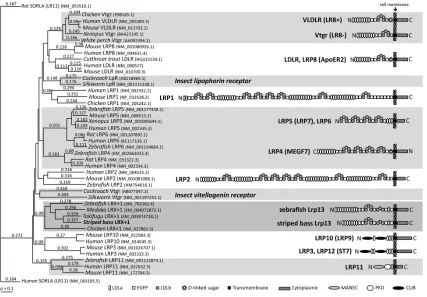
Anderson, Levitt, & Banaszak, 1998. ... 32 Figure 3. (Left) Nonreducing 7.5% acrylamide gel stained with Coomassie brilliant blue (CBB) and ligand blot (LB) of white perch ovary membrane proteins. The LB in lane 2 was prepared using 0.25 µg/ml of DIG-labeled VtgAa/b and the LB in lane 3 was performed in the presence of a 200-fold excess molar ratio of unlabeled VtgAa/b. (Right) Nonreducing 5% acrylamide gel Western blot (WB) of white perch ovary membrane proteins. The WB in lane 4 was prepared with α-WpLrp13 and the WB in lane 5 was performed with α-WpLr8−. Numbers to the left of gels or blots indicate the sizes of molecular weight markers (kDa). Positions of VtgAar, VtgAbr, pLDLR, Lr8−, and Lrp13 are indicated. From Reading et al., 2014 (J Lipid Res). ... 33 Figure 4. (Left) ClustalW dendrogram showing relationships between low-density lipoprotein receptor family polypeptide sequences. GenBank accession numbers are provided.
protein 13 (Lrp13). Receptor proteins for C-type Vtg are detected in ligand blots of trout ovarian membrane, but not in white perch. Receptor proteins that universally bind multiple Vtg subtypes are also detected in the ovarian membrane preparations of both species. LR8: lipoprotein receptor (LR) with 8 ligand binding (LB) repeats; LR7+1: LR with 7+1 LB repeats; LR13+1: LR with 13+1 LB repeats. From Hiramatsu et al., 2015 (Gen Comp Endocr). ... 35 Figure 6. The vitellogenin receptor binding domain of complete type Morone VtgAb features that are similarly conserved in Gallus, Xenopus, Oreochromis, Danio, and human apoE and apoB synthetic peptides (Dyer et al., 1995). Beyond overall sequence similarity, the
sequences above contain highly conserved cysteines that form disulfide bonds crucial for presentation of the positive residues (lysine [K] and arginine [R]) at positions 181 and 183. Identified as essential for receptor binding, 181K is noted in pink (Li et al., 2003). ... 36 Figure 7. Vitellogenin polypeptide sequences (N=70) from an array of fishes were aligned by ClustalW to generate a dendrogram (Fig. 7). While approved gene names vary, it is clear that vitellogenins group by type and that the Aa and Ab types are present in Acanthomorph fishes which represent more derived teleosts. ... 37 Figure 8. In white perch, yolk proteins derived from VtgC are minor components of the total egg yolk (< 5%), whereas in striped bass they are major components of the egg yolk
(~25%). [Williams et al., 2014 (J Exp Zool Part A); Schilling et al., 2014 (J Proteome Res)] 38 Figure 9. Average survival duration of food-restricted white perch and striped bass larvae. Dashed boxes indicate approximate time of hatching (~2 days) and onset of first feeding (~4 days in white perch, ~8 days in striped bass). [Mansuetti, 1964; Eldridge, et al., 1981; North & Houde, 2003]. ... 39
CHAPTER 2
Figure 2. Gene ontology graph of (A) Cellular Component (4th level GO terms), (B)
Molecular Function (3rd level GO terms), and (C) Biological Process (2nd level GO terms) of annotated genes in the white perch ovary transcriptome. The number of GOs in each class is shown and sections that contained 50-100 entries are represented by dark color, 100 and up by light color, and the predominant class is indicated by white. ... 61 Figure 3. Venn diagram depicting white perch ovary proteins uniquely and commonly
detected using the white perch ovary transcriptome (blue) and the striped bass ovary
transcriptome (red) as reference databases. ... 62 Figure 4. Gene ontology graph of (A) Cellular Component (6th level GO terms), (B)
Molecular Function (3rd level GO terms), and (C) Biological Process (2nd level GO terms) of annotated genes in the white perch ovary proteome using the striped bass ovary
transcriptome as the reference database. The number of GOs in each class is shown and sections that contained 50-100 entries are represented by dark color, 100 and up by light color, and the predominant class is indicated by white. ... 63 Figure 5. Venn diagram depicting the 114 white perch ovary proteins identified only in the cytosolic fraction (blue), the 169 proteins found only in the membrane fraction (red), and the 599 proteins found in both fractions using the striped bass ovary transcriptome as the
CHAPTER 3
Figure 1. Venn diagrams depicting the total number of white perch plasma proteins
detected by nanoLC-MS/MS in (A) male (left) and female (right) Initial Control (IC) plasmas, (B) IC male plasma (left) and 17β-estradiol (E2)-induced male plasma (right), (C) IC female plasma (left) and E2-induced female plasma (right), and (D) IC male (left) and E2-induced female plasma (right) using the AUGUSTUS predicted open reading frames from the striped bass genome as a reference database. ... 122 Figure 2. Western blotting of male white perch IC and E2-induced plasma using polyclonal antisera raised against two unique synthetic peptides per target protein (VtgAa, VtgAb, and VtgC). No reactivity for any of the vitellogenins was seen in IC plasma, while all three forms of vitellogenin were detected in male white perch plasma following induction with E2. ... 123 Figure 3. Modulated modularity clustering (MMC) of two-way ANOVA residual values from the white perch plasma protein expression dataset (left) and a table listing MMC module(s) with significant Gene Ontology (GO) enrichment within MMC modules using DAVID (right) for: The entire white perch plasma protein expression data set (A), IC male and IC female white perch plasma protein expression data (B), IC male and E2-induced male white perch plasma protein expression data (C), IC female and E2-induced female white perch plasma protein expression data (D), and E2-induced male and E2-induced female white perch plasma protein expression data (E). An asterisk after the Contig ID indicates that the protein varied significantly by at least one two-way ANOVA comparison after Benjamini-Hochberg false discovery rate correction. ... 124
CHAPTER 4
ovary tissues sampled across one reproductive year during pre-vitellogenesis (PreVG), early-vitellogenesis (EVG), mid-vitellogenesis (MVG), and post-vitellogenesis (PostVG). The mean ± standard error of the mean is shown. “N.Q.” indicates that the native peptide was not quantifiable. Levels not connected by the same letter are significantly different at α = 0.05. ... 164 Figure 3. Absolute quantification by selected reaction monitoring of white perch vitellogenins in the A) liver, B) plasma, and C) ovary tissues sampled across one reproductive year during pre-vitellogenesis (PreVG), early-vitellogenesis (EVG), mid-vitellogenesis (MVG), and post-vitellogenesis (PostVG). The mean ± standard error of the mean is shown. “N.Q.” indicates that the native peptide was not quantifiable. Levels not connected by the same letter are significantly different at α = 0.05. ... 165 Figure 4. Results of Western blotting for the three white perch vitellogenins in female liver, plasma, and ovary tissues sampled across one reproductive year during pre-vitellogenesis (PreVG), early-vitellogenesis (EVG), mid-vitellogenesis (MVG), and post-vitellogenesis (PostVG). ... 166 Figure 5. Results of Western blotting for the two white perch vitellogenin receptors in female liver, plasma, and ovary tissues sampled across one reproductive year during
Supplemental Figure 1. Extraction ion chromatogram depicting the co-elution of heavy (blue) and light (red) VtgAb peptides from post-vitellogenic white perch ovary tissues. ... 176 Supplemental Figure 2. ClustalW dendrogram showing relationships between
CHAPTER 1
Vertebrate Vitellogenins and Their Receptors: A Comprehensive Review of the Literature and Recent Insights
Justin Schilling1
1Department of Applied Ecology, North Carolina State University, Raleigh, North Carolina
27695, United States
Correspondence: Justin Schilling, 127 David Clark Laboratories, Department of Applied Ecology, North Carolina State University, Raleigh, NC 27695-7617, USA Telephone: 919-515-3830
E-mail: [email protected]
Vitellogenins and their receptors have crucial and diverse physiological roles
Vitellogenins (Vtgs) and their receptors (Vtgrs) are crucial components of a dynamic receptor-mediated system that underlies egg buoyancy and the deposition of nutrients and structural components in growing oocytes of oviparous vertebrates. Produced by the liver in response to estrogen, Vtgs are secreted as 300-600 kDa homodimers into the bloodstream and then bind Vtgrs that are expressed exclusively on the surface of growing oocytes. These Vtgrs mediate endocytosis of Vtgs into the ooplasm where they are processed into yolk proteins and deliver structural and metabolic substrates required during embryogenesis.
Vtgs are members of the large lipid transfer (LLT) protein (LLTP) superfamily, which includes insect apolipoprotein [apolipophorin-II/I (apoLp-II/I)], microsomal triglyceride transfer protein (MTP), and apolipoproteins B and E (apoB, apoE). Vtgs and other LLTPs play crucial roles in immunity, reproduction, development, aging, lifespan regulation, and diseases in vertebrates (Brandt, Zwaan, Beekman, Westendorp, & Slagboom, 2005). Dysfunctional lipidation, receptor binding, and lipid cargo processing of apoB can lead to metabolic disorders such as atherosclerosis, obesity, and type 2 diabetes (Boren et al., 1998; Mahley & Rall, 2000; Olofsson & Boren, 2005; Taskinen, 2005).
are complex lipoprotein yolk precursors that are loaded primarily with phospholipids in addition to carbohydrates, vitamins, and metal ions that are used as structural and metabolic substrates for the embryo before it can feed on its own (Finn, 2007a). As a shuttle of metal ions, Vtgs play a central role in embryonic trace mineral homeostasis in oviparous
vertebrates (Montorzi, Falchuk, & Vallee, 1995; Richards, 1997).
Vtgrs are members of the lipoprotein receptor superfamily and are similarly conserved across phyla. Recent studies have indicated that differential Vtg expression, accumulation, and processing affect egg buoyancy and viability, yet the underlying
mechanisms remain unknown (Finn, 2007c; 2007a; Finn, Kolarevic, Kongshaug, & Nilsen, 2009). Thus, given the diverse and significant physiological roles of the Vtg-Vtgr system, a better understanding of their molecular underpinnings will have direct relevance to not only animal health and egg quality but to human health as well, given the high degree of
conservation among LLTPs and their receptors.
Structural anatomy of the Vtg-Vtgr system
Vtgs are grouped into complete and incomplete types. From the N-terminus, a complete type vertebrate Vtg consists of a signal peptide, a lipovitellin heavy chain (LvH), a phosvitin (Pv), a lipovitellin light chain (LvL), and a von Willebrand factor type D domain (Vwfd) that is cleaved into a beta component (β’-c) and a C-terminal coding region (CT) in teleosts, while an incomplete type Vtg lacks Pv and/or CT domains.
largely of serine residues that can bind metal ions and transport them into growing oocytes (Finn, 2007b; Ghosh & Thomas, 1995). Within oocytes, phosphates play the aforementioned role of enhancing Vtg plasma solubility, but have also been shown to contribute to the
osmolyte pool responsible for governing oocyte hydration (Finn, 2007c). Trace minerals play a central role in embryogenesis, serving as catalytic or structural enzyme co-factors (Rowe & Eckhert, 1999).
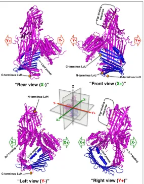
In response to changes in photoperiod and temperature, complete type Vtgs are secreted into the bloodstream and bind ovarian Vtgrs as 400-600 kDa homodimers. A crystal structure of a complete type lamprey vitellogenin exists (Fig. 2) (Anderson, Levitt, & Banaszak, 1998; Finn, 2007c; Raag, Appelt, Xuong, & Banaszak, 1988). Depicted as a monomer, the large lipid binding pocket is clearly visible. The putative receptor binding peptide is located in an amphipathic α-helix nested within a β sheet and presents basic residues (lysine [K], arginine [R]) to acidic ligand binding residues (aspartic acid [D], glutamic acid [E]) (See Fig. 2) on Vtgrs (Li, Sadasivam, & Ding, 2003). Vtgrs belong to the low density lipoprotein receptor (LDLR) related proteins (LRPs) superfamily (Smolenaars et al., 2006; Van der Horst, Roosendaal, & Rodenburg, 2009).
O-linked sugar domain exist in other species, white perch LR8 has only been identified without an O-linked sugar domain (Finn, 2007a; Hiramatsu, Chapman, Lindzey, Haynes, & Sullivan, 2004b; Hiramatsu, Matsubara, et al., 2002b).
Recently, we used affinity purification coupled to tandem mass spectrometry (AP-MS/MS) (Fig. 3) and Western blotting (Fig. 4) to identify and characterize the molecular structure of an additional Vtgr from white perch ovarian tissue, Lrp13 (Reading et al., 2014). Possessing additional LDLR Class B YWxD (LDLb), epidermal growth factor-like precursor (EGF), and LDLR Class A ligand binding repeat (LDLa) motifs, this receptor appears to represent a novel class of vertebrate lipoprotein receptor (Fig. 5). An additional
distinguishing feature of Lrp13 is that its cytoplasmic tail lacks any apparent endocytosis signal, raising the question of how this liganded-receptor complex is internalized,
compartmentalized, and processed. Intriguingly, in silico analysis of the protein sequence indicates several potential phosphorylation sites and kinase binding sites on the intracellular domain of Lrp13 that may be related to its localization and trafficking, which may be different from that of the LR8 receptor.
with a source of water until osmoregulatory and drinking mechanisms are fully developed (Finn, 2007a; 2007c; Finn & Kristoffersen, 2007). However, it is likely that concordant neofunctionalization of Vtgrs was required as well, although receptor specificity and
multiplicity have not been fully addressed in Vtg-Vtgr studies to date (Dieckmann, Dietrich, & Herz, 2010).
Structural and functional conservation among LLTPs and their receptors
A recent comparative genomic and phylogenetic study has placed Vtgs as a basal member of the LLTP superfamily (Hayward, Takahashi, Bendena, Tobe, & Hui, 2010). Vtg is ancestral to and shares considerable structural homology with apoB and apoE and LR8 shares a similar relationship with VLDLR and LDLR (Babin, Bogerd, Kooiman, Van
& Mahley, 1982; Weisgraber & Mahley, 1996; Wilson, Wardell, Weisgraber, Mahley, & Agard, 1991). This secondary structure is required for bioactivity (Nikoulin & Curtiss, 1998; Zaiou et al., 2000). The native receptor-binding domain is stabilized by an adjacent
amphipathic α-helix that does not interact directly with the receptor (Weisgraber & Mahley, 1996). This structural arrangement can be mimicked by synthetic peptides comprising the receptor-binding motif of apoB (11 amino acids) or apoE (15 amino acids) which have full binding activity for the LDLR (Cardin, Bowlin, & Krstenansky, 1988; Clay, Anantharamaiah, Mistry, Balasubramaniam, & Harmony, 1995; Dyer & Curtiss, 1991; Dyer, Smith, & Curtiss, 1991; Segrest et al., 1998). The corresponding synthetic monomer is unable to bind LDLR. The sequences flanking these native receptor-binding peptides are thought to be required for it to assume a proper 3D conformation. Therefore, the receptor-binding domains of apoB and apoE both utilize amphipathic structures to present tight clusters of homologous basic residues in a specific surface array as the basis for receptor recognition.
The receptor-binding sites of teleost complete type Vtgs resemble that of apoB. Chicken egg yolk very low-density lipoprotein (VLDL) was just as effective as native white perch Vtg in displacing 125I-Vtg from perch ovarian membranes (Tao, Berlinsky, & Sullivan, 1996). Chickens incorporate apoB into VLDL but they do not produce apoE. The laying hen expresses a ~95 kDa receptor on the oocyte surface that binds Vtgs and VLDL and
mediates their uptake by endocytosis (Hayashi, Ando, Stifani, & Schneider, 1989). This receptor also binds human apoE incorporated into lipid vesicles with an affinity similar to that which it exhibits for native complete type Vtgs (Steyrer, Barber, & Schneider, 1990).
receptor-receptor binding protein (VRBP), which are conserved in all chordates, seem to be essential for full receptor-binding activity. This expectation was confirmed when the VRBP was
localized to an 85-residue stretch of the Lv domain of teleost Vtgs (Fig. 6) (Li, Sadasivam, & Ding, 2003).
The gene encoding LR8 belongs to a diverse superfamily that encompasses several lipoprotein receptors, including LDLR and VLDLR (H Bujo, 1994; Jingami & Yamamoto, 1995). The cysteine-rich binding domain of LDLR is comprised of seven ligand-binding repeats (LBRs), whereas this domain within VLDLR and LR8 consists of eight such LBRs. The high cysteine content of these receptors is responsible for the proper folding of this domain into a rigid structure that presents clusters of acidic ligand-binding residues located at the C-termini of each LBR. These negatively charged residues may interact with positively charged residues (K, R) in the receptor-binding sites of apoB, apoE, and Vtg (Stifani, Menn, Rodriguez, & Schneider, 1990). It remains unknown whether or not other negatively charged residues distributed among the LBRs play a role in ligand binding.
2008; Babin et al., 1995; 1999; Babin & Vernier, 1989; Babin et al., 1997; Tao et al., 1996). Among lipoprotein receptors, ligand binding occurs through different LBRs. For example, LBR 5 is essential for binding of apoE, LBRs 2-7 cooperatively bind apoB, and repeats 1-3 are responsible for binding Vtg (Li, Sadasivam, & Ding, 2003a; Russell, Brown, & Goldstein, 1989). Vertebrate Vtgrs share an underlying ligand recognition mechanism as evidenced by the Vtgrs of chicken, Xenopus, and fish binding each other's complete type Vtgs (Stifani et al., 1988,1990ab; LeMenn and Nunez-Rodriguez, 1991; Tyler and Lancaster 1993;
Berlinsky et al., 1995; Tao et al., 1996). Additionally, our laboratory has shown that complete type Vtgs from five species of fishes both closely and distantly related to white perch bound to the perch LR8 (Hiramatsu et al., 2002a). This mechanism extends to receptor-mediated uptake of Vtg by the oocytes. For example, killifish (Fundulussp.) oocytes bind Xenopus
complete type Vtgs in vitro, a process that can be inhibited by the addition of complete type Vtgs from either Fundulus or Xenopus (Kanungo, Petrino, & Wallace, 1990). Among all oviparous vertebrates, both the receptor-binding motif of complete type Vtgs and the ligand binding sites of Vtgrs have changed little during the course of evolution.
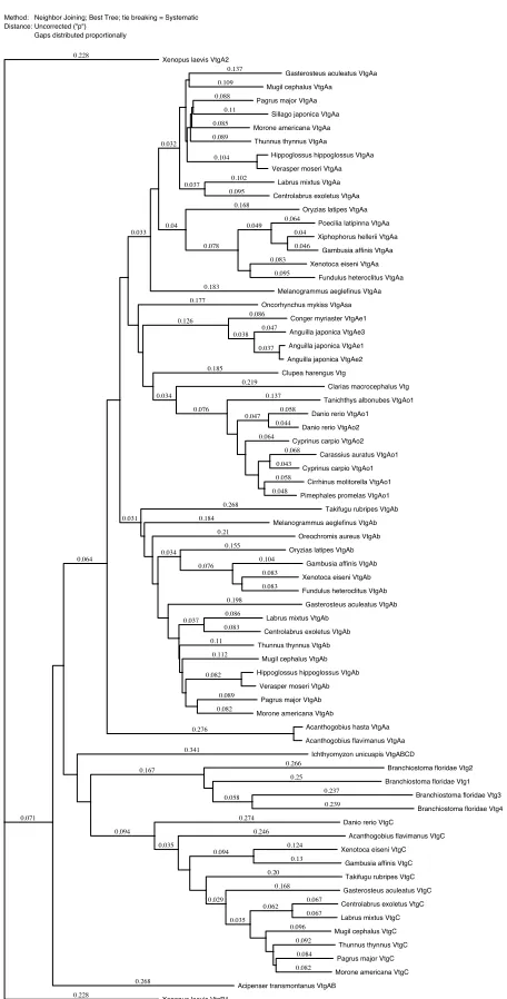
Vitellogenin polypeptide sequences (N=70) from an array of fishes were aligned by ClustalW to generate a dendrogram (Fig. 7). While approved gene names vary, it is clear that vitellogenins group by type and that the Aa and Ab types are present in Acanthomorph fishes which represent more derived teleosts.
Vtg lipidation
been shown to have dramatic effects on LLTP size, shape, surface charge, and availability of crucial receptor binding residues on the particle surface (Aviram et al., 1988; McKeone, Patsch, & Pownall, 1993; Ren et al., 2010; Rowe & Eckhert, 1999).
Given the high degree of conservation among LLTPs and their receptors, apolipoproteins and their receptors can serve as models for the Vtg-Vtgr system. For instance, the crucial role of basic residues (lysine and arginine) in receptor binding of
plasma lipoproteins was established three decades ago and subsequently confirmed in Vtgs (Finn, 2007a; 2007c; Finn et al., 2009; Innerarity et al., 1983; Li, Sadasivam, & Ding, 2003; Weisgraber et al., 1978). Additionally, core triglyceride content of LDL particles has been shown to affect the structure of apoB and its interactions with LDLR (Anderson et al., 1998; Aviram et al., 1988; Finn, 2007c; McKeone et al., 1993; Raag et al., 1988). Variable
apolipoprotein lipidation results in changes in overall particle size, surface charge, and in the local conformation of basic lysine residues of apoB on the LDL surface that constitute the receptor binding domain (Aviram et al., 1988; Li, Sadasivam, & Ding, 2003; Nigon, Lesnik, Rouis, & Chapman, 1991). LDL particles of intermediate size bind LDLR with higher affinity than both smaller and larger particles (Nigon et al., 1991; Smolenaars et al., 2006; Van der Horst et al., 2009).
Comprising up to 20% of Vtg mass, lipids are the dominant Vtg cargo. The majority of these lipids are phospholipids that can be utilized as structural components during
Reading et al., 2011; Richardson et al., 2005). With the data currently available, Pv domain size does not appear to correlate with lipid cargo size, however, a relationship between Pv domain size and lipid type has been suggested (Finn, 2007a; Hiramatsu, Chapman, Lindzey, Haynes, & Sullivan, 2004a; Silversand & Haux, 1995).
Vtg lipid cargo is comprised of polar lipids (~70-80%), specifically phophatidylcholine (PC), along with neutral lipids (~20-30%), primarily triacylglycerides (TAG) (Finn, 2007a; Hiramatsu, Chapman, Lindzey, Haynes, & Sullivan, 2004b; Hiramatsu, Matsubara, et al., 2002b). Relatively short Pv domains appear to be associated with eggs that lack large oil globules and contain a high percentage of PC, while longer Pv domains are primarily found in fishes that spawn eggs with large oil droplets that contain a higher percentage of neutral lipids(Anderson et al., 1998; Finn, 2007a; Finn & Kristoffersen, 2007; Finn, Fyhn,
required, each Vtg type may be loaded with a distinct lipid cargo that in turn could underlie receptor specificity and subsequent differential processing.
Implications of proportional vitellogenin accumulation
The proportional Vtg ratios within post-vitellogenic oocytes has been measured for barfin flounder, striped bass, and white perch (Sawaguchi et al., 2008; Williams et al., 2014). Barfin flounder spawn pelagic eggs in saltwater that have a post-vitellogenic proportional ratio of LvA : LvB : LvC of about 9 : 15 : 1 (Sawaguchi et al., 2008). Striped bass spawn their eggs with a post-vitellogenic proportional ratio of 1.4 : 1.4 : 1 in fresh or brackish water (Williams et al., 2014). Interestingly, the proportional abundance of the adhesive eggs spawned by white perch in fresh or brackish water as measured by selected reaction monitoring tandem mass spectrometry is 5 : 35.7 : 1.
While the proportional Vtg ratios do not appear to correlate to egg buoyancy or salinity, they may relate to other aspects of early life history. Considering the data that are currently available, it appears that VtgC is the most variable form of vitellogenin within the post-vitellogenic oocyte, ranging from ~2.5% in perch to 26% in striped bass (Fig. 8) (Schilling et al., 2014; Williams et al., 2014). The ratio of complete type Vtgs, on the other hand, appears to fall within a much narrower range of 1 : 1 to 1 : 1.7 for VtgAa : VtgAb, respectively.
In mosqitofish, for example, the VtgC-derived yolk proteins are the last components of the egg yolk that remain to be consumed by yolk-sac fry (Sawaguchi, Ohkubo, Koya, & Matsubara, 2005).
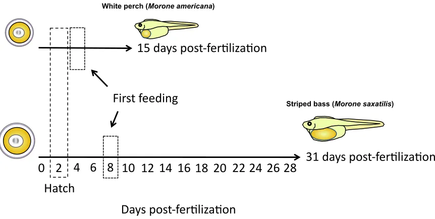
Depending upon temperature, fertilized white perch and striped bass larvae both hatch around 48 hrs post-fertilization (Eldridge, Whipple, Eng, Bowers, & Jarvis, 1981; Mansueti, 1964; North & Houde, 2003). When food is then restricted or deprived, white perch larvae survive for up to 15 days post-fertilization while striped bass larvae survive for up to 31 days post-fertilization (Fig. 9) (Eldridge et al., 1981; Mansueti, 1964). Additionally, the time to first feeding of these two species differs, with the white perch and the striped bass larvae beginning to feed at 3-5 days and 7-9 days post-fertilization, respectively. Therefore, although both of these closely related species share similar time frames during the earliest stages of development (i.e. from fertilization to hatch), the striped bass larvae appear to have an extended developmental window from hatching to first feeding and also these larvae have yolk stores that allow them to survive in the absence of food for twice as long as white perch after hatch. This disparity in early developmental stages post-hatch may relate to differences in VtgC yolk content of the white perch and striped bass eggs, which is 2.5-5% and 26%, respectively.
Summary and organization of dissertation
experiments that utilized methods such as protein expression and purification, nanoLC-MS/MS, and AP-MS/MS to further characterize white perch liver, plasma, and ovary tissues.
CHAPTER 2: Machine learning support vector machines are highly effective at discerning the sub-cellular fraction (i.e., cytosolic or membrane) from which a protein originated based upon nanoLC-MS/MS data. The simple fractionation method
utilized in this study effectively revealed an abundance of mitochondrial proteins.
CHAPTER 3: Male and female white perch have sex-specific plasma protein profiles both before and after induction with estradiol-17β (E2) as analyzed by machine learning support vector machines. In addition, the relatively uncharacterized latrophilin and seven transmembrane domain-containing protein 1 (Eltd1), and kininogen 1 (Kng1) are shown to be E2-responsive proteins in white perch.
CHAPTER 4: Selected reaction monitoring (SRM) absolute quantification tandem mass spectrometry indicates that 1) VtgAb is the predominant Vtg in white perch and the only Vtg detectable in the pre-vitellogenic liver, 2) of the three Vtgs, only VtgC can be detected in the pre-vitellogenic white perch ovary by SRM, and 3)
Chemical Society. As of March 3 2015, CHAPTER 3 is under a second round of review at
References
Anderson, T. A., Levitt, D. G., & Banaszak, L. J. (1998). The structural basis of lipid interactions in lipovitellin, a soluble lipoprotein. Structure, 6(7), 895–909.
Aviram, M., Lund-Katz, S., Phillips, M. C., & Chait, A. (1988). The influence of the
triglyceride content of low density lipoprotein on the interaction of apolipoprotein B-100 with cells. J. Biol. Chem., 263(32), 16842–16847.
Babin, P. J. (2008). Conservation of a vitellogenin gene cluster in oviparous vertebrates and identification of its traces in the platypus genome. Gene, 413(1-2), 76–82.
doi:10.1016/j.gene.2008.02.001
Babin, P. J., & Vernier, J. M. (1989). Plasma lipoproteins in fish. J. Lipid Res., 30(4), 467– 489.
Babin, P. J., Bogerd, J., Kooiman, F. P., Van Marrewijk, W. J. A., & Van der Horst, D. J. (1999). Apolipophorin II/I, Apolipoprotein B, Vitellogenin, and Microsomal Triglyceride Transfer Protein Genes Are Derived from a Common Ancestor. J. Mol. Evo., 49(1), 150– 160. doi:10.1007/PL00006528
Babin, P. J., Thisse, C., Durliat, M., Andre, M., Akimenko, M. A., & Thisse, B. (1997). Both apolipoprotein E and A-I genes are present in a nonmammalian vertebrate and are highly expressed during embryonic development. P. Natl. Acad. Sci. USA, 94(16), 8622–8627.
Baker, M. E. (1988). Is vitellogenin an ancestor of apolipoprotein B-100 of human low-density lipoprotein and human lipoprotein lipase? Biochem. J., 255(3), 1057.
Boren, J., Lee, I., Zhu, W., Arnold, K., Taylor, S., & Innerarity, T. L. (1998). Identification of the low density lipoprotein receptor-binding site in apolipoprotein B100 and the
modulation of its binding activity by the carboxyl terminus in familial defective apo-B100.
J. Clin. Invest., 101(5), 1084–1093. doi:10.1172/JCI1847
Brandt, B. W., Zwaan, B. J., Beekman, M., Westendorp, R. G. J., & Slagboom, P. E. (2005). Shuttling between species for pathways of lifespan regulation: A central role for the vitellogenin gene family? BioEssays, 27(3), 339–346. doi:10.1002/bies.20161
Cardin, A. D., Bowlin, T. L., & Krstenansky, J. L. (1988). Inhibition of lymphocyte proliferation by synthetic peptides homologous to human plasma apolipoproteins B and E. Biochem.
Bioph. Res Co., 154(2), 741–745.
Clay, M. A., Anantharamaiah, G. M., Mistry, M. J., Balasubramaniam, A., & Harmony, J. A. (1995). Localization of a domain in apolipoprotein E with both cytostatic and cytotoxic activity. Biochemistry-US, 34(35), 11142–11151.
Dashti, N., Gandhi, M., Liu, X., Lin, X., & Segrest, J. P. (2002). The N-Terminal 1000
Residues of Apolipoprotein B Associate with Microsomal Triglyceride Transfer Protein to Create a Lipid Transfer Pocket Required for Lipoprotein Assembly. Biochemistry-US,
41(22), 6978–6987. doi:10.1021/bi011757l
Davis, L. K., Hiramatsu, N., Hiramatsu, K., Reading, B. J., Matsubara, T., Hara, A., et al. (2007). Induction of three vitellogenins by 17beta-estradiol with concurrent inhibition of the growth hormone-insulin-like growth factor 1 axis in a euryhaline teleost, the tilapia
(Oreochromis mossambicus). Biol. Reprod., 77(4), 614–625.
doi:10.1095/biolreprod.107.060947
Dieckmann, M., Dietrich, M. F., & Herz, J. (2010). Lipoprotein receptors--an evolutionarily ancient multifunctional receptor family. Biol. Chem., 391(11), 1341–1363.
doi:10.1515/BC.2010.129
Durliat, M., Andre, M., & Babin, P. J. (2000). Conserved protein motifs and structural
that binds the LDL receptor. J. Biol. Chem., 266(34), 22803–22806.
Dyer, C. A., Smith, R. S., & Curtiss, L. K. (1991). Only multimers of a synthetic peptide of human apolipoprotein E are biologically active. J. Biol. Chem.,266(23), 15009–15015.
Eldridge, M. B., Whipple, J. A., Eng, D., Bowers, M. J., & Jarvis, B. M. (1981). Effects of Food and Feeding Factors on Laboratory-Reared Striped Bass Larvae. T. Am. Fish. Soc., 110(1), 111–120. doi:10.1577/1548-8659(1981)110<111:EOFAFF>2.0.CO;2
Finn, R. N. (2007a). The maturational disassembly and differential proteolysis of paralogous vitellogenins in a marine pelagophil teleost: a conserved mechanism of oocyte
hydration. Biol. Reprod., 76(6), 936–948. doi:10.1095/biolreprod.106.055772
Finn, R. N. (2007b). Vertebrate Yolk Complexes and the Functional Implications of Phosvitins and Other Subdomains in Vitellogenins. Biol. Reprod., 76(6), 926–935.
Finn, R. N. (2007c). Vertebrate yolk complexes and the functional implications of phosvitins and other subdomains in vitellogenins. Biol. Reprod., 76(6), 926–935.
doi:10.1095/biolreprod.106.059766
Finn, R. N., Fyhn, H. J., Henderson, R. J., & Evjen, M. S. (1996). The sequence of catabolic substrate oxidation and enthalpy balance of developing embryos and yolksac larvae of turbot (Scophthalmus maximus L.). Comp. Biochem. Physiol. A, 115(2), 133–151. doi:10.1016/0300-9629(96)00026-6
Finn, R. N., Kolarevic, J., Kongshaug, H., & Nilsen, F. (2009). Evolution and differential expression of a vertebrate vitellogenin gene cluster. BMC Evol. Biol., 9, 2.
doi:10.1186/1471-2148-9-2
Ghosh, P., & Thomas, P. (1995). Binding of metals to red drum vitellogenin and
incorporation into oocytes. Mar. Environ. Res., 39(1–4), 165–168. doi: 10.1016/0141-1136(94)00035-N
Bujo, H., Hermann, H., Kaderli, M, O., Jacobsen, L., Sugawara, S., Nimpf, J.,Yamamoto, T., Schneider, W, J. (1994). Chicken oocyte growth is mediated by an eight ligand binding repeat member of the LDL receptor family. EMBO J., 13(21), 5165.
Hayashi, K., Ando, S., Stifani, S., & Schneider, W. J. (1989). A novel sterol-regulated surface protein on chicken fibroblasts. J. Lipid Res., 30(9), 1421–1428.
Hiramatsu, N., Chapman, R. W., Lindzey, J. K., Haynes, M. R., & Sullivan, C. V. (2004a). Molecular Characterization and Expression of Vitellogenin Receptor from White Perch
(Morone americana). Biol. Reprod., 70(6), 1720–1730.
Hiramatsu, N., Chapman, R. W., Lindzey, J. K., Haynes, M. R., & Sullivan, C. V. (2004b). Molecular characterization and expression of vitellogenin receptor from white perch
(Morone americana). Biol. Reprod., 70(6), 1720–1730.
doi:10.1095/biolreprod.103.023655
Hiramatsu, N., Hara, A., Hiramatsu, K., Fukada, H., Weber, G. M., Denslow, N. D., & Sullivan, C. V. (2002a). Vitellogenin-Derived Yolk Proteins of White Perch, Morone
americana: Purification, Characterization, and Vitellogenin-Receptor Binding1. Biol.
Reprod., 67(2), 655–667.
Hiramatsu, N., Matsubara, T., Hara, A., Donato, D. M., Hiramatsu, K., Denslow, N. D., & Sullivan, C. V. (2002b). Identification, purification and classification of multiple forms of vitellogenin from white perch (Morone americana). Fish Physiol. Biochem., 26(4), 355– 370. doi:10.1023/B:FISH.0000009266.58556.9a
receptor. Curr. Opin. Lipidol., 6(2), 104–108.
Kanungo, J., Petrino, T. R., & Wallace, R. A. (1990). Oogenesis in Fundulus heteroclitus. VI. Establishment and verification of conditions for vitellogenin incorporation by oocytes in vitro. J. Exp. Zool., 254(3), 313–321. doi:10.1002/jez.1402540310
Lalazar, A., Weisgraber, K. H., Rall, S. C., Giladi, H., Innerarity, T. L., Levanon, A. Z., et al. (1988). Site-specific mutagenesis of human apolipoprotein E. Receptor binding activity of variants with single amino acid substitutions. J. Biol. Chem., 263(8), 3542–3545.
Li, A., Sadasivam, M., & Ding, J. L. (2003). Receptor-ligand interaction between vitellogenin receptor (VtgR) and vitellogenin (Vtg), implications on low density lipoprotein receptor and apolipoprotein B/E. The first three ligand-binding repeats of VtgR interact with the amino-terminal region of Vtg. J. Biol. Chem., 278(5), 2799–2806.
doi:10.1074/jbc.M205067200
Mahley, R. W., & Rall, S. C. (2000). Apolipoprotein E: far more than a lipid transport protein.
Annu. Rev. Genom. Hum. G., 1, 507–537. doi:10.1146/annurev.genom.1.1.507
doi:10.2307/1350789
McKeone, B. J., Patsch, J. R., & Pownall, H. J. (1993). Plasma triglycerides determine low density lipoprotein composition, physical properties, and cell-specific binding in cultured cells. J. Clin. Invest., 91(5), 1926–1933. doi:10.1172/JCI116411
Montorzi, M., Falchuk, K. H., & Vallee, B. L. (1995). Vitellogenin and Lipovitellin: Zinc Proteins of Xenopus laevis Oocytes. Biochemistry-US, 34(34), 10851–10858. doi:10.1021/bi00034a018
Ohkubo, N., & Matsubara, T. (2002). Sequential utilization of free amino acids, yolk proteins and lipids in developing eggs and yolk-sac larvae of barfin flounder Verasper moseri.
Mar. Biol., 140(1), 187–196. doi:10.1007/s002270100647
Nigon, F., Lesnik, P., Rouis, M., & Chapman, M. J. (1991). Discrete subspecies of human low density lipoproteins are heterogeneous in their interaction with the cellular LDL receptor. J. Lipid Res., 32(11), 1741–1753.
content of plasma vitellogenin from two Salmo species: rainbow trout (Salmo gairdneri) and sea trout (Salmo trutta). Comp. Biochem. Physiol. B, 81(4), 869–876.
North, E. W., & Houde, E. D. (2003). Linking ETM physics zooplankton prey and fish early life histories to striped bass Morone saxatilis and white perch M. americana recruitment.
Mar. Ecol.-Prog. Ser., 260, 219-236.
Ohkubo, N., Sawaguchi, S., Hamatsu, T., & Matsubara, T. (2006). Utilization of free amino acids, yolk proteins and lipids in developing eggs and yolk-sac larvae of walleye pollock
Theragra chalcogramma. Fisheries Sci., 72(3), 620–630.
doi:10.1111/j.1444-2906.2006.01192.x
Olofsson, S.-O., & Boren, J. (2005). Apolipoprotein B: a clinically important apolipoprotein which assembles atherogenic lipoproteins and promotes the development of
atherosclerosis. J. Intern. Med., 258(5), 395–410. doi:10.1111/j.1365-2796.2005.01556.x
Plack, P. A., & Pritchard, D. J. (1968). Effect of oestradiol 3-benzoate on the concentrations of retinal and lipids in cod plasma. Biochem. J., 106(1), 257–262.
Raag, R., Appelt, K., Xuong, N. H., & Banaszak, L. (1988). Structure of the lamprey yolk lipid-protein complex lipovitellin-phosvitin at 2.8 A resolution. J. M. Biol., 200(3), 553– 569.
Raffaï, R., Weisgraber, K. H., MacKenzie, R., Rupp, B., Rassart, E., Hirama, T., et al. (2000). Binding of an antibody mimetic of the human low density lipoprotein receptor to apolipoprotein E is governed through electrostatic forces. Studies using site-directed mutagenesis and molecular modeling. J. Biol. Chem., 275(10), 7109–7116.
Rall, S. C., Weisgraber, K. H., Innerarity, T. L., & Mahley, R. W. (1982). Structural basis for receptor binding heterogeneity of apolipoprotein E from type III hyperlipoproteinemic subjects. P. Natl. Acad. Sci. USA, 79(15), 4696–4700.
Reading, B. J., Hiramatsu, N., & Sullivan, C. V. (2011). Disparate binding of three types of vitellogenin to multiple forms of vitellogenin receptor in white perch. Biol. Reprod., 84(2), 392–399. doi:10.1095/biolreprod.110.087981
Reading, B. J., Hiramatsu, N., Schilling, J., Molloy, K. T., Glassbrook, N., Mizuta, H., et al. (2014). Lrp13 is a novel vertebrate lipoprotein receptor that binds vitellogenins in teleost fishes. J. Lipid Res., 55(11), 2287–2295. doi:10.1194/jlr.M050286
Richards, M. P. (1997). Trace mineral metabolism in the avian embryo. Poultry Sci., 76(1), 152–164.
Richardson, P. E., Manchekar, M., Dashti, N., Jones, M. K., Beigneux, A., Young, S. G., et al. (2005). Assembly of lipoprotein particles containing apolipoprotein-B: structural model for the nascent lipoprotein particle. Biophys. J., 88(4), 2789–2800.
doi:10.1529/biophysj.104.046235
Rowe, R. I., & Eckhert, C. D. (1999). Boron is required for zebrafish embryogenesis. J. Exp.
Biol., 202(12), 1649–1654.
Russell, D. W., Brown, M. S., & Goldstein, J. L. (1989). Different combinations of cysteine-rich repeats mediate binding of low density lipoprotein receptor to two different proteins.
J. Biol. Chem., 264(36), 21682–21688.
Sawaguchi, S., Ohkubo, N., Amano, H., Hiramatsu, N., Hara, A., Sullivan, C. V., & Matsubara, T. (2008). Controlled accumulation of multiple vitellogenins into oocytes during vitellogenesis in the barfin flounder, Verasper moseri. Cybium Int. J. Ichthyol.,
32(suppl 2), 262.
Schilling, J., Nepomuceno, A., Schaff, J. E., Muddiman, D. C., Daniels, H. V., & Reading, B. J. (2014). Compartment Proteomics Analysis of White Perch (Morone americana) Ovary Using Support Vector Machines. J. Proteome Res., 13, 1515-1526.
doi:10.1021/pr401067g
Schneider, W. J. (2009). Receptor-mediated mechanisms in ovarian follicle and oocyte development. Gen. Comp. Endocr., 163(1–2), 18–23. doi: 10.1016/j.ygcen.2008.11.032
Segrest, J. P., Jones, M. K., Mishra, V. K., Pierotti, V., Young, S. H., Boren, J., et al. (1998). Apolipoprotein B-100: conservation of lipid-associating amphipathic secondary structural motifs in nine species of vertebrates. J. Lipid Res., 39(1), 85–102.
Silversand, C., & Haux, C. (1995). Fatty acid composition of vitellogenin from four teleost species. J. Comp. Physiol. B, 164(8), 593–599. doi:10.1007/BF00389799
Smolenaars, M. M. W., Madsen, O., Rodenburg, K. W., & Van der Horst, D. J. (2006). Molecular diversity and evolution of the large lipid transfer protein superfamily. J. Lipid Res., 48(3), 489–502. doi:10.1194/jlr.R600028-JLR200
oogenesis: The piscine receptor for vitellogenin. BBA - Lipid Lipid Met., 1045(3), 271– 279. doi:10.1016/0005-2760(90)90130-P
Takahashi, S., Sakai, J., Fujino, T., Hattori, H., Zenimaru, Y., Suzuki, J., et al. (2004). The very low-density lipoprotein (VLDL) receptor: characterization and functions as a peripheral lipoprotein receptor. J. Atheroscler. Thromb., 11(4), 200–208.
Takahashi, Y., Itoh, F., Oohashi, T., & Miyamoto, T. (2003). Distribution of apolipoprotein E among lipoprotein fractions in the lactating cow. Comp. Biochem. Phys. B, 136(4), 905– 912. doi:10.1016/j.cbpc.2003.09.004
Tao, Y., Berlinsky, D. L., & Sullivan, C. V. (1996). Characterization of a vitellogenin receptor in white perch (Morone americana). Biol. Reprod., 55(3), 646–656.
Taskinen, M.-R. (2005). Type 2 diabetes as a lipid disorder. Curr. Mol. Med., 5(3), 297–308.
Van der Horst, D. J., Roosendaal, S. D., & Rodenburg, K. W. (2009). Circulatory lipid
transport: lipoprotein assembly and function from an evolutionary perspective. Mol. Cell.
Biochem., 326(1-2), 105–119. doi:10.1007/s11010-008-0011-3
Weisgraber, K. H., Innerarity, T. L., & Mahley, R. W. (1978). Role of lysine residues of plasma lipoproteins in high affinity binding to cell surface receptors on human fibroblasts. J. Biol. Chem., 253(24), 9053–9062.
Williams, V. N., Reading, B. J., Amano, H., Hiramatsu, N., Schilling, J., Salger, S. A., et al. (2014). Proportional accumulation of yolk proteins derived from multiple vitellogenins is precisely regulated during vitellogenesis in striped bass (Morone saxatilis). J. Exp. Zool.,
321(6), 301–315. doi:10.1002/jez.1859
Wilson, C., Wardell, M. R., Weisgraber, K. H., Mahley, R. W., & Agard, D. A. (1991). Three-dimensional structure of the LDL receptor-binding domain of human apolipoprotein E.
Science, 252(5014), 1817–1822.
Zaiou, M., Arnold, K. S., Newhouse, Y. M., Innerarity, T. L., Weisgraber, K. H., Segall, M. L., et al. (2000). Apolipoprotein E;-low density lipoprotein receptor interaction. Influences of basic residue and amphipathic alpha-helix organization in the ligand. J. Lipid Res.,
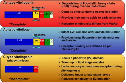
Figure 1. The primary domains of the three forms of vitellogenins are depicted. Yolk proteins derived from Aa-type vitellogenin are cleaved into free amino acids during final oocyte maturation. Ab-type vitellogenin receives partial or no proteolysis on lipovitellin heavy chain and are utilized during late embryonic growth. Adapted from Hiramatsu et al., 2002 (Biol. Reproduction, Fish Physiol Biochem); Reading et al., 2009 (Mar Biotech).
Aa-type vitellogenin
Phosphate
Pv LvL β’ Ct
LvL
Ab-type vitellogenin
C-type vitellogenin
(phosvitin-less)
Phosphate
Pv LvL β’ Ct
! Degradation of lipovitellin heavy chain (LvH) during ovarian maturation
! Osmotic effector during oocyte hydration
! Provides free amino acids to early embryos
! Receptor-binding site differs from VtgAb
“Complete”
“Complete”
“Incomplete”
! Intact LvH remains after oocyte maturation
! Provides large lipoprotein to late embryos and larvae
! Receptor binding site defined as per tilapia VtgAb
! Lacks a phosvitin (Pv) domain
! Taken up in lipid stage oocytes
! Lacks an oocyte membrane receptor during vitellogenesis
! Delivered intact to late-stage larvae
! Reduced sensitivity to E2 induction
LvH
Figure 2. 3D representation of a partial lamprey (Ichthyomyzon unicuspis) complete type vitellogenin. The lipovitellin heavy domain (LvH) is colored purple while the lipovitellin light domain (LvL) is blue. The LR8 binding sequence is in yellow (Li et al., 2003). The large lipid binding pocket and putative Zn2+ binding site are also indicated (pdb: 1LSH). From Anderson, Levitt, & Banaszak, 1998.
Figure 4. (Left) ClustalW dendrogram showing relationships between low-density lipoprotein receptor family polypeptide sequences. GenBank accession numbers are provided. Numbers above each branch are p-distances. (Right) Models representing linear domain structures of low-density lipoprotein receptor family members as defined at the bottom. From Reading et al., 2014 (J Lipid Res).
Fig. 1. (Left)ClustalW dendrogram showing relationships between low-density
Figure 5. Multiple vitellogenins (Vtg) and their receptors involved in Vtg-derived yolk formation of white perch and cutthroat trout. The A-type Vtgs (VtgAa, VtgAb and VtgAs) bind the ‘classical’ LR8-type Vtg receptor (Vtgr) and/or low-density lipoprotein receptor related protein 13 (Lrp13). Receptor proteins for C-type Vtg are detected in ligand blots of trout ovarian membrane, but not in white perch. Receptor proteins that universally bind multiple Vtg subtypes are also detected in the ovarian membrane preparations of both species. LR8: lipoprotein receptor (LR) with 8 ligand binding (LB) repeats; LR7+1: LR with 7+1 LB repeats; LR13+1: LR with 13+1 LB repeats. From Hiramatsu et al., 2015 (Gen Comp Endocr).
No receptor ? VtgAa VtgAb VtgC VtgAa VtgAb VtgC VtgAs VtgAs VtgC VtgC VtgC VtgAa VtgAb VtgAs VtgAs Unknown receptor Unknown receptor Unknown receptor Vtgr (LR8) Lrp13 (LR7+1) Lrp13 (LR13+1) Vtgr (LR8)
White perch Cutthroat trout
Figure 6. The vitellogenin receptor binding domain of complete type Morone VtgAb features that are similarly conserved in Gallus, Xenopus, Oreochromis, Danio, and human apoE and apoB synthetic peptides (Dyer et al., 1995). Beyond overall
sequence similarity, the sequences above contain highly conserved cysteines that form disulfide bonds crucial for presentation of the positive residues (lysine [K] and arginine [R]) at positions 181 and 183. Identified as essential for receptor binding, 181K is noted in pink (Li et al., 2003).
Distance:
Method: Neighbor Joining; Best Tree; tie breaking = Systematic Distance: Uncorrected ("p")
Gaps distributed proportionallyXenopus laevis VtgA2Gasterosteus aculeatus VtgAaMugil cephalus VtgAaPagrus major VtgAaSillago japonica VtgAaMorone americana VtgAaThunnus thynnus VtgAaHippoglossus hippoglossus VtgAaVerasper moseri VtgAaLabrus mixtus VtgAaCentrolabrus exoletus VtgAaOryzias latipes VtgAaPoecilia latipinna VtgAaXiphophorus hellerii VtgAaGambusia affinis VtgAaXenotoca eiseni VtgAaFundulus heteroclitus VtgAaMelanogrammus aeglefinus VtgAaOncorhynchus mykiss VtgAsaConger myriaster VtgAe1Anguilla japonica VtgAe3Anguilla japonica VtgAe1Anguilla japonica VtgAe2Clupea harengus VtgClarias macrocephalus VtgTanichthys albonubes VtgAo1Danio rerio VtgAo1Danio rerio VtgAo2Cyprinus carpio VtgAo2Carassius auratus VtgAo1Cyprinus carpio VtgAo1Cirrhinus molitorella VtgAo1Pimephales promelas VtgAo1Takifugu rubripes VtgAbMelanogrammus aeglefinus VtgAbOreochromis aureus VtgAbOryzias latipes VtgAbGambusia affinis VtgAbXenotoca eiseni VtgAbFundulus heteroclitus VtgAbGasterosteus aculeatus VtgAbLabrus mixtus VtgAbCentrolabrus exoletus VtgAbThunnus thynnus VtgAbMugil cephalus VtgAbHippoglossus hippoglossus VtgAbVerasper moseri VtgAbPagrus major VtgAbMorone americana VtgAbAcanthogobius hasta VtgAaAcanthogobius flavimanus VtgAaIchthyomyzon unicuspis VtgABCDBranchiostoma floridae Vtg2Branchiostoma floridae Vtg1Branchiostoma floridae Vtg3Branchiostoma floridae Vtg4Danio rerio VtgCAcanthogobius flavimanus VtgCXenotoca eiseni VtgCGambusia affinis VtgCTakifugu rubripes VtgCGasterosteus aculeatus VtgCCentrolabrus exoletus VtgCLabrus mixtus VtgCMugil cephalus VtgCThunnus thynnus VtgCPagrus major VtgCMorone americana VtgCAcipenser transmontanus VtgABXenopus laevis VtgB1
Gasterosteus aculeatus VtgAa Mugil cephalus VtgAa Pagrus major VtgAa
Sillago japonica VtgAa Morone americana VtgAa Thunnus thynnus VtgAa
Hippoglossus hippoglossus VtgAa Verasper moseri VtgAa
Labrus mixtus VtgAa Centrolabrus exoletus VtgAa
Oryzias latipes VtgAa Poecilia latipinna VtgAa Xiphophorus hellerii VtgAa
Gambusia affinis VtgAa Xenotoca eiseni VtgAa
Fundulus heteroclitus VtgAa Melanogrammus aeglefinus VtgAa Oncorhynchus mykiss VtgAsa
Conger myriaster VtgAe1 Anguilla japonica VtgAe3 Anguilla japonica VtgAe1 Anguilla japonica VtgAe2 Clupea harengus Vtg
Clarias macrocephalus Vtg Tanichthys albonubes VtgAo1 Danio rerio VtgAo1 Danio rerio VtgAo2 Cyprinus carpio VtgAo2
Carassius auratus VtgAo1 Cyprinus carpio VtgAo1
Cirrhinus molitorella VtgAo1 Pimephales promelas VtgAo1 Takifugu rubripes VtgAb Melanogrammus aeglefinus VtgAb
Oreochromis aureus VtgAb Oryzias latipes VtgAb
Gambusia affinis VtgAb Xenotoca eiseni VtgAb Fundulus heteroclitus VtgAb
Gasterosteus aculeatus VtgAb Labrus mixtus VtgAb Centrolabrus exoletus VtgAb Thunnus thynnus VtgAb
Mugil cephalus VtgAb Hippoglossus hippoglossus VtgAb Verasper moseri VtgAb Pagrus major VtgAb Morone americana VtgAb
Acanthogobius hasta VtgAa Acanthogobius flavimanus VtgAa
Ichthyomyzon unicuspis VtgABCD Branchiostoma floridae Vtg2 Branchiostoma floridae Vtg1
Branchiostoma floridae Vtg3 Branchiostoma floridae Vtg4 Danio rerio VtgC
Acanthogobius flavimanus VtgC Xenotoca eiseni VtgC
Gambusia affinis VtgC Takifugu rubripes VtgC Gasterosteus aculeatus VtgC Centrolabrus exoletus VtgC Labrus mixtus VtgC Mugil cephalus VtgC Thunnus thynnus VtgC Pagrus major VtgC Morone americana VtgC Acipenser transmontanus VtgAB Xenopus laevis VtgA2
Xenopus laevis VtgB10.0160.268 0.2680.064 0.0640.020.0190.276 0.2760.0120.031 0.0310.033 0.0330.0150.0120.183 0.1830.032 0.0320.04 0.040.0090.037 0.0370.0050.0020.137 0.1370.109 0.1090.0060.104 0.1040.0020.089 0.0890.0010.085 0.0850.088 0.0880.11 0.110.0150.0160.102 0.1020.095 0.0950.168 0.1680.078 0.0780.049 0.0490.0180.0640.0640.0240.04 0.040.046 0.0460.083 0.0830.095 0.0950.177 0.1770.0070.126 0.1260.0150.086 0.0860.038 0.0380.0470.0470.037 0.0370.0090.0060.185 0.1850.034 0.0340.219 0.2190.076 0.0760.137 0.1370.0150.047 0.0470.0120.0580.0580.044 0.0440.064 0.0640.0190.0170.0080.068 0.0680.043 0.0430.058 0.0580.048 0.0480.268 0.2680.0060.184 0.1840.0120.21 0.210.0080.034 0.0340.0150.155 0.1550.076 0.0760.104 0.1040.0150.083 0.0830.083 0.0830.198 0.1980.0170.037 0.0370.0010.0860.0860.083 0.0830.11 0.110.0040.112 0.1120.0050.082 0.0820.0160.0170.0210.089 0.0890.082 0.0820.0130.0120.341 0.3410.0210.167 0.1670.094 0.0940.266 0.2660.0130.25 0.250.058 0.0580.237 0.2370.239 0.2390.274 0.2740.035 0.0350.246 0.2460.0170.094 0.0940.0240.124 0.1240.13 0.130.20 0.200.029 0.0290.168 0.1680.035 0.0350.062 0.0620.0230.0670.0670.067 0.0670.096 0.0960.0110.092 0.0920.0050.084 0.0840.082 0.0820.228 0.2280.071 0.0710.228 0.228
Morone saxatilis
Striped bass
Morone americana
White perch
VtgAb
VtgC
VtgAa
Figure 8. In white perch, yolk proteins derived from VtgC are minor components of the total egg yolk (< 5%), whereas in striped bass they are major components of the egg yolk (~ 25%). [Williams et al., 2014 (J Exp Zool A); Schilling et al., 2014 (J
Figure 9. Average survival duration of food-restricted white perch and striped bass larvae. Dashed boxes indicate approximate time of hatching (~2 days) and onset of first feeding (~4 days in white perch, ~8 days in striped bass). [Mansuetti, 1964; Eldridge, et al., 1981; North & Houde, 2003].
Hatch&
15&days&post.fer2liza2on&
31&days&post.fer2liza2on&
0& 2& 4& 6&
8& 10& 12& 14& 16&
18&
20& 22&
24&
26&
28&
First&feeding&
Striped bass (Morone saxatilis)CHAPTER 2
Compartment Proteomics Analysis of White Perch (Morone americana) Ovary Using Support Vector Machines
Justin Schilling 1, Angelito Nepomuceno 2, Jennifer E. Schaff 3, David C. Muddiman 2, Harry
V. Daniels 1 and Benjamin J. Reading 1,*
1 Department of Applied Ecology, College of Agriculture and Life Sciences, North
Carolina State University, 127 David Clark Labs, Raleigh, North Carolina, United States
2 W. M. Keck FT-ICR Mass Spectrometry Laboratory, Department of Chemistry, North
Carolina State University, Raleigh, North Carolina, United States
3 Genomic Sciences Laboratory, North Carolina State University, Raleigh, North
Carolina, United States
Reprinted with permission from Schilling, J., Nepomuceno, A., Schaff, J. E., Muddiman, D. C., Daniels, H. V., & Reading, B. J. (2014). Compartment proteomics analysis of white perch
(Morone americana) ovary using support vector machines. Journal of Proteome Research,
13, 1515−1526. doi:10.1021/pr401067g. Copyright 2014 American Chemical Society.
KEYWORDS: compartment proteomics; support vector machines; modulated modularity clustering; ovary; oocytes; vitellogenin; transcriptome; mitochondria; alternatively spliced variants
characterize the cytosolic and membrane fractions of white perch ovary tissues by semiquantitative tandem mass spectrometry using label-free quantitation based on
normalized spectral counts. FASP depletes both low-molecular-weight and high-molecular-weight substances that could interfere with protein digestion and subsequent peptide separation and detection. Membrane proteins are notoriously difficult to characterize due to their amphipathic nature and association with lipids. The simple fractionation we employed effectively revealed an abundance of proteins from mitochondria and other membrane-bounded organelles. We further demonstrate that support vector machines (SVMs) offer categorical classification of proteomics data superior to that of parametric statistical methods such as analysis of variance (ANOVA). Specifically, SVMs were able to perfectly (100% correct) classify samples as either membrane or cytosolic fraction during cross-validation based on the expression of 242 proteins with the highest ANOVA p-values (i.e. those that were not significant for enrichment in either fraction). The white perch ovary cytosolic and membrane proteomes and transcriptome presented in this study can support future
investigations into oogenesis and early embryogenesis of white perch and other members of the genus Morone.
INTRODUCTION