Type of the Paper (Review)
1
Title:
Role of PhaC type I and type II enzymes during
2
PHA biosynthesis
.
3
Valeria Mezzolla,1 Oscar Fernando D’Urso2 and, Palmiro Poltronieri 3*
4
1 Bioesplora, Department of Biological and Environmental Science and Technologies, Ecotekne, Lecce;
5
[email protected]; [email protected];
6
2 Bioesplora, Department of Biological and Environmental Science and Technologies, Ecotekne, Lecce;
7
8
3 Institute of Sciences of Food Productions (ISPA-CNR), via Monteroni km 7, 73100 Lecce, Italy;
9
* Correspondence: [email protected]; Tel.: +39-0832-422609
10
11
12
Abstract: PHA synthases (PhaC) are grouped into four classes based on the kinetics and
13
mechanisms of reaction. The grouping of PhaC enzymes into four classes is dependent on substrate
14
specificity, according to the preference in forming short chain length (scl) or medium chain length
15
(mcl) polymers: class I, class III, and class IV produce scl-PHAs depending on propionate, butyrate,
16
valerate and hexanoate precursors, while class II phaC synthesize mcl-PHAs based on the alkane (C6
17
to C14) precursors.
18
PHA synthases of class I, in particular PhaCCs from Chromobacterium USM2 and PhaCCn/RePhaC1
19
from Cupriavidus necator/R. eutropha, have been analysed and the crystal structures of the C-domains
20
have been determined. PhaCCn/RePhaC1 was also studied by small angle X-ray scattering (SAXS)
21
analysis. Models have been proposed for dimerization, catalysis mechanism, substrate recognition
22
and affinity, product formation and product egress route. The assays based on amino acid
23
substitution by mutagenesis have been useful to validate the hypothesis on the role of amino acids in
24
catalysis and in accommodation of bulky substrates, for the synthesis of PHB co-polymers and
25
medium chain length-PHA polymers with optimized chemical properties.
26
Keywords: PhaC synthase, classification, dimerization, substrate binding, exit cavity, C3-C14
27
alkanes, polymer composition.
28
29
1. Introduction
30
Polyhydroxyalkanoates (PHAs) are biodegradable polyesters produced in several Gram-negative
31
and Gram-positive bacteria, in archea and cyanobacteria [1-8]. PHAs are polymers containing
32
various alkanes, such as3-hydroxy propionate (3HP), butyrate (3HB), valerate (3HV), hexanoate
33
(3HHx), heptanoate (3HHp), octanoate (3HO), nonanoate (3HN), decanoate (3HD), dodecanoate
34
(3HDD), in addition to 4-hydroxybutyrate (4HB) or 5-hydroxyvalerate (5HV), and produced
35
through the availability of the corresponding CoA thioester substrates [9-14].
36
PhaC synthases, the polymerizing enzymes, are grouped into four classes based on substrate
37
specificity, and the preference in forming short chain length (scl-) or medium chain length (mcl-)
38
polymers: class I, class III, and class IV produce principally scl-PHAs, while class II phaC synthesize
39
mcl-PHAs [7, 15].
40
The PHA biosynthesis genes include: PhaPs, genes coding for Phasins, the granule assembling
41
proteins, PhaM, encoding the activator and accelerator of the catalytic activity of PHA synthase,
42
encoded by PhaC; phaA, encoding the acetoacetyl-CoA β-thiolase, phbB coding for the
43
acetoacetyl-CoA reductase, phaG, coding for 3-hydroxyacyl-carrier protein-CoA transferase, phaJ,
44
encoding the enoyl-CoA hydratase, and PhaZ, encoding the PHA depolymerising enzyme.
45
PhaC proteins possess the so called lipase box, G-X-S-X-G, and a secondary structure containing the
46
α/β hydrolase fold, a characteristic succession of alpha helices and beta strands, typical of lipases.In
47
PhaC enzymes, the lipase box has the conserved sequence G-G/S-X-C-X-G/A-G, renamed the PhaC
48
box consensus sequence. The Cys in the lipase box-like sequence is the catalytic amino acid, forming
49
the covalently bond intermediate, Cys-S-H3B [7, 16].
50
PHA synthase activity is based on the catalytic triad C-H-D, cysteine, histidine, aspartate, also
51
responsible for catalysis in lipases (S-H-D). In the catalytic triad, the negatively charged Asp447 in
52
PhaCCs from Chromobacterium spp. [17, 18] and Asp480 in PhaCCn from Cupriavidus necator [19, 20]
53
assist to enhance the basicity of His477 and His508, respectively, by formation of a direct hydrogen
54
bond. Previous reports suggest that the Asp residue of the triad acts as a general base catalyst to
55
accelerate deprotonation of the 3-hydroxyl group of HB in the step involving elongation of the PHA
56
product.
57
PHA synthases are grouped into four classes based on the kinetics and mechanism of reaction. The
58
grouping of PhaC enzymes into each class is dependent on the structure of the PhaC, alone or in
59
association to other subunits, and the substrate specificity: class I, class III, and class IV produce
60
scl-polymers depending on propionate (3HP), butyrate (3HB, 4HB), valerate (HV) and hexanoate
61
(HH) precursors (C3 to C6 carbons), while class II phaC enzymes synthesize mcl-polymers
62
depending on hexanoate (3HH), heptanoate (3HHp), octanoate (3HO), decanoate (3HD),
63
undecanoate (3HUD), dodecanoate (3HDD) (C6 to C12), and availability of the corresponding CoA
64
thioester substrates, originating from three different metabolic pathways [9, 10, 20].
65
While some bacterial species produce mainly 3HB polymers, other species can synthesize various
66
PHAs, depending on availability of intermediate precursors [1, 3, 9]. The structure and properties of
67
the polymers are affected by the monomers that are incorporated. PHAs can be molded into films
68
and hollow bodies. Polyhydroxybutyrate (PHB) is brittle, fragile, and stiff, with low elongation
69
ability, and a break point below 15%. The incorporation of 3-hydroxyvalerate or other co-monomers
70
can decrease the brittleness of PHB. The thermal, rheological and barrier properties of PHAs show
71
good application potential in thermoplastic materials. The synthesis of copolymers is a frequent
72
strategy to improve the properties of PHAs, to improve the plastics flexibility and lower the glass
73
transition temperature (Tg) and the melting temperature (Tm) [21]. P3HB-4HB polymers and
74
conventional thermoplastic used for packaging show high tensile strength and higher elongation at
75
break. HBV-containing mcl-PHAs are elastic, have low melting point, a relatively low degree of
76
crystallinity, and with various tensile strength. PHB copolymer containing 3-hydroxyvalerate unit
77
P(3HB-co-3HV) has been developed with improved mechanical properties. To this aim, either
78
optimization of substrate availability (feedstock, successive addition of precursors), and efficiency
79
of enzymes, through genetic engineering and selection of PHA synthases, have been applied.
80
81
Class I and class II PHA synthases
82
Classes I and II PHA synthases are formed by a single protein (PhaC), of about 60 kDa.
83
In class I, the active PhaC enzyme is a dimer, with catalytic (CAT) domains facing each other, with
84
N-domains making direct contacts, sustaining protein interaction and dimerization. It was
85
hypothesized that a partially folded catalytic domain is partially occupied by the lid and cat domain
86
secondary structure, that change their conformation in presence of activation factors, to open the
87
catalytic domain and to allocate the 3HB-CoA in its binding site (Chek et al., 2017).
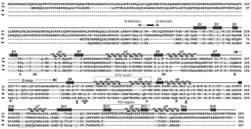
PhaC sequences differ in their length. In Cupriavidus necator (formerly Ralstonia eutropha) the class I
89
enzyme PhaCCn/RePhaC, the sequence is composed of a 191 amino acids N-terminal domain
90
proteolytically cleaved after arginine, and a C-domain, containing the catalytic site, composed of 398
91
amino acids, for a total of 589 residues (Figure 1). In C. necator, PhaCCn contains Cys319, Asp480 and
92
His508, located between the beta 6 and alpha 3 turn, the beta 10 and alpha 7 turn, and at the end of the
93
beta 11 strand, respectively. In the between of Cys319 and Asp480 is located the D-loop and the
94
helix-turn-helix motif (HTH), formed by α4, α5, α6, and β7-β8 stretches [18, 19]. (Wittenborn et al.
95
,2016, Kim et al., 2017a). PhaCCn/RePhaC1 was also studied by small angle X-ray scattering (SAXS)
96
analysis that confirmed the previous findings on the protein assembled as a dimer [22].
97
The PhaC synthase from Chromobacterium spp., phaCCs, was extensively studied [16, 17]. PhaCCs has a
98
peculiarity of utilization of 3HB, 3HV, and 3HH, producing scl-PHA polymers with mixed
99
composition, with ability to incorporate C5 and C6 alkanes into the PHA polymer. PhaCCs was found
100
highly active, with fast polymerization rate [17]. PhaCCs is shorter in length (for about 29 amino
101
acids) in respect to PhaCCn and this produces differences in numbering of amino acids. PhaCCs
102
structure has a substrate-binding site hidden by a partially disordered protein domain, the CAP
103
domain [16, 17]. Cysteine291, at the end of the ß6 sheet, is followed by the CAP domain, containing
104
the LID structure, that close the accessibility of the substrate access pocket. The β sheets in the CAP
105
domain have been renumbered with Greek letters in the PhaCCs sequence. Thus, from the N-terminal
106
sequence, up to the β6-α3 turn, the two PHA synthases conserve the same numbering in their
107
secondary structures, but the successive α-β turns are differently numbered. In PhaCCs, the core
108
subdomain contains six ß-strands (ß8 to ß13) and four a-helices (α4 to α7), whose number does not
109
correspond to those in the PhaCCn-CAT structure.
110
In PhaCCs thecatalytic triad is composed of Cys291, His477 (located after the β9 sheet, in respect to the
111
β11 sheet in PhaCCn) and Asp447 (located at the turn formed by ß8 strand and α4 helix, the ß8-α4 fold,
112
in respect to the β10-α7 fold in PhaCCn).
113
In other species, such as Delftia acidovorans (previously Comamonas acidovorans) the PHA synthase
114
contains a large insert of 40 amino acid residues shown to improve the specific activity of the
115
enzyme, located in the α/β hydrolase fold, following the catalytic cysteine after the β6 turn [25].
116
Aeromonas spp., such as A. caviae, A. hydrophila, and A. punctata, possess PhaC enzymes belonging to
117
class I. The enzyme PhaCCc from Caulobacter crescentus (C. vibrioides) [26], displayed to accommodate
118
alkanes with various alkyl side-chain length.
119
Class II phaC synthases have been extensively studied, and are widely distributed in bacteria: in
120
Pseudomonas spp. (P. putida, P. mendocina, P. oleovorans, P. campisalis; P. stutzeri) there are two phaC
121
genes, of which PhaC1 is the active enzyme under physiological conditions. PhaC synthases have
122
been reported in Halomonas spp., such as H. campisalis, Halomonas sp. O-1 and Halomonas elongata
123
DSM2581 [27], and in P. stutzeri, that can be exploited in polymerization of mcl-PHAs [28]. There are
124
two PHA synthases, PhaC1 and PhaC2, in P. oleovorans, of which PhaC2 has a higher affinity for
125
3-hydroxyhexanoate (3HH) monomers.
126
Class II phaC enzymes differ from PhaCCn, as prototype of class I PHA synthases, for about 28 amino
127
acids, reaching the C-terminal (1-559) with about 30 amino acids shorter sequence. The catalytic triad
128
has been renumbered as Cys296, Asp452, His453 and His480 in Pseudomonas spp., prototype for Class II
129
PHA synthases.
130
Class III and Class IV PHA synthases
132
Class III PHA synthases are made of two subunits, namely, a catalytic subunit PhaC (40–53
133
kDa) and a second subunit PhaE (ranging from 20 to 40 kDa), which form the PhaEC complex, in
134
which the PhaE subunit is necessary for PHA polymerization. Class III phaC are structured as
135
tetramers, such as phaEC from Allochromatium vinosum, (with catalytic triad Cys149, Asp302, His331),
136
whose enzyme activity has been studied using substrate analogs [15]. The authors performed
137
molecular docking and in silico studies, that are in agreement with the crystal structure of synthases
138
available [15], based on homology models built using CPHmodels3.0, SWISS-MODEL and
139
I-TASSER, performing structure-guided sequence profiles. The results of their analysis, referring to
140
PhaCCn and PhaC class III from Allochromatium vinosum, describe the presence of an active site of
141
cysteine, that is buriedin a pocket: the authors, by comparison with other enzymes with known
142
crystal structure (lipases), postulated the presence of the substrate entrance and product exit
143
channels [15].
144
There is also an Archaeal type, PhaC class IIIA: this group is represented by Haloarcula marismortui:
145
Archea present good perspectives of exploitation for polymer production, given by the easiness of
146
PHB extraction.
147
Class IV PHA synthases from Bacillus spp. are composed of a catalytic subunit PhaC (41.5 kDa) and a
148
PhaR (22 kDa) subunit, similarly to class III synthases composed of phaE and phaC units. Class IV
149
PHA synthases are classified as B. megaterium type (IVm) [23], Bacillus cereus type (IVc) [22], and B.
150
bataviensis type (IVb), with 33% homology to the other phaC sequences [5]
.
In E. coli expressing151
PhaRC from B. cereus YB-4, the biosynthesized PHA undergoes synthase-catalyzed alcoholytic
152
cleavage using endogenous and exogenous alcohols. This alcoholysis is thought to be shared among
153
class IV synthases, and this reaction is useful for regulation of PHA molecular weight and for
154
modification of the PHA carboxy terminus.
155
The catalytic cysteine in the active site is C151 in B. cereus, and C147 in B. megaterium type IV enzymes.
156
As shown for PhaCYB4 from B. cereus YB-4, the involvement of Cys151, Asp306, and His335 in
157
polymerization activity was shown by site-directed mutagenesis [5]
.
158
159
Diversity and spread of phaC in bacteria
160
PHA synthase genes can be identified in environmental bacterial strains for a preliminary screening,
161
before knowledge on PHA synthesis ability due to the presence of the gene, using PCR amplification
162
with conserved primers [7, 29].Through PCR analyses, phaC genes were detected in a collection of
163
bacterial strains isolated from soils and from marine environments. In samples of environmental
164
strains, we amplified phaC gene sequences in colonies from environmental isolates, and performed
165
DNA sequencing of ribosomal DNA to identify the strains at species level (unpublished results):
166
with this method several species were classified for ability to produce PHA, namely P. oleovorans, P.
167
fluorescens, P. sihuiensis, P. putida, Comamonas testosteroni, Aeromonas hydrophila, as well as Cupriavidus
168
necator. It is envisaged that PCR screening using various different primer sets can be optimized to
169
find new phaC polymorphisms and potential novel PHA synthase sequences. Quelas reported the
170
presence in Bradyrhizobium japonicum USDA110 of five polyhydroxyalkanoate (PHA) synthases
171
(PhaC), distributed into four different PhaC classes [30], and characterized the requirements for two
172
of the genes in legume nodules under various physiological conditions.
176
Figure 1. Amino acid sequence and secondary structure of Cupriavidus necator PhaCCn, aligned with the PHA
177
synthase of class II (P. aeruginosa), class IVm and class IVb, from Bacillus megaterium and B. cereus, respectively.
178
179
Crystal structure
180
In two publications appeared almost contemporarily, two teams reported on the crystal structure of
181
PhaCCn-CAT, the catalytic domain of PhaC from Cupriavidus necator [18, 19, 24]. PhaCCn-CAT was
182
shown to dimerize, and to adopt a partially open form maintaining a narrow substrate access to the
183
active site. PhaCCn needs PhaM, the primer of PHA synthesis, to start and accelerate polymer
184
synthesis, and this may be due to increased accessibility of 3HB-CoA substrate to the active site.
185
Wittenborn obtained the crystal structure of PhaCCn(C319A), a construct in which the active site
186
cysteine (Cys319) was mutated to alanine to improve protein stability in the absence of detergent.
187
During the crystallization, proteolysis of PhaCCn occurred after the N-domain (R192), leading to the
188
crystal structure of the C-domain: PhaCCn-CAT is formed by two Core subdomains (G143-F352,
189
L450-A589), flanking on both sides a Dimerization domain (A353-L549) containing the dimerization loop
190
(D-loop) and the Helix-loop-helix (HTH) domain; in addition, in the terminal Core sub-domain there
191
is an Extended C-terminal region (EC: R521-A589), that is missing in class IV PhaC (figure 1), and a
192
Protruding Structure, PS, that elongates from the Extended C-region.
193
The catalytic domain of PhaCCn contains the residues 201–368 and 378–589 (with residues 369–377
194
devoid of any structure), showing an α/β-hydrolase fold, featuring a central mixed β-sheet flanked
195
by α-helices on both sides. This architecture is similar to that seen in lipases. The CAT domain in
196
the PhaCCn sequence is structured by the presence of the β1-4 sheets, the α1 helix, the β5 sheet, α2
197
helix, β6 sheet facing the lipase box, followed by the α3-β7 fold: after this structure there is the
198
D-loop; after the β8 sheet, there is the helix-loop-helix (HTH), composed of the α4 and α5 helices
199
facing each other, and the β9-α6 fold, where the Dimerization subdomain ends (L449); as for the other
200
amino acids of the catalytic triad, the aspartate is located between the β10-α7 fold, and the histidine
201
is located after the β11 sheet. The active site of PhaCCn is accessible via a water-filled channel, with a
202
Two PhaC monomers interact through the dimerization surfaces (A353-E445), containing hydrophobic
204
amino acids, by means of interaction between one monomer helix-loop-helix motif (HTH) and the
205
D-loop of the second monomer [18, 19].
206
In the report on the crystal structure obtained from the catalytic domain of PhaC from
207
Chromobacterium sp. USM2, PhaCCs-CAT was compared to the PhaCCn-CAT crystal structure [17].
208
Considering the two structures described, in PhaCCs-CAT a difference in the accessibility of the
209
active site has been evidenced. Chek showed that in the PhaCCs-CAT dimer, the CAP and LID
210
domains close the access to the substrate binding site [17]. The structure proposed by Chek and
211
colleagues describing a PhaCCs active site covered by the CAP subdomain, differs from the partially
212
open form of the PhaCCn catalytic domain reported by Wittenborn. The CAP domain occupies
213
partially the access to the substrate binding pocket, and the LID domain needs to slide away in order
214
to free the access for 3OH-alkanoyl-S-CoA units. Both catalytic domains of PhaCCs and PhaCCn form a
215
dimer mediated by the CAP subdomain. The difference between the closed and partially open form
216
is provided by the conformation of the CAP subdomain. The CAP subdomain undergoes a
217
conformational change during catalytic activity with rearrangement of the dimeric form.
218
The main difference between the two crystal structures was found in the folding of αB’ and ηB’
219
helices and their linker loop of PhaCCs-CAT, while the corresponding positions in PhaCCn-CAT,
220
show a long α4 helix that presents a partial access to the active site. The region Leu402–Asn415
221
forming the α4 helix in PhaCCn-CAT is conserved among Class I and II PHA synthases, whereas the
222
corresponding segment, Leu369–Lys382 of PhaCCs-CAT, displays a disordered structure.
223
224
Catalytic mechanism
225
The models proposed for the available PhaC structures, hypothesize the presence of a substrate
226
entrance tunnel, that accommodates HB-CoA, with a size of about 12.5-13 Å, and a product egress
227
tunnel, positioned perpendicularly to the entrance tunnel. Various catalytic mechanisms for PHA
228
synthases have been proposed, in the context of dimerization of PHA synthases of class I and II [17].
229
One mechanism is referred to as the non-processive ping-pong model: this mechanism requires
230
two cysteines in the active sites in the dimer, for PHA chain elongation, with chain transfer from one
231
cysteine to the second active site. The ping-pong mechanism requires two thiol groups located at a
232
distance short enough to shuttle back and forth the growing (3HB)n chain between the two thiols.
233
The dimeric structures described by Wittenborn and by Kim for PhaCCn-CAT, and by Chek for
234
PhaCCs, show that the two active sites are too distant (33 and 28.1 Angstrom, respectively) for
235
successive chemical reactions.
236
The distance between the active sites in the dimer seems to favor the mechanism based on a
237
single active site for each elongation reaction. In the model described by Chek, the dimer, composed
238
by two units of phaCCs through the contacts between the two CAP domains and the two N-domains,
239
presents two channels leading to the two active sites. The dimeric structure proposed by Chek [17],
240
favors the involvement of one active site for each processing step. In the model, the substrate enters
241
the substrate-binding tunnel, while chain product is elongated along a path near the protein surface,
242
with a sliding mechanism of the PHA polymer under synthesis along a V shaped cavity within the
243
enzyme. In the proposed structure, the enzyme moves along the extremity of the forming polymer to
244
add new 3HB units, rather than hosting the polymer into a product egress channel. The mechanism
245
involves a processive model that requires a single active site for PHA chain elongation and a
246
non-covalent intermediate, in addition to a covalent intermediate bound to the Cys residue at the
247
active center during the catalytic cycle. The enzyme dimer, through interactions with other partners,
248
with substrate, phasins and phaM, move the CAP domains to flip away, opening the active site
249
entrance, and freeing the product channel, and the two Core units contemporarily accept the
250
that allows the intermediates to be located in the enlarged cavities partially freed from the CAP
252
occupancy. The arrangement of the dimer, different from that of the PhaCCn-CAT dimer, may
253
allow to the CAP subdomains to undergo a conformational change during catalytic activity with
254
rearrangements in the dimer, that facilitate substrate entry, intermediate product formation, and
255
product exit from the active site. According to the crystal structure of the PhaCCn-CAT dimer [18, 19]
256
the substrates enter through the substrate-binding tunnel: the first 3HB-CoA is attacked by the
257
nucleophilic Cys-SH to produce a 3HB-Cys covalent bond, as in the aforementioned model, and
258
frees CoA-SH, that is released from the product egress tunnel. A second 3HB-CoA attacks 3HB-Cys
259
thioester bond with the hydroxyl group in 3HB to produce a (3HB)2-CoA intermediate, reaction that
260
frees the Cys residue in the active center. The Cys residue again attacks the thioester bond of the
261
(3HB)2-CoA intermediate to produce (3HB)n+1, covalently bound to the Cys residue and release of
262
free CoA. In this model, the growing 3HB polymer is bound to the enzyme at the end of each cycle.
263
This model cannot allow to position large molecules such as (3HB)n-CoA intermediate within the
264
substrate binding site, that has a cavity of 12.5 Angstrom.
265
An alternative model has been proposed with a succession of reactions slightly different. The
266
model proposed for PhaCCn, by Wittenborn, implies that newly entered 3HB-CoA produces
267
3HB-Cys; then (3HB)2-CoA enters the active site to produce (3HB)3-CoA, which is again released
268
from the active site. When a new (3HB)2-CoA substrate binds, the HB hydroxyl group is
269
deprotonated by His508, facilitated through modulation of the histidine basicity by Asp480. The newly
270
formed HB alkoxide attacks the Cys-HB thioester, generating a noncovalent, CoA-bound
271
intermediate. However, if the (3HB)3-CoA produced is held in the active site and attacked by the
272
active Cys residue again to produce (3HB)3-Cys, chain elongation would then require an
273
inter-subunit reaction. Again, (3HB)n-Cys adducts would require a larger active site cavity.
274
275
Mutation and amino acid substitution studies
276
Several studies focused on PHB synthases with mutations enabling the enzymes to accelerate the
277
reaction kinetics [31-33] and ability to accept bulk substrates as precursors for the production of
278
mcl-PHAs and grafted copolymers.
279
Nomura and Taguchi [34] reviewed the attempts to engineer various classes of PHA synthases,
280
either by mutagenesis or by evolution, in class I and Class II enzymes. The methods utilized either
281
random mutagenesis, intragenic suppression mutagenesis, gene shuffling, random mutagenesis
282
combined with Site-specific saturation mutagenesis and recombination, localized semi-random
283
mutagenesis, PCR-mediated random chimeragenesis, intragenic suppression mutagenesis,
284
site-specific saturation mutagenesis.
285
Many authors described mutations in amino acids positioned in various domains of different PHA
286
synthases, most often finding a decrease in production of mcl-PHA and higher synthesis of scl-PHA.
287
Beneficial effects of mutagenesis studies of Glu130 and Ser477 have been described [35-37]. For
288
instance, the E130D substitution and S477X mutation in type II PHA synthase showed an
289
enhancement of PHA production and alteration of polymer molecular weight.
290
A mutational study of PhaCCs reported by Chuah [40], showed that in PhaCCs Ala479 is a critical
291
residue required for substrate specificity, as determined by various site-specific mutational assays
292
both in vivo and in vitro, and production tests of copolymers such as P(3HB-co-3HHx). In PhaCCn and
293
in other Class I enzymes this position corresponds to the conserved residue Ala517. In the structure
294
proposed by Chek, Ala479 is located within α5 helix and the side chain protrudes into a depression of
295
the molecular surface formed by loops (β4-α1, β9-α5 and α5-β10 loops) from the core subdomain,
296
and is partially covered by the helix ηB’ and the following loop of the LID region from the CAP
297
subdomain: the A479 mutation results in weakening of the interactions between the LID region and
298
the active site. Since Ala479 is surrounded by polar residues (Ser475 and Arg490), it is supposed that
300
replacement of Ala479 with Ser or Thr facilitates hydrogen-bonding interactions with the polar
301
residues and stabilization of α5 helix harboring the active residue His477, important for enzyme
302
activity.
303
A mutagenesis study of class I PHA synthases showed that the F420S mutation in PhaCCn
304
increased the specific activity with a shortened lag phase [41]. This residue corresponds to Phe387 of
305
PhaCCs, which is conserved among Class I and II PHA synthases, and is located in αC helix of the
306
CAP domain. Phe387 is involved in dimerization by participating in an intermolecular nonpolar
307
interaction linking the αC helix to the LID region, suggesting that the mutation may affect the
308
conformational stability and/or conformation transition of the LID region.
309
The CAP subdomain provides αC and αD helices as building blocks of the active site cavity filled
310
with a cluster of water molecules. In the structure obtained by Chek [17], the C-terminal portion of
311
the LID region of the CAP subdomain is disordered and is followed by αC helix docked to the core
312
subdomain. Two highly conserved residues, Trp392 and Asp395, are present in αC helix. Trp392 of
313
PhaCCs is located in the αC helix of the CAP subdomain and faces Site B of the channel.
314
Amara and Rhem attempted to modify the activity of PhaC from Pseudomonas species [37]. The
315
conserved residue Trp398 was replaced, such as Trp398Phe and Trp398Ala, and the mutation resulted
316
in inactivation of the enzyme. Using the threading model of enzyme structure, the authors located
317
the Trp residue as exposed on the surface, in agreement with the results shown by Chek for class I
318
enzymes [42-46].
319
Tyr412 in PhaCCs, and Tyr446 in the α6 helix in PhaCCn, are residues conserved in Class I, III and IV
320
PHA synthases, while Phe occupies this position in Class II synthases: in addition to this amino acid
321
position, there is a second substitution that seems to have a role in accommodating larger substrates.
322
Tyr438 is conserved in Class I, III and IV enzymes, while in Class II PhaC this position is occupied by
323
His: this may contribute to a reduction of size, eliminating the bulky side chain (phenol ring), and
324
determining changes in polar interactions with other amino acids facing the substrate entrance
325
tunnel; these amino acids interactions may account for the property to accommodate large substrates
326
in class II enzymes.
327
PhaC1 and PhaC2 from Pseudomonas stutzeri [39], have been applied to produce mcl-PHAs in
328
engineered bacteria. PsPhaC2 with four point mutations, at E130D, S325T, S477G, and Q481K was used to
329
accommodate substrates with various shapes and structures, to produce mcl-PHAs and block
330
copolymers. The putative catalytic residues Cys296, Asp452, His453 and His480 were replaced by
331
site-specific mutagenesis [37]. Considering the Pseudomonas mcl-PHA synthases, the His480Gln
332
substitution did not affect enzyme activity, posing the doubt that His is not a component of the
333
catalytic triad. As for a second conserved histidine, when His453 was replaced by Gln, the modified
334
enzyme showed only 24% of wild-type in vivo activity, which make suppose that His453 might be
335
part of the catalytic triad in class II PHA synthases [37]. However, no other study confirmed the
336
involvement of His453 in class II PhaC2 catalysis.
337
Sheu studied the increase of PHA synthase thermostability and activity, using chimeric
338
constructs, indicating that some amino acid substitutions may stabilize the enzyme at higher
339
temperature [31].
340
341
Production of PHA in fermentors
342
Various companies are involved in production of bioplastics for industrial applications. The
343
methods are various, either using patented strains, engineered PHA synthases, and growth
344
the endpoint step of PHA synthesis, and bacteria collection, various methods have been established,
346
from lipid staining [47] and analysis of fluorescence intensity, to physic-chemical analyses (Raman,
347
FTIR spectra). Since bacterial cultures require sterilization that is costly at industrial scale, methods
348
based on halophilic strains have been proposed as to circumvent the sterilization process. Extraction
349
of PHAs from bacteria requires costly procedures, therefore researchers used Archea or
350
cyanobacteria that have PHA granules easily extracted, decreasing the costs of production.
351
352
Progress and advancements in PHA field
353
Recent advancements on PHA granule structure and composition have been achieved [48].
354
The high molecular weight storage PHB consists of > 103 3HB residues (storage PHB). PHB granules
359
in vivo are covered by a surface layer that is distinct from the polymer core. PHA granules are
360
structured through the action of various proteins on the surface. The granules, named also
361
carbonosomes, represent supramolecular complexes with specific functions. In addition to Phasins
362
(such as PhaP2, PhaP3, PhaP4), among the proteins identified during PHA granule isolation, there
363
are the PHB synthase (PhaC1), PhaM, the activator of PhaC, Acetyl-CoA acetyltransferase, and
364
acylCoA synthetase: their presence may be explained by the need to avoid accumulation of CoA-SH,
365
produced by the PHA synthase during polymer synthesis, since an excess of CoA would inhibit the
366
enzyme. The most accurate model for PHA synthesis within bacterial cell is the Scaffold Model: it
367
assumes that PHB synthase of nascent PHB granules is attached to a scaffold within the cell. PHB
368
granules have been localized in the cell centre, along with the length axis of the bacteria. PhaM, that
369
specifically interacts with PhaC1 and with phasin PhaP5, interacts also with DNA and with the
370
nucleoid in vitro and in vivo, and this may explain why PHB granules have been found attached to
371
the bacterial nucleoid.
372
373
Conclusions
374
In this review, we reported on the classification of PHA synthases, the proposed structures and role
375
of individual amino acids in the catalysis and mechanism of activity of class I and class II PHA
376
synthases, presenting the information available on the other types of enzymes. We have reviewed
377
the engineering attempts and the effect of modification of key amino acids on the enzymatic activity
378
and product formation. It is expected that PHA synthases may be further improved to produce
379
effectively and at convenient costs tailor-made polymers.
380
381
Acknowledgments: No funds have been provided for covering the costs to publish in open access.
382
Author Contributions: V.M. and O.F.D. provided the information on research data on environmental strains.
383
P.P. wrote and reviewed the manuscript.
384
Conflicts of Interest: “The authors declare no conflict of interest."
385
References
386
1. Kumar P, Jun HB, Kim BS. Co-production of polyhydroxyalkanoates and carotenoids through
387
bioconversion of glycerol by Paracoccus sp. strain LL1. Int J Biol Macromol. 2018; 107, 2552-2558. doi:
388
10.1016/j.ijbiomac.2017.10.147
389
2. Park, D.H., and Kim, B.S. Production of poly(3-hydroxybutyrate) and
390
poly(3-hydroxybutyrate-co-4-hydroxybutyrate) by Ralstonia eutropha from soybean oil. New Biotechnol.
391
2011; 28, 719-24. doi: 10.1016/j.nbt.2011.01.007.
392
3. Kumar, P., Singh, M., Mehariya, S., Patel, S.K., Lee, J.K., Kalia, V.C. Ecobiotechnological approach for
393
exploiting the abilities of Bacillus to produce co-polymer of Polyhydroxyalkanoate. Indian J Microbiol.
394
2014, 54, 151-7. Doi: 10.1007/s12088-014-0457-9
395
4. Hermann-Krauss, C., Koller, M., Muhr, A., Fas, H., Stelzer, F., Braunegg, G. Archaeal production of
396
polyhydroxyalkanoate (PHA) co- and terpolyesters from biodiesel industry-derived by-products.
397
Archaea 2013, 129268. Doi: 10.1155/2013/129268
5. Tsuge, T., Hyakutake, M., Mizuno, K. Class IV polyhydroxyalkanoate (PHA) synthases and
399
PHA-producing Bacillus. Appl Microbiol Biotechnol. 2015, 99, 6231-40. doi: 10.1007/s00253-015-6777-9
400
6. Ansari, S., and Fatma, T. Cyanobacterial polyhydroxybutyrate (PHB): Screening, optimization and
401
characterization. PLoS One 2016; 11, e0158168. Doi: 10.1371/journal.pone.0158168
402
7. Tan G-Y.A., Chen, C.-L., Li, L., Ge, L., Wang, L., Ningtyas Razaad, I.M., Li, Y., Zhao, L., Mo, Y., Wang,
403
J.-Y. Start a research on biopolymer polyhydroxyalkanoate (PHA): A review. Polymers (Basel) 2014, 6,
404
706-754. Doi: 10.3390/polym6030706
405
8. Poltronieri, P., Mezzolla, V.; D'Urso, O.F. PHB production in biofermentors assisted through biosensor
406
applications. Proceedings (Basel), 2017, 1, 4. Doi:10.3390/ecsa-3-E014
407
9. Chen, G.-Q., Hajnal, I., Wu, H., Lv, L., Ye, J. Engineering biosynthesis mechanisms for diversifying
408
Polyhydroxyalkanoates. Trends Biotechnol. 2015, 33:565-574, doi: 10.1016/j.tibtech.2015.07.007 565
409
10. Chen, G.-Q., and Hajnal, I. The ‘PHAome’. Trends Biotechnol. 2015, 33, 559–564. Doi:
410
10.1016/j.tibtech.2015.07.006
411
11. Le Meur, S., Zinn, M., Egli, T., Thöny-Meyer, L., Ren, Q. Production of medium-chain-length
412
polyhydroxyalkanoates by sequential feeding of xylose and octanoic acid in engineered Pseudomonas
413
putida KT2440. BMC Biotechnol. 2012, 12, 53. Doi: 10.1186/1472-6750-12-53.
414
12. Anjum, A., Zuber, M., Zia, K.M., Noreen, A., Anjum, M.N., Tabasum, S. Microbial production of
415
polyhydroxyalkanoates (PHAs) and its copolymers: a review of recent advancements. Int J Biol
416
Macromol 2016, 89, 161–174.
417
13. Meng, D.C., Shen, R., Yao, H., Chen, J.C., Wu, Q., Chen, G.Q. Engineering the diversity of polyesters.
418
Curr Opin Biotechnol. 2014; 29, 24-33. Doi: 10.1016/j.copbio.2014.02.013.
419
14. Mezzolla, V; D'Urso, OF, Poltronieri, P. Optimization of polyhydroxyalkanoate production by
420
recombinant E. coli supplemented with different plant by-products. Biotechnol Indian J. 2017, 13, 138.
421
15. Zhang, W., Chen, C., Cao, R., Maurmann, L., Li, P. Inhibitors of polyhydroxyalkanoate (PHA)
422
synthases: synthesis, molecular docking, and implications. Chembiochem. 2015, 16, 156–166. Doi:
423
10.1002/cbic.201402380
424
16. Bhubalan, K., Chuah, J.A., Shozui, F., Brigham, C.J., Taguchi, S., Sinskey, A.J., Rha, C., Sudesh, K.
425
Characterization of the highly active polyhydroxyalkanoate synthase of Chromobacterium sp. strain
426
USM2. Appl Environ Microbiol. 2011, 77, 2926–2933.
427
17. Chek, M.F., Kim, S.Y., Mori, T., Arsad, H., Samian, M.R., Sudesh, K., Hakoshima, T. Structure of
428
polyhydroxyalkanoate (PHA) synthase PhaC from Chromobacterium sp. USM2, producing
429
biodegradable plastics. Sci Rep. 2017, 7, 5312. DOI: 10.1038/s41598-017-05509-4
430
18. Wittenborn, E.C., Jost, M., Wei, Y., Stubbe, J., Drennan, C.L. Structure of the Catalytic Domain of the
431
Class I Polyhydroxybutyrate Synthase from Cupriavidus necator. J. Biol. Chem 2016, 291, 25264-25277.
432
19. Kim, J., Kim, Y.-J., Choi, S. Y., Lee, S. Y. & Kim, K.-J. Crystal structure of Ralstonia eutropha
433
polyhydroxyalkanoate synthase C-terminal domain and reaction mechanisms. Biotechnol. J 2017, 12,
434
1600648. DOI: 10.1002/biot.201600648
435
20. Kumar, P., Ray, S., Kalia, V.C. Production of co-polymers of polyhydroxyalkanoates by regulating the
436
hydrolysis of biowastes. Bioresource Technol. 2016, 200, 413–419. doi: 10.1016/j.biortech.2015.10.045
437
21. Zou, H., Shi, M., Zhang, T., Li, L., Li, L., Xian, M. Natural and engineered polyhydroxyalkanoate
438
(PHA) synthase: key enzyme in biopolyester production. Appl Microbiol Biotechnol. 2017, 101,
439
7417–7426. DOI: 10.1007/s00253-017-8485-0
440
22. Kihara, T., Hiroe, A., Ishii-Hyakutake, M., Mizuno, K., Tsuge, T. Bacillus cereus-type
441
polyhydroxyalkanoate biosynthetic gene cluster contains R-specific enoyl-CoA hydratase gene. Biosci
442
Biotechnol Biochem. 2017, 81, 1627-1635. doi: 10.1080/09168451.2017.1325314.
443
23. Hyakutake M, Tomizawa S, Mizuno K, Abe H, Tsuge T. Alcoholytic cleavage of
444
polyhydroxyalkanoate chains by class IV synthases induced by endogenous and exogenous ethanol.
445
Appl Environ Microbiol. 2014; 80, 1421-9. doi: 10.1128/AEM.03576-13
24. Kim, Y-J, Choi, S.Y., Kim, J., Jin, K.S., Lee, S.Y., Kim, K-J. Structure and function of the N-terminal
447
domain of Ralstonia eutropha polyhydroxyalkanoate synthase, and the proposed structure and
448
mechanisms of the whole enzyme. Biotechnol. J. 2017, 12, 1600649. DOI: 10.1002/biot.201600649
449
25. Tsuge, T., Imazu, S., Takase, K., Taguchi, S., Doi Y. An extra large insertion in the
450
polyhydroxyalkanoate synthase from Delftia acidovorans DS-17: its deletion effects and relation to
451
cellular proteolysis. FEMS Microbiology Letters 2004, 231, 77-83. Doi: 10.1016/S0378-1097(03)00930-3
452
26. Qi, Q., and Rehm, B.H. Polyhydroxybutyrate biosynthesis in Caulobacter crescentus: molecular
453
characterization of the polyhydroxybutyrate synthase. Microbiology 2001, 147, 3353-8.
454
27. Ilham, M., Nakanomori, S., Kihara, T., Hokamura, A., Matsusaki, H., Tsuge, T., Mizuno, K.
455
Characterization of polyhydroxyalkanoate synthases from Halomonas sp. O-1 and Halomonas elongata
456
DSM2581: Site-directed mutagenesis and recombinant expression. Polymer Degrad. Stability 2014, 109,
457
416-423. Doi: 10.1016/j.polymdegradstab.2014.04.024
458
28. Chen, J.-Y., Song, G., Chen, G.-Q. A lower specificity PhaC2 synthase from Pseudomonas stutzeri
459
catalyses the production of copolyesters consisting of short-chain-length and medium-chain-length
460
3-hydroxyalkanoates. Antonie van Leeuwenhoek 2006, 89, 157–167. Doi: 10.1007/s10482-005-9019-9
461
29. Montenegro, E.M.D.S., Delabary, G.S., Silva, M.A.C.D., Andreote, F.D., Lima, A.O.S. Molecular
462
diagnostic for prospecting polyhydroxyalkanoate-producing bacteria. Bioengineering (Basel) 2017; 4, 52.
463
Doi: 10.3390/bioengineering4020052.
464
30. Quelas, J.I., Mongiardini, E.J., Pérez-Giménez, J., Parisi, G., Lodeiro, A.R. Analysis of two
465
polyhydroxyalkanoate synthases in Bradyrhizobium japonicum USDA 110. J. Bacteriol. 2013, 195, 3145-55.
466
Doi: 10.1128/JB.02203-12.
467
31. Sheu, D.S., Chen, W.M., Lai. Y.W., Chang, R.C. Mutations derived from the thermophilic
468
polyhydroxyalkanoate synthase PhaC enhance the thermostability and activity of PhaC from
469
Cupriavidus necator H16. J Bacteriol. 2012, 194, 2620–2629. Doi: 10.1128/JB.06543-11
470
32. Takase, K., Matsumoto, K., Taguchi, S., Doi, Y. Alteration of substrate chain-length specificity of type II
471
synthase for polyhydroxyalkanoate biosynthesis by in vitro evolution: in vivo and in vitro enzyme
472
assays. Biomacromolecules 2004, 5, 480–485. Doi: 10.1021/bm034323+
473
33. Chen, C., Cao, R., Shrestha, R., Ward, C., Katz, B.B., Fischer, C.J., Tomich, J.M., Li P. Trapping of
474
intermediates with substrate analog HBOCoA in the polymerizations catalyzed by class III
475
polyhydroxybutyrate (PHB) synthase from Allochromatium vinosum. ACS Chem Biol. 2015, 10,
476
1330-1339. Doi: 10.1021/cb5009958.
477
34. Nomura, C.T., and Taguchi, S. PHA synthase engineering toward superbiocatalysts for custom-made
478
biopolymers. Applied Microbiol Biotechnol. 2007, 73, 969–979. DOI: 10.1007/s00253-006-0566-4
479
35. Matsumoto, K., Takase, K., Aoki, E., Doi, Y., Taguchi, S. Synergistic effects of Glu130Asp substitution
480
in the type II polyhydroxyalkanoate (PHA) synthase: enhancement of PHA production and alteration
481
of polymer molecular weight. Biomacromolecules 2005, 6, 99–104.
482
36. Matsumoto, K., Aoki, E., Takase, K., Doi, Y., Taguchi, S. In vivo and in vitro characterization of
483
Ser477X mutations in polyhydroxyalkanoate (PHA) synthase 1 from Pseudomonas sp. 61-3: effects of
484
beneficial mutations on enzymatic activity, substrate specificity, and molecular weight of PHA.
485
Biomacromolecules 2006, 7, 2436–2442.
486
37. Amara, A.A., and Rehm, B.H. Replacement of the catalytic nucleophile cysteine-296 by serine in class II
487
polyhydroxyalkanoate synthase from Pseudomonas aeruginosa-mediated synthesis of a new polyester:
488
identification of catalytic residues. Biochem. J. 2003, 374, 413-21.
489
38. Zou, H., Shi, M., Zhang, T., Li, L., Li, L., Xian, M. Natural and engineered polyhydroxyalkanoate
490
(PHA) synthase: key enzyme in biopolyester production. Appl. Microbiol. Biotechnol. 2017; 101,
491
7417-7426. doi: 10.1007/s00253-017-8485-0.
492
39. Chen, J.Y., Liu, T., Zheng, Z., Chen, J.C., Chen, G.-Q. Polyhydroxyalkanoate synthases PhaC1 and
493
PhaC2 from Pseudomonas stutzeri 1317 had different substrate specificities. FEMS Microbiol Lett 2004,
494
234, 231–237. Doi: 10.1016/j.femsle.2004.03.029
495
40. Chuah, J.A., Tomizawa, S., Yamada, M., Tsuge, T., Doi, Y., Sudesh, K., Numata, K. Characterization of
496
site-specific mutations in a short-chain-length/medium-chain-length polyhydroxyalkanoate synthase:
497
In vivo and in vitro studies of enzymatic activity and substrate specificity. Appl. Environ. Microbiol. 2013,
498
79, 3813–3821. Doi: 10.1128/AEM.00564-13
41. Normi, Y.M., Hiraishi, T., Taguchi, S., Sudesh, K., Najimudin, N., Doi, Y. Site-directed saturation
500
mutagenesis at residue F420 and recombination with another beneficial mutation of Ralstonia eutropha
501
polyhydroxyalkanoate synthase. Biotechnol. Lett. 2005, 27, 705–712.
502
42. Tsuge, T., Saito, Y., Kikkawa, Y., Hiraishi, T., Doi, Y. Biosynthesis and compositional regulation of
503
poly(3-hydroxybutyrate)-co-(3-hydroxyhexanoate) in recombinant Ralstonia eutropha expressing
504
mutated polyhydroxyalkanoate synthase genes. Macromol Biosci. 2004, 4, 238–242. DOI:
505
10.1002/mabi.200300077
506
43. Shozui, F., Matsumoto, K., Sasaki, T., Taguchi, S. Engineering of polyhydroxyalkanoate synthase by
507
Ser477X/Gln481X saturation mutagenesis for efficient production of 3-hydroxybutyrate-based
508
copolyesters. Appl Microbiol Biotechnol. 2009, 84, 1117–1124. Doi: 10.1007/s00253-009-2052-2
509
44. Tsuge, T., Watanabe, S., Shimada, D., Abe, H., Doi, Y., Taguchi, S. Combination of N149S and D171G
510
mutations in Aeromonas caviae polyhydroxyalkanoate synthase and impacton polyhydroxyalkanoate
511
biosynthesis. FEMS Microbiol Lett. 2007, 277, 217–222. Doi: 10.1111/j.1574-6968.2007.00958.x
512
45. Gao, X., Yuan, X.X., Shi, Z.Y., Guo, Y.Y., Shen, X.W., Chen, J.C., Wu, Q., Chen, G.-Q. Production of
513
copolyesters of 3-hydroxybutyrate and medium-chain-length 3-hydroxyalkanoates by E. coli
514
containing an optimized PHA synthase gene. Microb Cell Factories 2012, 11, 130. Doi:
515
10.1186/1475-2859-11-130
516
46. Shen, X.W., Shi, Z.Y., Song, G., Li, Z.J., Chen, G.-Q. Engineering of polyhydroxyalkanoate (PHA)
517
synthase PhaC2Ps of Pseudomonas stutzeri via site-specific mutation for efficient production of PHA
518
copolymers. Appl Microbiol Biotechnol. 2011, 91, 655–665. Doi: 10.1007/s00253-011-3274-7
519
47. Choi, J,E,, Na, H.Y., Yang, T.H., Rhee, S.K., Song, J.K. A lipophilic fluorescent LipidGreen1-based
520
quantification method for high-throughput screening analysis of intracellular poly-3-hydroxybutyrate.
521
AMB Express 2015, 5, 131. doi: 10.1186/s13568-015-0131-6
522
48. Jendrossek, D., and Pfeiffer, D. New insights in the formation of polyhydroxyalkanoate granules
523
(carbonosomes) and novel functions of poly(3-hydroxybutyrate). Environ Microbiol. 2014, 16, 2357-73.
524
doi: 10.1111/1462-2920.12356