_____________________________________________________________________________________________________
www.sciencedomain.org
Comparative Evaluation of Smear Layer Removal
Efficacy Using QMix 2in1, Chitosan, Smear Clear
and Glyde
Shaheen Venghat
1*and Mithra N. Hegde
11
Department of Conservative Dentistry and Endodontics, A. B. Shetty Memorial Institute of Dental Sciences, Deralakatte, Mangalore, India.
Authors’ contributions
This work was carried out in collaboration between both authors. Author MNH designed the study, wrote the protocol and wrote the first draft of the manuscript. Author SV managed the literature searches and the experimental process. Both authors read and approved the final manuscript.
Article Information
DOI: 10.9734/BJMMR/2016/23185 Editor(s): (1) Emad Tawfik Mahmoud Daif, Professor of Oral & Maxillofacial Surgery, Cairo University, Egypt. Reviewers: (1) Neha Sisodia, Ram Manohar Lohia Hospital, India. (2)Jorge Paredes Vieyra, University of Baja California, Mexico. Complete Peer review History:http://sciencedomain.org/review-history/12928
Received 18th November 2015 Accepted 16th December 2015 Published 9th January 2016
ABSTRACT
Aim and Objective: The effects of four endodontic irrigants and on a smear layer created by hand and rotary instrumentation were evaluated in vitro in the middle and apical thirds of root canals. Materials and Methods: Forty eight mature extracted mandibular premolar teeth with a single root canal and a closed apex were distributed randomly into four groups of 12 teeth each. Whilst cleaning and shaping up to size F5 using Protaper Universal System, the root canals were irrigated with 3 mL of 5.25% NaOCl, between each file size. Group 1 (G1) were irrigated with a final flush of QMix 2in1. The teeth in group 2 (G2) were irrigated with a final flush of 0.2%Chitosan, group 3 (G3) with Smear Clear and group 4 (G4) with Glyde. The teeth were split longitudinally and prepared for examination by scanning electron microscopy.
Results: Specimens irrigated with a final flush of Glyde (G4) or 0.2%Chitosan (G2) were cleaner than with QMix and Smear Clear, showing very clean root canal surfaces in the middle one-third but in the apical one-third the smear layer was not completely removed, especially at the openings of the dentinal tubules. Statistical analysis showed no significant difference in the cleanliness of root
canal wall between G1, G2, G3 and G4.
Conclusion: Irrigation with QMix 2in1, Smear Clear, 0.2%Chitosan, and Glyde and 6% did not remove all the smear layer from the root canal system. All these irrigants showed less effectiveness in removal of smear layer from apical 3rd. Glyde showed maximum efficacy in removal of removal of smear layer followed by 0.2%Chitosan, Smear Clear and then QMix 2in1.
Keywords: Chitosan; instrumentation irrigant; NaOCl; QMix; smear clear; smear layer.
1. INTRODUCTION
Effective root canal treatment relies upon the root canal system being completely cleaned and disinfected, followed by complete obturation of the root canal space. Mechanical instrumentation alone will not completely eradicate bacteria from root canal. Endodontic Smear layer produced while instrumentation contains tooth structure and some inorganic contents which are non-specific [1]. The organic components may comprise of reacted coagulated proteins, necrotic or viable pulp tissue, odontoblastic processes, saliva, blood cells, and microorganisms. Smear plugs created by pushing smear into dentinal tubules up to 40 microns deep, can embalm bacteria and prevent adequate cleaning of the root canal system [2]. The decision to remove the smear layer in endodontic treatment has proponents and detractors. Many in-vitro studies have shown that removal of the smear layer increases dentin permeability and its removal has been the subject of many investigations of how it may affect the root canal seal quality [3].
Distinctive irrigants have been utilized to uproot the smear layer. Sodium hypochlorite is an irrigant solution used universally in root canal treatment because of its bactericidal properties and capacity of dissolution of organic tissues; however Sodium hypochlorite has not been appeared to be viable in removing the smear layer [4]. Some irrigants like citric acid, phosphoric acid, maleic acid and EDTA have been accounted for as suitable for clearing the smear layer. While, studies have demonstrated that combined use of NaOCL and EDTA cleared the smear layer only partially [5].
EDTA is a standout amongst the most broadly utilized irrigant for elimination of endodontic smear layer. It responds with calcium particles presents in dentine results in chelation. It improves dentine decalcification at normal profundities of 20–30 µm in 5 min. Since EDTA has its own particular disadvantages, search for
more biocompatible irrigants than EDTA, which is less unsafe impact on periapical tissues proceeds [6].
Chitosan is a naturally occurring polysaccharide, which has been utilized different field of dental exploration due to its alluring properties like biocompatibility, biodegradability, bio adhesion. It has extraordinary chelating capacity for different metal particles and has been connected widely in distinctive modern purposes. Chitosan is acquired by the deacetylation of chitin, which is found in shells of Crustaceans and has turned out to be biologically convincing for distinctive applications as a result of its bounty in nature and low assembling cost. Applications for chitosan are being seen more in the fields of medicine and pharmaceuticals. Chitosan is likewise an antibacterial and antitumour specialists, drug carrier, wound mending quickening agent, protein and cell transporter, chromatography tar, water purifier, iron and calcium retention quickening agent [7].
An experimental antimicrobial root canal irrigant (QMix) and its modifications containing a mixture of a bisbiguanide antimicrobial agent, a polyamino carboxylic acid calcium-chelating agent, saline, and a surfactant have been found to be more effective than BioPure MTAD against bacterial biofilms (Dr Markus Haapasalo, personal communication, August 2010. Smear Clear (Sybron Endo, Orange CA) is an EDTA solution recently introduced to the market that consists of 17% EDTA, cetrimide, and a special surfactant. The introduction of the surfactant seems to reduce the contact angle of the EDTA solution when placed on the dentin surface and enhances cleaning efficacy [8].
2. MATERIALS AND METHODS
Forty-eight extracted maxillary and mandibular single-rooted noncarious human teeth were used for this study. Teeth with previous coronal restorations or root canal treatment were excluded. The teeth were randomly divided into 4 groups of 12 teeth each according to the type of irrigants used during and after instrumentation. After preparing a conventional access preparation for each tooth, a K- file (size 10 or 15) was used to determine the working length by penetrating the apical foramen and pulling back into the clinically visible apical foramen. The working length of each tooth was 21 to 25 mm. Each canal was instrumented using a combination of passive step-back and rotary 0.06 taper nickel titanium files. The apical foramen of each tooth was enlarged to a size 30 file. Irrigants were delivered using a 30-G side-vented needle inserted to 1 mm above the apical seat. Initial rinse was done using 5.25% Sodium Hypochlorite. To determine the effect of experimental and control solutions as a final rinse on the surface of instrumented root canals, the canals were treated with 5 ml of one of the following solutions 1) QMix 2 in 1(Dentsply), 2). 0.2% Chitosan 3) Smear Clear (Roth International Ltd., Chicago, IL) 4) Glyde, (17%EDTA).
Group(n) Initial irrigant Final rinse
1 5.25% NAOCL QMix 2 in 1
2 5.25% NAOCL 0.2% Chitosan
3 5.25% NAOCL Smear Clear
4 5.25% NAOCL Glyde
The roots were grooved using diamond disks and splitted longitudinally into two halves by chisel. All specimens were fixed in buffered
formalin for 24 hours. The specimens then were dehydrated in a graded series of ethanol solutions, critical point dried, attached to
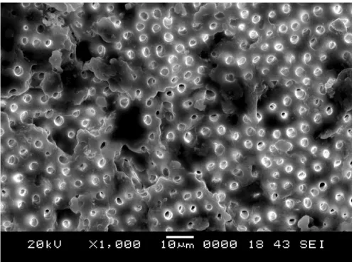
coded stubs, and coated with gold. The specimens were examined under a Scanning electron Microscope for debris and smear layer coverage. Three photographs filmed at ×1000 and ×2000 were taken randomly at the coronal, middle, and apical level. Each field was graded from 0 to 3 according to Rome et al. [9] as follows:
0 = No smear layer, dentinal tubules open, free of debris.
1 = Root canal surface covered with residue only at the opening of the dentinal tubules. 2 = Root canal surfaces with a thin covering
of residue on dentinal tubules with visible tubules only in a few regions.
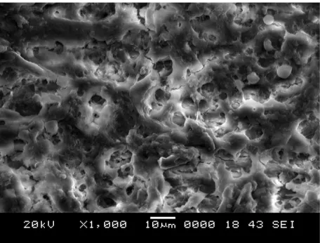
3 = Heavy smear layer, outlines of dentinal tubules totally covered with smear layer.
3. RESULTS
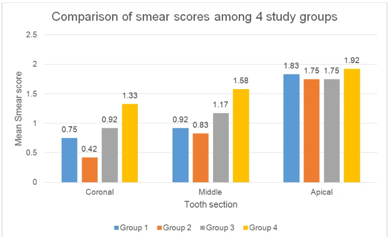
Removal of smear layer from the surfaces of root canals revealed the presence of more abundant and larger dentinal tubules in the coronal third of root canals compared with those seen in the middle and apical thirds of the root canal system. The dentinal tubules in the apical third of the canals were smaller and fewer than those observed in the rest of the root canals (Graph 1). In addition, removal of the smear layer showed the presence of many lateral canals in the apical thirds of the root canal systems. EDTA shown maximum removal of smear layer followed by 0.2% chitosan, Smear clear and Qmix 2 in 1. Any how there was no statistical significance difference seen in removal of smear layer among all the groups (Table 1).
Table 1. Mean difference of scores in coronal third, middle third and apical third
Group Middle 3rd -
Coronal 3rd
Apical 3rd - Coronal 3rd
Apical 3rd - Middle 3rd
1 Mean Difference -0.167(0.718) -1.083(0.900) -0.917(0.793)
Z -0.816 -2.598 -2.598
p-value 0.414(NS) 0.009* 0.009*
2 Mean Difference -0.417(0.669) -1.333(0.778) -0.917(0.793)
Z -1.890 -2.889 -2.598
p-value 0.059(NS) 0.004* 0.009*
3 Mean Difference -0.250(0.452) -0.833(0.718) -0.583(0.515)
Z -1.732 -2.640 -2.646
p-value 0.083(NS) 0.008* 0.008*
4 Mean Difference -0.250(0.754) -0.583(0.669) -0.333(0.492)
Z -1.134 -2.333 -2.000
Graph 1. Comparison of smear scores among 4 study groups
Fig. 1. SEM MICROGRAPHS SCORE 0 = No smear layer. No smear layer on the surface of the root canals; all tubules were clean and open
4. DISCUSSION
Micro-organisms in the root canals are the prime causative factors in the development of pulp and periapical lesions [10]. Eradication of the microbes is one of the significant goals for successful root canal treatment. It is compulsory to chemically debride teeth with complex internal
anatomy that can be missed by instrumentation of the root canals. Consequently the utilization of irrigants during root canal treatment is of prime need [11].
the aggressiveness of the irrigant and the manner in which the irrigant is delivered [11]. For example, the presence of a vapor lock in a closed-canal system precludes optimal delivery of an irrigant to the apical third of the canal wall [12]. This variable was not examined in the present study because the objective was to evaluate irrigant effectiveness rather than the
efficacy of canal irrigation. The bar charts in Fig. 2 show that the efficacy of smear layer
removal: coronal third >middle third > apical third. These results are consistent with the general findings from the endodontic literature that the apical third of the canal is more difficult
to clean. When the contribution from different canal levels was taken into consideration, the effectiveness of smear layer removal with the respective final irrigant is in the following descending order:
Glyde > 0.2% Chitosan> Smear clear >QMix 2in1
Although the presence of a vapor lock in a closed-canal system [13] may affect the efficacy of smear layer removal from the apical third of the canal well, the presence of a film of irrigant between the air bubble and the canal wall still
Fig. 2. SEM MICROGRAPHS SCORE 1 = Root canal surface covered with residue only at the opening of the dentinal tubules
Fig. 4. SEM MICROGRAPHS SCORE 3 = Heavy smear layer. Smear layer covered the root canal surface and the tubules
permits some form of smear layer removal in a less efficient manner. On the contrary, the ability to clear debris from the canal walls is more dependent on the flow of the irrigants [14] and the manner in which the irrigant is agitated [15] instead of the aggressiveness of the irrigants. Because there is only limited flow of irrigants by manual delivery of an irrigant through a side-vented needle without additional agitation, it is not surprising that there are no differences in the five experimental groups in terms of clearing of debris from the canal walls. In the future, the efficiency ofdebris clearance from the canal space should be evaluated in a closed-canal system in conjunction with agitation devices such as sonic and ultrasonic agitation systems as well as devices that incorporate an apical negative pressure approach [16,17].
The primary motivation behind this examination was to assess the viability of an irrigant solution with contents equipped for cleaning and disinfecting the dentin, clearing the smear layer, opening the dentinal tubules and permitting the antibacterial agents to enter the whole root canal anatomy.
The endodontic smear layer has been depicted as one that is formed during instrumentation, comprising of dentin as well as necrotic and suitable tissue, including leftovers of odontoblastic procedures, pulp tissue and microorganisms [18]. The smear layer assumes an essential part in the lateral sealing of the root canal, as a barrier that can meddle with attachment and infiltration of the root canal sealer into the dentinal tubules. Pashley et al had portrayed the smear layer as a permeable
structure which was porous to even expansive molecules such as albumin [19].
EDTA is one of the most commonly used chelating agent which reacts with the calcium ions in dentine and structures dissolvable calcium chelates [20,21]. It has been accounted for that EDTA demineralize dentine to a profundity of 20–30 mm in 5 min (von der Fehr & Nygaard-Ostby 1963); on the other hand, Fraser (1974) expressed that the chelating impact was verging on irrelevant inthe apical end of root canal [22,23].
The combined use of sodium hypochlorite and EDTA has demonstrated compelling in clearing smear layer shaped during root canal preparation (Goldman et al. 1982, Baumgartner & Mader 1987, Abbott et al. 1991) [24,25]. 17% EDTA follows up on the inorganic parts of the smear layer, causes the decalcification of peritubular and intertubular dentine, and leaves the collagen particle uncovered. In this manner, the utilization of NaOCl disintegrates the collagen, further aiding the passages to the intra dental tubules which are uncovered (Goldman et al. 1982, Baumgartner & Mader 1987). The 0.2% chitosan solution, even in such a low fixation, had the capacity to clear smear layer and give factually comparable results to those of the arrangements with higher focuses (15% EDTA and 10% citrus extract [25,5].
included, the chemical structure of chitosan and the pH of the solution (Guibal et al. 2000, Rhazi et al. 2000). Two models are accounted for in the writing as could be allowed activity instruments. One of them, known as the bridge model, depends on the hypothesis that two or more amino groups of a chitosan molecule associated to the same metal ion (Blair & Ho 1981). The other model backings the hypothesis that only one amino group of the substance's structure is included in the coupling, that being the metal ion "attached" to the amino group [27,28].
5. CONCLUSION
Effective instrumentation and irrigation are prerequisites for successful endodontic treatment. Instrumentation of root canal leads to formation of smear layer in root canal walls. Smear layer formed alters the dentine permeability and effect adversely on adhesion of intracanal medicaments and obturation materials to the root canal wall. Removal of smear layer lead to better obturation and better treatment outcome. Within the limitations of the current study, it could be concluded that, Chitosan could remove the smear layer as good as EDTA which is still considered as gold standard in clearing smear layer in a low concentration of 0.2% and better than or equal to Qmix 2in1 and Smear clear. Additional properties of chitosan like antibacterial and antifungal effectiveness enhance its importance to be used as a root canal irrigant.
CONSENT
It is not applicable.
ETHICAL APPROVAL
All authors hereby declare that all experiments have been examined and approved by the appropriate ethics committee and have therefore been performed in accordance with the ethical standards laid down in the 1964 Declaration of Helsinki.
COMPETING INTERESTS
Authors have declared that no competing interests exist.
REFERENCES
1. Gambarini G, Laszkiewicz J. A scanning electron microscopic study of debris and
smear layer remaining following use of GT rotary instruments. Int Endod J. 2002; 35(5):422-7.
2. Takeda FH. A comparative study of the removal of smear layer by three endodontic irrigants and two types of laser. International Endodontic Journal. 1999; 32(1):32-9.
3. Abott PV. An SEM study of the effects of different irrigation sequences and ultrasonic. International Endodontic Journal. 1991;24:308-31.
4. Silva PV, Guedes DFC, Nakadi FV, Pécora JD, Cruz-Filho AM. Chitosan: A new solution for removal of smear layer after root canal instrumentation. International Endodontic Journal. 2013;46:332–338. 5. Razali Nor Anis, Masudi Masudi, Ariffin
Ariffin, Alam Mohammad Khursheed. Scanning Electron Microscope (SEM) study of two lubrication agent in removing
smear layer on human dentine.
International Medical Journal. 2013;20(4): 513–517.
6. Lim TS. Light and scanning electron microscopic evaluation of Glydetm file prep in smear layer removal. International Endodontic Journal. 2003;36:336-343. 7. Ballal MV, Kundabala M, Bhat KS, et al.
Susceptibility of Candida albicans and
Enterococcus faecalis to Chitosan,
Chlorhexidine gluconate and their combination in vitro. Aust Endod J. 2009;35:29-33.
8. Lin Dai, Khaled Khechen, Sara Khan, Brian Gillen, Bethany A Loushine, Courtney E, Wimmer BS, James L Gutmann, David Pashley, Franklin R Tay. The effect of QMix, an experimental antibacterial root canal irrigant, on removal of canal wall smear layer and debris. JOE. 2011;37(1):1-4.
9. Raffaele Paragliola, Vittorio Franco, Cristiano Fabiani, Luciano Giardino, Flavio Palazzi, Nicoletta Chieffi, Hani F Ounsi, Simone Grandini. Comparison of smear layer removal using four final-rinse protocols. International Dentistry. 2004;1: 4-7.
10. Khademi M Feizianfard. The effect of EDTA and citric acid on smear layer removal of mesial canals of first mandibular molars, a scanning electron microscopic study. Journal of Research in Medical Sciences. 2004;9(2):80-88. 11. Sidney Ricardo Dotto, Rosana Maria
de Oliveira, Manoel Eduardo de Lima Machado, José Luiz Martins. Evaluation of ethylenediaminetetraacetic acid (EDTA) solution and gel for smear layer removal. Australian Endodontic Journal. 2007;33(2): 62-5.
12. Clarissa Teles roDrIgues Norberti bernarDIne Marco Antonio Hungaro DuarTe Clovis Monteiro bramanTe
Flaviana Bombarda de anDraDe
Evaluation of EDTA, apple vinegar and SmearClear with and without ultrasonic activation on smear layer removal in different root canal levels. Dental Press Endod. 2013;3(1):43-8.
13. Teixeira CS, Felippe MCS, Felippe WT. The effect of application time of EDTA and NaOCI on intracanal smear layer removal. International Endodontic Journal. 2005;38: 285–290.
14. Parag M Wani, Rajesh R Shetty, Pradnya S Kale, Pritesh G Jagtap. Comparative evaluation of phosphoric acid, EDTA and saline, with and without ultrasonic irrigation for smear layer removal. Journal of Dental Research and Scientific Development. 2014;1(1):1-5.
15. Gin Chen, Yu-Chao Chang. Effects of liquid- and paste-type EDTA on smear-layer removal during rotary root-canal instrumentation. 2011;6(1):41–47.
16. Kuah HG, Lui JN, Tseng PS, Chen NN. The effect of EDTA with and without ultrasonics on removal of the smear layer. J Endod. 2009;35(3):393-396.
17. Agrawal, Vineet. An in vitro scanning electron microscopic study comparing the efficacy of passive ultrasonic and syringe irrigation methods using sodium hypochlorite in removal of debris from the root canal system. Journal of the Irish Dental Association. 2014;6(1):47–52. 18. Torabinejad M, Handysides R, Khademi
A, Bakland LK. Clinical implications of the smear layer in endodontics: A review. Oral Surg Oral Med Oral Path Oral Radiol Endo. 2002;94:658–666. Of Macromolecular Science 32,629–40.
19. Sen BH. The effect of different concentrations of EDTA on instrumented root canal walls. Oral Surgery, Oral Medicine, Oral Pathology, Oral Radiology and Endodontology. 2009;108:622–627. 20. Hulsmann M. Chelating agents in root
canal treatment: Mode of action and indications for their use. International Endodontic Journal. 2003;36(12):810–830. 21. Mohammad Ali Mozayeni. Effect of 17% EDTA and MTAD on intracanal smear layer removal: A scanning electron microscopic study. Australian Endodontic Journal. 2009;35(1):13–17.
22. Prado M, Gusman H, Gomes BPFA, Sima˜o RA. Scanning electron microscopic investigation of the effectiveness of phosphoric acid in smear layer removal when compared with EDTA and citric acid. Journal of Endodontics. 2011;37:255–258. 23. Saleh IM. The effects of dentine
pretreatment on the adhesion of root-canal sealers. International Endodontic Journal. 2002;35:859–866.
24. Teixeira CS. The effect of application time of EDTA and NaOCl on intracanal smear layer removal: an SEM analysis. International Endodontic Journal. 2005;38: 285–290.
25. Berg MS. A comparison of five irrigating solutions: A scanning electron microscopic study. Journal of Endodontics. 1986; 12(5):192–197.
26. Akncbay H, Senel S, Ay ZY. Application of chitosan gel in the treatment of chronic periodontitis. Journal of Biomedical Materials Research. Part B, Applied Biomaterials. 2007;80:290–296.
27. Blair HS, Ho TC. Studies in the adsorption and diffusion of ions in chitosan. Journal of Chemical Technology and Biotechnology. 1981;31:6–10.
28. Cruz-Filho AM, Sousa-Neto MD, Savioli RN, Silva RG, Vansan LP, Pecora JD. (2011). Effect of chelating solutions on the microhardness of root canal lumen dentin. Journal of Endodontics. 2014;11:1–11. _________________________________________________________________________________
© 2016 Venghat and Hegde; This is an Open Access article distributed under the terms of the Creative Commons Attribution License (http://creativecommons.org/licenses/by/4.0), which permits unrestricted use, distribution, and reproduction in any medium, provided the original work is properly cited.
Peer-review history: