Scholarship@Western
Scholarship@Western
Electronic Thesis and Dissertation Repository
7-7-2014 12:00 AM
Functional Co-substituted Poly[(amino acid ester)phosphazene]
Functional Co-substituted Poly[(amino acid ester)phosphazene]
Biomaterials
Biomaterials
Amanda L. Baillargeon
The University of Western Ontario
Supervisor
Dr. Kibret Mequanint
The University of Western Ontario
Graduate Program in Biomedical Engineering
A thesis submitted in partial fulfillment of the requirements for the degree in Master of Engineering Science
© Amanda L. Baillargeon 2014
Follow this and additional works at: https://ir.lib.uwo.ca/etd
Part of the Biomaterials Commons
Recommended Citation Recommended Citation
Baillargeon, Amanda L., "Functional Co-substituted Poly[(amino acid ester)phosphazene] Biomaterials" (2014). Electronic Thesis and Dissertation Repository. 2249.
https://ir.lib.uwo.ca/etd/2249
This Dissertation/Thesis is brought to you for free and open access by Scholarship@Western. It has been accepted for inclusion in Electronic Thesis and Dissertation Repository by an authorized administrator of
(Thesis format: Monograph)
by
Amanda Lee Baillargeon
Graduate Program in Biomedical Engineering
A thesis submitted in partial fulfillment of the requirements for the degree of
Master of Engineering Science
The School of Graduate and Postdoctoral Studies The University of Western Ontario
London, Ontario, Canada
ii
Abstract
The development of new and improved biomaterials is essential for tissue engineering and regenerative medicine applications. Amino acid-based polyphosphazenes are being explored as scaffold materials for tissue engineering applications due to their non-toxic degradation products and tunable material properties. This work focuses on the synthesis of non-functional and novel non-functional poly[(amino acid ester)phosphazene]s using a facile method of thermal ring opening polymerization followed by one-pot room temperature substitution. The family of polyphosphazenes developed in this work is based on alanine (PP-A’s), L-phenylalanine (PP-F’s), and L-methionine (PP-M’s) with L-glutamic acid imparting the functionality. Characterization of these materials demonstrated that the one-pot substitution was successful in developing mono- and co-substituted poly[(amino acid ester)phosphazene]s. Cytotoxicity studies on two-dimensional films showed these materials to be compatible with NIH-3T3 fibroblasts over the five-day study. The PP-F’s also showed significantly enhanced cell viability over tissue culture polystyrene at day 1 (p<0.001) and
day 3 (p<0.01). As a proof of concept, electrospinning of PP-A100 blended with
poly(caprolactone) (PCL) (50% PP-A100/50% PCL) was conducted to fabricate
three-dimensional scaffolds. Overall, this study has shown that poly[(amino acid ester)phosphazene]s can be synthesized with good yields and better reaction conditions, leading to materials with promising cytocompatibility for use in biomedical applications.
Keywords
I would like to acknowledge my supervisor, Dr. Kibret Mequanint, for his continued support and guidance throughout my Master’s degree. I really appreciated the amount of time he designated weekly to meet with his students and ensure that they were progressing well with their research; it definitely pushed me to always have new findings to share with him. I would also like to thank the Natural Sciences and Engineering Research Council (NSERC) of Canada and the Strategic Training Program in Vascular Research supported by the Canadian Institutes for Health Research (CIHR) for the financial aid and skills training.
I would like to thank Dr. Darryl Knight and Dr. Kalin Panev for their help in synthetic chemistry problems, experimental setup, and elucidation of NMR spectra. I also would like to acknowledge Dr. Shigang Lin for his help with the design and implementation of the cell culture experiments. I would like to thank Ryan Guterman of the Ragogna research group for his help in running TGA, DSC, and FTIR analyses. Lastly, I would like to thank Somiraa Said for her guidance with the electrospinning and planning of cell culture studies. All of the helpful feedback and discussions from other lab members (especially Tierney Deluzio) were greatly appreciated.
iv
Table of Contents
Abstract ... ii
Acknowledgments... iii
Table of Contents ... iv
List of Tables ... viii
List of Figures ... x
List of Appendices ... xvi
List of Abbreviations ... xvii
Chapter 1 ... 1
1 Scope and Thesis Outline ... 1
1.1 Scope ... 1
1.2 Thesis Outline ... 2
Chapter 2 ... 3
2 Literature Review, Objectives, and Rationale ... 3
2.1 Tissue Engineering Methodology ... 3
2.2 Requirements of Tissue Engineering Scaffolds ... 5
2.2.1 Natural Polymers ... 7
2.2.2 Biodegradable Synthetic Polymers ... 8
2.2.3 Biomimetic Materials ... 9
2.2.4 Scaffold Fabrication ... 10
2.3 Synthesis of Polyphosphazenes ... 11
2.3.1 Polymerization to Poly[(dichloro)phosphazene] ... 13
2.3.2 Macromolecular Substitution of Poly[(dichloro)phosphazene] with Organic Nucleophiles... 18
v
2.4.1 In Vitro and In Vivo Biocompatibility ... 21
2.4.2 Biodegradability ... 23
2.4.3 Mechanical Properties ... 28
2.5 Objectives and Rationale ... 29
Chapter 3 ... 32
3 Materials and Methods ... 32
3.1 Thermal Ring Opening Polymerization (TROP) ... 32
3.1.1 Materials ... 32
3.1.2 Equipment ... 32
3.1.3 Method ... 33
3.2 Macromolecular Substitution of Non-Functional Amino Acid Esters ... 35
3.2.1 Materials ... 35
3.2.2 Sample Nomenclature ... 35
3.2.3 Methods... 35
3.3 Preparation of Glutamic Acid Ethyl Ester (E*) ... 40
3.3.1 Materials ... 40
3.3.2 Methods... 41
3.4 Macromolecular Co-substitution of Non-Functional Amino Acids with Functional Glutamic Acid ... 43
3.4.1 Materials ... 43
3.4.2 Methods... 43
3.5 Conjugation of a Model Compound to the Functional Polyphosphazenes ... 48
3.5.1 Materials ... 48
3.5.2 Methods... 48
3.6 Material Characterization ... 49
vi
3.6.2 Fourier-Transform Infrared Spectroscopy (FTIR) ... 49
3.6.3 Thermogravimetric Analysis (TGA) ... 50
3.6.4 Differential Scanning Calorimetry (DSC) ... 50
3.6.5 Gel Permeation Chromatography (GPC) ... 50
3.7 Cytotoxicity Studies of Polymeric Films Using MTT Assays... 51
3.7.1 Thin Film Preparation ... 51
3.7.2 Fibroblast (NIH-3T3) Cell Study ... 51
3.7.3 MTT Assay Protocol ... 51
3.8 Cell Morphology and Adhesion Studies on Polymer Films Using Confocal Microscopy ... 52
3.8.1 Thin Film Preparation ... 52
3.8.2 Cell Fixation, Staining, and Imaging ... 52
3.9 Electrospinning of Polyphosphazenes and Polyphosphazene/ Polycaprolactone Blends ... 53
3.9.1 Electrospinning Parameters ... 53
3.9.2 Scanning Electron Microscopy (SEM) Imaging of Electrospun Fibers ... 54
Chapter 4 ... 55
4 Results and Discussion ... 55
4.1 Optimization of Thermal Ring Opening Polymerization ... 55
4.2 Comparison of the Three Macromolecular Substitution Methods Using Non-Functional Amino Acid Esters ... 57
4.2.1 FTIR, 1H NMR, and 31P NMR Characterization ... 58
4.2.2 TGA, DSC, and GPC Characterization ... 70
4.3 Esterification of Fmoc- and Boc-Glu(OBzl)-OH ... 78
4.4 TFA Deprotection of Boc-Glu(OBzl)-OEt ... 81
4.5 Macromolecular Co-Substitution of Functional and Non-Functional Amino Acid Esters ... 82
vii
4.5.2 TGA, DSC, and GPC Characterization ... 89
4.6 Hydrogenation of Glutamic Acid Co-Substituted Polyphosphazenes for Model Compound Conjugation ... 95
4.7 Cytotoxicity and Cell Proliferation on Non-Functional and Functional Two-Dimensional Polyphosphazene Films ... 96
4.8 Electrospinning of Poly[(amino acid ester)phosphazene]s and PCL Blend Materials ... 100
4.9 Novelty of This Work ... 102
5 Conclusions and Future Directions ... 103
5.1 Conclusions ... 103
5.2 Future Directions ... 105
References ... 106
Appendices ... 117
viii
List of Tables
Table 2.1: Summary of in vitro degradation studies of poly[(amino acid ester)phosphazene]s
and their co-substituted polyphosphazenes. The ester refers to the chain attached to the carboxyl terminus of the amino acid. The detailed degradation profiles can be found in the cited references. ... 25
Table 4.1: Summary of the time-varied polymerizations of hexachlorocyclotriphosphazene (trimer) to poly[(dichloro)phosphazene] (linear precursor) with their respective yields and extent of conversion. ... 57
Table 4.2: Summary table of the yields, 1H NMR, 31P NMR, and FTIR analyses of PP-A100,
PP-F100, and PP-M100 synthesized by the three methods (LT 2S = low temperature
two-step reaction, 1P RT = one-pot room temperature reaction, and RT 2S = room temperature two-step reaction). Note: As described in the methods section, the mass of poly[(dichloro)phosphazene] (PDCP) is approximate since it cannot be dried fully and therefore the percentage yields shown are underestimated since yield calculations are based on PDCP as the limiting reagent. ... 58
Table 4.3: Summary of thermal data from TGA and DSC analysis for PP-A100, PP-F100, and
PP-M100 materials synthesized via either the 1P RT or RT 2S method. The decomposition
temperatures are presented as the peak decomposition temperatures rather than onset, meaning they are the values of maximal rate of change in weight % for all phases of the decomposition (i.e. maxima of the derivative of weight % plot)... 70
Table 4.4: GPC analysis of PP-A100, PP-F100, and PP-M100 materials using triple detection
and refractive index detection. A (-) indicates that analysis using that detection method was not possible and the relative molecular weights are calculated based on polystyrene standards. ... 75
Table 4.5: Summary of percent yields, 1H NMR, 31P NMR and FTIR characterization of the co-substituted poly[(amino acid ester)phosphazene]s. ... 84
Table 4.6: Summary of thermal data from TGA and DSC analysis for PP-A90E*10, PP-F90E*10,
ix
decomposition temperatures rather than onset, meaning they are the values of maximal rate of change in weight % for all phases of the decomposition (i.e. maxima of the derivative of weight % plot). ... 90
Table 4.7: GPC analysis of PP-A90E*10, PP-F90E*10, and PP-M90E*10 materials using triple
x
List of Figures
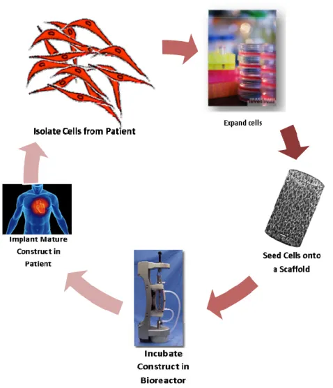
Figure 2.1: A diagrammatic representation of the in vitro tissue engineering process; where a
cell-seeded scaffold is matured in vitro in a bioreactor prior to implantation into the
patient. ... 5
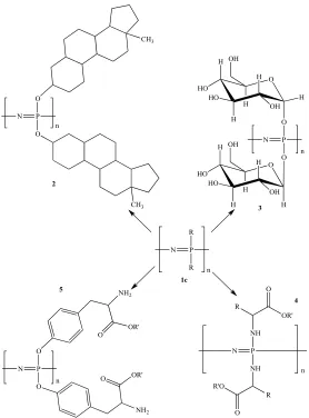
Figure 2.2: Structures of various polyphosphazenes including steroidal substituents (2), carbohydrates (3), amino acid esters (4), and side chain bound amino acid esters (5), to name a few. ... 12
Figure 3.1: The glass vials and stainless steel vial rack that were manufactured in-house for the facile thermal ring opening polymerizations. The schematic on the right side shows approximately dimensions of the glass vials and stoppers used as the reaction vessels. .. 33
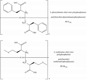
Figure 3.2: Chemical structures of the L-phenylalanine ethyl ester (PP-F100) and
L-methionine ethyl ester (PP-M100) based polyphosphazene materials. ... 38
Figure 3.3: Chemical structures of Fmoc- and Boc-protected glutamic acid that will be esterified to their ethyl ester amino acid derivatives. ... 41
Figure 3.4: Chemical structures of the co-substituted poly[(amino acid ester)phosphazene]s based on the non-functional amino acid esters, Ala-OEt, Phe-OEt, Met-OEt, and side chain protected functional amino acid ester, Glu(OBzl)-OEt. ... 45
Figure 4.1: 31P NMR of the time-varied facile polymerization of hexachlorocyclotriphosphazene (1) to purified poly[(dichloro)phosphazene] (5) at 230°C. Spectra (2), (3), and (4) are for 21, 23, and 25 hour polymerizations, respectively, without purification. ... 56
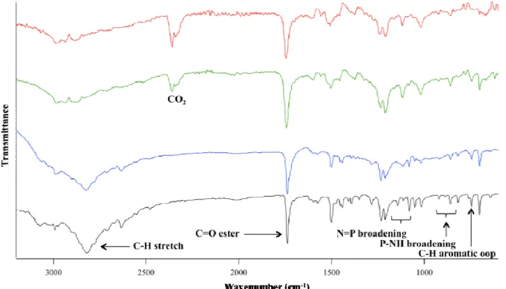
Figure 4.2: FTIR comparison of PP-A100’s prepared from three different synthetic methods.
The spectra correspond to L-alanine ethyl ester hydrochloride salt (black), and PP-A100’s
xi
Figure 4.3: FTIR comparison of PP-F100’s prepared from three different synthetic methods.
The spectra correspond to L-phenylalanine ethyl ester hydrochloride salt (black), and PP-F100’s from LT 2S method (blue), 1P RT method (green), and RT 2S method (red). ... 60
Figure 4.4: FTIR comparison of PP-M100’s s prepared from three different synthetic methods.
The spectra correspond to L-methionine ethyl ester hydrochloride salt (black), and PP-M100’s from LT 2S method (blue), 1P RT method (green), and RT 2S method (red). ... 61
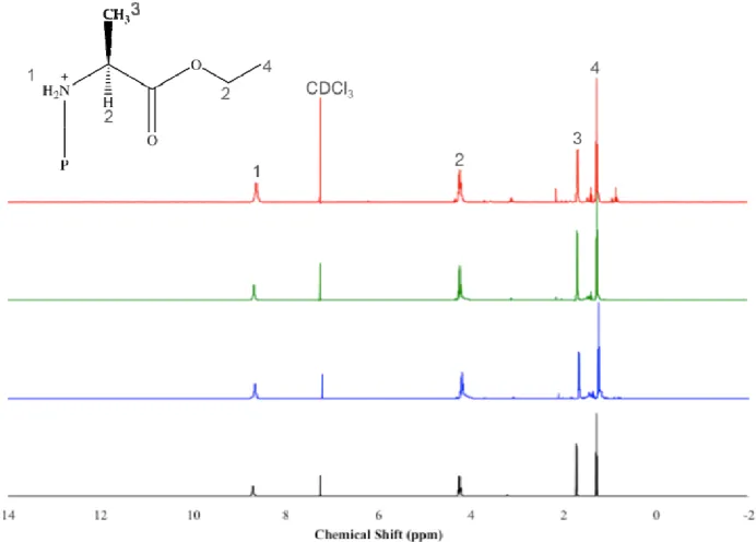
Figure 4.5: 1H NMR comparison of PP-A100’s prepared from three different synthetic
methods. The spectra correspond to L-alanine ethyl ester hydrochloride salt (black), and PP-A100’s from LT 2S method (blue), 1P RT method (green), and RT 2S method (red).
All small, unlabeled peaks are impurities that cannot be removed from dissolution/reprecipitation techniques. ... 62
Figure 4.6: 1H NMR comparison of PP-F100’s prepared from three different synthetic
methods. The spectra correspond to L-phenylalanine ethyl ester hydrochloride salt (black), and PP-F100’s from LT 2S method (blue), 1P RT method (green), and RT 2S
method (red). All small, unlabeled peaks are impurities that cannot be removed from dissolution/reprecipitation techniques. ... 63
Figure 4.7: 1H NMR comparison of PP-M100’s prepared from three different synthetic
methods. The spectra correspond to L-methionine ethyl ester hydrochloride salt (black), and PP-M100’s from LT 2S method (blue), 1P RT method (green), and RT 2S method
(red). All small, unlabeled peaks are impurities that cannot be removed from dissolution/reprecipitation techniques. ... 64
Figure 4.8: 31P NMR of the transition from hexachlorocyclotriphosphazene (cyclic trimer) to poly[(dichloro)phosphazene] (PDCP) and finally to the methionine ethyl ester and benzyl protected glutamic acid ethyl ester co-substituted polyphosphazene (PP-M90E*10). ... 65
Figure 4.9: TGA analysis for the PP-A100, PP-F100, and PP-M100 materials prepared using the
xii
Figure 4.10: DSC thermograms of PP-A100, PP-F100, and PP-M100 materials prepared using
the 1P RT and RT 2S methods. The glass transition temperatures (Tgs), which were
collected from the second heating cycle, range from -41.8 to +46.3°C. ... 73
Figure 4.11: Detector signal versus retention volume (mL) traces from GPC analysis of PP-M100 synthesized via the 1P RT method. Detector traces correspond to refractive index
(red), right angle light scattering (green), low angle light scattering (black) and viscometer (blue). ... 76
Figure 4.12: GPC traces from the refractive index detector for PP-A100 (----1P RT, - - - RT
2S), PP-F100 (---- 1P RT, - - - RT 2S), and PP-M100 (---- 1P RT, - - - RT 2S)... 76
Figure 4.13: 1H NMR spectra of the esterification with ethanol and deprotection with 25% TFA in DCM of Glu(OBzl)-OH (black). The ethyl esterified product, Boc-Glu(OBzl)-OEt, is shown blue and the Boc deprotected product, TFA-/H3N+
-Glu(OBzl)-OEt, is shown in green. Unlabeled peaks in the Boc-Glu(OBzl)-OEt and TFA-/H3N+
-Glu(OBzl)-OEt spectra are equivalent to the labeled peaks with the same chemical shift in the Boc-Glu(OBzl)-OH spectrum. ... 81
Figure 4.14: FTIR spectra of TFA-/H3N+-Glu(OBzl)-OEt (black), PP-A90E*10 (red), and
PP-A100 synthesized via the 1P RT method (blue). ... 85
Figure 4.15: FTIR spectra of TFA-/H3N+-Glu(OBzl)-OEt (black), PP-F90E*10 (red), and
PP-F100 synthesized via the 1P RT method (blue). ... 85
Figure 4.16: FTIR spectra of TFA-/H3N+-Glu(OBzl)-OEt (black), PP-M90E*10 (red), and
PP-M100 synthesized via the 1P RT method (blue). ... 86
Figure 4.17: 1H NMR spectra of TFA-/H
3N+-Glu(OBzl)-OEt (black), PP-A90E*10, (red), and
PP-A100 from the 1P RT method (blue). ... 87
Figure 4.18: 1H NMR spectra of TFA-/H3N+-Glu(OBzl)-OEt (black), PP-F90E*10 (red), and
PP-F100 from the 1P RT method (blue). ... 87
Figure 4.19: 1H NMR spectra of TFA-/H3N+-Glu(OBzl)-OEt (black), PP-M90E*10 (red), and
xiii
Figure 4.20: TGA analysis for the PP-A90E 10, PP-F90E 10, and PP-M90E 10 polymers. The
first graph represents the weight percent vs. temperature, where as the second graph shows the derivative of weight percent vs. temperature. ... 91
Figure 4.21: DSC thermograms of PP-A90E*10, PP-F90E*10, and PP-M90E*10 polymers. The
glass transition temperatures (Tgs), which were collected from the second heating cycle,
range from -49.4 to +44.5°C. ... 92
Figure 4.22: GPC traces from the refractive index detector for PP-A90E*10 (black), PP-F90E*10
(blue), and PP-M90E*10 (red). ... 94
Figure 4.23: Fibroblast metabolic activity as determined by MTT assay grouped by day to compare metabolic activity levels between materials and TCPS. Cells were seeded at 8,000 cells/well on 2-D polyphosphazene films (PP-A100, PP-F100, PP-M100, PP-A90E*10,
PP-F90E*10, andPP-M90E*10) that were either unconditioned or preconditioned in HBSS at
room temperature overnight. The cells were cultured for 1, 3, and 5 days with 5 replicates for each. The data are presented as the mean ± standard deviation and are normalized to TCPS at Day 1. (** = p<0.01, *** = p<0.001) ... 98
Figure 4.24: Fibroblast metabolic activity as determined by MTT assay grouped by material to show non-cytotoxicity from day 1 to day 5. Cells were seeded at 8,000 cells/well on 2-D polyphosphazene films (PP-A100, PP-F100, PP-M100, PP-A90E*10, PP-F90E*10, and
PP-M90E*10) that were preconditioned in HBSS at room temperature overnight. The cells
were cultured for 1, 3, and 5 days with 5 replicates for each. The data are presented as the mean ± standard deviation and are normalized to TCPS at Day 1. ... 98
Figure 4.25: Confocal microscopy images showing the morphology and spreading of fibroblasts on the unconditioned and preconditioned polyphosphazene materials after 3 days of culture. The cells were seeded at a density of 30,000 cells/well on the polymer coated glass coverslips in a 24-well tissue culture plate. Untreated glass cover slips were used as a positive control in these experiments. The fibroblasts were stained for F-actin (green) and nuclei (blue). Scale bar = 20 μm (panel B). ... 99
xiv
List of Schemes
Scheme 2.1: Scheme showing the synthesis and functionalization of poly[(dichloro)phosphazene] (1b) in the overall synthesis of polyphosphazenes from hexachlorocyclotriphosphazene (1a) via the thermal ring opening polymerization method.15 ... 14
Scheme 2.2: Reaction scheme for the solution phase thermal ring opening polymerization of hexachlorocyclotriphosphazene (cyclic trimer) to linear poly[(dichloro)phosphazene] using a sulfamic acid catalyst and calcium sulfate dehydrate promoter – The Magill Method. ... 15
Scheme 2.3: Reaction scheme for the phosphorus pentachloride (PCl5) initiated bulk and
solution phase polycondensation of trichlorotrimethylsilylphosphoranimine (Cl3P=NSiMe3) to poly[(dichloro)phosphazene] ([NPCl2]n) – The “Living” Cationic
Polymerization Method. ... 16
Scheme 2.4: Proposed cationic polymerization step-wise mechanism of the PCl5 initiated
polycondensation reaction of Cl3P=NSiMe3 to [N=PCl2]n. ... 17
Scheme 2.5: Reaction scheme for the synthesis of poly[(dichloro)phosphazene] from the polycondensation reaction of Cl3P=NP(O)Cl2 – The De Jaeger Method. ... 18
Scheme 3.1: Reaction scheme for the facile thermal ring opening polymerization (TROP) of hexachlorocyclotriphosphazene to linear poly[(dichloro)phosphazene] (PDCP). ... 34
Scheme 3.2: Reaction scheme for the low-temperature macromolecular substitution of L-alanine ethyl ester with poly[(dichloro)phosphazene] (PDCP) forming poly[bis(ethyl alanato)phosphazene] (PP-A100). ... 37
Scheme 3.3: Reaction scheme for the one-pot room temperature synthesis of poly[bis(ethyl alanato)phosphazene] (PP-A100) from L-alanine ethyl ester hydrochloride
xv
Scheme 3.4: Reaction scheme for the room temperature two-step substitution of poly[(dichloro)phosphazene] (PDCP) with L-alanine ethyl ester (H-Ala-OEtHCl). ... 40
Scheme 3.5: Expected reaction schemes for the ethyl esterification of Fmoc-Glu(OBzl)-OH and Boc-Glu(OBzl)-OH to Fmoc-Glu(OBzl)-OEt and Boc-Glu(OBzl)-OEt, respectively, using carbodiimide coupling chemistry. ... 42
Scheme 3.6: Boc deprotection of glutamic acid ethyl ester (Boc-Glu(OBzl)-OEt) using a 1:3 mixture of trifluoroacetic acid (TFA) in dichloromethane (DCM). ... 43
Scheme 3.7: One-pot three-stage room temperature reaction of a co-substituted poly[(amino acid ester)phosphazene] (PP-A90E*10 shown here) from the functional amino acid ester
(TFA- NH3+-Glu(OBzl)-OEt), non-functional amino acid ester (H-Ala-OEtHCl) and
poly[(dichloro)phosphazene] precursor (PDCP). ... 47
xvi
List of Appendices
Appendix 1: Summary of literature values for the characterization and thermal properties of poly[(amino acid ester)phosphazene]s. GPC data are relative to polystyrene standards in all cases. ... 117
Appendix 2: Enlarged regions of the PP-A100 FTIR spectra showing P=N and P-NH peak
broadening. The spectra correspond to L-alanine ethyl ester hydrochloride (black), and PP-A100’s made by the LT 2S (blue), 1P RT (green), RT 2S (red) reactions. ... 119
Appendix 3: Enlarged regions of the PP-F100 FTIR spectra showing P=N and P-NH peak
broadening. The spectra correspond to L-phenylalanine ethyl ester hydrochloride (black), and PP-F100’s made by the LT 2S (blue), 1P RT (green), RT 2S (red) reactions. ... 120
Appendix 4: Enlarged regions of the PP-M100 FTIR spectra showing P=N and P-NH peak
xvii
List of Abbreviations
2-D Two-dimensional
3-D Three-dimensional
3T3 Fibroblast cell line
1P RT One-pot room temperature
Å Angstrom
Ala, A L-alanine
ANOVA Analysis of variance
ATR Attenuated total reflectance
Boc t-Butyloxycarbonyl
Boc-Glu(OBzl)-OH N-- t-butyloxycarbonyl -L-glutamic acid γ-benzyl ester
Boc-Glu(OBzl)-OEt N-- t-butyloxycarbonyl -L-glutamic acid ethyl ester γ-benzyl ester
Bzl Benzyl
CDCl3 Deuterated chloroform
Da Daltons, g/mol
DAPI 4’-6’-diamidino-2-phenylindole dihydrochloride
DCM Dichloromethane
DI Deionized
DMAP 4-(Dimethylamino)pyridine
DMF N,N-Dimethylformamide
DMSO Dimethyl sulfoxide
DMSO-d6 Deuterated DMSO
dn/dc Differential index of refraction, Refractive index increment
DSC Differential scanning calorimetry
xviii
EDCI N-(3-dimethylaminopropyl)-N’-ethylcarbodiimide hydrochloride
EtOAc Ethyl acetate
EtOH Ethanol
F-actin Filamentous actin
Fmoc Fluorenylmethyloxycarbonyl
Fmoc-Glu(OBzl)-OH N--fluorenylmethyloxycarbonyl-L-glutamic acid γ-benzyl ester
FTIR Fourier transform infrared
GAG Glycosaminoglycans
Glu, E L-glutamic acid
GPC Gel permeation chromatography
H-Ala-OEt•HCl L-alanine ethyl ester hydrochloride
HBSS Hank’s balanced salt solution
HepG2 Human liver carcinoma cell line
H-Met-OEt•HCl L-methionine ethyl ester hydrochloride
H-Phe-OEt•HCl L-phenylalanine ethyl ester hydrochloride
LT 2S Low temperature two-step
MC3T3-E1 Mouse osteoblast precursor cell line
MeOH Methanol
Met, M L-methionine
Mn Number average molecular weight
MPa Megapascal
MTT 3-(4,5-dimethylthiazol-2-yl)-2,5-diphenyltetrazolium bromide
Mw Weight average molecular weight
N Newton
NIH-3T3 Mouse fibroblast cell line
xix
PBS Phosphate-buffered saline
PCL Poly(caprolactone)
Pd/C Palladium on carbon
PDCP Poly[(dichloro)phosphazene]
PDI Polydispersity index
Phe, P L-phenylalanine
PLA Poly(lactic acid)
PLAGA Poly(lactic acid –co– glycolic acid)
PMN Polymorphonuclear leukocytes
PNEA Poly[bis(ethyl alanato)phosphazene] (literature)
PNEF Poly[bis(ethyl phenylalanato)phosphazene] (literature)
PNEM Poly[bis(ethyl methionato)phosphazene] (literature)
PP-A100 Poly[bis(ethyl alanato)phosphazene] (this work)
PP-A90E*10 90% Ala-OEt / 10% Glu(OBzl)-OEt polyphosphazene
PP-F100 Poly[bis(ethyl phenylalanato)phosphazene] (this work)
PP-F90E*10 90% Phe-OEt / 10% Glu(OBzl)-OEt polyphosphazene
PP-M100 Poly[bis(ethyl methionato)phosphazene] (this work)
PP-M90E*10 90% Met-OEt / 10% Glu(OBzl)-OEt polyphosphazene
iPrOH iso-Propanol, 2-Propanol
psi Pounds per square inch
RALS/LALS Right angle light scattering/Low angle light scattering
RI Refractive index
RT 2S Room temperature two-step
SDH Succinic dehydrogenase
SDS Sodium dodecyl sulfate
xx
TCPS Tissue culture polystyrene
Td Decomposition temperature
TEA, NEt3 Triethylamine
TFA Trifluoroacetic acid
Tg Glass transition temperature
TGA Thermogravimetric analysis
THF Tetrahydrofuran
Tm Melting temperature
TROP Thermal ring opening polymerization
UV Ultraviolet
wt % Weight by weight percent
Chapter 1
1
Scope and Thesis Outline
1.1
Scope
As the fields of tissue engineering and regenerative medicine continue to emerge as promising alternatives to current treatment options for failed or otherwise diseased tissues, so too has the development of suitable biomaterials.1 Materials-mediated tissue engineering is the most frequently employed strategy, where a porous scaffold, which is ideally degradable, acts as a support for the developing tissue either in vitro or in vivo.
The scaffolds themselves must possess specific material and biological characteristics, such as desirable mechanical properties, degradation rates that parallel tissue formation, and biocompatibility, to ensure their success in tissue engineering applications.1 As such,
numerous natural and synthetic materials have been investigated for their use in biomedical applications.2-4 Natural materials, like cellulose, chitosan, and collagen, have been extensively studied due to their inherent biocompatibility and favourable cell-material interactions; however, they fall short as ideal cell-materials due to their uncontrolled degradation rates, poor mechanical properties, and batch-to-batch variability.4 Synthetic polymers, on the other hand, have shown to be an improvement over natural materials with their tunable and well-controlled mechanical properties and degradation rates, ease of processing, and availability.5, 6 That being said, degradable synthetic polymers, like poly(acids), have their own drawbacks such as the formation of acidic degradation products.7-9 Another limitation of synthetic materials is their inability to instruct cells since they do not contain the biochemical cues that are present on the native extracellular matrix, which direct proper tissue formation.10, 11 Therefore, degradable biomaterials that can be tailored with respect to mechanical and degradation properties to suit their intended applications and that degrade into non-toxic products are being actively pursued.1, 5
biomaterials.12, 13 Since these materials are based on amino acids, it is also possible to incorporate functionality into the polymers, which would allow for subsequent attachment of biomolecules shown to improve cell-material properties, potentially overcoming the main inadequacy of current synthetic materials. Current synthetic methods for the fabrication of poly[(amino acid ester)phosphazene]s are cumbersome14 and development of a facile synthesis technique would make studying these materials more accessible. With increased accessibility of poly[(amino acid ester)phosphazene]s, these materials could be studied for their utility in a wider range of applications not just orthopaedics, which has been the main application to date.15 Also, the facile synthesis would allow for the development of novel co-substituted poly[(amino acid ester)phosphazene]s with functional handles for biomolecule conjugation. In this work, a facile synthesis of poly[(amino acid ester)phosphazene]s was developed and the resulting materials were characterized extensively, investigated for cytotoxicity, and explored for their potential in fabricating three-dimensional constructs.
1.2
Thesis Outline
Chapter 2
2
Literature Review, Objectives, and Rationale
*2.1
Tissue Engineering Methodology
There are four main strategies of tissue engineering that could be explored for tissue repair and remodeling, cell transplantation, scaffold transplantation, and two forms of cell-seeded scaffold transplantation. The cell transplantation strategy involves simply injecting cells, whether they are stem cells or primary cells, to the damaged area and allowing the cells to interact with the natural extracellular matrix (ECM) and mature into working tissue in vivo. In this case, the host body serves as the “bioreactor” that
produces the necessary mechanical stimulation and other signals to guide the maturation and remodeling process. A limitation of this technique is that despite injection of the cells to the damaged tissue, functional tissue generation is not guaranteed.16 The rejection of the cells by the host’s immune system can also be problematic as cell sources are typically from donors or sources other than the patients themselves.17, 18 The new cells are also required to secrete their own ECM support system to fully integrate with the existing tissue and if the necessary cues are not received by the cells this may not occur.19
The second strategy of tissue engineering tackles the development of new tissues from a scaffold-based approach. In this approach, an unseeded scaffold, which can be based on natural4, 5, 10 or synthetic material1, 5, is implanted directly into the patient and relies on the already present neighboring cells to migrate and infiltrate the scaffold. The types of scaffolds used in this technique can range from natural polymers to synthetic materials, both of which maybe either stable or degradable. If cell migration cues on the scaffold are missing or the cells do not interact favourably with the material, infiltration may be poor, leading to a tissue with limited functionality and poor success rates.20
* Parts of this chapter have been published: Baillargeon, Amanda L; Mequanint, Kibret, Biodegradable
The last two paradigms of tissue engineering both rely on the use of an external cell source and scaffold to create the new tissue. The first type, cell-seeded scaffold implantation, involves transplanting the scaffold into the patient following seeding the cells onto the scaffold and allowing them to begin to adhere. In this method, the new tissue relies on the patient’s body to act as the “bioreactor” for in vivo tissue maturation
and remodeling, while the cell-seeded scaffold receives signals from the surrounding tissues and experiences mechanical forces and other stimuli to properly guide the tissue maturation process. In the second type, in vitro tissue engineering (Figure 2.1), the
cell-seeded scaffold is allowed to mature in an artificial bioreactor in vitro prior to
implantation in the patient. In the bioreactor, the scaffold and cells are subjected to appropriate forces, such as pulsatile flow and shear forces simulating those experienced in native vessels for the development of tissue-engineered blood vessels, in order to improve the maturation process to form completely functional tissues prior to implantation.16 These stimuli, along with other factors including chemical signals, have been shown to affect the development of functionally viable tissues, especially the phenotype of vascular smooth muscle cells in vascular tissue engineering, and therefore are important in generating useful tissue-engineered graft materials.21 These two methods often include the incorporation of biomolecules alongside the cells and scaffold that can improve cell-material interactions and enhance signaling for proper development of a functional tissue. These biomolecules are typically growth factors and other small protein molecules that can direct the proliferation, migration, and adhesion of cells onto the scaffold material.22 The scaffolds themselves serve as a temporary template for the
Figure 2.1: A diagrammatic representation of the in vitro tissue engineering process;
where a cell-seeded scaffold is matured in vitro in a bioreactor prior to implantation
into the patient.
2.2
Requirements of Tissue Engineering Scaffolds
In order for a scaffold to be successful in tissue engineering applications it should mimic the natural ECM of the tissue that is being repaired, regenerated, or replaced in both material and biological properties.23, 24 By mirroring the ECM, the likelihood of developing functional tissues is improved by encouraging proper cell proliferation, migration, adhesion, and tissue assembly. The scaffolds themselves can be based on either natural polymers, such as collagen, elastin, chitosan, and hyaluronic acid, or synthetic polymers designed to mimic natural materials.4, 5, 10 Another key factor to consider is the wide range of ECM morphology and structural arrangement across tissue types, which is important for directing proper cell growth, and should therefore be closely emulated in the natural and synthetic scaffolds using appropriate fabrication methods.25
The material properties that are important to consider are mechanical properties, porosity, processability, degradability, surface chemistry, and sterilizability. In terms of mechanical properties, the materials must be physically strong in order to withstand normal forces experienced within the specific type of tissue being engineered.26 For example, in blood vessels the tissues experience shear forces and pulsatile blood flow, so the materials should have adequate tensile strength and elastic modulus to endure these forces.27 The natural ECM is highly porous allowing for substantial cell infiltration and the development of complex tissues, likewise, the scaffold materials should be porous and give cells the necessary space to penetrate, explore, and adhere throughout the 3-D scaffold.28-30 Along with high porosity, there should also be a high surface area-to-volume ratio and appropriate pore diameters to accommodate the type of cells seeded into the structure. This ensures that the cells are not only capable of infiltrating through the pores, but also have sufficient surface area on which they can adhere. It is also important that the pores of the scaffold are uniformly distributed across the 3-D construct and that the pores are interconnected, allowing the cells to explore freely throughout the entire matrix without encountering blockages. By selecting an appropriate fabrication technique, such as freeze drying, solvent casting/particulate leaching, or electrospinning, the desired characteristics of the scaffold can be achieved, such as a specific average pore size and structural arrangement,.29-32 In solvent casting/particulate leaching methods, for example, the pore size of the resulting scaffold can be well controlled by selecting a porogen of appropriate diameter and selectively dissolving these out of the scaffold once the polymer matrix is fully set.33, 34 Some limiting factors to consider when selecting a
fabrication method is the processability of the material, including properties such a melting temperature (Tm), glass transition temperature (Tg), decomposition temperature
(Td), and solubility in appropriate solvents. For instance, solvent casting/particulate
material for the proliferating cells.32, 35 Cells not only migrate along the scaffold surface, but they also form focal adhesions and interact with the material itself. In order to improve cell-material interactions and encourage the proliferation and adhesion of the cells throughout the scaffold, it is important to include surface chemistry that is favourable for cells on the materials.30, 36 In synthetic polymers this can be realized by incorporating functional handles on the material surface that allow for the attachment of biomolecules known to enhance cell-material interactions and cell proliferation. Lastly, as these materials will eventually be implanted in patients, they must be sterilizable to minimize the chance of bacterial and other small organismal infections in the patient from the tissue-engineered constructs.
The biological requirements of scaffolds have been touched upon in the material properties discussion, although the key traits they must possess include biocompatibility and promotion of cell proliferation and adhesion. Considering the fact that these materials will undoubtedly be used in vivo, they must not elicit immune response in the patients
that would be harmful to their well-being, including any degradation products in the case of degradable materials. As such, materials are studied extensively in vitro and in vivo
prior to clinical trials, exploring cytoxicity, cell morphology, and inflammatory response, to name a few.37 The tissue-engineered product, including the scaffold, must also interact favourably and incorporate well with the existing tissues.
In conclusion, it is necessary to consider both the material and biological properties of both natural polymers and synthetic materials used for scaffold fabrication, like those described in the proceeding sections, to ensure they are well-suited for their intended tissue engineering application.
2.2.1 Natural Polymers
Natural materials for scaffolds are often based on ECM components such as collagen3, 29,
38, 39, fibrinogen40-42, hyaluronic acid43-45 and glycosaminoglycans (GAGs)46, 47 since these
materials have the bioactivity and compatibility required of a scaffold biomaterial. Other natural materials such as chitosan, gelatin, and cellulose4, 48 are also known to be highly
other biomedical applications. The main advantage of natural materials is that they intrinsically contain biological cues and signal molecules required for proper cell adhesion and migration, which are lacking with unmodified synthetic materials, thus limiting suitable cell-material interactions. Recently, Chen et al.25 developed knit
silk-collagen scaffolds for tendon tissue engineering applications, that when seeded with human embryonic stem cell-derived mesenchymal stem cells caused overexpression of scleraxis; a tendon differentiation marker protein by the cells ideal for tendon regeneration. Also, He et al. 49 fabricated aligned electrospun scaffolds of cellulose
nanocrystal reinforced nanocomposites for tissue engineering applications. The scaffolds showed improved tensile and elastic properties and displayed non-toxic behavior in toxicity assays with primary human dental follicle cells. The main drawbacks of natural materials are their inadequate mechanical strength, uncontrolled degradation rates, poorly defined structure, batch-to-batch variability, and limitations with purification and sterilization processes leading to immunorejection.1, 4 These limitations have directed researchers to investigate synthetic polymers as an alternative to natural materials.
2.2.2 Biodegradable Synthetic Polymers
Biodegradability is a desirable feature of a biomaterial used in tissue engineering since the goal for it is to act as a temporary scaffold, holding the growing tissue in place until the natural ECM has developed sufficiently. Beyond that point, the scaffold should break down into non-toxic degradation products capable of being disposed of by the body, leaving only the newly formed tissue. Synthetic polymers have the advantages of being relatively inexpensive and readily available over natural materials. They also have tunable material properties that can be matched to the intended application, whereas natural polymers have set material properties that limit their use in a range of applications. There are a wide variety of synthetic biodegradable polymers that have been, and continue to be, explored, including polyesters, polyanhydrides, polyacetals, and poly (α-amino acids).50-55 Recently, Kim et al.56 fabricated a poly(ethylene glycol)
morphology showing progress towards improved in vivo endothelialization of
tissue-engineered small diameter vascular grafts, which has previously been a limitation in that field. Amino acid-based synthetic polymers have been of great interest for biomedical applications due to the fact that part of their degradation products are naturally occurring amino acids, which are handled well by the body. Poly(ester amide)s53, 55 are one branch of these polymers and have been shown not only to be biocompatible with various cell types, but are also capable of being formed into 3-D constructs via various scaffold fabrication techniques. Despite their improvement over natural polymers with regards to degradation and mechanical properties, synthetic polymers have their own limitations. Control of mechanical and degradation properties of synthetic polymers are limited when the synthesis of materials is based on a single monomer, such as the case with polyesters. Another common problem with synthetic polymers, such as poly(lactic acid) (PLA), is the formation of acidic products during the degradation process which leads to diminished mechanical strength of the material and compromised cell function in the acidic environment.7-9 Unfunctionalized synthetic materials have a tendency to interact poorly with cells due to the lack of biological signaling molecules present on the material surface, unlike the native ECM which is rich in biomolecules for proper cell-material interactions. The quest for biomaterials with tunable degradation rates and mechanical properties, but that also maintain cell function and lack the formation of toxic degradation products is an active area of research.6, 57
2.2.3 Biomimetic Materials
In order to improve the efficiency of interactions between synthetic polymers and cells, the materials are often modified, either chemically or physically, prior to seeding cells into the scaffold materials. Typical biological cues present on the natural ECM include hormones, growth factors, and other small protein molecules, which direct cell adhesion, proliferation, and migration. These molecules can be incorporated into synthetic materials via bulk or surface modifications, although bulk modifications can significantly alter the material properties of polymers.11 Surface alterations, on the other hand, only slightly
achieved via adsorption, which relies on non-covalent bonding (hydrophobic interactions, hydrogen bonding, etc.). The main disadvantage of this method is that the biochemical signals can leach away from the scaffold when exposed to culture media, potentially resulting in a diminished effect of the biomolecule on cell-material interactions. When chemical modification is used instead, the molecules are retained at the scaffold surface by covalent linkages, which are not disrupted when the scaffold is submerged in culture media, leading to prolonged biomolecule-mediated cell-material interactions.11 Chemical linking of the bioactive compound to the material is limited by the availability of functional groups on the surface of the materials which can be reacted with available reactive sites on the biomolecule. This can be overcome by developing biomaterials that contain reactive groups as functional handles for biomolecule conjugation, like the carboxylic acid group of glutamic acid that has been explored in this work.
2.2.4 Scaffold Fabrication
or rotating. The syringe pump begins to push the polymer solution into the needle tip where the voltage induces a charge on the solution. Within the needle tip there is a fine balance between the surface tension forces keeping the solution together and the electrostatic repulsive forces driving the solution to spread. When the electrostatic forces exceed the surface tension, a polymer jet escapes the needle and begins to travel towards the grounded collector.60, 61 During flight, the polymer jet experiences whipping instability, which increases travel time and path length to the collector. This increase in travel time allows sufficient time for the majority of the solvent to evaporate, leaving only the polymer behind as fibers on the collector and decreasing fiber diameter.30, 62, 63 By optimizing the solution and processing variables (e.g. solvent, concentration, voltage, distance to the collector, flow rate, needle diameter, etc.), fibers with well-defined diameters and morphology are achievable, which in turn affects the construct’s mechanical properties.63-65 Overall, electrospinning is an ideal process for developing 3-D scaffolds for tissue engineering applications due to its relatively inexpensive setup and adaptability for numerous applications.
2.3
Synthesis of Polyphosphazenes
P R R N n 1c P NH NH N O OR' O R'O R R n P O O N n O H HO H HO H OH H H OH O H HO H HO H H OH H OH P O O N n NH2 NH2 OR' O OR' O P O O N n CH3 CH3 2 3 5 4
Figure 2.2: Structures of various polyphosphazenes including steroidal substituents (2), carbohydrates (3), amino acid esters (4), and side chain bound amino acid esters (5), to name a few.
The synthesis of polyphosphazenes, as shown in Scheme 2.1, are typically synthesized via a two-step reaction, beginning with the thermal ring opening polymerization of hexachlorocyclotriphosphazene (1a), the cyclic trimer, to the linear
2.3.1 Polymerization to Poly[(dichloro)phosphazene]
2.3.1.1 Thermal Ring Opening Polymerization – Bulk Phase
Although the thermal ring polymerization of the trimer (1a) to linear poly[(dichloro)phosphazene] was attempted in the late 1800s by H. N. Stokes69, a useful material that was soluble and capable of being functionalized was not realized until the 1960s. The initial thermal ring opening polymerization performed by Stokes led to a product that was insoluble, due to crosslinking, and that was readily susceptible to hydrolysis when exposed to moisture. In 1965, Allcock and Kugel70 were able to synthesize linear poly[(dichloro)phosphazene] through a well-controlled thermal ring opening polymerization from the cyclic trimer hexachlorocyclotriphosphazene according to Scheme 2.1. The product obtained was soluble, allowing it to be modified further by macromolecular substitution of the reactive P-Cl bonds with organic and organometallic nucleophiles. The thermal ring opening polymerization technique developed by Allcock
et al. is the most commonly used route to prepare the linear poly[(dichloro)phosphazene]
N P N P N P Cl Cl Cl Cl Cl Cl
250oC , 120h
P Cl Cl N n 1b 1a NaOR
H2NR HN
R2 MR
P OR OR N n P NHR NHR N n P NR2 NR2 N n P R R N n
Scheme 2.1: Scheme showing the synthesis and functionalization of poly[(dichloro)phosphazene] (1b) in the overall synthesis of polyphosphazenes from hexachlorocyclotriphosphazene (1a) via the thermal ring opening polymerization method.15
Despite the success of this reaction, researchers have explored other ways of forming linear poly[(dichloro)phosphazene] in order to overcome some of the limitations of the thermal ring opening polymerization since it requires specialized equipment, the use of extreme temperatures, and high purity of the starting trimer.
2.3.1.2 Thermal Ring Opening Polymerization – Solution Phase
In 1990, Magill and co-workers71 developed a new solution phase ring opening polymerization method for the synthesis of poly[(dichloro)phosphazene], which eliminated the need for a specialized oven that allowed for continuous rocking of the sample throughout the reaction. Also, the new Magill method substantially decreased the reaction time from 5 days down to roughly one hour. The Magill method used a three-neck round bottom flask as the reaction vessel, which allowed for the attachment of a reflux condenser and a gas inlet for N2 bubbling (to protect the hydrolytically sensitive
polymer from moisture). In their method, they dissolved the trimer in 1,2,4-trichlorobenzene and added small amounts of sulfamic acid (HSO3(NH2)) and calcium
sulfate dihydrate (gypsum, CaSO42 H2O) as catalyst and promoter, respectively
(Scheme 2.2). They allowed the mixture to react at reflux with dry N2 bubbling and
reaction because if it was allowed to proceed beyond this point crosslinking occurred and the linear poly[(dichloro)phosphazene] was no longer useful as it was insoluble. They obtained yields of approximately 35% using this method. A major drawback of this method over the bulk polymerization is that it uses solvents, which are expensive and can be problematic for biocompatibility if they are not fully removed prior to cell seeding and implantation, and the necessity of the experimenter to be ever present, unlike the bulk phase TROP that can be controlled by a self-timed oven.
N P N P N P Cl Cl Cl Cl Cl Cl P Cl Cl N n trace HSO3(NH2) (catalyst)
trace CaSO4 2H2O (promoter)
dry N2 bubbling 1,2,4-trichlorobenzene
210oC, 45 - 60 min
Scheme 2.2: Reaction scheme for the solution phase thermal ring opening polymerization of hexachlorocyclotriphosphazene (cyclic trimer) to linear poly[(dichloro)phosphazene] using a sulfamic acid catalyst and calcium sulfate dehydrate promoter – The Magill Method.
2.3.1.3 “Living” Cationic Polymerization
Polymerization methods that do not rely on the thermal ring opening of hexachlorocyclotriphosphazene have also been reported in the synthesis of poly[(dichloro)phosphazene].72-80 In 1995, Allcock and co-workers 75 proposed a room temperature synthesis of linear poly[(dichloro)phosphazene] that overcame the obstacle of high temperature synthesis experienced with the previously described ring opening polymerization, both in the bulk and solution phase. In their research, they developed a synthesis of poly[(dichloro)phosphazene] from the phosphorus pentachloride (PCl5)
initiated polycondensation of trichlorotrimethylsilylphosphoranimine (Cl3P=NSiMe3).
This reaction proceeded with trace amounts up to 2 equivalents of PCl5 initiator for every
1 equivalent of Cl3P=NSiMe3, leading to the elimination of trimethylsilyl chloride
(Me3SiCl) as a by-product (Scheme 2.3). The use of trace amounts of PCl5 allowed for
product. In bulk phase preparation, freshly distilled Cl3P=NSiMe3 and trace PCl5 were
reacted via a heterogeneous reaction in an evacuated, heat-sealed Pyrex tube, similar to those used for the bulk phase thermal ring opening polymerizations previously discussed, at room temperature over 24 hours.70 Molecular weight control proved challenging in bulk phase polymerization, even as the ratio of initiator to monomer was varied, leading the researchers to look into solution phase polycondensation polymerization. In solution phase preparation, PCl5 was added to a solution of Cl3P=NSiMe3 dissolved in
dichloromethane (DCM) under nitrogen. The mixture was stirred and allowed to react for 24 hours at 25°C. The proposed reaction mechanism of the “living” cationic polymerization is presented in Scheme 2.4. The polycondensation reaction described above has also been studied in numerous other solvents to determine solvent effects and reaction kinetics.72, 73 Although the reaction times of the polycondensation of Cl3P=NSiMe3 are much less than those used in bulk thermal ring-opening
polymerizations, they require the use of PCl5 which is a highly moisture sensitive
compound that reacts violently with water and poses a potential safety concern. Also, the hydrolysis of PCl5 leads to the formation of hydrogen chloride (HCl), which could cause
problems with cell viability, as in the case of polymers that hydrolyze into acidic by-products, if the starting material is not reacted completely and remains in the product due to insufficient purification.81
P N
Cl Cl
Cl
Si CH3 CH3
CH3
Bulk Phase Solution Phase
PCl5 24h, RT, stirring evacuated Pyrex tube
PCl5 DCM 24h, 25oC, stirring
dry N2 atmosphere
P
Cl Cl
N
n
+ Me3SiCl
Scheme 2.3: Reaction scheme for the phosphorus pentachloride (PCl5) initiated bulk
and solution phase polycondensation of trichlorotrimethylsilylphosphoranimine (Cl3P=NSiMe3) to poly[(dichloro)phosphazene] ([NPCl2]n) – The “Living” Cationic
P N
Cl Cl
Cl
SiMe3
trace PCl5/DCM
P N
Cl Cl
Cl
PCl3 PCl6
n Cl3P=NSiMe3
Cl3P N P N
Cl
Cl
PCl3 PCl6
n
- Me3SiCl - n Me3SiCl
Scheme 2.4: Proposed cationic polymerization step-wise mechanism of the PCl5
initiated polycondensation reaction of Cl3P=NSiMe3 to [N=PCl2]n.
2.3.1.4 One-Pot De Jaeger Polymerization
In the early 1980s, De Jaeger and co-workers79, 80 developed an alternative route to poly[(dichloro)phosphazene] ([NPCl2]n) using
trichloro-N-(dichlorophosphoryl)-monophosphazene (Cl3P=NP(O)Cl), rather than the standard thermal ring opening
polymerization. In their synthesis, Cl3P=NP(O)Cl monomer is reacted in the bulk
through a thermal polycondensation reaction that releases P(O)Cl3 (Scheme 2.5). This
reaction is performed in a three-neck reaction vessel with a dry nitrogen gas inlet, motorized stirring, and two condensers (one with oil cooling at 130°C and the other with traditional water cooling). The monomer was added to the vessel at 245°C and allowed to react for approximately 18 hours, at which point trichlorobiphenyl was added as a solvent to decrease the viscosity of the reaction mixture and improve the conversion of the monomer to poly[(dichloro)phosphazene]. The reaction mixture dissolved in trichlorobiphenyl was allowed to react for up to another 34 hours at 276°C. It is not well documented how the poly[(dichloro)phosphazene] polymer is collected from the reaction solution, although it is presumed that the solution was concentrated under reduced pressure and the polymer was reprecipitated with a miscible non-solvent. This method gives almost quantitative conversion of the monomer to linear poly[(dichloro)phosphazene] and an easily removable P(O)Cl3 byproduct. Drawbacks of
not ideal if the ultimate intention is for these materials to be scaled-up for use in the development of clinically viable tissue-engineered grafts.
P N Cl Cl Cl P O Cl Cl
1) stirring, 245oC for 18h
2) add trichlorobiphenyl stirring, 276oC for up to 34h
P Cl Cl N n + P Cl O Cl Cl n (n-1)
Scheme 2.5: Reaction scheme for the synthesis of poly[(dichloro)phosphazene] from the polycondensation reaction of Cl3P=NP(O)Cl2 – The De Jaeger Method.
Overall, the thermal ring opening polymerization involves the least synthetic chemistry and has the fewest by-products and potential solvent contaminants, which is ideal for materials intended for biomedical applications. Therefore, a facile thermal ring opening polymerization that does not require expensive equipment like rocking machinery within the oven or the use of a glovebox would be ideal in synthesizing polyphosphazenes for biomedical applications such as tissue engineering.
2.3.2 Macromolecular Substitution of Poly[(dichloro)phosphazene] with Organic Nucleophiles
Once high molecular weight linear poly[(dichloro)phosphazene] is synthesized, the polymer can be modified by substituting the phosphorus-bound chlorine atoms with organic side groups. The polymer undergoes a macromolecular substitution (Scheme 2.1, reaction of compound 1b into four potential polyphosphazene structures) when subjected to organic and organometallic nucleophiles, forming a large class of polyphosphazenes as shown in Figure 2.2.66, 67 All of the polyphosphazenes shown in
Figure 2.2 have one type of side chain throughout the entire polymer, although it has been shown that developing co-substituted materials with well-defined ratios of side chains are possible by controlling the amount and order of addition of the nucleophiles.67,
68, 82 The modification of type and ratio of the side chains of the polymer affords the
The main substitution method for amino acid esters that has been described in the literature entails a two-step reaction in which the free base of the amino acid is generated prior to substitution by reacting the amino acid ester hydrochloride salt with a non-nucleophilic base, typically triethylamine (TEA, NEt3).12, 15, 55, 67, 83-94 Harry Allcock and
Along with the reflux and room temperature mixing two-step substitution reaction described above, Allcock and co-workers also investigated the success of a two-step reaction at low temperatures as well.95 In the low temperature synthetic scheme, the amino acid ester hydrochloride salt was reacted with excess triethylamine in refluxing dry benzene for 3.5 hours, at which point it was cooled and filtered. In a separate flask, poly[(dichloro)phosphazene] was dissolved in dry benzene and cooled to 0°C. The filtrate containing the free base of the amino acid ester was then added to this mixture drop-wise and the solution was allowed to stir for an additional 6 hours at 0°C, followed by 10 hours at room temperature. The final solution was filtered to remove insoluble salts and the solution was concentrated by rotary evaporation. The substituted polymer was collected as a solid by precipitation with n-heptane. This process of dissolution in
benzene and repricipitation in n-heptane was performed twice more to satisfactorily
purify the polymer. Another low temperature substitution method was studied by Parnigotto and co-workers,77, 78 which used THF as the solvent rather than carcinogenic benzene and was successful in the synthesis of poly[(amino acid ester)phosphazene]s. An adapted two-step low temperature substitution was attempted in this work based on the low temperature variations reported in the literature.
2.4
Suitability of Polyphosphazene Biomaterials
to their unique tunability of degradation and mechanical properties and non-toxic degradation products, making them useful in a wide range of biomedical applications including tissue engineering.12, 15, 68
2.4.1 In Vitro and In Vivo Biocompatibility
The cytocompatibility of amino acid ester functionalized polyphosphazene biomaterials was first studied by Laurencin et al.88 who compared rat primary osteoblast adhesion to
poly[(ethyl glycinato)phosphazene] (PNEG) with well-known poly(lactic acid-co-glycolic acid) (PLAGA) and poly(anhydrides). Data from this study showed that the osteoblast cells adhered to the PNEG material to the same extent as the control materials for a period of 8 hours. The degradation of PNEG did not influence cell proliferation as it promoted cell growth to the same extent as the PLAGA control material. In a follow-up study89, similar experiments on other ethyl glycinato/methyl phenoxy co-substituted polyphosphazenes using MC3T3-E1 cells (osteoblast precursor cell line from mice) were conducted. The results from this study also suggested that cells responded favourably to polyphosphazene materials, especially those with a high ratio of ethyl glycinato substituents, and that cell adhesion and proliferation characteristics were not diminished in comparison to tissue culture plate and PLAGA controls. The polymers with 50% and greater of ethyl glycinato substituents demonstrated improved cell growth in comparison to the tissue culture plate and the polymer with 25% ethyl glycinato substitution was only slightly less efficient than the tissue culture plate, although all of these were better than the PLAGA control, which has been widely accepted as a biocompatible material. Studies on co-substituted amino acid ester based polyphosphazenes containing an ethyl alanato substituent along with aryloxy substituents such as poly[(ethyl alanato)1(ethyl
oxybenzoate)1 phosphazene] (PNEAEOB) and poly[(ethyl alanato)1(propyl
oxybenzoate)1phosphazene] (PNEAPOB) demonstrated that neither PNEAEOB nor
Gumusderelioglu and Gur103 performed a study that investigated the cytotoxicity of poly[bis(ethyl-4-aminobutyro)phosphazene] by analyzing the activity level of succinic dehydrogenase (SDH) through an MTT assay method. SDH plays a critical role in cellular metabolism and is therefore a good indicator of cytotoxicity.104 For these experiments extracts collected from the incubation of the polymeric films with growth medium were added to 3T3 and HepG2 cells. For the negative control, extracts were collected from a polyethylene centrifuge tube that was incubated with the growth medium but lacked a sample of polymeric film. It was shown that poly[bis(ethyl-4-aminobutyro)phosphazene] extracts did not significantly decrease cell viability in Swiss 3T3 and HepG2 cells in comparison to negative controls. The material maintained cell viability, as demonstrated by SDH activity level, greater than 80% of that of the control for all time points and for both cell types. This study was successful in showing the cytocompatibility of the material with respect to 3T3 and HepG2 cells, which are commonly used cell lines to study fibroblast and hepatocyte biology, respectively. The cytocompatibility of electrospun matrices of co-substituted poly[(amino acid ester)phosphazene]s towards rat endothelial cells was investigated by Carampin and co-workers.105 They studied poly[(ethyl phenylalanato)1.4(ethyl glycinato)0.6phosphazene]
for both cell adhesion and growth properties in comparison to a fibronectin-coated polystyrene tissue culture plate as the control. They found that the polymer only slightly improved cell adhesion (7% increase) in comparison to the culture plates, but that the polymer enhanced growth of the adhered cells by approximately 17%. These results reinforced the notion that polyphosphazenes could act as a biocompatible material for use in biomedical applications such as tissue engineering.
All of the aforementioned studies involved only in vitro analyses of the cytocompatibility
of the polyphosphazenes despite the fact that their end goal is to be used as a biomaterial
in vivo. Towards this end, in vivo studies of alanine-modified polyphosphazenes on rat
implantation, [poly(ethyl alanato)phosphazene] (PNEA) induced a moderate inflammatory response that initially decreased to minimal at four weeks but then slightly increased to mild at 12 weeks. As for poly[(ethyl alanato)1(p-methyl
phenoxy)1phosphazene] (PNEAmPh), the material caused a moderate inflammatory
response at two weeks, which gradually decreased to a minimal response after 12 weeks. The poly[(ethyl alanato)1(p-phenyl phenoxy)1phosphazene] (PNEAPhPh) material
elicited a mild initial response at two weeks, which slowly decreased to a minimal response at 12 weeks. Overall, the inflammatory responses for the PNEAmPh and PNEAPhPh were minimal suggesting that the materials are suitable for bone tissue engineering. The PNEA material triggered a greater inflammatory response than the two co-substituted polyphosphazenes although the response decreased over the time span of the study suggesting that, it too, is a good candidate. All three materials elicited immune responses that were acute and did not pose a long-term threat to the animals.
The potential utility of polyphosphazenes is not limited to bone tissue engineering. Langone et al.106 conducted in vivo biocompatibility of polyphosphazenes as tubular
nerve guides in rat models. Comparative studies of poly[(ethyl alanato)1.4(imidazolyl)0.6
phosphazene] (PNEAIL) nerve guides with traditional biostable silicone guides suggested the absence of inflammatory response to the polyphosphazene material after 30 and 60 days of implantation. Upon removal of the implanted nerve guide, it was noted that the stumps of the rat sciatic nerve, which had initially been transected, had reattached with components comprised primarily of nerve fiber bundles, akin the natural nerve tissue. This in vivo study suggested that PNEAIL was a biocompatible material, especially for
use in nerve regeneration strategies, and highlights its potential utility in the future.
2.4.2 Biodegradability
ammonia, and phosphates.84 Unlike the acidic products produced from the hydrolysis of other polymers, the ammonia and phosphates act as a buffering system and prevent fluctuations in pH, which could otherwise be detrimental to the tissue.108 Although the exact mechanism of degradation is not known, there are several pathways that have been proposed.84 Overall, the first two steps of the degradation result in the hydrolysis of the ester of the amino acid, forming an alcohol, and detachment of the amino acid from the polyphosphazene backbone forming the amino acid itself. The backbone of the polyphosphazene is then hydrolyzed to phosphates and ammonia. The formation of phosphates during the degradation process was verified through the addition of silver nitrate or zirconyl chloride, which forms a yellow silver phosphate or white zirconyl phosphate precipitate, respectively.84 The amino acids and ammonia degradation products can be demonstrated by ninhydrin test, which detects ammonia, primary, and secondary amines, whereas as 1H nuclear magnetic resonance (NMR) spectroscopy can be utilized for detecting alcohols.
Table 2.1: Summary of in vitro degradation studies of poly[(amino acid
ester)phosphazene]s and their co-substituted polyphosphazenes. The ester refers to the chain attached to the carboxyl terminus of the amino acid. The detailed degradation profiles can be found in the cited references.
In view of this, the degradation rates of poly[(amino acid ester)phosphazene]s with different amino acids and different esters of the amino acids were studied in solution- and solid-state, although solid-state degradation is more representative of how degradation would occur with in vivo scaffold materials and is the method that will be discussed.84
The effect of changing the ester group was investigated using glycine based poly[(amino acid ester)phosphazene]s including poly[bis(methyl glycinat-N-yl)phosphazene] (PNMG), poly[bis(ethyl glycinat-N-yl)phosphazene] (PNEG), poly[bis(tert-butyl
increased on the order of PNMF < PNMV < PNMA < PNMG. This trend was observed since the hydrophobicity of the polymer increased as larger non-polar side chain amino acids, like phenylalanine, were incorporated into the polyphosphazene. This study was a good initial demonstration of the biodegradability and hydrolysis properties of different poly[(amino acid ester)phosphazene]s, although a more suitable degradation medium would be phosphate buffer solution (PBS) at 37°C, which is more representative of the body fluid pH, temperature, and ion concentrations.
The effect that types of side groups had on the degradation rates of L-alanine co-substituted polyphosphazenes, specifically PNEA, poly[(ethyl alanato)1(ethyl
glycinato)1phosphazene] (PNEAEG), PNEAmPh, and PNEAPhPh was also reported in a
separate study.83 As may be expected the ethyl glycinato substituted phosphazene (PNEAEG) had the fastest molecular weight decline, where as the biphenyl substituted phosphazene (PNEAPhPh) had the slowest molecular weight decline. The PNEAEG material hydrolyzed so quickly that molecular weight could not be evaluated beyond week two of the degradation study. It was noted that the pattern of molecular weight decline showed a quicker degradation rate for the smaller, more hydrophilic substituent polymers as compared to those substituted with large bulky hydrophobic substituents. Compared to imidazolyl side groups, increasing the amount of ethyl glycinato groups increased the degradation rate of the polymer, indicating that the incorporation of less sterically hindered, more hydrophilic groups causes the polymers to degrade more quickly.89 The results of the study were successful in demonstrating the tunability of
degradation properties of co-substituted polyphosphazenes, which is a key requirement in the development of a biomaterial for tissue engineering applications. Overall, this study effectively showed the ability to tune degradation rates of poly[(amino acid ester)phosphazene]s through careful selection of side group substituents. In vivo



![Figure 3.4: Chemical structures of the co-substituted poly[(amino acid ester)phosphazene]s based on the non-functional amino acid esters, Ala-OEt, Phe-OEt, Met-OEt, and side chain protected functional amino acid ester, Glu(OBzl)-OEt](https://thumb-us.123doks.com/thumbv2/123dok_us/7771024.1279536/66.612.124.522.64.616/figure-chemical-structures-substituted-phosphazene-functional-protected-functional.webp)
![Figure 4.1: 31P NMR of the time-varied facile polymerization of hexachlorocyclotriphosphazene (1) to purified poly[(dichloro)phosphazene] (5) at 230°C](https://thumb-us.123doks.com/thumbv2/123dok_us/7771024.1279536/77.612.118.513.69.313/figure-varied-facile-polymerization-hexachlorocyclotriphosphazene-purified-dichloro-phosphazene.webp)