MISCHLER, ADAM. A Pluripotent Stem Cell Model of Human Placental Development. (Under the direction of Dr. Balaji Rao).
One of the early events in early embryo development is the specification of the
trophectoderm and inner cell mass in the blastocyst-stage embryo. The trophectoderm is the
precursor to the placenta, which plays a critical role during pregnancy. The placenta regulates the
exchange of gases and nutrients between the mother and the developing fetus. The
trophectoderm gives rise to all the trophoblast cell types in the placenta – the cytotrophoblast
(CTB), syncytiotrophoblast (STB), and the extravillous cytotrophoblast (EVT). The CTB during
early gestation is a multipotent cell that can differentiate to form the multinucleate STB that is in
contact with maternal blood during later gestation, and the EVT which play a critical role in
remodeling uterine arteries to enable efficient perfusion of the placenta with maternal blood.
Abnormalities with early human trophoblast development are associated with several
pregnancy-associated pathologies such as preeclampsia, recurrent loss of pregnancy, placenta
accreta, and intrauterine growth restriction. Despite its critical importance to maternal and fetal
health, early human trophoblast development remains poorly understood. Legal restrictions and
ethical considerations limit research on human embryos and placental samples from early
gestation are scarce. In this context, the use of trophoblast derived from human embryonic stem
cells (hESCs) as a model system for early human trophoblast development has emerged as an
attractive alternative since generation of trophoblast-like cells upon treatment with Bone
Morphogenetic Protein 4 was first reported. However, whether bona fide trophoblasts can be
obtained from hESCs has been a subject of intense debate. In previous studies, we have shown
that trophoblasts with characteristics consistent with their in vivo counterparts can indeed be
obtained from hESCs. Here, we expand upon those studies to rigorously establish the use of
trophoblast are downregulated, similar to levels associated with primary trophoblast of 6-9 weeks.
This analysis also identified epigenetic factors potentially involved in early placental development.
Next, we developed a chemically defined and serum-free medium for trophoblast differentiation
of hESCs. We show that the sphingolipid, sphingosine-1-phosphate (S1P) plays an important role
in trophoblast differentiation in the context of this culture system. Further analysis revealed the
role of receptor-mediated signaling in effect of S1P, and chemical agonists for the S1P receptors
(S1PR)1-3 could replace S1P in our culture system. Subsequently, using our culture system we
showed that Rho/ROCK and YAP signaling are necessary for differentiation of hESCs to
functional trophoblast. Finally, using our culture system, we successfully derived human
trophoblast stem cells (hTSCs) from hESCs. Specifically, we showed that two distinct hTSC states
can be maintained in culture – one expressing markers consistent with the trophectoderm of the
blastocyst-stage embryo and the second with similarities to cytotrophoblasts found in the
placental villi, similar to the human trophoblast stem cells derived from primary samples. The use
of hTSCs from hESCs can significantly accelerate research on trophoblast biology, including
knockout studies on genes implicated in development of the trophectoderm, and developing
trophoblast models of placental disorders through derivation of hTSCs from induced pluripotent
© Copyright 2019 by Adam Mischler
by Adam Mischler
A dissertation submitted to the Graduate Faculty of North Carolina State University
in partial fulfillment of the requirements for the degree of
Doctor of Philosophy
Chemical Engineering
Raleigh, North Carolina 2019
APPROVED BY:
_______________________________
_______________________________
Dr. Balaji Rao Dr. Jason Haugh Committee Chair
_______________________________
_______________________________
ii
DEDICATION
To my mom and dad, Ken and Cindy Mischler, for their understanding of my path towards
research, and their love and support throughout the process. Also, to Heather Barton, the love of
my life, and the one who kept me sane throughout these past few years. Your love and dedication
are astounding, and I cannot wait to see what the future has in store for us.
“Success in not final, failure is not fatal: it is the courage to continue that counts”
iii
BIOGRAPHY
Adam Mischler was born in Oshkosh, Wisconsin on October 3rd, 1988, who later moved
to the infamous city of Manitowoc, Wisconsin. Upon graduating high school, he attended the
great University of Wisconsin where he completed a Bachelor’s in Chemical & Biological
Engineering and Chemistry in the Spring of 2012. During his time at UW-Madison, he became
interested in the complexities of research through Dr. Daniel Klingenberg, and later fell in love
with the possibilities of stem cell biology through Dr. Sean Palecek. Through his passion for
research, he decided to attend North Carolina State University for his Ph.D. in Chemical &
Biomolecular Engineering in Fall of 2012. He joined the research group of Dr. Balaji Rao in
January of 2013, where he worked on the differentiation of trophoblasts from human embryonic
iv
ACKNOWLEDGMENTS
I would like to acknowledge my advisor, Dr. Balaji Rao for his guidance through the long
slog of my Ph.D. career. Without his enthusiasm, patience, and passion I would have not have
been able to achieve as high of quality as this work. I would also like to thank my committee
members: Dr. Haugh, Dr. Reeves, and Dr. Piedrahita.
I would like to thank all the group members I have worked with over the years: Dr. Carlos
Cruz (for being a great lab mate and friend), Dr. Karthik Tiruthani, Dr. Prasenjit Sarkar, Dr. Kevin
Carlin, Kaitlyn Bacon, John Bowen, Jenna Meanor, Nikki McArthur, Jessica Mahinthakumar,
Victoria Karakis, Thomas McDonald, and Alison Waldman. I would like to especially thank Dr.
Prasenjit Sarkar for his mentorship during my early years of graduate school.
I would like to give thanks to all my friends that I have made over the years. Also, to my
family for their support and love throughout entire life. They have helped me become the person
I am today. Lastly, I would like to thank Heather Barton, for being my rock in life. You are the
v
TABLE OF CONTENTS
LIST OF TABLES ... viii
LIST OF FIGURES ... ix
Chapter 1: Introduction ... 1
1.1 Early Development of the human placenta ... 1
1.1.1 Origin of the placenta ... 1
1.1.2 Lacunar stage ... 3
1.1.3 Early villous stage ... 4
1.1.4 Formation of the definitive placenta ... 6
1.2 Trophoblast research ... 7
1.2.1 Mouse trophoblast model ... 7
1.2.2 Human trophoblasts ... 8
1.2.3 Trophoblast from human embryonic stem cells ... 11
1.3 Trophoblast role on disease ... 13
1.4 Overview ... 14
1.5 References ... 17
Chapter 2: Identification of epigenetic factor proteins expressed in human embryonic stem cell-derived trophoblast and in human placental trophoblasts ... 27
2.1 Abstract ... 27
2.2 Introduction ... 27
2.3 Experimental procedures ... 29
2.3.1 Cell culture and differentiation ... 29
2.3.2 Subcelluar fractionation, in-gel digestion, LC-MS/MS and data analysis ... 29
2.3.3 Placental sample collection ... 31
vi
2.3.5 Flow cytometry ... 32
2.3.6 Immunofluorescence ... 33
2.4 Results ... 33
2.4.1 Identification of epigenetic factor proteins in hESC-derived trophoblasts ... 33
2.4.2 Expression of BAF155, BAF60A and BAF57 in first trimester human placentas... 40
2.4.3 Expression of DNMT1, DNMT3B and ING5 in first trimester human placentas... 41
2.5 Discussion ... 47
2.6 Conclusion ... 51
2.7 References ... 52
Chapter 3: A chemically defined medium reveals a role for sphingosine-1-phosphate mediated signaling in trophoblast differentiation of human embryonic stem cells ... 59
3.1 Summary ... 59
3.2 Introduction ... 59
3.3 Results ... 62
3.3.1 Initial S1P treatment of hESCs leads to formation of CTB-like cell with potential of subsequent formation of both EVT- and STB-like cells ... 62
3.3.2 S1P, Rho/ROCK signaling, and YAP are necessary for TB differentiation from hESCs ... 66
3.3.3 Formation of TBs from hESCs acts through receptor-mediated S1P signaling ... 69
3.4 Discussion ... 73
3.5 Materials & Methods ... 76
3.5.1 Culture of hESCs ... 77
vii
3.5.3 RNA isolation, cDNA synthesis and quantitative PCR ... 78
3.5.4 Immunofluorescence ... 79
3.6 References ... 81
Chapter 4: Derivation of trophoblast stem cells from human embryonic stem cells trophoblast stem cells ... 88
4.1 Summary ... 88
4.2 Introduction ... 88
4.3 Results ... 90
4.3.1 Reduced induction time of hESCs retains stem cell potential ... 90
4.3.2 Establishment of CDX2+ hTSCs from day-3 treated hESCs ... 92
4.3.3 Directed differentiation of hTSCsCDX2 into STB and EVTs ... 92
4.3.4 Formation of villous-like hTSCs from hESCs and hTSCsCDX2 in TSCM ... 93
4.3.5 Directed differentiation of hTSCsP63 into STB and EVTs ... 94
4.4 Discussion ... 97
4.5 Materials & Methods ... 100
4.5.1 Culture of hESCs ... 101
4.5.2 Differentiation of hESCs to hTSCs ... 101
4.5.3 Culture of hTSCs ... 102
4.5.4 Differentiation of hTSCCDX2 and hTSCsP63 ... 102
4.5.5 Immunofluorescence ... 103
4.5.6 Confocal image analysis ... 104
4.6 References ... 105
Chapter 5: Conclusions and future work ... 109
viii
LIST OF TABLES
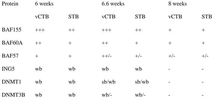
Table 2.1: Expression of epigenetic factor proteins in early human placental trophoblasts.... 41
Table 3.1: Key resources table... 76
Table 3.2: List of primers used for quantitative PCR analysis ... 79
ix
LIST OF FIGURES
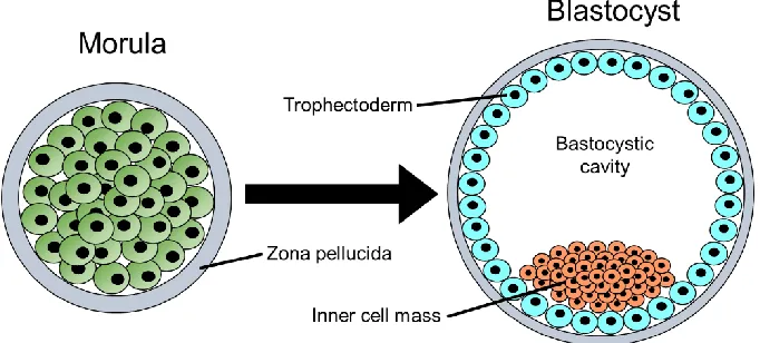
Figure 1.1 Transition of the morula to the blastocyst around day 3 p.c. ... 2
Figure 1.2 Stages of placental development... 3
Figure 1.3 Schematic of extravillous trophoblast invasion in human placenta ... 5
Figure 1.4 Schematic representation of EVT invasion and artery remodeling during normal and
preeclamptic pregnancy ... 14
Figure 2.1 Schematic of experimental design for analysis of the nuclear proteome during hESC differentiation to trophoblasts using SILAC and quantitative
mass spectrometry ... 35
Figure 2.2 Nuclear proteins modulated during hESC differentiation to trophoblasts ... 36
Figure 2.3 Study of the expression of BAF155, BAF60A, BAF57, ING5, and DNMT1 in day 0 undifferentiated H9 cells and day 12 differentiated H9 cells using
immunofluorescence. ... 38
Figure 2.4 Study of expression of BAF155, BAF60A, BAF57, ING5, DNMT1, and DNMT3B in day6-differentiated H9 cells and day12-differentiated H1 cells
using immunofluorescence ... 43
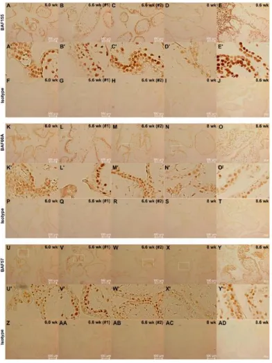
Figure 2.5 Study of the expression of BAF155, BAF60A, and BAF57 in first trimester
human placentas using immunohistochemistry ... 46
Figure 2.6 Study of expression of ING5, DNMT1, and DNMT3B in first trimester
human placentas using immunohistochemistry ... 48
Figure 3.1 Initial S1P treatment of hESCs leads to formation of CTB-like cell with
potential of subsequent formation of both EVT- and STB-like cells ... 64
Figure 3.2 S1P, Rho/ROCK signaling, and YAP are necessary for TB differentiation
from hESCs. ... 67
Figure 3.3 Formation of TBs from hESCs acts through receptor-mediated S1P
x
Figure 4.1 Establishment of CDX2+ hTSCs from day-3 treated hESCs ... 91
Figure 4.2 Directed differentiation of hTSCsCDX2 into STB and EVTs ... 93
Figure 4.3 Formation of villous-like hTSCs from hESCs and hTSCsCDX2 in TSCM ... 95
Figure 4.4 Directed differentiation of hTSCsP63 into STB and EVTs ... 96
1
CHAPTER 1: Introduction
1.1 Early development of the human placenta 1.1.1 Origin of the trophoblast
One of the key early events in human development is the specification of the
trophectoderm and inner cell mass during transition of the embryo from the morula to the
blastocyst stage. Following fertilization, the zygote continuously divides into a ball of cells, the
morula. Cells of the morula are totipotent with little contact of neighboring cells and encapsulated
by the zona pellucida1. Around day 3 postcoitus (p.c), the cells start to flatten due to compaction,
which results in forming tight cellular junction and ionic gradients. Ionic gradients allow water to
be drawn out to the center of the morula, while the tight cellular junctions allow for redistribution
of cytoskeleton and protein expression between cells. Taken together, this results in
asymmetrical division of cells and transition of the morula to the blastocyst which contains two
distinct populations – the trophectoderm (TE) that gives rise to all trophoblast (TB) cells that form
the placenta, and the inner cell mass (ICM) that gives rise to the embryo proper (Figure 1.1)2.
This specification is the earliest fate decision of the embryo, where trophoblast cells are
committed to their restricted lineage. Genetic studies about this lineage separation have been
restricted to mouse models, where the trophoblast specification is dependent on the transcription
factor, CDX2, while the ICM is similar morula stage, with the expression of the transcription factors
OCT4 and SOX23. Other fate determining factors such as polarization and epigenetic
modifications play a role in trophoblast formation4–8. Once the blastocyst is formed and enters
2
Figure 1.1: Transition of the morula to the blastocyst around day 3 p.c.
The first step of implantation in humans occurs around day 6-7 p.c. and is called
apposition, where the blastocyst locates an implantation site and attaches to the endometrium
lining(Figure 1.2A)9. At this stage the blastocyst can be dislodged from the uterine surface
without damage through flushing of the uterine lumen. Once full attachment of the blastocyst
takes place, a strong physical connection between the trophoblast and the endometrial epithelium
takes place and the blastocyst cannot be dislodged10,11. During apposition, the ICM usually faces
the endometrium, however the ICM is able to migrate along the inside face of the TE to align itself
to the side of apposition10. Upon attachment, TBs at the embryonic pole rapidly proliferate into a
double layer. The TBs facing the maternal tissue undergoes differentiation by cellular fusion of
neighboring cells forming the syncytiotrophoblast (STB). Upon differentiation the embryo invades
into the maternal endometrium (Figure 1.2B)9. The inner trophoblast layer remains epithelial and
comprises the stem cell compartment of the trophoblast called the cytotrophoblast (CTB). The
invasion of the blastocyst progresses through interstitial implantation, where the STB penetrates
the intercellular gaps between endometrial cells without initiating apoptosis by inducing lysis of
3
1.1.2 Lacunar stage
Over the following days the syncytiotrophoblast invades further into the endometrium and
subsequent trophoblastic proliferation and fusion occurs upon trophoblast contact of maternal
tissues. By day 12 p.c. the whole blastocyst has fully invaded into the endometrium, closing the
uterine epithelium over the implantation site. The outer surface of the blastocyst is completely
composed of the STB with the inner surface covered by a layer of CTBs. Since trophoblast
proliferation and differentiation to the STB occurred at the embryonic pole, this section is
considered thicker at this point compared to the antiimplantation pole. This size difference is
Figure 1.2: Stages of placental development; (A) Blastocyst implantation (6 to 7 days p.c.); (B) Prelacunar period (7 to 8 days p.c.); (C) Lacunar period (8 to 9 days p.c.); (D) Formation of primary villus (12 to 15 days p.c.); (E) Formation of secondary villus (15 to 21 days p.c.); (F) Formation of tertiary villus (18 days to term).
CP, primary chorionic plate; CT, cytotrophoblast; D, decidua; E, endometrial epithelium; EB, embryoblast; EG,
endometrial gland; EM, extraembryonic mesoderm; IVS, intervillous space; L, lacunae; SA, spiral artery; ST,
syncytiotrophoblast; T, trabeculae; TS, trophoblastic shell; X, extravillous trophoblast (X cells). (Modified from Frank et al.)9
4 never made up by the thinner trophoblast section during subsequent developmental steps. This
allows the thicker trophoblast section to subsequently form the placenta, whereas the thinner
regressively transforms to a smooth chorion. Over the course of blastocyst invasion, small
vacuoles of water within the STB start to appear due to the rapid increase in mass of the STB.
These vacuoles quickly expand to form a system of lacunae, while the separated pillars of STB
are the trabeculae, forming the lacunar stage in development (Figure 1.1C)1,10. Around the same
time as lacunae form, the CTBs beneath the STB proliferate and expand within the trabeculae.
Formation of the lacunar stage subdivides the trophoblasts into three layers, the primary chorionic
plate, which faces the original blastocystic cavity; the lacunar system with the trabeculae; and the
cytotrophoblastic shell, which faces the endometrium that is the forerunner of the basal plate.1
1.1.3 Early villous stage
Expansion of the CTBs within the trabeculae allows for longitudinal growth and branching
of the trabeculae that protrude into the lacunae forming the primary villi (Figure 1.2D). The primary villi are made up of a core of CTBs surrounded by the STB. Further growth and branching
of the CTBs initiate the development of primitive villous tree structures. Upon formation of the
primary villi, the CTBs start to slowly lose expression of the trophectoderm marker, CDX2, but
maintain another bipotential trophoblast stem cell marker, P6312. This transcriptional and
physiological change leads to the CTBs to become villous CTBs (vCTBs). Some vCTBs
continually expand and penetrate past the STB and into the endometrium, in which they spread
laterally to form the cytotrophoblastic shell and anchor the blastocyst9. When the primary villi
maintain contact with the cytotrophoblastic shell, they are called anchoring villi. VCTBs of the
anchoring villi characterized as collumn cytotrophoblast (cCTBs). In addition, the exposure of
cCTBs to endometrial tissue initiates differentiation to extravillous trophoblasts (EVTs) that
migrate into the endometrium to start trophoblast invasion. This key event is responsible for
adaptation of maternal vessels and further anchorage of the developing placenta13,14. During
5 After the formation of the primary villi, the extraembryonic mesenchyme originating from
the embryonic disk expands out forming a new layer to the chorionic plate. Extraembryonic
mesenchyme cells continue to expand within the center of the primary villi and establish a
connective tissue core forming secondary villi (Figure 1.2E). The extraembryonic mesenchyme expansion is restricted by the vCTBs and cCTBs and never reach the cytotrophoblast shell. Soon
after the formation of the secondary, the mesenchyme differentiates to form cells associated with
the formation of hemangioblastic cell cords15,16. By around day 20 p.c., the first fetal capillaries
start to appear, changing the secondary villi into tertiary villi structures (Figure 1.2F). Until term, this is the general structure of all placental villi. Only new villous sprouts correspond to primary
and secondary villi.
6
1.1.4 Formation of the definitive placenta
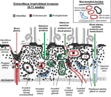
As the fetal capillaries establish within the villi, the maternal arteries begin to circulate
directly to the placenta. One of the key events that begin this process is through the EVTs. The
EVTs differentiate from the proliferative cCTBs located at the cytotrophoblastic shell and invade
into the maternal tissue. This invasion occurs along two routes; the interstitial cells that migrate
within the maternal stromal cells, and the endovascular cells that invade the maternal spiral
arteries (Figure 1.3). The interstitial cells invade deep into the first third of the myometrium providing anchoring of the placenta, where they undergo a differentiation into a large
multinucleated cell giant cell. This differentiation process is to ensure the tight restriction of
trophoblast invasion into the maternal tissue17. Further, the interstitial EVTs play an important
role in the communication with maternal uterine Natural Killer cells, macrophages, and the
endometrium. Interstitial EVTs also remodel spiral arteries deep within the endometrium. The
arteries change from narrow flexible vessels with high flow resistance to highly dilated, low
resistance stiff vessels18,19. The interstitial EVTs affect the immunomodulation of cells by
secreting specific factors that aid in apoptosis of T-cells, reduce cytotoxicity of uterine Natural
Killer cells, and reduce the activation of lymphocytes1. Lastly, the interstitial EVTs help promote
the endometrial stroma to undergo differentiation to decidual cells through paracrine actions20–25.
The decidualization aids in the immune-response of the invading EVTs and regulates the amount
of invasion. Further, the decidualization of the endometrium allows for the detachment of the
placenta during birth.
The endovascular EVTs initially propagate at the spiral artery interface within the
intervillous space and plug the outflow. This initial plugging only allows plasma to perfuse through
lessening the oxidative stress and allowing villi to grow normally. Around the end of the first
trimester (10-12 weeks), plugging of the spiral arteries dissipates and induction of remodeling the
spiral arteries takes place26,27. This marks the transition of the vascular connection between
7 to occur, the endovascular EVTs migrate along the arterial lumens and remodel the arteries. The
narrow, flexible vascular smooth muscle and endothelial cells are replaced by the endovascular
EVTs to create an expanded stiff arteries allowing high perfusion flow into the intervillous space
for nutrient-gas exchange on the villi22,28–30.
Once fetal blood enters the intervillous place nutrients-gas permeate through the tertiary
villi to the fetoplacental vessels which in turn transfer to the developing fetus along the umbilical
cord. Over the course of pregnancy, the network of tertiary villi continues to expand within the
hemotrophic system, with new sprouting villi occurring throughout pregnancy. As the growth of
new villi occurs, the expansion of the STB lowers the maternofetal diffusion distance and creates
a high surface area network for nutrient-gas exchange needed for healthy fetal growth. This
encapsulates the formation of the definitive placenta.
1.2 Trophoblast research 1.2.1 Mouse Trophoblast Model
One key model system for studying trophoblasts is the mouse. Mouse trophoblast stem
cells (mTSCs) were first derived from blastocysts and the extra-embryonic ectoderm. These cells
provide a key in vitro model for studying the signaling mechanisms and function of TBs31. MTSCs
grow indefinitely and differentiate to all lineages under continuous induction of fibroblast growth
factor 4 (FGF4) and transforming growth factor β1 (TGFβ1). The trophoblast transcription factors
CDX2, TEAD4, TFAP2C and GATA3 were found to be essential for specification and
determination of trophoblasts32–37. CDX2 was the first to be implicated to TE development; where
loss of CDX2 in mouse embryos does not affect blastocyst formation but does prevent the
blastocyst implantation. In addition, CDX2 ectopic induction in mouse embryonic stem cells
(mESCs) promoted mTSC fate. Expression of CDX2 has an essential role in the function and
specification of TE, though other factors may compensate the specification upon loss of CDX23,38.
8 induction of GATA3 in mESCs lead to formation of mTSCs36. Loss of GATA3 via RNAi-mediated
knockdown led to inhibition of the morula to blastocyst transition39.
In preimplantation mouse embryos, TEAD4 has an imperative role in establishment of the
TE, where loss of TEAD4 prevents TE-specific gene expression including CDX2 and GATA334.
The signaling mechanisms in mediating TEAD4 have revolved around the hippo signaling
pathway, specifically nuclear localization of YAP. Signaling mechanisms involving YAP and
TEAD4 have been proposed to lead to specification of TE and ICM, where the ICM lacks TEAD433.
Findings associated with transcription factor expression and their associated signaling pathways
have led to a better understanding of TE specification. However, mouse models cannot be directly
compared to human models. Whereas both systems share similarities in morphology of placenta
and hemochorial gas-nutrient exchange, they differ from each other in gestational length, litter
size, trophoblast cell types, organization, and invasion40.
1.2.2 Human Trophoblasts
Human trophoblast research has been limited due to the scarcity of human samples and
ethical concerns associated with studying human embryos41–44
.
Until very recently, when firsttrimester placenta samples were obtained, isolated trophoblasts underwent rapid differentiation
and could not be maintained45–53
.
Due to this restriction, most in vitro studies are carried outwith choriocarcinoma-derived trophoblast cell lines such as BeWo, JEG-3, and HTR-8/SVneo54–
56. These cells are easily maintained and do perform similar functions of trophoblasts cells such
as cellular syncytialization and invasion. However, these cell lines still show differences in their
transcriptome compared to primary samples. Further, these choriocarcinoma cell-lines lack the
ability to be multipotent and differentiate into both STB and EVTs57–59
.
Lastly, many of the cellsdo not meet the trophoblast criteria proposed by Lee et al.60. Recently another human trophoblast
progenitor cell line was established from the chorion and a human ESC (hESC) line named
9 of Lee et al. was not tested60. In addition, the established cell lines represent a mesenchymal
morphology, where CTBs are epithelial. Due to these key differences, studies using these cells
and choriocarcinoma-derived lines must be met with skepticism.
Recently, two model systems isolated from primary samples have been reported that
could have significant influence on trophoblast research. The first system is the establishment of
the trophoblast organoid63,64. The organoid system provides a 3D model that could potentially
recapitulate the physiology of the primary villi structure found in early placenta formation and the
formation of cell columns that a 2D model could not generate. The 3D model in organoid
structures allows for the study of the complex dynamic between the CTBs and STB, and
differentiation into the EVTs that occurs during in vivo placentation. 3D epithelial organoids have
been achieved from various tissues such as the endometrium, but organoids of the human
placenta could never be established in the past65,66. The trophoblast organoids established were
able to be continuously propagated through the establishment of the CTBs and an inner layer of
the STB. Embedding the organoid into the extracellular matrix, Matrigel, or inhibition of Wnt
signaling leads to the spontaneous differentiation of the CTBs to EVTs63,67. These organoids
could allow a better understanding into cell column formation and what effect the endometrium
microenvironment has on trophoblast function. Further, analysis on the histotrophic nutrition
system within the organoid or their response to extraembryonic mesoderm cells could provide
crucial insight into the kinetics of the gas-nutrient exchange within the villi and formation of more
complex villi structures.
Though the trophoblast organoid systems provide new areas to explore and understand
early placenta development, limitations around the organoids remain. The trophoblast organoids
developed have physiological differences compared to human primary villi in that the CTBs are
on the periphery surrounding the STBs. In primary samples the STB encapsulate the CTBs,
wherein the CTBs differentiate and fuse to replenish the STB. This physiological difference from
10 established. Further transcriptome differences in primary villi and trophoblast organoids shows
that further work is required for optimization of culture conditions64.
The second key system reported was the establishment and maintenance of the human
trophoblast stem cell (hTSC) in vitro derived from primary placental samples and blastocysts68.
This study showed that the transcriptome of the hTSCs were very similar to primary human CTBs
and met all the criteria associated with human trophoblast cells60. Further, they showed ability to
form the EVTs and STB (2D and 3D) with similar transcriptomes to their primary counterparts. In
addition, studies on hTSCs revealed that the activation of Wnt (wingless/integrated) and inhibition
of TGF-β are essential for derivation and maintenance of cells. In contrast, mTSCs require the
opposite conditions – presence of TGF-β and inhibition of Wnt signaling69–71. In mTSCs, FGF4
and FGFR2c are essential for maintenance of cells, FGFR2c is not expressed in human
blastocysts72. HTSCs were found to express the FGFR2 spliced variant FGFR2b, associated with
FGF10, thus showing the stark difference between the two models and the potential in revealing
the key signaling involved in trophoblast maintenance. Lastly, when the hTSCs were injected into
immune-deficient mice, they mimicked key features of trophoblast invasion during implantation.
Though ethical reasons prohibit the examination of hTSC injected into human blastocysts, the
hTSCs established show comprehensive data corresponding to human CTBs. Though the
development of hTSCs provides a powerful tool to examine the mechanistic signaling and
functionality of each trophoblast cell type, limitations remain. The hTSCs established lost
expression of the early trophectoderm marker, CDX2, though maintained the vCTB marker, P63.
This maintenance of P63 shows that the hTSCs represent cell associated with vCTBs, rather than
trophectoderm, which could limit studying trophoblast signaling, differentiation, and functionality
associated with implantation and maintenance of cell associated with the blastocyst and lacunar
stages during development. Lastly, culture conditions associated with differentiation of STB and
EVTs are undefined and suboptimal in establishing what key mechanistic signaling is responsible
11 better understanding trophoblasts and could lead to substantial insight into developmental
disorders associated with trophoblasts.
1.2.3 Trophoblast from human embryonic stem cells
Human embryonic stem cells (hESCs) were first established by the immunosurgery of
human blastocysts73–75. Most hESCs are derived from discarded embryos during in vitro
fertilization. HESCs have great potential for regenerative medicine due to their ability to form all
tissue types found in the body and be continuously maintained. TBs were first derived from
hESCs through treatment with Bone Morphogenetic Protein 4 (BMP4); under these conditions,
differentiated cells expressed key TB markers76. The differentiation of TBs from hESCs has since
been optimized in other studies12,43,77–90. HESCs exposed to BMP4 undergo morphological
differentiation in large flattened cells proceeding from the periphery inwards. During BMP4
treatment, hESCs lose expression associated with ICM, and undergo upregulation of transcription
factors associated with TB, with little upregulation of mesoderm, endoderm or ectoderm82,89,90.
Extended exposure of BMP4 leads to syncytialization of cells and secretion of placental hormones
associated with formation of STB78,79. Through these studies, BMP4 treatment with inhibition of
Activin/Nodal and FGF2 signaling (BAP treatment) provided became the accepted condition for
forming TBs79. However, these cells exhibit key differences in transcriptome, epigenetics, and
ability to form EVTs compared to primary samples. One of the key morphological differences is
that the cells from BAP treatment become STB-like cell type that do not form mesenchymal
HLA-G expressing cells. Though these cells have been found to have invasive capabilities, their
transcriptome differs significantly from primary samples. Further all cells treated continuously
with BMP4 showed high methylation at the ELF5 promoter region showing epigenetics more
associated with hESCs than human TBs42,60,91,92. HESCs differentiated from BAP or BMP4
treatment show increasingly higher heterogeneity over longer induction periods, leading to
confusion as to what key mechanistic pathways cause cellular differentiation. Lastly, TB
12 induction leads to extraembryonic mesoderm phenotype over TB91. In the study is claimed that
hESCs are epigenetically restricted from forming TBs, an analog of mESCs and TB formation.
However, further studies have addressed the issues raised by Bernardo et al., but seeds of doubt
about hESC ability to form TBs remain12,79,93.
Over the past few years, we have established a protocol for developing homogenous
development of STB and EVTs through a two-step protocol involving the inhibition/activation of
Activin/Nodal signaling upon passaging of BMP4 treated hESCs77,81. In addition to homogenous
cellular populations of EVTs and STB, we have shown the cells to have hypomethylation of ELF5
promoter region and high expression of HLA-G in EVTs. These key advances are essential to
verifying the potential for TB differentiation from hESCs, still maintenance of CTBs from hESCs
remains elusive.
The media systems involved in the differentiation of TB from hESCs are all derived from
feeder cells or involve complex component additives such as fetal bovine serum or knockout
serum replacement (KOSR) or bovine serum albumin (BSA). These additive, including BSA, are
associated with lipid species that aid in cell growth but confound mechanistic analysis of the
signaling pathways involved in the differentiation process94–97. A variety of defined culture
systems have been used for maintenance of undifferentiated hESCs including Essential 8,
mTeSR1, XFT, HESCO, NBF, and FTDA98–103. Defined culture conditions are an invaluable tool
to understand hESCs differentiation and signaling since all known exogenous signaling inputs are
known. However, all these media formulations vary in terms of what signaling pathways are
induced/inhibited. For example, mTeSR1, and HESCO contain Wnt signaling activators while the
other media systems do not. Further feeder-cell conditioned media contains serum-associated
lipid factors such as sphingosine-1-phosphate (S1P) and lysophosphatidic acid (LPA), while the
defined media formulations do not104. S1P has shown to interact with the Hippo signaling
pathways, specifically inhibiting Lats1/2 and allowing YAP translocation to the nucleus and
13 differentiation, but previous studies looking at TB formation using serum-free media have shown
to form mesoderm, extraembryonic mesoderm and partial endoderm91,109–111. It is important to
use a defined culture to understand TB formation from hESCs; this in turn will enable the
derivation of hTSCs from hESCs, if such a transition is theoretically possible.
1.3 Trophoblast role on disease
Human trophoblast research has come a long way over the past few years with the
establishment of the hTSC and the organoid systems. However, as previously discussed these
are associated with significant limitation, and early human trophoblast development remains
unclear. The National Institute of Health recently coined the placenta “the least understood organ”,
and with good reasoning. Improper trophoblast development within the placenta is responsible
for a number of pregnancy disorders such as preeclampsia, placental accreta, infarcts, and
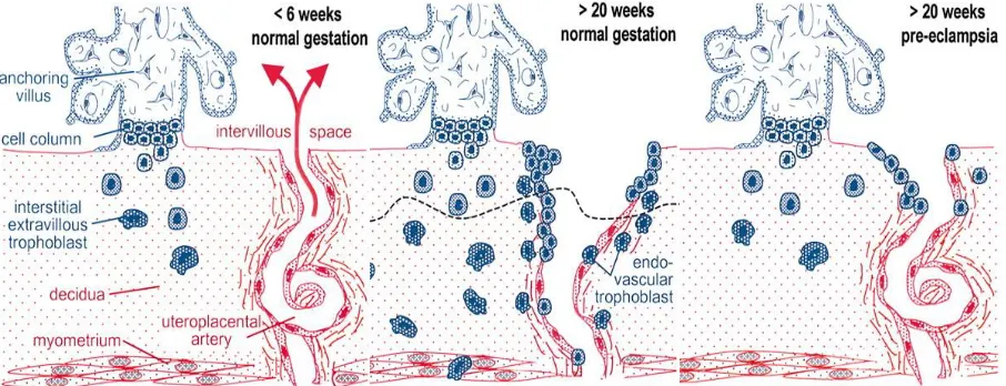
intrauterine growth restriction1,112–117. A common factor in several placental disorders is the extent
of invasion associated with the EVTs. When the EVTs are overly invasive, such as in placental
accreta, they migrate too far into the myometrium and can cause the placenta to attach strongly
to the uterus; this can cause hemorrhaging during labor118,119. When EVTs are not invasive
enough, such as in preeclampsia and intrauterine growth restriction, they do not fully remodel the
spiral arteries causing insufficient exchange of nutrients and gases required for the developing
fetus (Figure 1.4)30,113,114. A mechanistic understanding of early trophoblast differentiation will
provide insight into these placenta-associated pregnancy disorders. This is particularly relevant
when in the United States, maternal deaths are on the rise – in contrast to the rest of the
developed world120. In addition to maternal deaths, approximately 70% of conceptions are lost
prior to birth due to implantation failure, miscarriage, or early pregnancy loss, with little
understanding as to what is affecting this low yield121. Obesity and cesarean section operations
have led to a higher incidence of pregnancy disorders and almost no pharmaceutical medicine is
available to help122–124. Moreover, environmental influence on the epigenetics of the placenta is
14 and later adult disease58,125. Thus overall, understanding early TB differentiation will have a
significant impact on maternal and fetal health during pregnancy and beyond.
Figure 1.4: Schematic representation of EVT invasion and artery remodeling during normal and preeclamptic pregnancy (modified from Kaufman et al.)30.
1.4 Overview
Development of the trophoblast cells in early gestation plays an important role in placental
development, which in turn has significant implications for fetal and maternal health both during
and after pregnancy. The ability to form and maintain hTSCs enables better understanding of the
functionality, mechanistic signaling in, and differentiation of TBs. Current models of the hTSCs
and TB organoids derived from primary placental samples may provide useful insight into
pathogenesis of TB defects occurring around the first trimester of pregnancy. However, these
models differ from primary tissue in important aspects as discussed earlier. Importantly, these
models do not capture the trophectoderm state in the blastocyst stage embryo. A model system
for TE cells would enable studies on formation of all TB cell types and give potential insight into
the implantation of the blastocyst. It is unknown if isolated first trimester primary TBs can be
reverted back to their TE state; therefore, hESCs become an attractive and indispensable tool.
HESCs are characteristically representative to the ICM of the blastocyst, and can be brought to a
15 describes the use of hESCs for developing an in vitro model system for early trophoblast
development, including cells that are consistent with the TE.
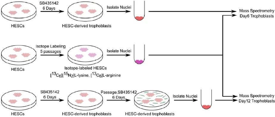
In Chapter 2, we describe the use of hESCs as a model to identify epigenetic factor
proteins during the differentiation to TBs. Using isotope labeling during cell culture and
quantitative proteomics, 30 epigenetic factors were identified as changing during the
differentiation process. Chief among the epigenetic factors were the down regulation of DNA
methyltransferases DNMT1, DNMT3A, and DNMT3B. The epigenetic factor proteins found from
hESC-derived trophoblasts were in turn analyzed through immunohistochemistry analysis in
6-9-week human placentas. The expression patterns found in primary placentas, largely matched
those found in the hESC-derived TBs. This validation enables further investigation into the
epigenetic factors involved in trophoblast development and the power of the hESC-model for
assessing TB epigenetics.
In Chapter 3, we discuss the development of a chemically defined media system for TB
differentiation from hESCs. During this process, it was found that S1P plays a vital role in enabling
hESCs to access the TB compartment and prohibiting gene expression to neural and mesoderm
cell types. Using this system, we were able to show that both inhibition ROCK and knockdown of
YAP eliminates the ability to form bipotent TBs from hESCs. The specific signaling mechanism
in which S1P acts during hESC differentiation to TB was found to be receptor mediated. This
system provides insight into the signaling pathways during hESCs differentiation to TB and
enabled development of a culture system for maintenance of hTSCs.
Chapter 4 described the maintenance of a TE-like stem cell derived from hESCs. These
cells maintain expression of the TE-marker CDX2, while showing the ability to adapt to a P63+
vCTB in the media system developed by Okae et al68. Further, the established vCTBs match
morphologically to primary cells in same media system and are able to differentiate to both EVTs
16 Derivation of bona fide hTSCs from hESCs can greatly accelerate TB research. This will
enable generation of hTSCs from pluripotent stem cells that are genetically modified (or from
different genetic backgrounds). In addition, ease of access to human pluripotent stem cell lines
and hESCs could lead more widespread research into TB development. Finally, the ability to
form TE from hESCs allows for exploration into molecular mechanisms involved during the
17
1.5 References
1. Benirschke, K., Baergen, R. N. & Burton, G. (Graham J. . Pathology of the human placenta [electronic resource]. (Springer, 2012).
2. Selwood, L. & Johnson, M. H. Trophoblast and hypoblast in the monotreme, marsupial and eutherian mammal: evolution and origins. BioEssays28, 128–145 (2006).
3. Strumpf, D. et al. Cdx2 is required for correct cell fate specification and differentiation of trophectoderm in the mouse blastocyst. Development132, 2093–2102 (2005).
4. Hemberger, M., Udayashankar, R., Tesar, P., Moore, H. & Burton, G. J. ELF5-enforced transcriptional networks define an epigenetically regulated trophoblast stem cell
compartment in the human placenta. Hum. Mol. Genet.19, 2456–67 (2010).
5. Ng, R. K. et al. Epigenetic restriction of embryonic cell lineage fate by methylation of Elf5.
Nat. Cell Biol.10, 1280–90 (2008).
6. Zernicka-Goetz, M., Morris, S. A. & Bruce, A. W. Making a firm decision: multifaceted regulation of cell fate in the early mouse embryo. Nat. Rev. Genet.10, 467–477 (2009).
7. Skamagki, M., Wicher, K. B., Jedrusik, A., Ganguly, S. & Zernicka-Goetz, M. Asymmetric localization of Cdx2 mRNA during the first cell-fate decision in early mouse development.
Cell Rep.3, 442–57 (2013).
8. Jedrusik, A. et al. Role of Cdx2 and cell polarity in cell allocation and specification of trophectoderm and inner cell mass in the mouse embryo. Genes Dev.22, 2692–2706
(2008).
9. Frank, H.-G. Placental Development. Fetal Neonatal Physiol. 101–113 (2017).
doi:10.1016/B978-0-323-35214-7.00010-X
10. Su, R.-W. & Fazleabas, A. T. Implantation and Establishment of Pregnancy in Human and Nonhuman Primates. doi:10.1007/978-3-319-15856-3_10
11. Aplin, J. D. & Ruane, P. T. Embryo–epithelium interactions during implantation at a glance. J. Cell Sci.130, 15–22 (2017).
12. Horii, M. et al. Human pluripotent stem cells as a model of trophoblast differentiation in both normal development and disease. Proc. Natl. Acad. Sci. U. S. A.113, E3882-91 (2016).
13. Pijnenborg, R. et al. In vivo analysis of trophoblast cell invasion in the human. Methods Mol. Med.122, 11–44 (2006).
14. Carter, A. M., Enders, A. C. & Pijnenborg, R. The role of invasive trophoblast in
18 20140070 (2015).
15. Demir, R., Kaufmann, P., Castellucci, M., Erbengi, T. & Kotowski, A. Fetal
vasculogenesis and angiogenesis in human placental villi. Acta Anat. (Basel).136, 190–
203 (1989).
16. Robin, C. et al. Human Placenta Is a Potent Hematopoietic Niche Containing
Hematopoietic Stem and Progenitor Cells throughout Development. Cell Stem Cell5,
385–395 (2009).
17. Velicky, P., Knöfler, M. & Pollheimer, J. Function and control of human invasive
trophoblast subtypes: Intrinsic vs. maternal control. Cell Adh. Migr.10, 154–62 (2016).
18. Harris, L. K. IFPA Gabor Than Award lecture: Transformation of the spiral arteries in human pregnancy: Key events in the remodelling timeline. Placenta32, S154–S158
(2011).
19. Chang, W.-L. et al. PLAC8, a new marker for human interstitial extravillous trophoblast cells, promotes their invasion and migration. Development145, (2018).
20. Gellersen, B., Brosens, I. & Brosens, J. Decidualization of the Human Endometrium: Mechanisms, Functions, and Clinical Perspectives. Semin. Reprod. Med.25, 445–453
(2007).
21. Gellersen, B. & Brosens, J. J. Cyclic Decidualization of the Human Endometrium in Reproductive Health and Failure. Endocr. Rev.35, 851–905 (2014).
22. Kam, E. P. Y., Gardner, L., Loke, Y. W. & King, A. The role of trophoblast in the physiological change in decidual spiral arteries. Hum. Reprod.14, 2131–2138 (1999).
23. Blanks, A. M. & Brosens, J. J. Progesterone action in the myometrium and decidua in preterm birth. Facts, views Vis. ObGyn4, 33–43 (2012).
24. Mori, M., Bogdan, A., Balassa, T., Csabai, T. & Szekeres-Bartho, J. The decidua-the maternal bed embracing the embryo-maintains the pregnancy. Semin. Immunopathol.38,
635–649 (2016).
25. Ramathal, C. Y., Bagchi, I. C., Taylor, R. N. & Bagchi, M. K. Endometrial decidualization: of mice and men. Semin. Reprod. Med.28, 17–26 (2010).
26. Moser, G. et al. Extravillous trophoblasts invade more than uterine arteries: evidence for the invasion of uterine veins. Histochem. Cell Biol.147, 353–366 (2017).
27. Weiss, G., Sundl, M., Glasner, A., Huppertz, B. & Moser, G. The trophoblast plug during early pregnancy: a deeper insight. Histochem. Cell Biol.146, 749–756 (2016).
19 extravillous trophoblast invasion. J. Reprod. Heal. Med.2, S9–S14 (2016).
29. Pijnenborg, R., Vercruysse, L. & Hanssens, M. The Uterine Spiral Arteries In Human Pregnancy: Facts and Controversies. Placenta27, 939–958 (2006).
30. Kaufmann, P., Black, S. & Huppertz, B. Endovascular Trophoblast Invasion: Implications for the Pathogenesis of Intrauterine Growth Retardation and Preeclampsia. Biol. Reprod.
69, 1–7 (2003).
31. Tanaka, S., Kunath, T., Hadjantonakis, A. K., Nagy, A. & Rossant, J. Promotion of trophoblast stem cell proliferation by FGF4. Science282, 2072–5 (1998).
32. Choi, I., Carey, T. S., Wilson, C. A. & Knott, J. G. Transcription factor AP-2γ is a core regulator of tight junction biogenesis and cavity formation during mouse early
embryogenesis. Development139, 4623–32 (2012).
33. Home, P. et al. Altered subcellular localization of transcription factor TEAD4 regulates first mammalian cell lineage commitment. Proc. Natl. Acad. Sci. U. S. A.109, 7362–7
(2012).
34. Nishioka, N. et al. Tead4 is required for specification of trophectoderm in pre-implantation mouse embryos. Mech. Dev.125, 270–283 (2008).
35. Niwa, H. et al. Interaction between Oct3/4 and Cdx2 determines trophectoderm differentiation. Cell123, 917–29 (2005).
36. Ralston, A. et al. Gata3 regulates trophoblast development downstream of Tead4 and in parallel to Cdx2. Development137, 395–403 (2010).
37. Yagi, R. et al. Transcription factor TEAD4 specifies the trophectoderm lineage at the beginning of mammalian development. Development134, 3827–36 (2007).
38. Nishiyama, A. et al. Uncovering Early Response of Gene Regulatory Networks in ESCs by Systematic Induction of Transcription Factors. Cell Stem Cell5, 420–433 (2009).
39. Home, P. et al. GATA3 Is Selectively Expressed in the Trophectoderm of
Peri-implantation Embryo and Directly Regulates Cdx2 Gene Expression. J. Biol. Chem.284,
28729–28737 (2009).
40. Soncin, F. et al. Comparative analysis of mouse and human placentae across gestation reveals species-specific regulators of placental development. (2018).
doi:10.1242/dev.156273
41. Golos, T. G., Giakoumopoulos, M. & Gerami-Naini, B. Review: Trophoblast differentiation from human embryonic stem cells. Placenta doi:10.1016/j.placenta.2012.11.019
20 to be or not to be? Reproduction147, D1-12 (2014).
43. Schulz, L. C. et al. Human Embryonic Stem Cells as Models for Trophoblast Differentiation. 10–16 (2008). doi:10.1016/j.placenta.2007.10.009
44. Tiruthani, K., Sarkar, P. & Rao, B. Trophoblast differentiation of human embryonic stem cells. Biotechnol. J.8, 421–33 (2013).
45. Aboagye-Mathiesen, G., Laugesen, J., Zdravkovic, M. & Ebbesen, P. Isolation and characterization of human placental trophoblast subpopulations from first-trimester chorionic villi. Clin. Diagn. Lab. Immunol.3, 14–22 (1996).
46. Caulfield, J. J., Sargent, I. L., Ferry, B. L., Starkey, P. M. & Redman, C. W. G. Isolation and characterisation of a subpopulation of human chorionic cytotrophoblast using a monoclonal anti-trophoblast antibody (NDOG2) in flow cytometry. J. Reprod. Immunol.
21, 71–85 (1992).
47. James, J. L., Stone, P. R. & Chamley, L. W. Cytotrophoblast differentiation in the first trimester of pregnancy: evidence for separate progenitors of extravillous trophoblasts and syncytiotrophoblast. Reproduction130, 95–103 (2005).
48. James, J. L., Stone, P. R. & Chamley, L. W. The isolation and characterization of a population of extravillous trophoblast progenitors from first trimester human placenta.
Hum. Reprod.22, 2111–9 (2007).
49. Manoussaka, M. S., Jackson, D. J., Lock, R. J., Sooranna, S. R. & Kumpel, B. M. Flow cytometric characterisation of cells of differing densities isolated from human term placentae and enrichment of villous trophoblast cells. Placenta26, 308–18 (2005).
50. Stenqvist, A.-C. et al. An efficient optimized method for isolation of villous trophoblast cells from human early pregnancy placenta suitable for functional and molecular studies.
Am. J. Reprod. Immunol.60, 33–42 (2008).
51. Tarrade, a et al. Characterization of human villous and extravillous trophoblasts isolated from first trimester placenta. Lab. Invest.81, 1199–211 (2001).
52. Trundley, A., Gardner, L., Northfield, J., Chang, C. & Moffett, A. Methods for isolation of cells from the human fetal-maternal interface. Methods Mol. Med.122, 109–22 (2006).
53. Whitley, G. S. J. Production of human trophoblast cell lines. Methods Mol. Med.121,
219–28 (2006).
54. Pattillo, R. A. & Gey, G. O. The establishment of a cell line of human hormone-synthesizing trophoblastic cells in vitro. Cancer Res.28, 1231–6 (1968).
21 trophoblast cells with extended lifespan. Exp. Cell Res.206, 204–11 (1993).
56. KOHLER, P. O. & BRIDSON, W. E. Isolation of Hormone-Producing Clonal Lines of Human Choriocarcinoma 1. J. Clin. Endocrinol. Metab. 32, 683–687 (1971).
57. Petroff, M. G., Phillips, T. A., Ka, H., Pace, J. L. & Hunt, J. S. Isolation and culture of term human trophoblast cells. Methods Mol. Med.121, 203–17 (2006).
58. Novakovic, B. et al. Wide-ranging DNA methylation differences of primary trophoblast cell populations and derived cell lines: implications and opportunities for understanding trophoblast function. Mol. Hum. Reprod. 17, 344–53 (2011).
59. Rothbauer, M. et al. A comparative study of five physiological key parameters between four different human trophoblast-derived cell lines. Sci. Rep.7, 5892 (2017).
60. Lee, C. Q. E. et al. What Is Trophoblast? A Combination of Criteria Define Human First-Trimester Trophoblast. Stem cell reports6, 257–72 (2016).
61. Genbacev, O. et al. Establishment of human trophoblast progenitor cell lines from the chorion. Stem Cells29, 1427–36 (2011).
62. Zdravkovic, T. et al. Human stem cells from single blastomeres reveal pathways of embryonic or trophoblast fate specification. Development142, 4010–25 (2015).
63. Haider, S. et al. Self-Renewing Trophoblast Organoids Recapitulate the Developmental Program of the Early Human Placenta. Stem cell reports11, 537–551 (2018).
64. Turco, M. Y. et al. Trophoblast organoids as a model for maternal–fetal interactions
during human placentation. Nature564, 263–267 (2018).
65. Boretto, M. et al. Development of organoids from mouse and human endometrium showing endometrial epithelium physiology and long-term expandability. Development
144, 1775–1786 (2017).
66. Turco, M. Y. et al. Long-term, hormone-responsive organoid cultures of human endometrium in a chemically defined medium. Nat. Cell Biol.19, 568–577 (2017).
67. Haider, S. et al. Notch1 controls development of the extravillous trophoblast lineage in the human placenta. Proc. Natl. Acad. Sci.113, E7710–E7719 (2016).
68. Okae, H. et al. Derivation of Human Trophoblast Stem Cells. Cell Stem Cell22, 50–63.e6
(2018).
69. Latos, P. A. & Hemberger, M. From the stem of the placental tree: trophoblast stem cells and their progeny. (2016). doi:10.1242/dev.133462
22 Culture Conditions in Mice. PLoS One9, 107308 (2014).
71. Erlebacher, A., Price, K. A. & Glimcher, L. H. Maintenance of mouse trophoblast stem cell proliferation by TGF-β/activin. Dev. Biol.275, 158–169 (2004).
72. Kunath, T. et al. Developmental differences in the expression of FGF receptors between human and mouse embryos. Placenta35, 1079–1088 (2014).
73. Thomson, J. A. et al. Embryonic stem cell lines derived from human blastocysts. Science
282, 1145–7 (1998).
74. Chen, A. E. et al. Optimal timing of inner cell mass isolation increases the efficiency of human embryonic stem cell derivation and allows generation of sibling cell lines. Cell Stem Cell4, 103–6 (2009).
75. Pera, M., Reubinoff, B. & Trounson, A. Human embryonic stem cells. J. Cell Sci.113, 5–
10 (2000).
76. Xu, R.-H. et al. BMP4 initiates human embryonic stem cell differentiation to trophoblast.
Nat. Biotechnol.20, 1261–4 (2002).
77. Sarkar, P. et al. Activin/Nodal Signaling Switches the Terminal Fate of Human Embryonic Stem Cell-derived Trophoblasts. J. Biol. Chem.290, 8834–8848 (2015).
78. Das, P. et al. Effects of fgf2 and oxygen in the bmp4-driven differentiation of trophoblast from human embryonic stem cells. Stem Cell Res.1, 61–74 (2007).
79. Amita, M. et al. Complete and unidirectional conversion of human embryonic stem cells to trophoblast by BMP4. Proc. Natl. Acad. Sci. U. S. A. 110, E1212-21 (2013).
80. Telugu, B. P. et al. Comparison of extravillous trophoblast cells derived from human embryonic stem cells and from first trimester human placentas. Placenta34, 536–43
(2013).
81. Sarkar, P. et al. Identification of Epigenetic Factor Proteins Expressed in Human Embryonic Stem Cell-Derived Trophoblasts and in Human Placental Trophoblasts. J. Proteome Res.15, 2433–2444 (2016).
82. Ezashi, T., Telugu, B. P. V. L. & Roberts, R. M. Model systems for studying trophoblast differentiation from human pluripotent stem cells. Cell Tissue Res.349, 809–24 (2012).
83. Xu, R.-H. In vitro induction of trophoblast from human embryonic stem cells. Methods Mol. Med.121, 189–202 (2006).
23 85. Chen, G. et al. Trophoblast Differentiation Defect in Human Embryonic Stem Cells
Lacking PIG-A and GPI-Anchored Cell-Surface Proteins. Cell Stem Cell2, 345–355
(2008).
86. Wu, Z. et al. Combinatorial signals of activin/nodal and bone morphogenic protein regulate the early lineage segregation of human embryonic stem cells. J. Biol. Chem.
283, 24991–5002 (2008).
87. Douglas, G. C., VandeVoort, C. A., Kumar, P., Chang, T.-C. & Golos, T. G. Trophoblast Stem Cells: Models for Investigating Trophectoderm Differentiation and Placental Development. Endocr. Rev.30, 228–240 (2009).
88. Erb, T. M. et al. Paracrine and epigenetic control of trophectoderm differentiation from human embryonic stem cells: the role of bone morphogenic protein 4 and histone deacetylases. Stem Cells Dev.20, 1601–14 (2011).
89. Marchand, M. et al. Transcriptomic signature of trophoblast differentiation in a human embryonic stem cell model. Biol. Reprod.84, 1258–71 (2011).
90. Sudheer, S., Bhushan, R., Fauler, B., Lehrach, H. & Adjaye, J. FGF inhibition directs BMP4-mediated differentiation of human embryonic stem cells to syncytiotrophoblast.
Stem Cells Dev.21, 2987–3000 (2012).
91. Bernardo, A. S. et al. BRACHYURY and CDX2 Mediate BMP-Induced Differentiation of Human and Mouse Pluripotent Stem Cells into Embryonic and Extraembryonic Lineages.
Cell Stem Cell9, 144–155 (2011).
92. Lee, H. J. et al. Lineage specific methylation of the Elf5 promoter in mammary epithelial cells. Stem Cells29, 1611–9 (2011).
93. Morgani, S. M. et al. Totipotent embryonic stem cells arise in ground-state culture conditions. Cell Rep.3, 1945–57 (2013).
94. Sarkar, P., Randall, S. M., Muddiman, D. C. & Rao, B. M. Targeted proteomics of the secretory pathway reveals the secretome of mouse embryonic fibroblasts and human embryonic stem cells. Mol. Cell. Proteomics11, 1829–39 (2012).
95. Bendall, S. C. et al. An enhanced mass spectrometry approach reveals human embryonic stem cell growth factors in culture. Mol. Cell. Proteomics8, 421–32 (2009).
96. Spector, A. A., John, K. & Fletcher, J. E. Binding of long-chain fatty acids to bovine serum albumin. J. Lipid Res.10, 56–67 (1969).
24 98. Chen, G. et al. Chemically defined conditions for human iPSC derivation and culture. Nat.
Methods8, 424–9 (2011).
99. Ludwig, T. E. et al. Derivation of human embryonic stem cells in defined conditions. Nat. Biotechnol.24, 185–187 (2006).
100. Liu, Y. et al. A novel chemical-defined medium with bFGF and N2B27 supplements supports undifferentiated growth in human embryonic stem cells. Biochem. Biophys. Res. Commun.346, 131–139 (2006).
101. Frank, S., Zhang, M., Schöler, H. R. & Greber, B. Small Molecule-Assisted,
Line-Independent Maintenance of Human Pluripotent Stem Cells in Defined Conditions. PLoS One7, e41958 (2012).
102. Tsutsui, H. et al. An optimized small molecule inhibitor cocktail supports long-term maintenance of human embryonic stem cells. Nat. Commun.2, 167 (2011).
103. Lu, J., Hou, R., Booth, C. J., Yang, S.-H. & Snyder, M. Defined culture conditions of human embryonic stem cells. (2006).
104. Miller, E. et al. Identification of serum-derived sphingosine-1-phosphate as a small molecule regulator of YAP. Chem. Biol.19, 955–62 (2012).
105. Maceyka, M., Harikumar, K. B., Milstien, S. & Spiegel, S. Sphingosine-1-phosphate signaling and its role in disease. Trends Cell Biol.22, 50–60 (2012).
106. Yu, F.-X. et al. Regulation of the Hippo-YAP pathway by G-protein-coupled receptor signaling. Cell150, 780–91 (2012).
107. Mendelson, K., Evans, T. & Hla, T. Sphingosine 1-phosphate signalling. Development
141, 5–9 (2014).
108. Tsai, H.-C. & Han, M. H. Sphingosine-1-Phosphate (S1P) and S1P Signaling Pathway: Therapeutic Targets in Autoimmunity and Inflammation. Drugs76, 1067–1079 (2016).
109. Yu, X. et al. Notch Signaling Activation in Human Embryonic Stem Cells Is Required for Embryonic, but Not Trophoblastic, Lineage Commitment. Cell Stem Cell2, 461–471
(2008).
110. Yu, P., Pan, G., Yu, J. & Thomson, J. A. FGF2 Sustains NANOG and Switches the Outcome of BMP4-Induced Human Embryonic Stem Cell Differentiation. Cell Stem Cell8,
326–334 (2011).
111. Zhang, P. et al. Short-term BMP-4 treatment initiates mesoderm induction in human embryonic stem cells. Blood111, 1933–41 (2008).
25 pregnancy. Wiener Medizinische Wochenschrift 162, 187–190 (2012).
113. Silva, J. F. & Serakides, R. Intrauterine trophoblast migration: A comparative view of humans and rodents. Cell Adh. Migr.10, 88–110 (2016).
114. Malhotra, S. S., Banerjee, P. & Gupta, S. K. Regulation of trophoblast differentiation during embryo implantation and placentation: Implications in pregnancy complications. J. Reprod. Heal. Med.2, S26–S36 (2016).
115. Burton, G. J. & Jauniaux, E. The cytotrophoblastic shell and complications of pregnancy.
Placenta60, 134–139 (2017).
116. Norwitz, E. R. Defective implantation and placentation: laying the blueprint for pregnancy complications. Reproductive BioMedicine Online13, 591–599 (Elsevier, 2006).
117. Shcherbakov, V. I. Apoptosis in the trophoblast and its role in pregnancy complications.
Biol. Bull. Rev.1, 325–335 (2011).
118. Duzyj, C. et al. Extravillous trophoblast invasion in placenta accreta is associated with differential local expression of angiogenic and growth factors: a cross-sectional study.
BJOG An Int. J. Obstet. Gynaecol.125, 1441–1448 (2018).
119. Adler, E., Madankumar, R., Rosner, M. & Reznik, S. E. Increased placental trophoblast inclusions in placenta accreta. Placenta35, 1075–1078 (2014).
120. MacDorman, M. F., Declercq, E., Cabral, H. & Morton, C. Recent Increases in the U.S. Maternal Mortality Rate: Disentangling Trends From Measurement Issues. Obstet. Gynecol.128, 447–55 (2016).
121. Macklon, N. S., Geraedts, J. P. M. & Fauser, B. C. J. M. Conception to ongoing pregnancy: thèblack box’ of early pregnancy loss.
122. Norman, R. J. & Clark, A. M. Obesity and reproductive disorders: a review. Reprod. Fertil. Dev.10, 55 (1998).
123. Klar, M. & Michels, K. B. Cesarean section and placental disorders in subsequent pregnancies – a meta-analysis. J. Perinat. Med.42, 571–583 (2014).
124. Kamara, M., Henderson, J., Doherty, D., Dickinson, J. & Pennell, C. The risk of placenta accreta following primary elective caesarean delivery: a case-control study. BJOG An Int. J. Obstet. Gynaecol.120, 879–886 (2013).
125. Januar, V., Desoye, G., Novakovic, B., Cvitic, S. & Saffery, R. Epigenetic regulation of human placental function and pregnancy outcome: considerations for causal inference.
Am. J. Obstet. Gynecol.213, S182–S196 (2015).
26
27
CHAPTER 2: Identification of Epigenetic Factor Proteins Expressed in Human Embryonic Stem Cell-derived Trophoblast and in Human Placental Trophoblasts
2.1 Abstract
Human embryonic stem cells (hESCs) have been used to derive trophoblasts through
differentiation in vitro. Intriguingly, mouse ESCs are prevented from differentiation to trophoblasts
by certain epigenetic factor proteins such as Dnmt1, thus necessitating the study of epigenetic
factor proteins during hESC differentiation to trophoblasts. We used stable isotope labeling by
amino acids in cell culture and quantitative proteomics to study changes in the nuclear proteome
during hESC differentiation to trophoblasts and identified changes in the expression of 30
epigenetic factor proteins. Importantly, the DNA methyltransferases DNMT1, DNMT3A, and
DNMT3B were downregulated. Additionally, we hypothesized that nuclear proteomics of
hESC-derived trophoblasts may be used for screening epigenetic factor proteins expressed by primary
trophoblasts in human placental tissue. Accordingly, we conducted immunohistochemistry
analysis of six epigenetic factor proteins identified from hESC-derived trophoblasts−DNMT1,
DNMT3B, BAF155, BAF60A, BAF57, and ING5−in 6−9-week human placentas. Indeed,
expression of these proteins was largely, though not fully, consistent with that observed in 6−9
week placental trophoblasts. Our results support the use of hESC-derived trophoblasts as a
model for placental trophoblasts, which will enable further investigation of epigenetic factors
involved in human trophoblast development.
2.2 Introduction
Molecular mechanisms underlying the development of trophoblast cell types in the human
placenta−the villous cytotrophoblasts (vCTBs), the syncytiotrophoblast (STB), and extravillous
cytotrophoblasts1 −remains poorly understood due to ethical and legal constraints on research