and 2 Lead to Long-term Changes in the Gene Expression Response to DNA Damage. (Under the direction of Dr. Michael Sikes).
Despite dramatic advances in treatment, cancer remains one of the world’s most significant public health challenges. In the United States alone, one in three women and one in two men will develop some form of cancer. Although the types of cancers and their root causes are varied, all cancers develop from the accumulation of critical genetic mutations and the loss of genomic stability. My research interests have focused on the molecular programs that cells employ to preserve genomic stability in the face of environmentally-induced DNA damage. Upon genotoxic insult, cells rapidly activate P53-dependent programs that pause the cell cycle and direct either DNA repair or apoptosis. Beyond P53, the long-term transcriptional programs that direct either cell recovery or senescence, depending on the success of DNA repair, are unclear. In this study, we used RNAi to investigate the potential of Upstream Stimulatory Factor 1 (USF1) and Upstream Stimulatory Factor 2 (USF2) stress-response transcription factors as directors of long-term DNA damage responses in the P53-deficient mouse B lymphocyte cell line, M12. Microarray analysis revealed 765 differentially expressed genes (≥1.50-fold change, n=3) in
transcription factor family including Transcription Factor p65 (REL-A), REL-B, c-REL, p50, IB and IB.The dramatic up-regulation of the genes for AID (Aicda, 7.99 ± 1.11), REL-A
(Rela 15.53 ± 0.39), p50 (Nfkb1 9.38 ± 3.07), and IB (Nfkbia 11.87 ± 0.87) were particularly
surprising as neither USF protein has previously been implicated in the regulation of these well-studied genes that are central to lymphocyte function. Usf1-/- thymic RNA and RNA harvested from M12 cells selectively depleted of either USF1 or USF2 exhibited significantly lower levels of NFB and Aicda expression than observed in M12 cells depleted of both USF1 and USF2.
Despite increased Aicda transcription, sequencing analysis of the functional Ig Vba9*/J1
rearrangement in 66 independent clones revealed no somatic hypermutation in long-term cultures of USF-depleted M12 cells, supporting previous studies that somatic hypermutation in germinal center B cells is a multifaceted response to antigenic and cytokine stimulation, and involves increased expression, activation and nuclear translocation of the AID protein. Taken together, the findings of my Master’s research provide the first evidence that USF1 and USF2 collaborate to
play a novel role in regulating long-term response to DNA damage. These findings also illustrate the significance of investigating USF’s regulatory role using models that overcome the
functional overlap shared by USF1 and USF2. Finally, by using our USF RNAi model cell line, I have provided the first evidence that loss of USF1 and USF2 tumor suppressor gene expression results in the overexpression of NFB and AID, suggesting a significant role for USF in
by
Kimberly Suzanne Bellingham-Johnstun
A thesis submitted to the Graduate Faculty of North Carolina State University
in partial fulfillment of the requirements for the degree of
Master of Science
Microbiology
Raleigh, North Carolina 2017
APPROVED BY:
_______________________________ _______________________________
Dr. Michael L. Sikes Dr. Scott M. Laster
Committee Chair
_______________________________ _______________________________
DEDICATION
BIOGRAPHY
ACKNOWLEDGMENTS
I would like to thank my friends and family that have supported me through this period in my life, especially my husband, Joel, who traveled this rocky road with me more closely than anyone else. I would also like to thank my mother and father, whose encouragement through all my schooling led me to greater heights. I would like to thank my lab-mates, both past and present, especially Jenn Stone and John King for not only helping me with the project, but for being great coworkers and friends. Of course, I cannot over-emphasize how much is owed to Dr. Michael Sikes. He was an outstanding mentor, both forgiving of mistakes and
TABLE OF CONTENTS
LIST OF TABLES ... vii
LIST OF FIGURES ... viii
CHAPTER 1: Literature Review ... 1
1.1: DNA stress during B cell development ... 1
1.2: Upstream Stimulatory Factors ... 3
1.3: USF as a tumor suppressor ... 6
1.4 USF as a stress response factor ... 9
1.5 USF regulation by phosphorylation ... 10
1.6: Activation-induced cytidine deaminase ... 12
1.7: Regulation of AID ... 14
1.8: Pathologies associated with AID ... 17
1.9: Introduction and Objectives of the Thesis Project ... 19
CHAPTER 2: Materials & Methods ... 24
2.1 Cell culture ... 24
2.2 RNA interference and transfection ... 24
2.3 Western blotting ... 26
2.4 Ionizing radiation treatment ... 27
2.5 RNA extraction ... 28
2.6 Affymetrix Microarray Hybridization ... 28
2.7 Bioinformatic Analysis of Gene Expression ... 29
2.8 Quantitative and reverse transcriptase PCR (qPCR and RT-PCR) ... 29
CHAPTER 3: Results ... 32
3.1: USF depletion ... 32
3.2: Transcriptomic analysis of USF depletion... 34
3.3: Gene Ontology clustering analysis ... 43
3.4: RT-QPCR validation of transcriptomic data ... 46
3.5: Aicda activity in USF-depleted cells ... 51
CHAPTER 4: Discussion ... 53
REFERENCES ... 62
LIST OF TABLES Chapter 3: Results
Table 1. List of Oligonucleotides……….31 Table 2: Selected GeneChip mouse whole transcript microarray determinations of fold
increase in gene expression in untreated USFKD M12 cells………...39 Table 3: Selected GeneChip mouse whole transcript microarray determinations of fold
decrease in gene expression in untreated USFKD M12 cells………..40 Table 4: Selected GeneChip mouse whole transcript microarray determinations of fold change in gene expression in USFKD M12 cells 7 days post-IR………41 Table 5: Selected GeneChip mouse whole transcript microarray determinations of fold
change in gene expression in sshRNA M12 cells 7 days post-IR………42 Appendix A
LIST OF FIGURES
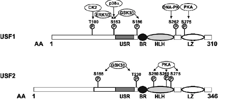
Figure 1. Functional domains and kinase sites of mouse USF1 and USF2………...5
Figure 2. Schematic of AID regulation………16
Figure 3. USF Knockdown in M12 cells……….33
Figure 4. Differential gene expression analysis of USF-depleted and irradiated M12 cells...35
Figure 5. Gene expression response to USF depletion and IR………36
Figure 6. Heat map of the clustering among the enriched Gene Ontology biological process terms annotated to the DEGs………...44
Figure 7. Loading control validation in USF-depleted and irradiated cells……….48
Figure 8. Quantitative PCR (qPCR) validation of microarray findings………...50
Figure 9. Representative sequence alignment of Vk region……….52
CHAPTER 1: Literature Review 1.1: DNA stress during B cell development
Humoral immunity is mediated by antibodies, the secreted form of the B cell antigen receptor or Immunoglobulin (Ig). Antibodies are multi-subunit protein complexes composed of two heavy (IgH) and two light (IgL) chains. Each Ig contains repeated domains (so-called Ig domains) of anti-parallel beta sheets linked by alpha helical loops. The enormous diversity of antigens that can be recognized by antibodies is generated early in B cell development through a program of somatic DNA rearrangements within the 5’ portion of each Ig gene. These rearrangements assemble the exon that encodes each Ig’s antigen binding domain from pools of subexonic variable (V), diversity (D) and joining (J) gene segments, and produce the capacity to recognize as many as 5x10¹³ different antigenic molecules1.
V(D)J recombination is initiated by the recombination activating gene 1 and 2 (or RAG1 and RAG2) proteins, which are only expressed in developing lymphocytes2. The RAG
proteins drive a stepwise assembly of the antigen binding exon, with IgH genes assembled by D-to-J, and then V-to-DJ joining, followed by IgL V-to-J joining (IgL genes lack D gene segments). For each pair of compatible gene segments, recombination begins when RAG1/2 complexes bind conserved recombination signal sequences (RSS) that flank each V, D and J coding segment, and nick the DNA at the RSS:coding junction, catalyzing a
transesterification reaction that breaks the DNA into a hairpinned coding end and blunt signal end3. The double-stranded breaks (DSBs) caused by RAG1/2 are repaired through the
Kinase (ATM), and Tumor Suppressor p53-binding Protein 1 (53BP1) to aggregate in the chromatin area surrounding DSB and create a “DNA damage repair focus”4. KU70/80
heterodimers, DNA-dependent Protein Kinase (DNA-PKcs), ARTEMIS, XRCC4-like Factor (XLF), and DNA Ligase 4 (LIG4) accumulate at this focus, stabilizing free DNA ends, recruiting additional DNA damage response (DDR) factors, and ligating the two blunt signal ends together (most typically as an extrachromosomal signal joint). Restoration of the chromosome by ligating the compatible coding junctions requires additional processing that dramatically increases antigen binding diversity5. This “junctional diversity” is generated
when RAG proteins at the coding ends, together with and DNA-PKcs again nick the DNA, opening the hairpinned coding ends, which may then be subjected to limited exonuclease activity, and/or templated and non-templated polymerization5. Deficiency of either NBS1,
y-H2AX, ATM, or 53BP1 leads to abnormalities in antigen receptor V(D)J recombination, while loss of KU80 and LIG4 causes a dramatic increase in DNA breaks and chromosomal translocations3,6,7.
After V(D)J recombination, B cells that complete development in the bone marrow are released into circulation as mature naïve B cells that express surface-bound Ig as their B cell receptor (BCR). Should the B cell bind its cognate antigen and become activated while trafficking through secondary lymphoid tissue, the Ig genes are again targeted for
diversification through the processes of somatic hypermutation (SHM) and class-switch recombination (CSR)8. During SHM, point mutations are introduced in the IgH and IgL
generation10. Mutations are introduced by Activation-Induced Cytidine Deaminase (or AID),
resulting in a C-to-T substitution. In mammals, CSR produces antibodies with different effector activities through DNA rearrangements that replace the 5’-most constant region exon of the IgH gene, Cμ and C, with either , or exons 11,12. Since the effector activities and
tissue localization activities of antibodies are encoded by the constant region exons, CSR alters antibody functionality without altering its antigen specificity.9. In CSR, DSBs occur
between palindromic switch (S) regions after a S-S synaptosome is formed. The S-S
synaptosome is created by AID driving C-to-A substitutions in the donor Sμ and an acceptor S, S or S region13. The combined DNA targeting activities of V(D)J recombination,
somatic hypermutation and class-switch recombination are absolutely essential for humoral immunity, and loss of either results in severe compromise of humoral immune efficacy. Each however also offers the potential for unintended harm to the host through tumorigenic mutations. Indeed, the vast majority of B cell lymphomas are linked to either AID- or RAG-mediated translocations.
1.2: Upstream Stimulatory Factors
Upstream Stimulatory Factor (USF) was first identified by the Sharp laboratory in 1985 as Major Late Transcription Factor (MLTF), a single 46 KDa polypeptide responsible for RNA Polymerase II activation of the adenovirus major late promoter14,1516. In a later
USF2a and USF2b, with USF2b having the potential to act as a dominant negative regulator of USF1/219, though the significance and tissue distribution of USF2b are unclear.
Both USF1 and USF2 are ubiquitously expressed, though concentrations vary between tissues20. USF1 and USF2 act as transcription factors by binding to canonical
CANNTG targets shared with other so-called E proteins (e.g. E47, E2A, MYC, MAX; all sharing a conserved bHLH “E protein” motif). As with other E proteins, USF binds DNA as a dimer, preferentially formed between USF1 and USF2, though both USF1 and USF2
homodimers are also active19. CANNTG E boxes are found in a wide array of genes
distributed throughout the genome. Chromatin immunoprecipitation (ChIP)-seq experiments suggest that USF proteins bind as many as 2500 different human genes21. Both contain a
highly-conserved USF-specific region (USR) in N-terminal region responsible for regulation of transcriptional activity18,22,23. However, USF1 and USF2 are not fully interchangeable, as
only USF2 can bind the pyrimidine-rich Inr elements in the core promoters of some targets24.
Likewise, studies in available model systems lacking either USF1 or USF2 (but not both) have linked USF2 specifically to prostate cancer, while USF1 appears to provide UV-response independent of USF225. The two USF proteins show significant preference for a
canonical CACGTG E box, though they will also bind E boxes with variations at the third and fourth nucleotides, or even noncanonical heptameric E boxes that preserve the
complexes bind competitively at select promoters. For example, expression of the CAD pyrimidine biosynthesis protein in dividing cells requires the binding of MYC:MAX heterodimers at the Cad gene promoter. In non-dividing cells, MYC:MAX is replaced by USF1:USF2 heterodimers, and expression of the Cad gene is repressed27. The widespread
nature of USF and MYC expression, together with their affinity for CACGTG binding sites suggests that competition between USF and MYC may occur at many of the USF target genes28.
1.3: USF as a tumor suppressor
The genetic lesion most associated with prostate cancer aggressiveness is localized to human Chromosome 1929. This Prostate Cancer Aggressiveness Locus (19q12-q13.1) is most
tightly linked to genes encoding Kallikrein 1 (KLK1) and Kallikrein 3 (KLK3) (or PSA), members of the kallikrein family of serine proteases, positioned at 19q13.3330. The Usf2 gene
sits at 19q13.11 and is also frequently lost in prostate cancers bearing 19q deletions31. PC3
human prostate cancer cell lines show loss of USF2 activity despite persistence of the Usf2
gene32, suggesting that linkage of Usf2 deletion and prostate cancer may underestimate
USF2’s prostate cancer association. Indeed, PC3 cells engineered to overexpress a recombinant USF2 transgene showed reduced tumorigenicity in xenograft transplants20.
Although the scope of USF2’s gene regulatory involvement in prostate biology has not been studied, USF2 assembles with Androgen Receptor (AR) to regulate transcription of AR target genes in prostate epithelial cells33. USF2 knock-out (KO) mice also support a role for USF2
transformation in rat embryonic fibroblasts (REFs) induced by the adenovirus onco-protein E1A22.
Though not as clearly demonstrated as USF2, USF1 also exhibits tumor suppressor activity. Both USF proteins can prevent RAS- and MYC-dependent transformation of REFs
22, and loss of both USF1 and USF2 transcriptional activities was shown in a panel of human
breast cancer cell lines (as noted in PC3 cells, this loss of USF transactivating activity was seen despite persistent expression of both USF proteins)34. USF1’s role in tumor suppression
is perhaps best understood in skin cells that have been exposed to UV radiation. In mouse skin cells treated with UV light, USF1 appears to bind and stabilize P5335, regulate G1/S and
G2/M transitions through transcriptional regulation of multiple cyclin and cyclin dependent kinase genes 36, and induce expression of pigmentation and nucleotide excision repair (NER)
factors37.
In addition to the evidence that links USF1 or USF2 to skin or prostate cancer, respectively, both USF proteins have been linked to a number of other cancer types. An analysis of human breast cancer cell lines found that while USF1 and USF2 protein levels were similar to those seen in untransformed and primary breast tissues, 75% of the breast cancer lines lacked detectable USF2 transactivating activity, and half also lacked USF1 activity34. Similar studies have linked changes in USF expression or activity to a number of
subcellular staining in ovarian tumors found that USF1 was mislocalized to a perinuclear compartment that appeared Golgi-related. The authors speculate that mislocalization of USF1 to the Golgi could account for the protein’s appearance in cancer serum samples39.
Though no attempt has been made to systematically define USF’s impact on genome-wide transcription, a number of genes critical to growth regulation have been defined as USF target genes. For example, the Tp53, Brca2 and Apc tumor suppressor genes are all direct targets of USF1/240. Whereas USF activates expression of tumor suppressor genes like Apc
and Brca241, they repress expression of human telomerase reverse transcriptase (hTert)42.
Deletion of USF leads to elevated hTERT in oral cancer cell models43, mimicking the
MYC-dependent upregulation of hTERT that is seen in over 85% of tumor cells44. USF2 also
represses expression of the cell cycle kinase CDK436. As with CAD and perhaps hTERT,
USF’s repression of the Cdk4 gene opposes activation by c-MYC36. Together, the repressive
role USF plays in regulating growth promoting genes like hTert and Cdk4, and the activating role it plays in regulating tumor suppressor genes like Brca2 result in a largely
antiproliferative function.
Paradoxically, whereas loss of USF activity is associated with prostate, breast and ovarian cancers, one study suggested that USF2 overexpression induces hyperproliferation in human dysplastic lung epithelium and non-small cell lung cancer cell lines45, though no
1.4 USF as a stress response factor
Stress-induced activation of USF is not limited to the UV radiation response. In cell models of inflammation, USF1 has been shown to counteract pro-inflammatory NFB
signals through induction of the gene for tumor necrosis factor alpha-induced protein-3 (TNFAPI3 or A20)46. Whereas USF1 normally suppresses expression of plasminogen
activator inhibitor 1 (PAI-1), it stimulates PAI-1 expression when activated at wound sites47.
USF1 is activated in response to fasting, and in turn regulates the genes for insulin48 and fatty
acid synthetase49, suggesting a potential linkage with Type II diabetes50.
The ability of USF to transduce environmental signals extends beyond stressor like DNA damage, tissue lesion or fasting. USF is essential for early embryonic, neuronal, reproductive and hematopoietic development programs and regulates a wide array of genes involved in the immune response. Attempts to generate mouse genetic models of USF deletion first confirmed its role in neuronal development. USF2-deficient mice in particular showed dramatically shortened male lifespan, reduced viability, growth and fertility.
Moreover, both USF1-/- and USF2-/- mice were prone to spontaneous epileptic seizures51. In
contrast, homozygous USF1-/-/USF2-/- mice died during early embryogenesis, as did mice
USF1 has been shown to promote transcription of the IgH and Igλ2 genes53, the C4
complement protein and 2 microglobulin54–56. USF1/2 heterodimers regulate expression of
the J chain and polymeric Ig receptor in plasma cells57. As with Ig expression in B cells, USF
regulates expression of multiple T cell receptor genes in developing T cells. Whereas it drives transcriptional activation of the D2 gene segment in Tcrd, our laboratory found that it
represses germline transcription of the Tcrb D2 gene segment58. During Tcrb assembly,
DJβ1 joints are preferentially formed over more distal DJβ2 joints. We found that USF occupies the D2 upstream promoter prior to recombination, leaving the D2 RSSs
untranscribed and therefore less accessible to V(D)J recombinase at the outset of gene assembly. However, as DJ1 recombination progresses, transient DNA breaks trigger
activation of DNA-PKcs, which phosphorylates USF1, leading to its displacement from the 5’Dβ2 promoter. In the absence of USF, the promoter is activated and D2 rearrangements
begin to accumulate26. This temporal delay in DJ2 assembly ensures that V gene segments
initially target the upstream DJ1 joint, allowing DJ2 to serve as a target for secondary
V-to-DJ rearrangements should V-V-to-DJ1 joints be non-functional due to frameshift.
1.5 USF regulation by phosphorylation
Given their ubiquitous and largely stable expression, the ability of USF1 and USF2 to respond to stressors suggests that function of the USF proteins is regulated
hypoxic cells60. However, the significance of such regulatory schemes has not been extended.
More intriguingly, two independent studies have shown that nuclear localization of USF2 declines in activated mast cells and in hepatocytes of infants suffering from biliary atresia61,62, mirroring the loss of nuclear USF1 seen in ovarian cancer63 and the lack of
detectable USF activity in breast cancer cell models34. The mechanisms that regulated altered
USF nuclear translocation in such stress conditions are unknown.
The diverse pathways by which USF1 and USF2 are phosphorylated, and the functional consequence of their phosphorylation are better understood. For example, DNA-PK phosphorylates USF1 at ser262 in the b-HLH-LZ domain, triggering subsequent lys237 acetylation and USF1 activation64. We have shown in lymphocyte cell lines exposed to IR,
that induction of DNA-PK results in USF1 displacement from the Tcrb 5’D2 promoter and
loss of promoter repression26. Conversely, DNA-PK induced in fasting adipocytes resulted in
enhanced USF1 binding at the fatty acid synthase (FAS) promoter and promoter activation64.
Whereas IR activates DNA-PK to alter USF1, UV light activates P38-MAP kinase, which phosphorylates USF1 at thr153. Similar to IR-mediated activation, phosphorylation at thr153 leads to lysine acetylation (in this case, lys199) and altered DNA binding affinity65. In
trigeminal ganglion neurons, USF1 transactivation is regulated by (MEK)/ERK MAP kinase-dependent phosphorylation at thr15366.
In cardiac muscle, USF1 is phosphorylated by PKC, which increases binding at the cardiac α-myosin heavy chain promoter67, by CDK2, which regulates multiple stages of the
binding activity, CDK2-phosphorylated c-MYC showed reduced DNA binding69,70. USF1 is
also phosphorylated by messenger-independent CK2 in pancreatic insulinoma cell lines, which regulates USF1’s heterodimerization and transactivation69. Both USF1 and USF2
contain putative PKA phosphorylation sites (two for USF1, three for USF2)71 that are linked
to USF’s transactivation potential7273. Likewise, both proteins are phosphorylated by
Glycogen synthase kinase-3 (GSK3), which regulates PI3K/Akt and Wnt/-catenin
pathways, and is implicated in a number of pathologies including type-2 diabetes and
cancer74. Phosphorylation of USF1 (again, at thr153) was critical for transactivation of genes
that promote apoptosis and cell cycle arrest75. In silico modeling of GSK3-phosphorylated
USF2 suggests adoption of a more open conformation that would facilitate DNA binding and transactivation potential. Functional studies using USF2 variants that mimic GSK3
phosphorylated forms found no impact on cell proliferation, but instead found increased
levels of cell migration31, suggesting that GSK3 regulation of USF proteins may influence
their role in tumorigenesis.
1.5: Activation-induced cytidine deaminase
Activation-induced cytidine deaminase (AID) is responsible for initiating both SHM and CSR9. In both cases, AID deaminates cytosines found on single-stranded DNA (ssDNA)
during transcription8, and is facilitated by AID’s interaction with RNA Pol II (RNAP II)76,77.
During SHM, AID preferentially targets the hotspot sequence RGYW/WRCY78, with a focus
on WRC (A/T, A/G, C) motifs in IgH and IgL genes79. SHM typically starts approximately
mutations occurring up to 1-2 kb downstream of these TSSs80. Mutations that occur within
the V region promoter can lead to a decrease in SHM by preventing transcription initiation80.
Mismatch repair (MMR) and base excision repair (BER) factors, like MSH2/MSH6 and UNG, help repair U*G mismatches that result from deaminating cytidine to uridine, either restoring the C*G basepair, or producing a new T*A basepair. Deficiencies in these genes are evidenced by the presence of unrepaired uracil bases within the Ig genes, or the accumulation of AID-mediated C*G to T*A transition mutations in genes other than IgH and IgL81. Such
AID off-target genes include Bcl6, h2afx and c-myc, all three of which are induced during germinal center activation78.
Although MMR and BER are typically error-free DNA repair mechanisms, their recruitment to AID-mediated DNA lesions, together with DNA Exonuclease 1 or AP
endonuclease 1, respectively, result in excision of a single-stranded or double-stranded patch of DNA containing the mismatch. Single-stranded ends are repaired by an error-prone DNA polymerase like Pol or Pol, while DSBs are first bound by Rad52/51 complexes, resected,
and then repaired by Pol82. The error-prone nature of SHM lesion repair is essential for
hypermutation of the Ig V sequences.
During CSR, synaptosomes between donor Sμ and a downstream acceptor Switch region are formed in part due to AID deaminating cytidines in the targeted S, S or Sε
region83. Switch regions are highly enriched in AID’s preferred WRC target sequences79.
deletes the sequence between S and target S breaks. Simultaneous targeting of AID to sites
outside of IgH can also lead to translocations like those between S and cMyc that result in
Burkitt’s Lymphoma78. CSR is essential for immune competence. The rarity of Burkitt’s-like
tumorigenic translocations is again facilitated by the affinity of AID for RNAP II and by its preference for the G-rich R-loops that the highly repetitive Switch regions form during their transcription83,84, causing RNAP II stalling and increased activating histone modifications
and chromatin accessibility85,86.
AID is expressed at low levels in most cells including embryonic cells, and
participates in pathways unrelated to SHM and CSR. Specifically, AID was found to promote DNA demethylation through the deamination of meC to thymidine87 and repair of the
subsequent mismatch though MMR and BER repair pathways88–90. Some have speculated
that this pathway may play a role in pluripotent tissues87. AID expression in transitional B
cells may also mediate tolerance91,92. However, the significance of AID activity in processes
beyond SHM and CSR, and the mechanisms by which they are mediated, remain highly speculative.
1.6: Regulation of AID
To safeguard against inappropriate alteration of the genome, AID is regulated on many levels, including expression, subcellular localization and activation. AID transcription is induced by a variety of cytokines including: T cell-derived cytokines like IL-4 and CD40L, T-cell independent cyokines like BAFF and APRIL, female sex hormones, and TLR
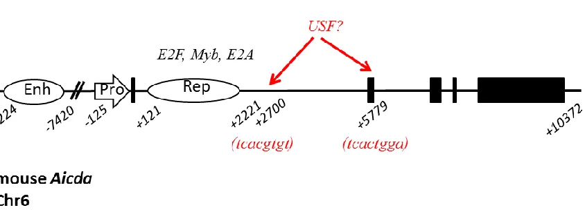
Aicda promoter and enhancer12,38,93–95. Transcription factors implicated in Aicda transcription
include NF-κB, C/EBP, Smad3/4, Sp1, Oct/Hox and STAT696. The first 2 kb downstream of
the Aicda transcription start site contains repressor elements critical to limiting expression in the absence of germinal center activation signaling. Transcription factors implicated in Aicda
repression include: PAX5, c-Myb, E2F, NFB and E47/E2-297. Stability of the processed
Aicda mRNA is further repressed by microRNAs miR-155 and miR-181b 98–101; in acute B
cell lymphoma lacking miR-155 increased AID protein levels and nuclear translocations lead to aggressive CSR85,98–100.
The AID protein is stabilized through several mechanisms. In the cytoplasm, where 90% of AID is found before GC induction102, AID is stabilized by heat shock protein 90
(HSP90)103, while eEF1A prevents AID from entering the nucleus104. Recruitment of
intranuclear AID to its ssDNA target is further regulated by a series of co-factors including mechanism for the regulation of AID activity is through the recruitment of ssDNA binding adaptor 14-3-3 protein105, RNA-processing and/or splicing factors, polypyrimidine-tract
binding protein (PTBP2)106, RNAPII stalling cofactor (Spt5)107, replication protein A
(RPA)108 and RNA exosomes109. RPA stabilizes AID’s interaction with ssDNA108 at
immunoglobin variable genes specifically10, while ar1-α provides a similar function at sites
Casellas et al, 2016
1.7: Pathologies associated with AID
Deregulated expression of AID in B cells is associated with chromatid breaks and translocations in nearly every chromosome111, and with 95% of lymphomas having a B cell
origin112, a role for AID in B cell pathogenesis is of particular interest. Translocations occur
when 1) there are two pairs of DSBs, 2) there is close proximity between the broken ends, and 3) there is joining of the heterologous DNA ends5. They usually occur at fragile sites,
which are large genomic regions prone to replicative stress113; fragile sites containing CpG
nucleotides may be more susceptible to AID-independent replication fork collapse114,
although it has also been proposed that AID expression in early B cells targets CpG sites, resulting in the generation of substrates for RAG- and ARTEMIS-dependent nicking115.
There are many translocations associated with AID from BCR:ATM116 to Myc:IgH. AID
overexpression is strongly correlated with oncogenesis, irrespective of cell type, and AID-mediated translocations have been identified in prostate cancer cells117. Indeed, AID
disregulation has been implicated in diverse pathologies including blast crisis progression in chronic myeloid leukemia101, prostate malignancies117, and gastric tumors118.
The most common oncogenic lesion associated with AID is the Myc:IgH
translocation found in Burkitt’s lymphoma. In this disease, a reciprocal translocation occurs between the coding region of the c-Myc proto-oncogene and the Igh V(D)J coding region119,
placing Myc expression under control of the strongly activated IgH 3’ enhancer120. The Myc
and Igh genes frequently colocalize in the nuclei of activated B cells, but not in the nuclei of resting B cells or other cell types121,85; this may imply that they share transcriptional
seem to be particularly susceptible to AID-dependent translocation due to the R-loops and other unstable structures, like G-quadruplexes, that form during transcription of the highly repetitive switch regions123. Multiple studies have confirmed that these translocations are
AID-dependent. First, overexpression of AID has been shown to drive DNA damage at the exon1-intron1 junction of Myc124. Second, translocations in B cell lines do not occur if UNG
is absent, indicating that the U:G mismatches created by AID are necessary125. Third, Balb/c
mice injected with IL-6 generated Myc-IgH rearrangements only when AID was present119,126,127.
Translocations are not the only aberration found due to AID expression. In fact, many cancers caused by improper AID expression are characterized by point mutations in both oncogenes and tumor suppressor genes128. Some genes associated with B cell tumorigenesis,
like Myc, Pim1, Pax5, Ocab (Pou2af1), H2afx, Rhoh are able to escape AID-induced mutations by mismatch and base excision repair78. When these repair mechanisms are
present, the mutation rate in off-target genes like c-Myc often doesn’t exceed background levels78. When tumor suppressor genes like Tp53 are absent, AID can induce rapid onset
mature B cell lymphoma111; p53 functionality can be impaired through normal processes like
replication errors, or through the induction of AID expression, which causes mutations in
Tp53 in human gastric cells118 and which is strongly implicated in Tp53 mutation in human
breast cancer cells129. Bcl6 is the most frequently mutated non-Ig gene in B cells, and is a
gene implicated in diffuse large B cell lymphoma78. Its mutation rate is ascribed to AID, as
frequency than that in AID-expressing cells78. Further, AID accounted for mutations in 25%
of 188 genes randomly surveyed in a germinal center B cell genome, though such off-target genes showed mutation rated of 100-fold lower than the Ig loci78.
1.8: Introduction and Objectives of the Thesis Project
Cancer arises from genome instability that arises after damaged DNA is either not repaired or improperly repaired. To avoid genome instability, cells have elaborate programs that are activated in response to damage130. The substitution error rate of human DNA
polymerase ε is around 10-8 errors per base pair, the vast majority of which are then repaired
by ubiquitous DNA repair enzymes. DNA can similarly be damaged during the process of recombination. But again, that damage is aggressively repaired, and DNA recombination is limited to meiosis in gametes and antigen receptor gene assembly in developing
lymphocytes131. More commonly, cancer-causing mutations arise after DNA is damaged by
As noted earlier, the ubiquitously expressed USF1 and USF2 stress response proteins were among the first mammalian transcription factors identified based on their involvement in regulating activity of the adenovirus major late promoter 16. They have long been
appreciated as tumor suppressors, though evidence supporting such a role is limited. Chiefly, USF’s tumor suppressor linkages include: (a) their activity being lost in a variety of cancers, including breast23, liver 132, colorectal40, prostate20, and oral cancers43, (b) the ability of USF2
overexpression to reduce in vivo tumorigenicity of mouse prostate cancer xenografts20, (c)
their binding activity at the promoters of other tumor suppressor genes including Apc, Brca2 and Tp5340, (d) their ability to repress expression of TERT133, and (e) their antagonism of the
transforming function of MYC134.
Much of the deciphering the molecular pathways that link USF to cancer has been conducted in the skin of USF1-deficient mice. These studies demonstrate that upon UV exposure USF1 is activated in keratinocytes and melanocytes by p38 MAPK, and drives expression of genes involved in nucleotide excision repair (NER)135 and melanin deposition
as part of the tanning response136. USF1 also appears to stabilize P53 protein levels through
nuclear protein:protein interaction35. Unfortunately, all of the studies examining USF1’s role
generated either during V(D)J recombination or by experimental exposure of cultured lymphocyte cell lines to sublethal doses of ionizing radiation26. In our lymphocyte cell
models however, we found that exposure to IR or prolonged V(D)J recombinase expression resulted in USF activation that persisted long after nonhomologous end-joining (NHEJ) repair programs had attempted to resolve DNA lesions26, suggesting that USF might be an
ideal candidate for directing the long-term transcriptional changes that guide cell recovery and limit genomic instability after DNA stress. To test its candidacy however, we would need to improve on existing genetic models of USF deficiency.
The two USF proteins are basic helix-loop-helix (bHLH) transcription factors and function primarily as USF1/USF2 heterodimers, binding E boxes at over 2500 target genes21.
As seen in the example of UV irradiation, stress responsiveness of USF is controlled through differential phosphorylation of USF1 by protein kinases including p38 (as well as ERK MAPK), DNA-PK, GSK3, CK2 and PKA64 (see Figure 1). Phosphorylation by these
protein kinases regulates USF1/2 heterodimerization, DNA binding, and transactivation potential of USF proteins137. Attempts to define the role of USF in DDR and cancer
development have been limited by difficulty in generating appropriate genetic model systems. Although USF1 knock-out mice are prone to seizures51, and USF2-deficient mice
are underweight and have reduced male sterility138, they each retain a significant degree of
early in embryogenesis51, no animal model deficient for USF activity has been developed.
Consequently, no profile of USF’s genomic stress-response yet exists.
Germinal center B cells are activated in response to antigen presentation, and each undergo two separate events that induced dsDNA breaks in their Ig heavy chain genes. These events, somatic hypermutation and class-switch recombination, are both essential for B cell function, but are also strongly linked to lymphomagenesis, illustrating the need for B cells to discriminate between DNA damage that is ultimately beneficial or dangerous. Building on our finding that USF activity is responsive to DNA breaks in cultured lymphocytes, we modeled USF deficiency using RNAi to simultaneously deplete both USF1 and USF2 from the M12 mouse germinal center B cell line. Cells were transduced with either a scrambled shRNA control lentivirus (sshRNA) or a mixture of USF1 and USF2 shRNA viruses, stably transduced clones were isolated based on their resistance to virally-encoded puromycin, and the transcriptomic response to USF depletion and ionizing radiation was measured using Affymetrix mouse whole transcript arrays.
Microarray analysis revealed 765 differentially expressed genes (≥1.50-fold change, 0.05
RT-qPCR for a number of immune- and cancer-related genes including Aicda, Blk, Blnk, and multiple NFB family members, none of which had previously been identified as USF target
genes. The value of using a genetic system that simultaneously targets both USF proteins was further supported by expression analyses in Usf1-/- thymus and USF single-gene KD cells, both of which showed trends in differential gene expression similar to but of significantly less magnitude than that seen in the USF1/USF2 double KD cells. These findings provide the first evidence that USF1 and USF2 collaborate to play a novel role in regulating long-term response to DNA damage and lymphocyte activity, and also illustrate the significance of investigating USF’s regulatory role using models that overcome the functional overlap
CHAPTER 2: Materials & Methods 2.1 Cell culture
The murine M12 germinal center B cell line has been previously described141. All
M12 cell lines used in this study were cultured at 37oC/5% CO
2 in RPMI 1640 media
supplemented with 10% fetal calf serum, 2 mM L-glutamine, 0.01% penicillin/streptomycin, and 50 mM -mercaptoethanol.
2.2 RNA interference and transfection
For depletion of USF2 in M12 cells, a validated Mission USF2 shRNA
(TRCN0000071575) in pLKO.1 backbone was purchased and prepared as described by the provider (Sigma). Two independent USF1 targeting shRNAs were designed using the BROAD Institute Genetic Perturbation Platform. Duplexed shRNA USF1shR1 (sense: 5’- CCGGCAGAAGTTAAGATGCGTACCTCTCGAGAGGTACGCATCTTAACTTCTGTTT TTG -3’, antisense:
5’-AATTCAAAAACAGAAGTTAAGATGCGTACCTCTCGAGAGGTACGCATCTTAACT TCTG-3’) and USF1 shR2 (sense:
5’-CCGGGAGGGCTCAACATAACGAAGTCTCGAGACTTCGTTATGTTGAGCCCTCTTT TTG-3’, antisense:
USF1shR1, USF2shR2 and sshRNA plasmids were independently transfected into 293T cells, together with third generation lentiviral packaging plasmids (kindly provided by Dr. Frank Scholle) using FugeneHD (Roche). Briefly, for shRNA plasmid transfections, 1 x 10⁷ 293T cells were seeded per 10 ml DMEM complete medium (supplemented with 10%
fetal calf serum, 2 mM L-glutamine, 0.01% penicillin/streptomycin, and 50 mM
-mercaptoethanol) and allowed to attach for 4 hours. USF1shR1, USF1shR2 and USF2shR (640 ng each) or sshRNA (2.56 g) was mixed together with pFG12 (640 ng), pMDLG (1.6
g), pREV (1.6 g) and pCMV-G (1.0 g) in 870 l serum free DMEM and 56 l
FugeneHD, vortexed briefly, and allowed to complex at room temperature for 10 minutes. DNA mixture was added dropwise to plated cells. The plate was swirled gently to mix, and returned to the incubator overnight. After overnight transfection, the culture medium was replaced with complete DMEM supplemented with 1% BSA. After 24 hours, the culture supernatant was harvested and stored at 4°C, while the plate was replenished with
BSA-supplemented medium and allowed to grow an additional 8 hours, at which point the culture supernatant was harvested, combined with the initial harvest and filtered (0.45 m).
The day before transduction, M12 cells were split to ensure growth. For lentiviral
transductions, 105 M12 cells were seeded in 500 l complete RPMI in each well of a 24-well
tissue culture plate. Filtered viral supernatant (500 l) and polybrene (8 g/ml final
concentration). Individual clones were manually isolated and screened for USF1 and USF2 expression by Western blotting.
2.3 Western blotting
For protein analysis, freshly growing M12 clones (1-5 x 10⁶ cells) were washed in ice-cold PBS, and nuclear or whole cell protein lysates were generated. For nuclear fraction isolates, cells were lysed in NP-40 Buffer (500 l of 10 mM HEPES pH 7.9, 1.5 mM MgCl2,
10 mM KCl, 0.5 mM DTT, 0.1% NP-40) supplemented with PMSF and HALT protease and phosphatase inhibitor cocktail (Thermo Scientific) for 2 min on ice. After lysis, cells were pelleted in a microcentrifuge (2 min @ 12,000 RPM @ 4oC), the nuclear pellet was
resuspended in Nuclear Lysis Buffer (100 l 10 mM HEPES pH 7.9, 25% glycerol, 0.42 M
NaCl, 1.5 mM MgCl2, 0.2 mM EDTA, 0.5 mM DTT) supplemented with PMSF and HALT protease and phosphatase inhibitor cocktail, and incubated 30 min on ice with frequent manual agitation. Nuclear lysate was recovered by microcentrifugation (20 min @ 12,000 RPM @ 4oC) and stored at -80oC. Whole cell lysates were generated by lysing pelleted cells in RIPA (1 ml of 150 mM NaCl, 1% NP-40, 0.5% NaDeoxycholate, 0.1% SDS, 50 mM Tris-HCl 8.0) at 4oC for 30 min with frequent manual agitation, and microcentrifuged at 12,000
RMP for 20 min at 4oC. Protein concentration in the recovered supernatant was assessed by Coomassie Protein Assay (Thermo Scientific) according to the manufacturer’s instructions.
For Western analysis, nuclear or whole cell lysates (15 g each) were electrophoresed
TransBlot Turbo device (Bio-Rad). The membrane was treated with 1X TBS 3% Casein Blocking Buffer (Bio-Rad) one hour at room temperature, and then incubated with rabbit anti-mouse polyclonal antibodies (Santa Cruz Biotechnology) against USF1 (sc-229), USF2 (sc-862), Sp1 (sc-59) or -actin (sc-130656) at a 1:5000 dilution in Blocking Buffer
overnight at 4°C. After washing 5X in TBST, the membrane was incubated with goat anti-rabbit HRP secondary antibody (Pierce, 32260) for 1hr at RT, rewashed (5X in TBST) before ECL Prime chemiluminescent detection (GE Heathcare) and autoradiographic exposure.
2.4 Ionizing radiation treatment
2.5 RNA extraction
Total cellular RNA was extracted from 1-5 x 10⁶ cells using RNeasy Plus (Qiagen) according to the manufacturer’s protocol. RNA levels were quantified using a NanoDrop
1000 spectrophotometer (Thermo Scientific), and stored at -80oC.
2.6 Affymetrix Microarray Hybridization
RNA quality was assessed using an AATI Fragment Analyzer (Advanced Analytical Technologies, Ankeny, IA). RNA samples (3-5 ul) are heated at 70oC for two minutes, placed on ice, and 2 ul of the denatured RNA is added to 22 ul of Normal Sensitivity RNA Marker-Diluent (Advanced Analytical Technologies, Ankeny, IA) in a 96 well plate. The 96 well plate is loaded on to the instrument and the RNA quality is assessed using the
PROSizeTM analytical software (Advanced Analytical Technologies, Ankeny, IA). Fragment
Analyzer provides a RNA Quality Number (RQN) to assesses the quality or integrity of total RNA on a scale of 1–10 (one is poor quality, 10 is excellent quality). The average RQN for the samples used in this experiment was 9.4.
and Stain Kit and user manual following protocol FS450-0001. Arrays were scanned in an Affymetrix Scanner 3000 and data was obtained using the GeneChip® Command Console software.
2.7 Bioinformatic Analysis of Gene Expression
The CEL files were preprocessed by computing the robust multiarray average (RMA) of background adjusted, quantile normalized and log2 transformed perfect match (PM) pixel intensity values (PMID: 12582260, PMID: 12925520). Microarray analysis was performed using a three-way (cell line, treatment and time) analysis of variance (ANOVA) model with a three-way interaction term in Partek Genomics Suite v6.6 software. For each cell line,
differentially expressed genes (DEGs) were detected by contrasting treatment group means versus the mean of the untreated controls and using a false discovery rate (FDR) < 0.05142 with an absolute fold change > 1.5. The DEGs were enriched for biological pathways
(FDR < 0.05) using gene ontology (GO) biological processes and the Kyoto Encyclopedia of Genes and Genomes (KEGG) knowledge base (PMID: 10592173) in the Database for Annotation, Visualization, and Integrated Discovery (DAVID) v6.7 Bioinformatics resource (PMID: 19131956, PMID: 19033363)143,144.
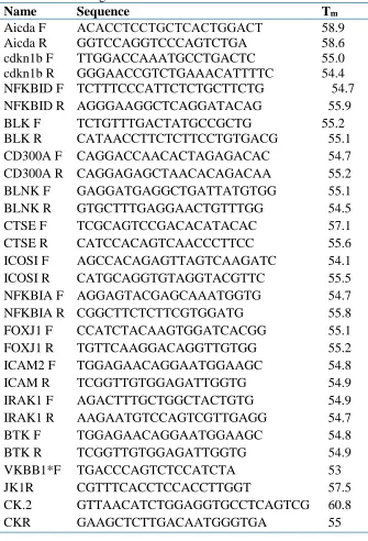
2.8 Quantitative and reverse transcriptase PCR (qPCR and RT-PCR)
For RT-qPCR analyses, each RNA sample (1 g) was reverse transcribed using
indicated primer pairs (Table 1). PCR reaction mixtures (20 l SensiMix Plus; Bioline) were
amplified (94oC, 20 sec; 60oC, 30 sec; 72oC, 30 sec) for 40 cycles, followed by melt-curve analysis using an iQ5 iCycler (Bio-Rad). Quantitation of target gene levels was achieved by
2-CT analysis145 normalizing gene expression levels in experimental samples to matched
Table 1. List of Oligonucleotides
Name Sequence Tm
Aicda F ΑCACCTCCTGCTCACTGGACT 58.9
Aicda R GGTCCAGGTCCCAGTCTGA 58.6
cdkn1b F TTGGACCAAATGCCTGACTC 55.0
cdkn1b R GGGAACCGTCTGAAACATTTTC 54.4
NFKBID F TCTTTCCCATTCTCTGCTTCTG 54.7
NFKBID R AGGGAAGGCTCAGGATACAG 55.9
BLK F TCTGTTTGACTATGCCGCTG 55.2
BLK R CATAACCTTCTCTTCCTGTGACG 55.1
CD300A F CAGGACCAACACTAGAGACAC 54.7
CD300A R CAGGAGAGCTAACACAGACAA 55.2
BLNK F GAGGATGAGGCTGATTATGTGG 55.1
BLNK R GTGCTTTGAGGAACTGTTTGG 54.5
CTSE F TCGCAGTCCGACACATACAC 57.1
CTSE R CATCCACAGTCAACCCTTCC 55.6
ICOSI F AGCCACAGAGTTAGTCAAGATC 54.1
ICOSI R CATGCAGGTGTAGGTACGTTC 55.5
NFKBIA F AGGAGTACGAGCAAATGGTG 54.7
NFKBIA R CGGCTTCTCTTCGTGGATG 55.8
FOXJ1 F CCATCTACAAGTGGATCACGG 55.1
FOXJ1 R TGTTCAAGGACAGGTTGTGG 55.2
ICAM2 F TGGAGAACAGGAATGGAAGC 54.8
ICAM R TCGGTTGTGGAGATTGGTG 54.9
IRAK1 F AGACTTTGCTGGCTACTGTG 54.9
IRAK1 R AAGAATGTCCAGTCGTTGAGG 54.7
BTK F TGGAGAACAGGAATGGAAGC 54.8
BTK R TCGGTTGTGGAGATTGGTG 54.9
VKBB1*F TGACCCAGTCTCCATCTA 53
JK1R CGTTTCACCTCCACCTTGGT 57.5
CK.2 GTTAACATCTGGAGGTGCCTCAGTCG 60.8
CHAPTER 3: Results 3.1: USF depletion
USF1 phosphorylation by P38 MAPK facilitates protective transcriptional responses to DNA damage elicited when keratinocytes and melanocytes are exposed to UV
radiation137,146. We have similarly shown that DSBs elicited by IR treatment of lymphocyte
cell lines trigger DNA-PK-dependent phosphorylation of USF1 that also regulates its trans -activation capacity26. An appropriate mouse model of USF deficiency does not yet exist.
While single-gene Usf1 or Usf2 knockout mice have been developed, the ability of the remaining USF protein to partially compensate for deletion of the other has prevented the mapping of USF target genes and pathways. At the same time, mice deficient for both Usf
genes or constitutively expressing a dominant negative USF transgene fail to develop beyond early embryogenesis. In light of USF’s ubiquitous expression, its involvement in responses to a wide array of stressor including DNA damage, and its function as a tumor
suppressor20,35,39,147, a better understanding of how USF regulates stress response and cancer prevention is urgently needed.
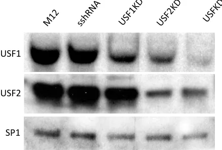
differences in cell viability or growth were observed among the populations of transduced cells. Puromycin-resistant transduced clones expressing short hairpinned RNAs (shRNAs) targeting either Usf1 or Usf2 alone, or in combination, or expressing a scrambled shRNA (sshRNA) control were isolated and screened for USF depletion by Western blotting of nuclear protein extracts (Fig. 3). Previous studies in USF2-/- mice showed that loss of USF2 led to reduced USF1 expression51. Consistent with this finding, we observed significantly
diminished USF1 protein levels in cells transduced with shRNAs targeting either Usf1 or
Usf2 (lanes 3 and 4), and still further USF1 reduction in cells transduced with shRNAs targeting both Usf genes (lane 5). In contrast, depletion of USF2 protein expression was less extensive and was only observed in the presence of Usf2 shRNAs.
USF1
USF2
SP1
3.2: Transcriptomic analysis of USF depletion
To test the functional significance of USF depletion on the longterm cellular response to DNA damage, we exposed USFKD and sshRNA control lines (n = 3) to a single sublethal (5 Gy) dose of ionizing radiation. Total RNA from untreated cells and cells allowed to recover from IR treatment for one or seven days was screened on Affymetrix GeneChip WT arrays to investigate the impact of genotoxic stress on the transcriptome (Fig. 4). We
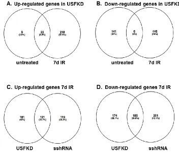
identified 9289 differentially expressed gene probes showing >1.5-fold change (<0.05 FDR) between knockdown and/or treated samples and untreated sshRNA controls (Fig. 4A). ChIP-chip studies suggest that USF proteins bind a wide array of sites across the genome, primarily at the promoter regions of transcriptionally active genes21. However, USF depletion had minimal impact on the pattern of gene expression in unirradiated cells. Untreated sshRNA and USFKD transcription patterns co-segregated in hierarchical Spearman and Ward
clustering analyses (Fig. 4B). Indeed, around 10% of the total DEG probes identified showed greater than 1.5-fold change in gene expression in the USF knockdown cells, corresponding to 31 genes with increased expression and 141 genes with decreased expression when compared with untreated sshRNA controls (Fig. 5A).
During the first day of recovery, the majority of transcriptional responses to IR are P53-dependent149. As expected, the absence of P53 activity in M12 resulted in only 360 DEG probes meeting the 1.5-fold change and <0.05 FDR thresholds of significance, and did not include genes previously linked to P53 induction (Fig. 4A). Interestingly, no DEG probes met the thresholds of significance in USFKD cells one day post-IR when compared with untreated USFKD cells, suggesting that USF is dispensable for the near-term transcriptional response to DNA damage. In contrast, USF-dependent responses were much more evident seven days post-IR. When normalized to untreated sshRNA controls, both sshRNA and USFKD cells yielded very similar numbers of DEG probes with >1.5-fold change in
expression (Fig. 4A, 5054 and 5035, respectively). However, analysis of the genes identified by these probes showed a markedly different pattern of gene expression seven days post-IR in USF knockdown cells than in sshRNA cells (Fig. 5B). Only half the 240 genes
upregulated in the control cells seven days post-IR, were also significantly upregulated in USF-depleted cells, along with an additional 181 genes. Likewise, of the 446 genes down-regulated in sshRNA cells seven days after IR, only 185 were similarly down-down-regulated in the knockdown cells, along with an additional 174 genes. The significance of USF’s involvement in such longer-term changes in gene expression is further underscored when the gene
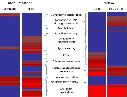
3.3: Gene Ontology clustering analysis
Clustering analyses identified 152 distinct Gene Ontology terms associated with DEGs in this study, grouped into 12 clusters of related biological function (Fig. 6) that were differentially enriched among experimental samples (Table 5). Irrespective of treatment, DEGs associated with MHC II-dependent antigen processing and presentation (e.g.:
GO:0002478, GO:0002495, GO: 0002504, GO: 0019886), antigen receptor production (e.g.: GO:0002377, GO:0002460, GO:0016445), lymphocyte differentiation (GO:0045580,
GO:0045619, GO:0045621) and the activation of an immune response (e.g.: GO:0002250, GO:0002757, GO:0001817, GO:0006955, GO:0019724, GO:0050863, GO:0051251, GO:0002253, GO:0050864, GO:0050865, GO:0051249) were enriched in USF-depleted cells. In contrast, DEGs associated with proliferation of immune cells (GO:0030888, GO:0032944, GO:0050670, GO:0070663) and protein tyrosine phosphorylation
Gene Ontology terms associated with response to ionizing radiation and DNA damage (e.g.: GO:0009314, GO:0009628, GO:0010212, GO:0006302, GO:0008630, GO:0042770), DSB repair (GO:0006302), chromatin regulation (e.g.: GO:0000070, GO:0006325, GO:0030261) and apoptosis (GO:0010941, GO:0042981, GO:0043067), as well as nucleic acid processing and transport and regulation of gene expression (e.g.: GO:0040029, GO:0006350, GO:0006397, GO:0032259, GO:0051169, GO:0051052) were enriched in 7 day post-IR sshRNA controls, but were absent from IR-treated USFKD cells. However, both sshRNA and USFKD cells were enriched 7 days after IR for GO terms associated with rRNA processing (GO:0006364, GO:0016072), intracellular transport (GO:0006886, GO:0034613, GO:0046907), ribosome biogenesis (GO:0042254), cell cycle progression (GO:0000278, GO:0007049, GO:0022402, GO:0022403), mitosis (GO:0000087, GO:0000278, GO:0000280, GO:0007067, GO:0051301) and DNA replication (GO:0006259, GO:0006260, GO:0006261, GO:0006270), suggesting that USF may be critical for the long-term genomic repair and stabilization, but not for regulating cell growth and division. A complete list of GO terms in each cluster is provided (Appendix 1).
As noted above, USF depletion impacted a number of biological processes associated with immune function. DEGs in USF-depleted cells included those for immune receptors (e.g.: Il2rg, Il12rb2, Tlr3, Tlr4, Tlr6, Tlr9, Cd19, Cd37, Cd55, Cd74, Fas, and Icosl),
Among the highest observed increases in gene expression upon USF depletion was seen for
Aicda, with or without IR treatment. DEGs that were enriched 7 days after IR in both USFKD and sshRNA cells included those associated with ribosomal function (e.g.:
downregulation of Imp4, Nsa2 and Bms1), DNA replication (e.g.: downregulation of Mcm2, Mcm4, Mcm5, Mcm7, Pole2 and Polq, but upregulation of Primpol and Polg2), and cell cycling (e.g.: downregulation of Ccna2, Cdc20, Cdc45, Nde1, Smc1a, Smc3). Conversely, those DEGs that were enriched only in the 7 days post-IR control cells included genes critical for response to DNA damage (e.g: downregulation of Trp53, Trp53bp, Rbl1, Brca2, Brcc3, Chek1 and Chek2 and upregulation of Bbc3, Gadd45b, Atg4a, Btg1 and Btg2). Because M12 cells are P53-deficient, repair of the DNA damage generated during IR exposure depends primarily on ATM/ATR signaling through the Checkpoint kinases encoded by Chek1 and
Chek2150. The failure to appropriately retard expression of DDR genes like Trp53, Rbl1, Brca2 and the Checkpoint kinase genes, and to enhance expression of the antiproliferative
Gadd45b and B cell translocation genes (Btg1 and 2)genes in irradiated USFKD cells that lack a P53-dependent DNA damage response suggests that USF activity is involved in limiting the cellular response to genotoxic trauma.
3.4: RT-QPCR validation of transcriptomic data
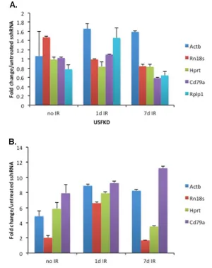
expression of a series of genes routinely used as PCR loading controls to assess potential impacts of USF depletion and IR exposure. In each case, PCR signals in treated and untreated USFKD samples were normalized to untreated sshRNA cells (Fig. 7A). None of the genes tested were altered by USF depletion. Whereas expression of Histone H1 and
Gapdh genes was significantly affected by IR treatment, expression of 18s rRNA (Rn18s),
-actin (Actb), Ig (Cd79a), Hprt and Rplp1 remained relatively constant between treatment
conditions. We next amplified Aicda, which was strongly induced in untreated (+3.47-fold) and 7 days post-IR USFKD microarray screens (+4.38-fold), and found that normalizing
Aicda qPCR signals and the qPCRS signals of the genes contained in Figure 8a to -actin and
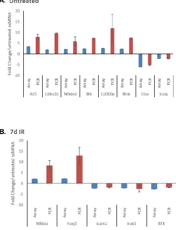
We next amplified a panel of representative DEGs identified by microarray, normalizing expression in each sample to untreated sshRNA, and using validated -actin as a loading
control. In each case, we were able to recapitulate the directionality of altered gene
3.5: Aicda activity in USF-depleted cells
Activation-induced cytidine deaminase is responsible for initiating both class-switch recombination and somatic hypermutation in germinal center B cells. Overexpression of the
Aicda gene in vitro has been shown to drive IgH:Myc translocations observed in Burkitt’s Lymphoma119, as well as enhanced somatic hypermutation of Igk antigen binding exons10. However, AICDA activity is regulated at multiple levels38, and somatic hypermutation in M12 cells has to date only been demonstrated in a single study141. To determine whether elevated Aicda expression in US-depleted M12 cells is sufficient to drive AICDA activity,
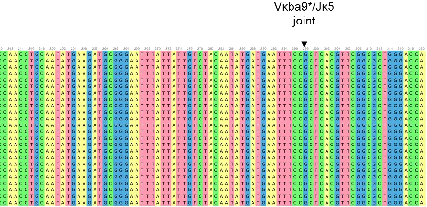
we compared sequence variation within the in-frame Igk Vba9/J5 rearrangement141 and downstream C region that had accumulated in untreated USFKD and sshRNA cells. PCR
products of the rearranged and control exons were subcloned and sequenced. Despite culture over multiple passages, analysis of the USFKD Vba9/J5 exon in 66 clones revealed no
alterations from the published M12 Vba9/J5 sequence (Fig. 9), matching the sequence
conservation seen in 33 C controls (data not shown). As such, our data are consistent with
Figure 9. Representative sequence alignment of a portion of the cloned Vκba9*/Jκ5 rearranged antigen binding exons in USFKD using Unipro Ugene v1.26. The
CHAPTER 4: Discussion
The current study establishes a role for USF transcription factors in regulating the longterm transcriptional response to DNA damage. The ability to properly respond to DNA damage and safeguard the genome is essential to our survival. If DNA lesions, particularly DSBs, are not resolved or are resolved incorrectly, mutations can accumulate that destabilize the genome and leave the cell susceptible to cancerous transformation. Some participants in the DDR pathway and their contributions are well understood, while others have gone greatly understudied. USF1 and USF2 were among the first mammalian transcription factors
identified, are known to bind both tumor suppressor gene and protooncogene
promoters40,134,151, and participate in the cellular response to UV-induced DNA damage. USF activity is diminished or lost from several cancers, including breast cancer34, prostate
cancer20, oral epithelial cancer43, and liver cancer152. Despite these many linkages to cancer, a
critical investigation of how the USF proteins contribute to the DNA damage response and cancer prevention has not been undertaken. This critical gap in knowledge is due in part to the absence of model systems in which both USF activities are targeted, and in part to the focus in DDR studies on activities initiated within the first few hours after genotoxic insult. Although USF is induced by UV-dependent phosphorylation in skin cells, its role in the immediate DDR appears limited35. However, our findings in lymphocytes show that DSBs can induce longterm changes in USF DNA binding activity26, suggesting that USF’s role in
DNA damage, but during the poorly-studied window in which cells must complete the repair response and either return to homeostasis or senesce and die.
To investigate USF’s role in DDR, we examined the transcriptional response of P53-deficient M12 germinal B cell lymphoma cell lines depleted of both USF1 and USF2 to sublethal ionizing radiation. The resulting data reveal many interesting elements of the role of USF. Despite USF’s widespread DNA binding profile21, only 940 DEG probes were
enriched in untreated USFKD cells relative to control cells transduced with a scrambled shRNA (Fig. 4), suggesting that either the USF proteins play a limited role in directing gene expression in the absence of stress, or that sufficient levels of USF1 or USF2 proteins remained in USFKD cells to mediate their regulatory functions. Annotation of DEGs identified in untreated USFKD cells to Gene Ontology biological processes reveals a strong association with immune-related functions (Fig. 6), including lymphocyte proliferation (NFB family genes, Aicda, the common gamma chain gene Il2rg), antigen recognition
(TLR and MyD88 genes), and immune cell activation (Btk, Fyn, Blk, Blnk). Strikingly, of these genes, only Il2rg has previously been shown to require USF activity153. Given that USF depletion altered the expression of a number of additional transcription factors implicated in
immune activity (e.g. Rela and Nfkb1 which encode the P65 and P50 subunits of NFB, the
IB gene Nfkbia, Nfatc1 and Stat5a, among others), the impact of USF depletion on
expression of immune-related genes would be expected to include a mixture of genes like
source of material for future studies to map both USF’s direct and indirect regulatory cascades. Irrespective of association with promoters of the genes listed above, our study clearly demonstrates that the expression of immune-related genes is particularly sensitive to reductions in USF activity. Since a variety of stimuli from DNA damage to metabolic stress have been shown to at least transiently alter USF activity through phosphorylation154, our data suggest that USF may be a critical unappreciated transcriptional regulator of the immune system.
As noted above, the immediate transcriptional response to DNA damage is dominated by P53, and is focused on pausing the cell cycle and mobilizing DNA repair machinery to the site(s) of DNA lesion155. In P53-deficient cells like M12148, survival after DNA damage is mediated by P38MAPK/MK2156. As expected, transcriptional response to treatment with 5
Gy IR was strongly impacted by P53-deficiency, and argues against a widespread impact of USF depletion on the immediate DDR. However, unlike the transcription profiles of
untreated USFKD samples which clustered together with sshRNA control samples, the transcription profiles of individual USFKD samples one day after IR were distinct from the controls (Fig. 4). This suggests that USF does in fact play a role, even if limited, in the early response to DNA damage. However, a more sensitive system (e.g. a system in which USF1 and USF2 are fully deleted) may be required in order to identify which genes are most impacted by loss of USF immediately following genotoxic stress.
cell viability, in which case our selection strategy for stable knockdown clones would bias against cells lacking USF2. Indeed, USF2-deficient mice show marked decreases in lifespan51, and erythroid stem cells expressing a USF dominant negative transgene fail to progress through erythropoiesis52. We do not however expect that USF2 deficiency would
prevent growth of the transformed M12 B cell line. Indeed, no lymphocyte defect was reported for USF2-deficient mice. A more likely explanation for the persistence of USF2 in our knockdown clones is the apparent positive impact USF1 deficiency has on USF2 expression51, which would be expected to limit the effectiveness of our USF2 RNAi targeting, and again supporting the need for a gene deletion approach to removing USF activity.
By far, the most striking impact of USF depletion was on the transcriptional profile of cells one week after they had been irradiated, well after most analyses of the DNA damage response. Although microarray identified over 5000 DEG probes in both USFKD and sshRNA RNAs seven days after IR when contrasted with untreated controls, the Gene Ontology profiles of the two cell types were markedly different. Control cells up- or down-regulated genes associated with DDR, apoptosis and chromatin regulation, whereas USF-depleted cells failed to alter expression of these genes, and instead up- or down-regulated expression of genes associated with immune development and function well beyond the number of genes seen in untreated USFKD cells.
However, contrasting the 1 day post-IR and 7 day post-IR sshRNA controls does reveal two critical groups of biological processes that differentially expressed during the DDR response and that are not seen in matched USF-depleted cells (Fig. 6). Specifically, genes associated with the response to ionizing radiation and chromatin regulation are altered between 1 and 7 days post-IR in sshRNA cells, but not between 7 days post-IR and untreated sshRNA, indicating that these genes are altered during the initial response to DNA damage, but then return to untreated levels within 7 days. In contrast, genes more broadly associated with the DNA damage response and with ribosome development and function are differentially expressed when comparing 7 day post-IR sshRNA profiles with untreated controls, but not when comparing 7 day post-IR with 1 day post-IR sshRNA profiles, indicating that these gene expression responses are activated early, but persist a week after irradiation. DEGs associated with both of these short-term and long-term transcriptional responses to IR are absent when USF is depleted, accounting for the paucity of DEGs in 1 day post-IR USFKD cells when contrasted with untreated knockdown cells and part of the divergence seen