C L I N I C A L T R I A L R E P O R T
MRI Changes After Platelet Rich Plasma Injection
in Knee Osteoarthritis (Randomized Clinical Trial)
This article was published in the following Dove Press journal: Journal of Pain Research
Seyed Ahmad Raeissadat1
Elham Ghorbani2
Morteza Sanei Taheri3
Reza Soleimani 3
Seyed Mansoor Rayegani 2
Marzieh Babaee2
Sara Payami 4
1Physical Medicine and Rehabilitation
Department, Shahid Modarres Hospital, Shahid Beheshti University of Medical
Sciences, Tehran, Iran;2Physical Medicine
and Rehabilitation Research Center, Shohada-e-Tajrish Hospital, Shahid Beheshti University of Medical Sciences,
Tehran, Iran;3Radiology Department,
Shohada-e-Tajrish Hospital, Shahid Beheshti University of Medical Sciences,
Tehran, Iran;4Department of Emergency
Medicine, Amir al momenin Hospital, Tehran Azad University of Medical Sciences, Tehran, Iran
Purpose: Few papers have studied the objective effects of PRP on cartilage. In this study, we investigated the effect of PRP on cartilage characteristics by special MRI sequencing in knee osteoarthritis (IRCT registration number: 2014020413442N6).
Patients and methods:In this double blind randomized clinical trial, patients with bilateral knees osteoarthritis-grade 1, 2, and 3 were included in the study. Each patient's knees were randomly allocated to either control or treatment groups. PRP was injected in two sessions with 4 week intervals in PRP group. The VAS (visual analog scale) and WOMAC (Western Ontario and McMaster Universities Arthritis Index) were utilized and MRI was performed for all patients, before, and 8 months after treatment. The MRI sequences taken were transverse 3D TRUFISP and coronal and sagittal fat saturated proton-density. Imaging was scored according to four cartilage characteristics.
Results:46 knees (from 23 patients) were included in this study. 23 knees in the case group and 23 knees in control group were studied. All patients were female with mean age of 57.57±5.9 years. Mean total WOMAC and VAS changes before and after treatment in control group were 11.61±8.5 and 1.3±1.1 respectively. In PRP group, mean total WOMAC and VAS changes showed better improvement with 20±12.3 and 3.2±1.6 respectively (P-value <0.05). In PRP group, all of the radiologic variables (patellofemoral cartilage volume, synovitis and medial and lateral meniscal
disintegrity), with the exception of subarticular bone marrow abnormality, had significant
improve-ment (P-value <0.05). In a comparison between the two groups, patellofemoral cartilage volume
and synovitis had significantly changed in the PRP group (P-value <0.05).
Conclusion: In this study, in addition to the effect of PRP on VAS and WOMAC, there
was a significant effect on radiologic characteristics (patellofemoral cartilage volume and
synovitis). For further evaluation, a longer study with a larger sample size is recommended.
Keywords: osteoarthritis, platelet rich plasma, PRP, magnetic resonance imaging, MRI, cartilage, knee
Introduction
Knee osteoarthritis is a progressive disease, which in addition to involving the tibiofemoral and patellofemoral joints, also affects their adjacent soft tissues. This disease is among the main causes of pain and disability which not only reduces the
quality of life, but also imposes afinancial burden on the individual as well as the
healthcare system. The disease prevalence varies among different demographics: 19.3% among Iranian rural populations, and 2.8% among Filipinos. In a study by
Framingham, this prevalence was estimated to be 19% among 50–60 year olds.1
Despite the high number of cases, a definite non-surgical cure has not been found yet.
Hyaluronic acid, ozone, laser, botulinum toxin A, and other medical treatments have Correspondence: Marzieh Babaee
Physical Medicine and Rehabilitation Research Center, Shohada-e-Tajrish Hospital, Tajrish Sq, Tehran 1989934148, Iran
Tel/Fax +98 212 273 1112 Email [email protected]
Journal of Pain Research
Dove
press
open access to scientific and medical research Open Access Full Text Article
Journal of Pain Research downloaded from https://www.dovepress.com/ by 118.70.13.36 on 24-Aug-2020
been suggested.2–6One of the more recent modalities cur-rently gaining attention is the various growth factor pro-ducts. These include platelet rich plasma and plasma rich in growth factors; which because of having more platelet con-tent compared to standard plasma, contains more growth
factors.7The growth factors stored in the alpha granules of
the platelets play an important role in biological processes. The preparation of these products using a laboratory cen-trifuge is feasible and their administration is among the
safest, most minimally invasive procedures.8,9
Various studies have shown the effectiveness of these products on the symptoms and functioning of patients with knee osteoarthritis. However, a number of studies have shown controversial results.10–12One reason for these differ-ences in results could be the study not being performed in a double-blinded manner, which could adversely affect the results. Most studies focus on the role that these products have on the subjective experiences of the patient regarding clinical symptoms, performance, and quality of life. Based on our research, a small number of studies have shifted their attention toward objective changes in the articular cartilage of
the knee and its surrounding tissues.13Special imaging
meth-ods for measuring cartilage have recently been employed in clinical management as well as in research. MRI is the most important noninvasive investigation method in the assess-ment of traumatic or degenerative lesions of the cartilage before and after performing treatments in the knee. This imaging method can show the structure of the joint in three dimensions, which can accurately and reliably measure the volume of cartilage, because of high soft tissue contrast.14–16 In some studies, MRI imaging has been utilized to evaluate intra-articular injections including viscosupplements, growth factors, stem cells etc., in which all except PRP (in a few controversial studies) have shown a positive effect on carti-lage structure.17–20In this study, we have therefore attempted to objectively assess the effect of PRP on knee osteoarthritis using the changes of cartilage in MRI sequences.
Materials and Methods
This study was a randomized, double blind clinical trial,
performed between 2016–2017 in Shohoda-e-Tajrish and
Shahid Modarres hospitals in Tehran. Patients with osteoarthritis visiting the physical and rehabilitation med-icine clinics of these two centers were included in the study based on the following inclusion and exclusion criteria.
Inclusion criteria: history of pain for the last 3 months according to the ACR criteria for knee osteoarthritis along
with radiologic evidence of joint destruction based on Kellgran-Lawrence criteria (including grades 1, 2, and 3). Exclusion criteria: 1) any contraindication for perform-ing an MRI includperform-ing aneurism clips, pacemakers, non MRI-compatible metallic devices in the body and claustro-phobia; 2) any form of knee injection such as viscosupple-ments, ozone etc. in the preceding 3 months; 3) having arthroscopic or open surgery in the past 6 months; 4) having
immunodeficiency, autoimmune disease, collagen vascular
disease, or diabetes; 5) history of cancer; 6) infection or
inflamed lesion in the knee; 7) platelet disorder or disease;
8) use of anticoagulant or anti-platelet medication 10 days before injection; 9) use of NSAIDs 2 days before injection; 10) corticosteroid knee injection 3 weeks before injection or use of systemic corticosteroids 2 weeks before the injection; 11) hemoglobin level less than 12 g/dL and platelets less than 150,000/microliter; 12) history of severe knee trauma; 13) age of less than 45 and higher than 65; 14) history of vasovagal shock; 15) pregnancy and lactation; 16) genu valgum or genu varum more than 20 degrees.
After patient selection, the aims, methods, current
scien-tific evidence, benefits and possible risks of the study were
described to the patients by a physical medicine and rehabi-litation doctor. After this stage, all participants who had signed the informed consent form were included in the study. The form and trial were approved by the ethics committee of Shahid Beheshti University of Medical Sciences (IRCT registration number: 2014020413442N6), this was conducted in accordance with the Declaration of Helsinki. After the patients entered the study, personal infor-mation such as age, gender, and grade of osteoarthritis (Kellgren-Lawrence Grading scale in plain radiograph) was recorded. Afterwards, for each patient, a VAS (Visual Analog Scale) for pain assessment as well as a WOMAC arthritis index (Western Ontario and McMaster Universities) for func-tional assessment were completed with the help of a physiatrist resident. The VAS is a psychometric tool, which is used to document the severity of symptoms of
a person’s illness. This tool consists of a 100 mm ruler,
which has descriptive drawings of severity on the two sides of the ruler. The patient marks his/her symptom severity based on the descriptions. The higher the number, the worse the symptoms. The WOMAC questionnaire is one of the questionnaires used in evaluating the function of patients with rheumatologic disease; in particular knee osteoarthritis. It covers the differentfields of stiffness, pain, and function in certain activities; where a higher score indicates worse function.21
Journal of Pain Research downloaded from https://www.dovepress.com/ by 118.70.13.36 on 24-Aug-2020
Knee MRI images of patients were taken by a Siemens AVANTO (1.5 Tesla) MR system using a circumferential knee
coil before (10–14 days before starting the treatment) and 8
months after treatment. The sequences taken were transverse 3D TRUFISP and coronal and sagittal fat saturated proton-density (PD). All images were reviewed by a radiologist and
cross-checked again in Shohada-e-Tajrish’s radiology
depart-ment. They were unaware of the case and control groups or whether the images were taken before or after treatment. All MR images were evaluated in an unpaired manner. Four different characteristics of the joint were scored in the follow-ing manner.
1) Volume of patellofemoral cartilage: the patellofemoral cartilage was evaluated using 3D slicer software (a free plat-form for viewing and analysis of images in a software
spe-cific manner). All axial images were manually segmented in
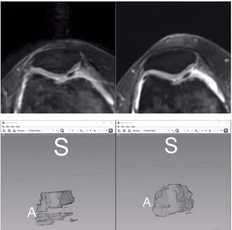
TRUFISP 3D sequence so that the cartilage area was sepa-rated from the nearby bone and soft tissue. Afterwards, the volume of the cartilage was calculated by multiplying the sum of the cartilage area of all the images by the thickness of the slide using 3D slicer22(Figure 1).
2) Subarticular bone marrow abnormality: subarticular bone marrow abnormality was scored in 15 different areas
using the WORMS score. This was defined by increases in
signal intensity in normal fat containing epiphyseal bone marrow in fat-saturated PD sequence. Each of these 15 regions were scored from 0 to 3 based on the level of abnormality; 0: no signal increase, 1: less than or equal to
25% of the area, 2: 25–50% of the area, 3: more or equal
to 50%.22
3) Internal and external meniscal integrity: the anterior and posterior horns and body of the medial and lateral
menisci were separately evaluated and scored from 0–3
based on sagittal and coronal images; 0: intact, 1: globular signal inside the meniscus which is not adjacent to the articular surface, 2: linear signal inside the meniscus which is not adjacent to the articular surface, 3: abnormal signal inside the meniscus which has reached the articular surface.23
4) Synovitis: the existence of signs of inflammation in
the synovium was scored as 0 or 1.22
The patients’knees were randomly (based on a random
table of numbers) divided into two groups. Both knees had exercise therapy prescribed to them, while the study knee also had a PRP injection (group A: PRP and exercise;
group B: exercise alone,Figure 2). In both knees, exercise
was taught by a physiatrist. The exercise protocol employed was multi-angle isometric exercise of muscles
around the knee (quadriceps, thigh abductor and adduc-tors) as well as stretching of the hamstrings 3 times a day, 10 times for each move and 10 seconds each time. After 4 weeks, strengthening exercises for the quadriceps, adduc-tors, and abductors were taught to the patients.
PRP was prepared using a Royagen kit. In order not to prolong the content, the details of the PRP protocol are
similar to those of our previous papers.24–26PRP
prepara-tions in this study contained leukocytes (LR-PRP) and
platelet concentrations were 4–6 times normal. In the
PRP group, 500 mg of paracetamol without codeine could be ingested for pain relief after injection. In case of further pain, paracetamol with codeine was prescribed as needed and patients were forbidden from using any forms of NSAIDs or steroids. The patients could restart all daily activities one week post injection. In the study group, exercise therapy commenced one week after injec-tion, which started with lower intensity and increased gradually. In the control group, exercise therapy started immediately and the patients in this group were also asked to use 500 mg of paracetamol without codeine in case of pain; and if the pain was not controlled, paracetamol with codeine was to be used. The second injection was
per-formed 28 days (4 weeks) after thefirst.
The prescribed medications and the importance of adherence to the prescribed drugs as well as refraining from use of drugs which affect platelet activity were explained. The patients were asked to contact the research-ers in case of any problems.
All patients were followed up 32 weeks after therapy, when the VAS and WOMAC forms were completed again for them and another MRI was performed.
Thefinal data were entered into SPSS-16 for analysis.
Normality of the variables’ distribution was assessed
using the Shapiro Wilk test. Comparisons for before and after each intervention were performed separately for each
group usingt-test, pairedt-test, and ANOVA for variables
with normal distribution. For non-parametric variables,
Wilcoxon signed rank, Mann–Whitney and Kruskal Wallis
tests were employed. Qualitative data were expressed using value and percentage. In order to assess the relation between
quantitative variables, Pearson’s correlation coefficient and
Spearman’s coefficient were utilized.
Results
46 knees (of 23 patients) were entered into this study, 23 of which entered the control group and 23 of which were in the intervention group. 2 knees from each group were
Journal of Pain Research downloaded from https://www.dovepress.com/ by 118.70.13.36 on 24-Aug-2020
eliminated because of failure to show up in follow up,
leaving 21 knees in each group after 8 months (Figure 2).
All patients were female, with an average age of 57.57±5.9
years and average BMI of 28.49±3.24 Kg/m2.
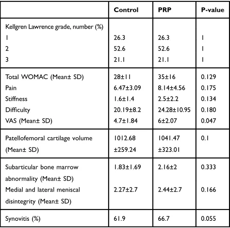
Except for VAS score, none of the characteristics had
any significant difference between groups (Table 1). The
average VAS showed a significant improvement inter-group
and between groups (P<0.05) (Table 2).
In addition, there was a meaningful difference between the before and after treatment WOMAC scores of both groups. There was also a difference between the after treatment WOMAC scores of both groups except for the
pain subgroup which had borderline P-Value (Table 2).
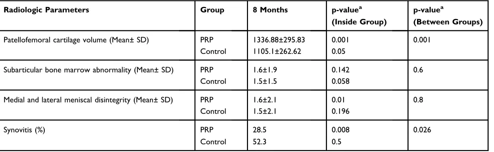
In the PRP group, patellofemoral cartilage volume, synovitis, and medial and lateral meniscal disintegrity
showed significant improvement after treatment. In the
Figure 1The patellofemoral cartilage was evaluated using 3D slicer software (a free platform for viewing and analysis of images in a software specific manner). Left picture before PRP injection and right picture after PRP injection.
Journal of Pain Research downloaded from https://www.dovepress.com/ by 118.70.13.36 on 24-Aug-2020
control group, only patellofemoral cartilage volume
showed meaningful improvement (Table 3).
In comparison of the two groups, improvement in patellofemoral cartilage volume and synovitis was better
in the PRP group (Table 3).
Because of the low number of patients, no relation was found between the radiologic parameters and grade of osteoarthritis.
Discussion
This clinical trial showed the positive effects of PRP on MRI
findings. In our study MRI changes including patellofemoral
cartilage volume and synovitis were better in the PRP group. At present, systematic reviews and meta analyses have shown the positive role of PRP in pain reduction and functional improvement in patients with mild to moderate
osteoarthritis. The findings of these studies have been
obtained in a subjective manner.10,12,27 Few studies have
compared osteoarthritis treatments such as hyaluronic acid, growth factors, stem cells etc., objectively; using
MR imaging.28–30 Furthermore, to the best of our
knowl-edge, few studies have assessed the effect of PRP on radiologic and MRIfindings.19,31,32
Figure 2CONSORTflow diagram.
Table 1Baseline Characteristics of Study Population
Control PRP P-value
Kellgren Lawrence grade, number (%)
1 26.3 26.3 1
2 52.6 52.6 1
3 21.1 21.1 1
Total WOMAC (Mean± SD) 28±11 35±16 0.129
Pain 6.47±3.09 8.14±4.56 0.175
Stiffness 1.6±1.4 2.5±2.2 0.134
Difficulty 20.19±8.2 24.28±10.95 0.180
VAS (Mean± SD) 4.7±1.84 6±2.07 0.047
Patellofemoral cartilage volume (Mean± SD)
1012.68 ±259.24
1041.47 ±323.01
0.1
Subarticular bone marrow abnormality (Mean± SD)
1.83±1.69 2.16±2 0.333
Medial and lateral meniscal disintegrity (Mean± SD)
2.27±2.7 2.44±2.7 0.166
Synovitis (%) 61.9 66.7 0.055
Journal of Pain Research downloaded from https://www.dovepress.com/ by 118.70.13.36 on 24-Aug-2020
In 2013, Halpern et al studied the effect of PRP on clinical
signs and MRIfindings in knees of 15 patients suffering from
osteoarthritis. 6 mL of PRP prepared from 20 mL of patient blood was injected. Clinical signs were assessed using WOMAC and VAS, while MRIs were performed via a 1.5 Tesla scanner and fast spin echo images in 3 planes; and afterwards reported by 2 radiologists. The patients were followed after 1 week as well as 1, 3, 6, and 12 months. After 6 and 12 months, the WOMAC score and VAS improved. From the 15 knees with MRI images, 12 (80%) did not show changes toward exacerbation of osteoarthritis in the patellofemoral joint. Furthermore, in 12 knees (80%), during one year no change was seen in the radiologic appear-ance of the lateral tibiofemoral joint. Similarly, no changes were reported in the involvements of the medial compart-ment in 73.3% of cases; although this did not have statistical
significance.19 The radiologic appearance of one knee
showed improvement after 1 year. In the end, the authors
expressed that some studies had shown 4–6% yearly
reduc-tion in cartilage volume in an osteoarthritic compartment,
which was in contrast with their current study results regard-ing PRP treatment. Moreover, it is possible that one injection of PRP can reduce cartilage loss. In comparison, in our study, patellofemoral cartilage volume and synovitis improved
further in the PRP group. Although Halpern’s study did not
show changes in the thickness of cartilage, it did not show
any loss of cartilage either. Thesefindings could be related to
the number of injections which was double in our study and single injection in Halpern’s study.
In 2018, Buendía-López studied the effect of PRP,
hya-luronic acid, and NSAIDS on MRI findings. 98 patients
(33 in the NSAID group, 32 in hyaluronic acid group, and 33 in the PRP group) completed the study. In the LP-PRP group (leukocyte poor PRP), an injection was performed once and the patients of all three groups were assessed with WOMAC and VAS 26 and 52 weeks into follow up. In addition, an X-ray and MRI were performed at the
begin-ning and end (52 weeks) of the study.31 In WOMAC and
VAS-pain, the results of the LP-PRP group were better. In this study the change of grade of osteoarthritis from grade
Table 2Mean± SD of WOMAC and VAS After 8 Months Treatment in Both Groups
WOMAC Parameters Group 8 Months p-valuea
(Inter Group)
p-valuea
(Between Groups)
Pain PRP 3.85±3.4 0.001 0.056
Control 4.04±2.45 0.001
Stiffness PRP 0.76±0.88 0.001 0.036
Control 1±1.1 0.036
Functional capacity PRP 10.15±8.3 0.001 0.038
Control 12.05±6.7 0.001
VAS PRP 2.76±2.07 0.001 0.001
Control 3.42±1.83 0.001
Note:a
P-Value <0.05 was considered significant.
Table 3Radiologic Parameters After Treatment in Both Groups
Radiologic Parameters Group 8 Months p-valuea
(Inside Group)
p-valuea
(Between Groups)
Patellofemoral cartilage volume (Mean± SD) PRP 1336.88±295.83 0.001 0.001
Control 1105.1±262.62 0.05
Subarticular bone marrow abnormality (Mean± SD) PRP 1.6±1.9 0.142 0.6
Control 1.5±1.5 0.058
Medial and lateral meniscal disintegrity (Mean± SD) PRP 1.6±2.1 0.01 0.8
Control 1.5±2.1 0.196
Synovitis (%) PRP 28.5 0.008 0.026
Control 52.3 0.5
Note:aP-Value <0.05 was considered significant.
Journal of Pain Research downloaded from https://www.dovepress.com/ by 118.70.13.36 on 24-Aug-2020
1 to 2 was 17%, while grade 2 did not show significant change. No increase in thickness of cartilage was observed; although no meaningful decrease was observed between the three groups either. This study was without a placebo group and a single LP-PRP injection with a concentration of 3.8
times was assessed in only grade 1 and 2. In Hart’s study,
which observed the effect of 9 PRP injections in one year
on MRI findings, no regeneration of cartilage was seen.
Patients with grade 2 and 3 osteoarthritis were entered into this study and the PRP had 2 to 2.5 times
concentration.32These differences in results of various
stu-dies could be due to the presence of leukocytes in some PRP preparations, PRP concentration, number of PRP injec-tions, follow up time, and grade of osteoarthritis.
In 2018, in addition to clinical findings, Ahmad
assessed the changes in cartilage using sonography in 2 groups of PRP and hyaluronic acid injections. In both groups, 3 injections were performed with a 2 week inter-vals between injections. In both groups, after 3 and 6 months of follow up, clinical improvement was observed; with the PRP group showing better improvement than the hyaluronic acid group. The improvements in the PRP group showed less effusion, reduced synovial hypertrophy,
and improved vascularity scores in ultrasound
assessment.33 Although the imaging method is different
from our study, the radiologic changes bear similarities with our study’s results.
In 2016, Smara also studied a platelet-derived product named autologous platelet lysates and its effects on cartilage in MRIs. 15 patients with grade 1 and 2 osteoarthritis had this product injected 2 times, every 3 weeks. After one year follow up, the thickness of cartilage on both the tibial and
femoral sides of the joint showed significant improvement.20
What is evident is that MRI imaging plays an impor-tant role in faster assessment of patients with
osteoarthri-tis. In Framingham’s study, it was shown that patients who
lack any signs of knee osteoarthritis in plain radiographs could have some MRI changes indicative of the beginning
of osteoarthritis.34 Therefore, MRI is one of the most
sensitive tools for evaluation of changes in osteoarthritis and its response to treatment. Use of MRI in order to assess the effect of treatment, in particular the somewhat newer modalities, can determine their actual effects. In this study, we tried to observe the visible effects of PRP. The presence of a control group, the design of the study as
a clinical trial, and being among the first studies using
MRI imaging to assess PRP treatment are the positive aspects of our study. The relatively small number of
subjects, shorter follow up time, assessment of mild to moderate grades of osteoarthritis, results being affected by other variables such as a high BMI as well as lack of comparison with other treatment methods can be among the shortcomings of this study. We attempted, in this research, to study the effects of PRP, as one of the more commonly accepted treatments for mild to moderate
osteoarthritis, on MRIfindings.
Conclusion
It was observed that compared to the control group, PRP was effective in the improvement of patellofemoral cartilage volume and synovitis. Even though, a longer study with a higher number of subjects and comparison with other treatment methods is recommended for further evaluation.
Data Sharing Statement
The authors intend to share individual participant data that underline the results reported in this article after deidentifi
ca-tion (text, tables, figures, and appendix). Study protocol,
statistical analysis plane, informed consent form and clinical study report will be made available and the data will be accessible by email to [email protected]. To gain access, data requestors will need to sign a data access agree-ment. Immediately following publication and ending 2 years following article publication they will be made available.
Acknowledgments
We thank Ms. Mahshid Zaman for her assistance with
collecting the patients’images and data.
An abstract of this paper titled“Volumetricfindings of
MRI after platelet rich plasma injection in knee
osteoar-thritis (A randomized clinical trial)”was presented at the
12th International Society of Physical and Rehabilitation Medicine Conference, as a conference talk with interim
findings; and was published in “Poster Abstracts” in the
Annals of Physical and Rehabilitation Medicine Journal
(https://doi.org/10.1016/j.rehab.2018.05.022);35 and was also presented as an oral presentation at the Annual
European Congress of Rheumatology EULAR 2018.36
Disclosure
The authors report no conflicts of interest in this work.
References
1. Felson DT The epidemiology of knee osteoarthritis: results from the Framingham Osteoarthritis Study. Paper presented at: Seminars in arthritis and rheumatism;1990. doi:10.1099/00221287-136-2-327
Journal of Pain Research downloaded from https://www.dovepress.com/ by 118.70.13.36 on 24-Aug-2020
2. Kon E, Filardo G, Drobnic M, et al. Non-surgical management of early knee osteoarthritis.Knee Surg Sports Traumatol Arthroscopy. 2012;20(3):436–449. doi:10.1007/s00167-011-1713-8
3. Raeissadat SA, Tabibian E, Rayegani SM, Rahimi-Dehgolan S, Babaei-Ghazani A. An inestigation into the efficacy of intra-articular ozone (O2-O3) injection in patients with knee osteoarthritis: a systematic review and meta-analysis.J Pain Res.2018;11:2537–2550. doi:10.2147/JPR. S175441
4. Raeissadat SA, Rayegani SM, Forogh B, Abadi PH, Moridnia M, Dehgolan SR. Intra-articular ozone or hyaluronic acid injection: which one is superior in patients with knee osteoarthritis? A 6-month randomized clinical trial. J Pain Res. 2018;11:111. doi:10.2147/JPR.S142755
5. Rayegani SM, Raeissadat SA, Heidari S, Moradi-Joo M. Safety and effectiveness of low-level laser therapy in patients with knee osteoar-thritis: a systematic review and meta-analysis. J Lasers Med Sci. 2017;8(Suppl 1):S12. doi:10.15171/jlms.2017.s3
6. Raeissadat SA, Rayegani SM, Sedighipour L, et al. The efficacy of electromyographic biofeedback on pain, function, and maximal thick-ness of vastus medialis oblique muscle in patients with knee osteoar-thritis: a randomized clinical trial. J Pain Res. 2018;11:2781. doi:10.2147/JPR.S169613
7. Raeissadat SA, Rayegani SM, Ahangar AG, Abadi PH, Mojgani P, Ahangar OG. Efficacy of intra-articular injection of a newly devel-oped Plasma Rich in Growth Factor (PRGF) versus hyaluronic acid on pain and function of patients with knee osteoarthritis: a single-blinded randomized clinical trial. Clin Med Insights. 2017;10:1179544117733452.
8. Raeissadat SA, Babaee M, Hashemi Z. Application of platelet-rich plasma and other platelet-derivative products in Iranian studies. koo-mesh.2019;21(3):408–422.
9. Raeissadat SA, Babaee M, Rayegani SM, et al. An overview of platelet products (PRP, PRGF, PRF, etc.) in the Iranian studies. Future Sci OA.2017;3(4):FSO231. doi:10.4155/fsoa-2017-0045 10. Laudy AB, Bakker EW, Rekers M, Moen MH. Efficacy of
platelet-rich plasma injections in osteoarthritis of the knee: a systematic review and meta-analysis.Br J Sports Med. 2015;49 (10):657–672. doi:10.1136/bjsports-2014-094036
11. Campbell KA, Saltzman BM, Mascarenhas R, et al. Does intra-articular platelet-rich plasma injection provide clinically superior outcomes com-pared with other therapies in the treatment of knee osteoarthritis? A systematic review of overlapping meta-analyses. Arthroscopy. 2015;31(11):2213–2221. doi:10.1016/j.arthro.2015.03.041
12. Khoshbin A, Leroux T, Wasserstein D, et al. The efficacy of platelet-rich plasma in the treatment of symptomatic knee osteoar-thritis: a systematic review with quantitative synthesis.Arthroscopy. 2013;29(12):2037–2048. doi:10.1016/j.arthro.2013.09.006
13. Meheux CJ, McCulloch PC, Lintner DM, Varner KE, Harris JD. Efficacy of intra-articular platelet-rich plasma injections in knee osteoarthritis: a systematic review.Arthroscopy.2016;32(3):495–505. doi:10.1016/j.arthro.2015.08.005
14. Gold GE, Chen CA, Koo S, Hargreaves BA, Bangerter NK. Recent advances in MRI of articular cartilage.Am J Roentgenol.2009;193 (3):628–638. doi:10.2214/AJR.09.3042
15. Hill CL, Hunter DJ, Niu J, et al. Synovitis detected on magnetic resonance imaging and its relation to pain and cartilage loss in knee osteoarthritis.Ann Rheum Dis.2007;66(12):1599–1603. doi:10.1136/ ard.2006.067470
16. Hunter DJ, Li J, LaValley M, et al. Cartilage markers and their association with cartilage loss on magnetic resonance imaging in knee osteoarthritis: the Boston osteoarthritis knee study. Arthritis Res Ther.2007;9(5):R108. doi:10.1186/ar2314
17. Cubukcu D, Ardic F, Karabulut N, Topuz O. Hylan G-F 20 efficacy on articular cartilage quality in patients with knee osteoarthritis: clinical and MRI assessment.Clin Rheumatol.2005;24(4):336–341. doi:10.1007/s10067-004-1043-z
18. Durmus D, Alayli G, Bayrak IK, Canturk F. Assessment of the effect of glucosamine sulfate and exercise on knee cartilage using magnetic resonance imaging in patients with knee osteoarthritis: a randomized controlled clinical trial. J Back Musculoskelet Rehabil. 2012;25 (4):275–284. doi:10.3233/BMR-2012-0336
19. Halpern B, Chaudhury S, Rodeo SA, et al. Clinical and MRI outcomes after platelet-rich plasma treatment for knee osteoarthritis.Clin J Sport Med.2013;23(3):238–239. doi:10.1097/JSM.0b013e31827c3846 20. Samara O, Al-Ajlouni J, Al-Najar M, et al. Intra-articular autologous
platelet lysates produce positive MRI structural changes in early and intermediate knee osteoarthrosis.PJR.2016;27:1.
21. Gandek B. Measurement properties of the Western Ontario and McMaster Universities osteoarthritis index: a systematic review.Arthritis Care Res (Hoboken).2015;67(2):216–229. doi:10.1002/acr.v67.2
22. Peterfy C, Guermazi A, Zaim S, et al. Whole-organ magnetic reso-nance imaging score (WORMS) of the knee in osteoarthritis. Osteoarthritis Cartilage. 2004;12(3):177–190. doi:10.1016/j.joca. 2003.11.003
23. Stoller DW, Martin C, Crues J 3rd, Kaplan L, Mink JH. Meniscal tears: pathologic correlation with MR imaging.Radiology.1987;163 (3):731–735. doi:10.1148/radiology.163.3.3575724
24. Raeissadat SA, Rayegani SM, Babaee M, Ghorbani E. The effect of platelet-rich plasma on pain, function, and quality of life of patients with knee osteoarthritis.Pain Res Treat.2013;2013.
25. Raeissadat SA, Rayegani SM, Hassanabadi H, et al. Knee osteoar-thritis injection choices: platelet-rich plasma (PRP) versus hyaluronic acid (a one-year randomized clinical trial).Clin Med Insights.2015;8 (CMAMD):S17894.
26. Raeissadat SA, Karimzadeh A, Hashemi M, Bagherzadeh L. Safety and efficacy of platelet-rich plasma in treatment of carpal tunnel syndrome; a randomized controlled trial. BMC Musculoskelet Disord.2018;19(1):49. doi:10.1186/s12891-018-1963-4
27. Chang K-V, Hung C-Y, Aliwarga F, Wang T-G, Han D-S, Chen W-S. Comparative effectiveness of platelet-rich plasma injections for treat-ing knee joint cartilage degenerative pathology: a systematic review and meta-analysis. Arch Phys Med Rehabil. 2014;95(3):562–575. doi:10.1016/j.apmr.2013.11.006
28. Forney MC, Gupta A, Minas T, Winalski CS. Magnetic resonance imaging of cartilage repair procedures.Magn Reson Imaging Clin. 2014;22(4):671–701. doi:10.1016/j.mric.2014.07.008
29. Wang Y, Hall S, Hanna F, et al. Effects of Hylan GF 20 supplementa-tion on cartilage preservasupplementa-tion detected by magnetic resonance ima-ging in osteoarthritis of the knee: a two-year single-blind clinical trial.BMC Musculoskelet Disord.2011;12(1):195. doi:10.1186/1471-2474-12-195
30. Woollard JD, Gil AB, Sparto P, et al. Change in knee cartilage volume in individuals completing a therapeutic exercise program for knee osteoarthritis. J Orthop Sports Phys Ther. 2011;41 (10):708–722. doi:10.2519/jospt.2011.3633
31. Buendía-López D, Medina-Quirós M, Marín MÁF-V. Clinical and radiographic comparison of a single LP-PRP injection, a single hya-luronic acid injection and daily NSAID administration with a 52-week follow-up: a randomized controlled trial. J Orthopaedics Traumatol.2018;19(1):3. doi:10.1186/s10195-018-0501-3
32. Hart R, Safi A, Komzák M, Jajtner P, Puskeiler M, Hartová P. Platelet-rich plasma in patients with tibiofemoral cartilage degeneration. Arch Orthop Trauma Surg. 2013;133(9):1295–1301. doi:10.1007/s00402-013-1782-x
33. Ahmad HS, Farrag SE, Okasha AE, et al. Clinical outcomes are asso-ciated with changes in ultrasonographic structural appearance after platelet-rich plasma treatment for knee osteoarthritis.Int J Rheum Dis. 2018;21(5):960–966. doi:10.1111/apl.2018.21.issue-5
34. Guermazi A, Niu J, Hayashi D, et al. Prevalence of abnormalities in knees detected by MRI in adults without knee osteoarthritis: popula-tion based observapopula-tional study (Framingham Osteoarthritis Study). BMJ (Clinical Research Ed).2012;345:e5339.
Journal of Pain Research downloaded from https://www.dovepress.com/ by 118.70.13.36 on 24-Aug-2020
35. Raeissadat S, Ghorbani E, Rayegani S, Taheri MS, Babaee M, Soleimani R. Volumetricfindings of MRI after platelet rich plasma injection in knee osteoarthritis (A randomized clinical trial).Ann Phys Rehabil Med.2018;61:e9. doi:10.1016/j.rehab.2018.05.022
36. Raeissadat S, Ghorbani E, Sanei M, Rayegani S, Babaee M, Soleimani R. AB0975 Does Platelet Rich Plasma Change the Volumetric Findings of MRI Imaging?(A Randomised Clinical Trial).BMJ Publishing Group Ltd.2018.
Journal of Pain Research
Dove
press
Publish your work in this journal
The Journal of Pain Research is an international, peer reviewed, open access, online journal that welcomes laboratory and clinicalfindings in thefields of pain research and the prevention and management of pain. Original research, reviews, symposium reports, hypothesis formation and commentaries are all considered for publication. The manuscript
management system is completely online and includes a very quick and fair peer-review system, which is all easy to use. Visit http:// www.dovepress.com/testimonials.php to read real quotes from pub-lished authors.
Submit your manuscript here:https://www.dovepress.com/journal-of-pain-research-journal
Journal of Pain Research downloaded from https://www.dovepress.com/ by 118.70.13.36 on 24-Aug-2020