Microstructural Evolution during Isothermal Aging
in Ni-Rich Ti-Zr-Ni Shape Memory Alloys
Adrian Mihai Sandu
1;*, Koichi Tsuchiya
1, Masayuki Tabuchi
2, Shinya Yamamoto
1,
Yoshikazu Todaka
1and Minoru Umemoto
11Dept. of Production Systems Engineering, Toyohashi University of Technology, Toyohashi 441-8580, Japan 2Hakodate Polytechnic College, Hakodate 042-8501, Japan
Microstructural evolution during isothermal aging at temperatures ranging from 673 K to 773 K was investigated by transmission electron microscopy (TEM) for Ni-rich Ti-Zr-Ni alloys. It was revealed that the aging is a two-stage process: the first stage is characterized by circular diffuse scattering in electron diffraction patterns, most likely attributed to short range ordered structure with no significant microstructural change. Second stage is characterized by a fine coherent precipitation followed by the coarsening. The precipitates had a lenticular shape and their habit wasf100gB2. The electron diffraction pattern of the sample containing the precipitates exhibited satellite spots at1=3h110iB2and
1=4h210iB2positions. High resolution TEM observations and fast Fourier transformation analysis revealed that theh100iB2electron diffraction pattern was composed of the reflection from 4 variants of the precipitates. The two-stage microstructural evolutions coincide well with the previously reported changes in mechanical properties and martensitic transformation behavior. [doi:10.2320/matertrans.48.432]
(Received September 21, 2006; Accepted December 7, 2006; Published February 25, 2007)
Keywords: shape memory alloys, martensitic transformation, aging, precipitation, short range ordering
1. Introduction
Superelasticity (SE), shape memory effect (SME) and biocompatibility of TiNi based shape memory alloys have found various applications in medical devices, such as guidewires and stents, as well as actuators for various appliances. The SE and SME are closely related to martensitic or R-phase transformation. Thus, the control of transformation temperatures and mechanical properties be-comes very important for practical applications. The proper-ties of TiNi can be modified by adequate thermomechanical treatment and by changing alloy composition. In the case of Ni-rich TiNi, aging at temperatures between 673773K results in the fine precipitation of Ti3Ni4 phase, which
significantly strengthens the material and thus leads to improved SME and SE properties.1) Addition of the third element is also common method to modify the properties. Addition of noble metal elements, such as, Au, Pt and Pd, or rare earth elements, such as, Hf increases martensitic transformation temperature,2)while the addition of the most of transition metal elements decreases the transformation temperatures with Zr being an exception. Also Zr addition may lead to an improvement of biocompatibility because of its high affinity with oxygen and ability to form stable oxides. A number of investigations subjecting Ti-Zr-Ni shape memory alloys have been carried out.3–9)However, limited information is available regarding the effect of aging on phase transformation and mechanical properties in Ti-Zr-Ni alloys, and if the precipitation behaviour similar to the one observed in a Ni-rich binary TiNi occurs during aging.
In our previous research it was revealed that the trans-formation temperatures decrease in the early stage of aging and then turn to increase in the latter stage.10)On the other hand, the hardness increased in the initial stage followed by a slight decrease after the longer aging. The aging conditions which give the highest hardness correspond roughly to the
one that gives the lowest martensitic transformation ature. Similar tendency can be seen for other aging temper-atures. It can be thus inferred that these two aging effects are mutually related and may have their origins in common. It was also revealed that the initial aging stage is accompanied by no significant change in microstructure, except for intensifying the diffuse scattering in electron diffraction patterns while the later aging stage was accompanied by fine precipitation.
The present report presents the details in microstructure during aging, such as, the morphology and structure of the precipitates as well as their effect on compression deformation.
2. Experimental Procedures
Ingots of Ti-6.0 mol%Zr-52 mol%Ni, Ti-6.0 mol%Zr-53 mol%Ni and Ti-11.1 mol%Zr-51.5 mol%Ni were prepared using a tri-arc melting furnace. They were homogenized in evacuated quartz tube for 605 ks (7 days) at 1073 K under an argon atmosphere followed by water quenching. The alloys were isothermally aged for various time durations up to 360 ks at temperatures from 673 to 773 K in an argon gas atmosphere. Cylindrical samples for compression test having a height of 3.9 mm and a diameter of 2.2 mm were cut from Ti-6.0 mol%Zr-53 mol%Ni ingot. Compression tests were performed on Shimadzu AGS-J apparatus imposing an 8% deformation for 5 consecutive cycles at room temperature. During the compression deformation the both ends of the samples were lubricated with BN powder to minimize the effect of friction. Strain was measured by video extensom-eter. After each compression cycle the sample was heated over austenitic transformation finish temperature. TEM samples were carefully cut from the ingots and from as-compressed samples as well. Specimens were electrochemi-cally polished using an electrolyte of 20% H2SO4 and 80%
methanol at 253 K. Microstructural observations were done using Hitachi H-800 and JEOL JEM-2010 transmission electron microscopes (TEM) both operated at 200 kV. *Graduate Student, Toyohashi University of Technology
3. Results and Discussion
3.1 Changes in microstructures by aging in Ti-6.0 mol%Zr-52 mol%Ni
Figure 1 shows a bright field (BF) image (a), selected area diffraction (SAD) pattern with the electron beam parallel to
½001B2 (b) and its key diagram (c) for Ti-6.0 mol%Zr-52 mol%Ni aged at 773 K for 3.6 ks. The SAD pattern exhibits circular diffuse scattering around the main spots of the B2 phase. They are slightly elongated in the directions parallel to
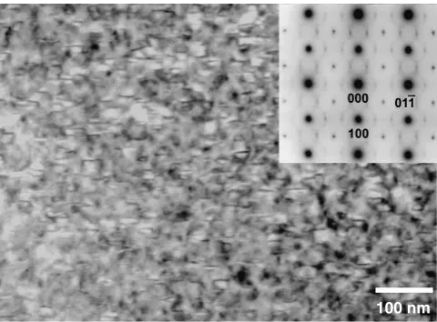
h110iB2and exhibit the four-fold symmetry around the main spots. The diffuse scattering is different from those due to the precursor effect of R-phase often observed in binary TiNi,11) and thus is most likely related to the short range ordered arrangement of Zr atoms. These features were essentially identical with the ones noted for the as-homogenized sample. TEM image from a different region of this sample is shown in Fig. 2. The BF image (Fig. 2(a)) exhibits only the bend contour and no second phase is seen. However, circular diffuse scattering are noted in the corresponding SAD pattern with the electron beam parallel to ½011B2 presented in Fig. 2(b). They are elongated in the directions parallel to
h100iB2as depicted in the key diagram (Fig. 2(c)).
Figure 3 shows a BF image (a), the corresponding SAD pattern (b) and its key diagram (c) of the sample aged at 773 K for 14.4 ks. Numerous flat precipitates withð100ÞB2or
ð010ÞB2 habit are seen in the BF image (Fig. 3(a)). The precipitates are surrounded by dark contrasts associated with
elastic strain fields. Also in the BF image, dark lobes with a line of no contrast in the middle can be seen as indicated by the arrows. They are a typical of precipitates with a circular perimeter accompanied by coherency strain around them. Thus, it is inferred that the precipitates are lenticular with their habit on f100gB2. The size of the precipitates is about 70 nm in diameter and about 20 nm thick. The SAD (Fig. 3(b)) indicates sharp satellite spots at1=3h110iB2 and diffuse satellite at 1=4h210iB2 elongated in h100iB2 direc-tions. The SAD pattern is clearly different from that of the aged TiNi with Ti3Ni4 precipitates, which exhibits satellite
spots at1=7h321iB2, and from that of the sputtered rich Ti-48.2 mol%Ni thin films aged at 745 K for 3.6 ks.12,13)Careful examination of the films revealed that the diffuse scattering rings are still present in SAD pattern shown in Fig. 3(b).
Figure 4 shows a distribution of the precipitates. These precipitates are uniformly distributed throughout the B2 grains and the region near the grain boundaries. This feature is also maintained near the Laves phase (Ti,Zr)2Ni (marked
by black arrow), which is occasionally found at the grain boundaries.
Figure 5 is a closer look of the area shown in Fig. 4 with corresponding SAD pattern (e==½111B2) as an inset for the sample aged at 773 K for 14.4 ks. The SAD pattern reveals additional spots situated at 1=3h1110iB2 direction. Also, the
100 nm
(b)
(a)
(c)
Fig. 1 Microstructure of Ti-6Zr-52Ni aged for 3.6 ks at 773 K: (a) Bright field image of B2 phase; (b) selected area diffraction pattern (e==
½001B2); (c) key diagram.
100 nm
(a)
000
100 011
_
(b)
(c)
Fig. 2 Microstructure of Ti-6Zr-52Ni aged for 3.6 ks at 773 K: (a) Bright field image of B2 phase; (b) selected area diffraction pattern, (e==
½011B2); (c) key diagram.
100 nm
010
100
(a)
(b)
(c)
Fig. 3 Microstructure of Ti-6Zr-52Ni aged for 14.4 ks at 773 K: (a) Bright field image of plate-like precipitates; (b) selected area diffraction pattern, (e==½001
B2); (c) key diagram.
500 nm
closer examination of the image revealed h110iB2 diffuse streaks connecting the B2 fundamental reflections.
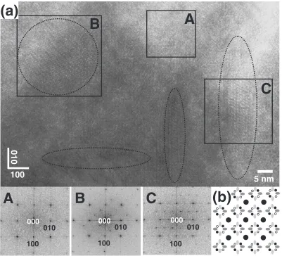
Figure 6(a) shows a high resolution image of a sample aged at 773 K for 144 ks. Fig. 6(b) is a key diagram of SAD pattern. The B2 main spots are indicated by large black circles; satellite spots are indicated by small black solid and open circles, and dark and light gray ellipses. The insets
denoted as A, B and C are the fast Fourier transformation (FFT) images obtained from the areas marked as A, B and C in Fig. 6(a), respectively. Area A contains only the B2 matrix. Areas B and C contain a precipitate with its habit in the foil plane and perpendicular to the foil plane, respec-tively. Approximate perimeters of the precipitates are out-lined by dotted lines in the figure. The FFT image for the area A exhibits the B2 reflections only. In the inset B, along B2 main spots, one can note the presence of a series of satellite reflections positioned at 1=3h110iB2, corresponding to the black small solid circles in the key diagram (Fig. 6(b)). No other satellite reflections can be noted in the FFT image, thus the satellites shown as the full small black circles in Fig. 6(b) come from the in-plane precipitates. In the inset C a series of satellites situated at1=4h210iB2 is seen; they correspond to the ones indicated as a dark gray ellipses in Fig. 6(b). No other spots are observed in inset C from Fig. 6. Thus, it is inferred that the reflections originate from the precipitate with its habit normal parallel to h010iB2. It should be also noted that the satellites are elongated in the directions perpendicular to the habit of the precipitates. The length of elongated spots corresponds well to the thickness of the precipitates (20nm). Hence the observed SAD as shown in Fig. 3(b) can be interpreted as the electron diffraction from 4 variants of the precipitates. The appearance of satellite reflections indicates that the crystal structure of the
precip-100 nm
Fig. 5 Bright field image of Ti-6Zr-52Ni aged for 14.4 ks at 773 K. Inset is the electron diffraction pattern, (e==½111
B2).
A
B
A
C
100 010
000
010
100
(b)
5 nm
100 010
000
100 010
000
B
C
(a)
[image:3.595.56.284.173.349.2] [image:3.595.98.499.393.757.2]itates is a super structure of the B2 phase. More detailed analysis of the precipitate structures is in progress.
Figure 7 shows a bright field (BF) image (a),½011B2SAD pattern (b) and its key diagram (c) for a sample aged in the same conditions as the one shown in Figure 6. Precipitates parallel to the trace ofð100ÞB2 can be seen, accompanied by coherency strain contrast. The SAD pattern exhibits the B2 main reflections which are indicated by black circles in the key diagram (Fig. 7(c)). Additional diffuse satellite spots are elongated parallel toh100iB2directions. They are situated at
1=3h0111iB2 positions and at 1=2h111iB2 between B2 main spots. The key diagram depicts them as dark gray ellipses. Fade streak-like reflections are also noted in Fig. 7(b). The key diagram indicates them as light gray segments. These reflections are found at 1=4h1111iB2 and they are oriented parallel to h11111iB2. The second family of streak-like reflections aligned parallel toh1111iB2 can be also seen.
Figure 8 depicts the BF image for the sample aged at 773 K for 360 ks. The precipitates grow to the much larger diameter of 160 nm and a thickness of 30nm. Martensite phase fills the space between the precipitates, suggesting that the coarse precipitates act as obstacles for the growth of martensite plates. Furthermore, one can note the absence of dark contrasts around the precipitates, indicating a dimin-ished coherency strain.
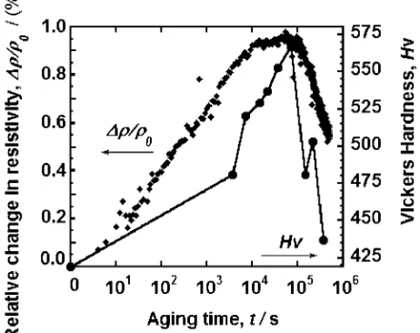
A drastic increase in microhardness was noted in the sample aged at 673 K up to 144 ks as shown in Fig. 9.
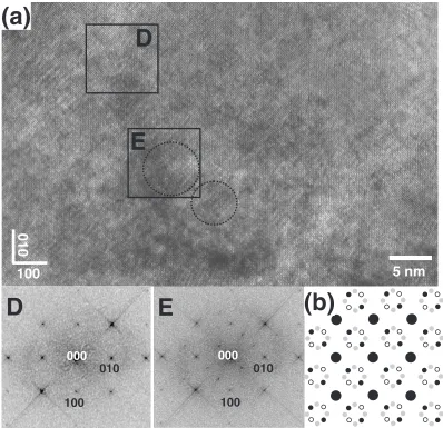
Concomitantly, a small but systematic variation in electrical resistivity was observed. In terms of microstructures, aging at 673 K for up to 144 ks did not alter the structure and the features of electron diffraction patterns. Figure 10 shows the TEM image of the sample aged at 673 K for 360 ks. Conventional TEM observations shown in Fig. 10(a) does not clearly reveal the precipitates but satellites are seen at
1=3h110iB2 and 1=4h210iB2 positions in the SAD pattern shown in Fig. 10(b). These positions are the same as those of the satellites shown in Fig. 3(b) but their shapes are spotty. HRTEM image with the electron beam parallel to½001B2is presented in Fig. 11(a). A precipitate is seen in the area delineated by a dotted circle of about 5 nm. The insets D and E show the FFT images for the corresponding areas in Fig. 11(a). The FFT image D exhibits only B2 reflections with no additional spots while the inset E exhibits the satellite spots at1=3h110iB2positions. These reflections are denoted as small black solid circles in the key diagram (Fig. 11(b)). It is difficult to assess the precipitates shape in these cases but the satellite spots shown in Fig. 10(b) suggest that they are spherical as delineated by the dotted lines in the area E in Fig. 11(a).
20 nm
000
100 011_
(b)
(a)
(c)
Fig. 7 Microstructure of Ti-6Zr-52Ni aged for 144 ks at 773 K: (a) Bright field image of B2 phase and precipitates; (b) selected area diffraction pattern, (e==½011
B2); (c) key diagram.
100 nm
Fig. 8 Bright field image of Ti-6Zr-52Ni aged for 360 ks at 773 K.
Fig. 9 Relative changes in electrical resistivity and microhardness as a function of aging time at 673 K; 0- electrical resistivity for the as-homogenized state;- the change of electrical resistivity with aging time.
20 nm
(a)
(b)
[image:4.595.49.288.73.205.2](c)
Fig. 10 Microstructure of Ti-6Zr-52Ni aged 360 ks at 673 K: (a) Bright field image. (b) selected area diffraction pattern (e==½001
[image:4.595.311.542.79.262.2] [image:4.595.55.283.260.431.2] [image:4.595.306.548.332.470.2]Electrical resistivity is sensitive to a change in the density of lattice defects, e.g., dislocations and vacancies, second phases and also to a change in short range or long range ordered atomic arrangements. Electrical resistivity increased with aging time up to 72 ks for aging at 673 K, as shown in Fig. 9. This is closely followed by the microhardness increase. TEM investigations showed no precipitation proc-ess for this aging duration. The SAD pattern revealed only a change in the diffuse scattering intensity. Moreover, it was shown that after aging at 673 K for 72 ks no transformation peaks were seen in DSC and the microhardness was the highest.10)Thus, it may be inferred the previously described evolutions may be related to a change in the short range ordered structures. For longer aging both resistivity and hardness decreased and this was accompanied by the appearance of precipitates as shown for the samples aged for 360 ks (Fig. 11).
3.2 Effect of deformation on microstructures in aged Ti-6.0 mol%Zr-53 mol%Ni
The microstructural changes during aging and their influence on mechanical behaviour were assessed for a Ti-6.0 mol%Zr-53 mol%Ni alloy. Cylindrical samples were compression tested for an imposed deformation of about
8% for five consecutive cycles.
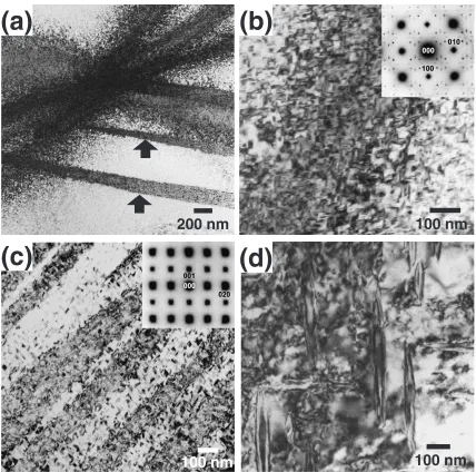
Figure 12 exhibits the BF micrograph for a sample aged 3.6 ks at 773 K. The SAD pattern shown as the inset of Fig. 12 reveals circular diffuse scattering similar to the ones observed in Fig. 1. The BF image also depicts a high density of dislocations introduced during the cyclic deformation. Since the sample exhibited only a small plastic strain,14)it can be inferred that the most of the dislocations may be introduced during the forward/reverse transformation as a lattice invariant shear. This does not contradict with the aligned structure seen in Fig. 12, which is parallel to the
f110gB2habit planes.
A low magnification BF image for the sample aged 144 ks at 773 K is presented in Fig. 13(a). The image exhibits the B2 matrix containing uniformly distributed plate-like precipi-tates. The black arrows indicate the presence of residual martensite.
The precipitates are accompanied by coherency strain contrast as seen in Fig. 13(b). They havef100gB2habit. The SAD pattern indicates sharp satellite spots at1=3h110iB2and diffuse satellite at 1=4h210iB2 elongated in h100iB2 direc-tions. This SAD pattern is similar to the one presented in Fig. 3(b). There are few dislocations seen in this area, although some areas with a higher density of dislocations
000
100
010
000
E
D
010
100
(a)
D
(b)
100
010
5 nm
[image:5.595.97.495.72.457.2]E
Fig. 11 Microstructure of Ti-6Zr-52Ni aged for 360 ks at 673 K: (a) high resolution TEM image (e==½001
were occasionally found in the same sample. Precipitates can be also found inside the residual martensite plates, as shown in Fig. 13(c), suggesting that the coherent precipitates do not interfere with martensitic transformation of the B2 matrix. Careful analysis of the SAD pattern shown as the inset also revealed the satellite spots having a very weak intensity. They are generated by the precipitates and accompany the martensite main reflections. Figure 13(d) shows the micro-structures of the sample aged at 773 K for 360 ks after compression cycles. The precipitates grew in the width to
500nm and thickness 50nm. They show no dark contrasts around them. The only dark contrasts are generated by the dislocations between the precipitates. The dislocations exhibited a random orientation in this case. This micro-structure is the manifestation of that the coarse precipitates does not act as the obstacles against the dislocation generation.
3.3 Ti-11 mol%Zr-52 mol%Ni alloy aging behaviour
The microstructural features after aging for an alloy with much higher Zr content (Ti-11.1 mol%Zr-51.5 mol%Ni) are
100 nm
200 nm
100 nm
(a)
(b)
(d)
[image:6.595.56.283.73.254.2]100 nm
(c)
Fig. 13 TEM bright field image of Ti-6Zr-53Ni aged at 773 K: (a) 144 ks, (b) higher magnification image of BF presented in (a); (c) precipitates inside martensite phase and (d) 360 ks.
[image:6.595.84.512.333.757.2]100 nm
Fig. 12 Bright field image of Ti-6Zr-53Ni aged for 3.6 ks at 773 K and compression tested for 5 cycles and corresponding electron diffraction pattern (inset,e==½001
presented in the followings. Figure 14 exhibits the BF micrograph for a sample aged 144 ks at 723 K. The SAD pattern shown as the inset of Fig. 14 reveals additional satellite spots situated at1=2h1111iB2. Diffuse elongated spots oriented parallel withh100iB2are also found. The BF image presents numerous precipitates uniformly distributed in B2 matrix. They have a diameter of around 30 nm and a thickness of 10 nm. The coherency strain contrasts can be observed around the precipitates. This is an indication that same type of precipitates as presented in Fig. 3–12 can be obtained in higher Zr content alloy after aging as well.
However, the distribution of the precipitates is not uniform after aging at 773 K for 144 ks. As shown in Fig. 15, the precipitates tend to form colonies. Their dimensions vary from 50 nm to 100 nm. Also, it seems that precipitation process may have an autocatalytic character as observed for the precipitation of Ti3Ni4 phase in binary TiNi alloys.15)
New precipitates tend to nucleate and grow in the elastic strain field generated by another precipitate.
4. Summary
Effect of isothermal aging on microstructure in Ti-6.0 mol%Zr-52 mol%Ni and Ti-11.1 mol%Zr-51.5 mol%Ni alloy was investigated. The influence of aging on micro-structure after cyclic compression deformation in Ti-6.0 mol%Zr-53 mol%Ni alloy was also assessed. These alloys exhibit two-stage aging behaviour. Namely, the first stage exhibits no microstructural changes during aging. This is associated with some intensification of the diffuse scatter-ing in electron diffraction patterns and an increase in electrical resistivity, probably due to a short range ordering process. The second stage is clearly related to the fine coherent precipitation accompanied by a decrease in elec-trical resistivity. The precipitates in the samples aged at 773 K are lenticular with f100gB2 habit. The electron
diffraction patterns are characterized by1=3h110iB2satellite spots, indicating that the structure is a super structure of the B2 phase. This is consistent with the high resolution electron microscopy and FFT analysis. Aging at 673 K produced the spherical precipitates but they exhibit similar characteristic satellite reflections in electron diffraction. The more detailed study on the structure of the precipitates is currently under way.
The matrix containing fine precipitates shows low dis-location density after cyclic compression loading. Residual martensite plates containing numerous precipitates were occasionally found, which suggests that the coherent precip-itates do not interfere with martensitic transformation.
REFERENCES
1) M. Nishida, C. M. Wayman and T. Honma: Metall. Trans.17(1986) 1505.
2) G. S. Fristov, J. van Hubeeck and Y. N. Koval: Scripta Mater.50(2004) 243.
3) J. Beyer and J. H. Mulder: MRS Symp. Proc.360(1995) 443. 4) L. Meisner and V. Sivokha: Journal de PhysiqueC8(1995) 765. 5) J. H. Mulder, J. H. Maas and J. Beyer:Proc. International Conference
on Martensitic Transformation, (Monterey Institute of Advanced Studies, 1996) 869.
6) L. L. Meisner, V. P. Sivokha and O. B. Perevalova: Phisica B262 (1999) 49.
7) S.-H. Kang, H.-J. Im, H.-W. Lee and T.-H. Nam: Metals and Materials International7(1999) 201.
8) S. K. Wu and S. F. Hsieh: J. Alloys. Comp.297(2000) 294. 9) V. P. Sivokha and L. L. Meisner: Phisyca B296(2001) 329. 10) A. Sandu, K. Tsuchiya, S. Yamamoto, Y. Todaka and M. Umemoto:
Mater. Science Forum539–543(2007) 3163.
11) Y. Murakami and D. Shindo: Phil. Mag. Lett.81(2001) 631. 12) T. Kikuchi, K. Ogawa, S. Kajiwara, T. Matsunaga, S. Miyazaki and
Y. Tomota: Philos. Mag.78A(1998) 467.
13) S. Kajiwara, T. Kikuchi, K. Ogawa, T. Matsunaga and S. Miyazaki: Philos. Mag. Lett.74(1996) 137.
14) A. M. Sandu, K. Tsuchiya, S. Yamamoto, Y. Todaka and M. Umemoto: Scripta Mater55(2006) 1079.
[image:7.595.49.289.71.248.2]15) J. K. Allafi, A. Dlouhy and G. Eggeler: Acta Mater.50(2002) 4255. 100 nm
Fig. 14 Bright field image of Ti-11.1Zr-51.5Ni aged for 144 ks at 723 K and corresponding electron diffraction pattern (inset,e==½011
B2).
100 nm
[image:7.595.313.541.71.240.2]