OncoTargets and Therapy 2018:11 2533–2543
OncoTargets and Therapy
Dove
press
submit your manuscript | www.dovepress.com 2533
O r i g i n a l r e s e a r c h
open access to scientific and medical research
Open access Full Text article
Prognostic factors for operable biliary tract
cancer: serum levels of lactate dehydrogenase,
a strong association with survival
ling Ma,1,* Jinrong Qiu,2,*
Yaodong Zhang,3 Tianzhu Qiu,1
Biao Wang,1 Wensen chen,4
Xiao li,5 Jing sun,1 Ke Wang,3
Xiangcheng li,3 Yanhong gu,1
Yongqian shu,1,6 Xiaofeng
chen1
1Department of Oncology, The First
affiliated hospital of nanjing Medical University, nanjing, People’s republic of china; 2Department of Biological Therapy,
eastern hepatobiliary surgery hospital affiliated to second Military Medical University, shanghai, People’s republic of china; 3liver Transplantation center,
The First affiliated hospital of nanjing Medical University, nanjing, People’s republic of china; 4Department of
infection Management Office, The First affiliated hospital of nanjing Medical University, nanjing, People’s republic of china; 5Department of Pathology, The
First affiliated hospital of nanjing Medical University, nanjing, People’s republic of china; 6Jiangsu Key lab of cancer
Biomarkers, Prevention and Treatment, collaborative innovation center for cancer Personalized Medicine, nanjing Medical University, nanjing, People’s republic of china
*These authors contributed equally to this work
Background: Biliary tract cancers (BTCs) are uncommon but fatal, with a low 5-year survival rate after surgical resection. This study was designed to investigate the prognostic factors for operable BTC.
Methods: Baseline demographics at diagnosis were retrospectively evaluated in 341 BTC patients undergoing radical surgery at The First Affiliated Hospital of Nanjing Medical University from January 2011 to December 2015. The association between prognostic factors and overall survival (OS) was determined by multivariate analysis using the Cox proportional hazards regression model.
Results: Our study showed that 341 patients were included in the analysis, of which 166 (48.7%) were males and 175 (51.3%) were females. Older age, depth of tumor invasion, positive surgical margin, lower hemoglobin, and higher lactic dehydrogenase (LDH) were associated with significantly worse OS using multivariate analysis. In the entire cohort,
the estimate of median OS in patients with LDH ,271 U/L was 36.291 months (95% CI;
30.989–41.594 months), and 30.736 months (95% CI; 19.154–42.318 months) in patients
with LDH $271 U/L (adjusted HR-1.505, 95% CI; 1.009–2.245, P = 0.045). Moreover, it
was investigated whether serum LDH retained its significance as a prognostic marker in BTC subgroups separately. The results showed that LDH was prognostic in patients with distal bile
duct (DBD) carcinoma undergoing radical surgery (HR-2.452, 95% CI; 1.167–5.152, P = 0.018).
However, there were no statistical differences between LDH and OS in multivariate analysis in the other three individual subgroups except for DBD carcinoma. This may be due to the limited number of patients in the study, indicating that a greater number of patients may be required for statistical significance.
Conclusion: Older age, depth of tumor invasion, positive surgical margin status, lower hemo-globin levels, and elevated serum LDH level are associated with poor survival in operable BTC patients. Serum LDH level is a cost-effective prognostic biomarker in patients with operable BTC and especially DBD carcinoma.
Keywords: biliary tract cancer, lactate dehydrogenase, tumor marker, prognosis, radical surgery
Introduction
Biliary tract cancers (BTCs) are rare but fatal. In the USA in 2017, the estimated number of new cancer cases from gallbladder and other biliary tissue was 11,740 (5,320 in males; 6,420 in females) and the estimated number of deaths was 3,830 (1,630 in males; 2,200 in females).1 In People’s Republic of China in 2015,
estimates of gallbladder cancer incidence and mortality were 52,800 (24,500 in males;
correspondence: Xiaofeng chen; Yongqian shu Department of Oncology, The First Affiliated hospital of nanjing Medical University, 300 guangzhou road, nanjing 210029, People’s republic of china
Tel +86 135 8517 2066; +86 25 6813 6428 Fax +86 25 8371 0040; +86 25 6813 6428 email [email protected]; [email protected]
Journal name: OncoTargets and Therapy Article Designation: Original Research Year: 2018
Volume: 11
Running head verso: Ma et al
Running head recto: Prognostic factors for operable biliary tract cancer DOI: 150502
OncoTargets and Therapy downloaded from https://www.dovepress.com/ by 118.70.13.36 on 25-Aug-2020
For personal use only.
Dovepress
Ma et al
28,300 in females) and 40,700 (18,800 in males; 21,800 in females), respectively.2 Tumors of the biliary tract typically
have a poor prognosis, with 5-year survival rates in the range of 5%–15%.3,4 According to the primary site, BTCs
encompass gallbladder carcinoma (GBC), distal bile duct (DBD) carcinoma, intrahepatic cholangiocarcinoma (IHC), and hilar cholangiocarcinoma.
Surgical resection is reported to be the only potential method for curative treatment of BTC.5,6 Data supporting an
adjuvant approach are sparse. The Phase III UK Advanced Biliary Cancer-02 (ABC-02) study confirmed the combina-tion of cisplatin with gemcitabine as the standard treatment for advanced BTC.7 Targeting angiogenesis and HER2/
neu blockade have been shown to be promising treatment strategies for BTC patients.8,9 However, prognostic factors
among BTC patients remain scarce. R0 resection and adju-vant chemotherapy may be prognosticators of long-term survival for patients with cholangiocarcinoma.10–12 Despite
surgical resection, recent reports concerning postoperative prognosis are unsatisfactory, with a 5-year survival rate of 27%–37% for DBD carcinoma, 22%–44% for IHC, and 11%–41% for hilar cholangiocarcinoma.13–17 Therefore, the
prognostic factors that predict long-term survival of BTC patients undergoing radical surgery are essential.
Serum tumor markers, including carbohydrate antigen 19-9 (CA19-9) and carcinoembryonic antigen (CEA), have been used as a complementary approach for the diagnosis of BTC in conjunction with radiology and histology/ cytology.18–20 Likewise, lactic dehydrogenase (LDH) is a
glycolytic enzyme that is essential for tumor maintenance and can be used as an attractive antitumor strategy by inhibiting glucose metabolism.21 LDH is reported to be associated
with the hypoxia-inducible factor (HIF) pathway, hypoxia, acidity, and neoangiogenesis.22 Serum LDH levels can
provide prognostic information on overall survival (OS) in patients with advanced pancreatic cancer, colorectal cancer, and melanoma.23–26 However, the prognostic roles of tumor
markers including CEA, CA19-9, and LDH have not yet been clearly elucidated in operable BTC.
The aim of this study was to investigate the role of tumor markers in predicting clinical outcome for BTC patients who have undergone radical surgery. It was hypothesized that elevated baseline serum LDH is prognostic of diminished OS in operable BTC, especially in operable DBD carcinoma.
Methods
study population and data collection
Patients who were diagnosed with BTC at The First Affiliated Hospital of Nanjing Medical University from
January 2011 to December 2015 were enrolled in this study. All patients were histologically confirmed with BTC. Due to a potentially different biology,27 ampullary tumors were
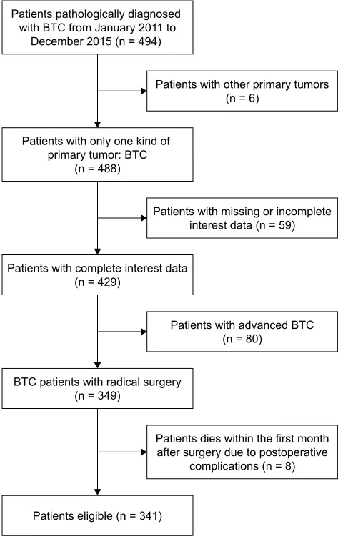
not included in this study. Exclusion criteria were as fol-lows: 1) BTC patients with concurrent primary tumors of other types, 2) patients with missing or incomplete interest data, 3) patients with advanced BTC, and 4) patients who expired within the first month after surgery due to postop-erative complications. In total, 341 patients were eligible for the analysis and were followed until December 2016 (Figure 1).
The baseline demographics that were evaluated in this study included age, sex, site of primary tumor (DBD, GBC, IHC, or hilar), depth of tumor invasion, lymph node status, metastasis, stage, histologic differentiation, lymphovascular invasion, perineural invasion, surgical margin status, hemoglobin, CEA, CA19-9, and LDH at diagnosis (prior to surgery). A CEA level of 4.7 ng/mL, CA 19-9 level of
3DWLHQWVZLWKRWKHUSULPDU\WXPRUV Q
3DWLHQWVZLWKFRPSOHWHLQWHUHVWGDWD Q
%7&SDWLHQWVZLWKUDGLFDOVXUJHU\ Q
3DWLHQWVZLWKPLVVLQJRULQFRPSOHWH LQWHUHVWGDWDQ
3DWLHQWVZLWKDGYDQFHG%7& Q
3DWLHQWVGLHVZLWKLQWKHILUVWPRQWK DIWHUVXUJHU\GXHWRSRVWRSHUDWLYH
FRPSOLFDWLRQVQ 3DWLHQWVSDWKRORJLFDOO\GLDJQRVHG
ZLWK%7&IURP-DQXDU\WR 'HFHPEHUQ
3DWLHQWVZLWKRQO\RQHNLQGRI SULPDU\WXPRU%7&
Q
3DWLHQWVHOLJLEOHQ
Figure 1 Flowchart on patient selection.
Abbreviation: BTc, biliary tract cancer.
OncoTargets and Therapy downloaded from https://www.dovepress.com/ by 118.70.13.36 on 25-Aug-2020
Dovepress Prognostic factors for operable biliary tract cancer
39 U/mL, and LDH level of 271 U/L were defined as cutoff values for normal levels according to the historical data and manufacturer’s recommendations.28,29 Stage classification
was based on pathological findings and documented accord-ing to the seventh edition of the Union for International Cancer Control tumor-node-metastasis staging system.30
The protocol was approved by the Ethics Committee of The First Affiliated Hospital of Nanjing Medical University prior to study initiation and conformed to the provisions of the Declaration of Helsinki (as revised in Fortaleza, Brazil, October 2013). Written informed consent was obtained from all patients.
statistics
Continuous variables were described as median (interquartile range [IQR]). OS was defined as the period from the day of operation to death or the last follow-up (December 2016). The OS of all patients in this study was analyzed and recorded. Survival distribution was estimated by the Kaplan–Meier method. The log-rank test was used to evaluate the equality of survivor functions across groups. The Cox proportional hazards model was used with a 95% CI for the univariate and multivariate analyses. The chi-square test was used to compare baseline characteristics of patients between the LDH groups. All statistical tests were two-sided. Statistical significance was defined as P , 0.05. All analyses were performed using SPSS software for Windows (version 19; IBM SPSS, Somers, NY, USA).
Results
Patients
A total of 341 BTC patients were available for our analysis. Patient and tumor characteristics of the entire cohort are presented in Table 1. Among the 341 patients, 146 (42.8%) patients were above 65 years of age and 195 (57.2%) patients were below 65 years of age. The study cohort consisted of 166 (48.7%) males and 175 (51.3%) females. The median serum levels of CEA, CA19-9, and LDH of the entire cohort were 3.40 ng/mL (IQR 2.11–5.27 ng/mL), 83.2 U/mL (IQR 24.8–417.2 U/mL), and 215 U/L (IQR 184–273 U/L), respectively.
Patient characteristics by primary site are reported in Table 2. Malignancies included 98 (28.7%) DBD carcinoma, 127 (37.2%) GBC, 38 (11.1%) IHC, and 75 (22.0%) hilar cholangiocarcinoma.
Outcomes in the entire cohort
To investigate whether the patient baseline characteristics and laboratory factors were associated with survival of
Table 1 Demographics for entire cohort of patients with biliary tract cancer (n = 341)
Variable Entire cohort
(N = 341) N (%)
age
,65 years
$65 years
195 (57.2) 146 (42.8) sex
Male Female
166 (48.7) 175 (51.3) Primary tumor site
Distal bile duct gallbladder intrahepatic hilar na
98 (28.7) 127 (37.2) 38 (11.1) 75 (22.0) 3 (0.9) Depth of tumor invasion
T1 T2 T3 T4 na
23 (6.7) 132 (38.7) 158 (46.3) 15 (4.4) 13 (3.8) lymph node status
negative Positive na
223 (65.4) 109 (32.0) 9 (2.6) Metastasis
negative Positive na
299 (87.7) 33 (9.7) 9 (2.6) seventh Uicc TnM stage
i ii iii iV na
41 (12.0) 138 (40.5) 100 (29.3) 46 (13.5) 16 (4.7) histologic differentiation
Well and moderately differentiated Poorly differentiated
na
151 (44.2) 169 (49.6) 21 (6.2) lymphovascular invasion
negative Positive na
296 (86.8) 39 (11.4) 6 (1.8) Perineural invasion
negative Positive na
172 (50.4) 163 (47.8) 6 (1.8) resection margin
negative Positive na
258 (75.7) 77 (22.6) 6 (1.8)
hemoglobin g/l, median (iQr) 125 (114–135)
cea ng/ml, median (iQr) 3.40 (2.11–5.27)
ca19-9 U/ml, median (iQr) 83.2 (24.8–417.2)
lDh U/l, median (iQr) 215 (184–273)
Abbreviations: na, not available; Uicc, Union for international cancer control; TnM, tumor node metastasis; iQr, interquartile range; cea, carcinoembryonic antigen; ca19-9, carbohydrate antigen 19-9; lDh, lactic dehydrogenase.
OncoTargets and Therapy downloaded from https://www.dovepress.com/ by 118.70.13.36 on 25-Aug-2020
Dovepress
Ma et al
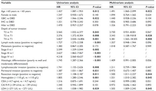
BTC patients undergoing radical surgery, univariate and multivariate analyses were performed. Univariate analysis revealed that better survival was associated with age ,65 years, with a tumor primary site from DBD, depth of tumor invasion,
negative lymph node status, early stage, well and moderately differentiated histological differentiation, absence of lympho-vascular invasion, absence of perineural invasion, negative surgical margin, hemoglobin $110 g/L, and LDH ,271 U/L
Table 2 Demographics by primary site for entire cohort of patients with biliary tract cancer (n = 338)
Variable Distal bile duct
(N = 98) N (%)
Gallbladder (N = 127) N (%)
Intrahepatic (N = 38) N (%)
Hilar (N = 75) N (%)
age
,65 years
$65 years
57 (58.2) 41 (41.8) 68 (53.5) 59 (46.5) 25 (65.8) 13 (34.2) 45 (60.0) 30 (40.0) sex Male Female 52 (53.1) 46 (46.9) 47 (37.0) 80 (63.0) 19 (50) 19 (50) 47 (62.7) 28 (37.3) Depth of tumor invasion
T1 T2 T3 T4 na 2 (2.0) 29 (29.6) 62 (63.3) 2 (2.0) 3 (3.1) 7 (5.5) 48 (37.8) 64 (50.4) 6 (4.7) 2 (1.6) 12 (31.6) 13 (34.2) 9 (23.7) 1 (2.6) 3 (7.9) 2 (2.7) 42 (56.0) 23 (30.7) 6 (8.0) 2 (2.7) lymph node status
negative Positive na 66 (67.3) 31 (31.6) 1 (1.0) 78 (61.4) 48 (37.8) 1 (0.8) 28 (73.7) 7 (18.4) 3 (7.9) 51 (68.0) 22 (29.3) 2 (2.7) Metastasis negative Positive na 93 (94.9) 4 (4.1) 1 (1.0) 110 (86.6) 16 (12.6) 1 (0.8) 28 (73.7) 7 (18.4) 3 (7.9) 67 (89.3) 6 (8.0) 2 (2.7) seventh Uicc TnM stage
i ii iii iV na 20 (20.4) 58 (59.2) 13 (13.3) 4 (4.1) 3 (3.1) 11 (8.7) 37 (29.1) 56 (44.1) 20 (15.7) 3 (2.4) 8 (21.1) 10 (26.3) 4 (10.5) 11 (28.9) 5 (13.2) 2 (2.7) 33 (44.0) 27 (36.0) 11 (14.7) 2 (2.7) histologic differentiation
Well and moderately differentiated Poorly differentiated na 49 (50.0) 43 (43.9) 6 (6.1) 56 (44.1) 65 (51.2) 6 (4.7) 16 (42.1) 20 (52.6) 2 (5.3) 30 (40.0) 41 (54.7) 4 (5.3) lymphovascular invasion negative Positive na 88 (89.8) 10 (10.2) 0 109 (85.8) 17 (13.4) 1 (0.8) 35 (92.1) 2 (5.3) 1 (2.6) 64 (85.3) 10 (13.3) 1 (1.3) Perineural invasion negative Positive na 45 (45.9) 53 (54.1) 0 84 (66.1) 42 (33.1) 1 (0.8) 22 (57.9) 15 (39.5) 1 (2.6) 21 (28.0) 53 (70.7) 1 (1.3) resection margin negative Positive na 76 (77.6) 22 (22.4) 0 100 (78.7) 26 (20.5) 1 (0.8) 31 (81.6) 6 (15.8) 1 (2.6) 51 (68.0) 23 (30.7) 1 (1.3)
hemoglobin g/l, median (iQr) 124.50 (111.00–134.25) 125.00 (113.00–134.00) 127 (117.75–139.25) 124.00 (113.00–135.00)
cea ng/ml, median (iQr) 3.65 (2.32–4.93) 3.42 (1.90–6.93) 3.30 (2.00–6.44) 3.10 (2.13–5.15)
ca19-9 U/ml, median (iQr) 130.90 (49.20–418.90) 40.01 (11.93–279.75) 69.60 (24.90–1,000) 204.00 (48.60–606.40) lDh U/l, median (iQr) 219.50 (183.00–300.25) 204.00 (177.00–250.00) 222.50 (182.00–273.00) 222.00 (194.00–267.0)
Abbreviations: na, not available; Uicc, Union for international cancer control; TnM, tumor node metastasis; iQr, interquartile range; cea, carcinoembryonic antigen; ca19-9, carbohydrate antigen 19-9; lDh, lactic dehydrogenase.
OncoTargets and Therapy downloaded from https://www.dovepress.com/ by 118.70.13.36 on 25-Aug-2020
Dovepress Prognostic factors for operable biliary tract cancer
(Table 3 and Figure 2). However, using univariate analysis, sex, metastasis, CEA, and CA19-9 were not statistically significant in OS in patients with radical surgery of BTC. Compared to LDH ,271 U/L, LDH $271 U/L was associ-ated with a worse OS using univariate analysis (HR-1.435, 95% CI; 1.038–1.982, P = 0.029). Moreover, LDH retained its significance as a prognostic marker in multivariate analysis (HR-1.505, 95% CI; 1.009–2.245, P = 0.045) in the entire cohort along with age, depth of tumor invasion, resection margin, and hemoglobin (Table 3).
comparison between high and low lDh
groups
For all patients included in the analysis, the estimate of median OS in patients with LDH ,271 U/L was 36.291 months (95% CI; 30.989–41.594 months) and 30.736 months (95% CI; 19.154–42.318 months) in patients with LDH $271 U/L (log-rank P = 0.028) (Figure 2E).
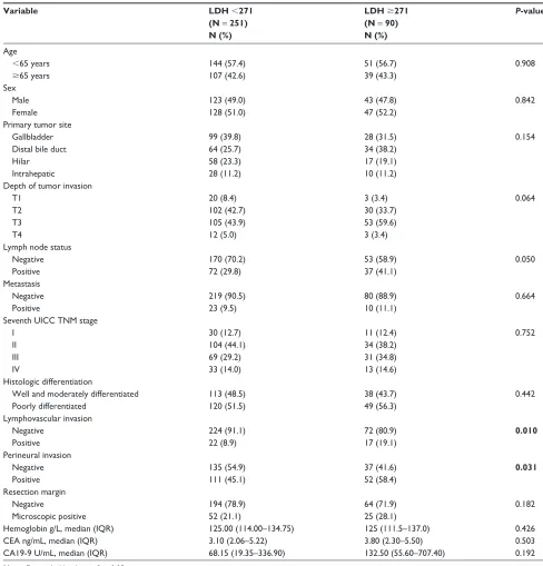
Patients showing a pretreatment serum LDH ,271 U/L were classified as LDH-low patients (251 patients, 73.6%, group A), whereas patients with pretreatment serum LDH level $271 U/L were classified as LDH-high patients
(90 patients, 26.4%, group B). The patients with positive lym-phovascular invasion and positive perineural invasion were more frequently found in the high LDH group (P = 0.010,
P = 0.031, respectively) (Table 4).
Outcomes by BTc site
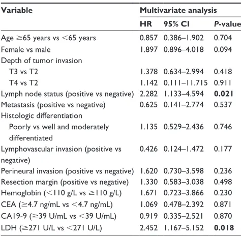
Considering the tumor primary site, it was further deter-mined whether serum LDH retained its significance as a prognostic marker in BTC subgroups separately. The results showed that LDH was prognostic in multivariate analysis for the DBD subgroup (HR-2.452, 95% CI; 1.167–5.152,
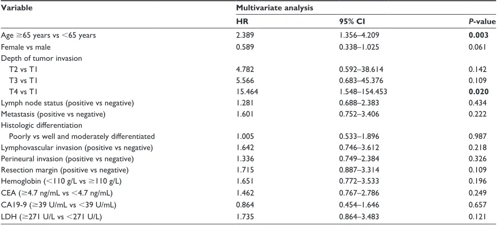
P = 0.018) (Table 5). The data showed a nonsignificant improvement in survival with LDH ,271 U/L compared with LDH $271 U/L in multivariate model in individual subgroups: GBC (Table S1) or hilar cancer (Table S2). Moreover, in multivariate analysis, age (P = 0.003) and depth of tumor invasion (P = 0.020) showed a significant association with OS in GBC (Table S1). Since there was a limited number of patients in the IHC subgroup, the cor-responding multivariate analysis was not available. Details of multivariate analysis outcomes for OS by BTC site are provided in Tables 5, S1, and S2.
Table 3 Univariate and multivariate analyses of patients with biliary tract cancer (n = 341)
Variable Univariate analysis Multivariate analysis
HR 95% CI P-value HR 95% CI P-value
age $65 years vs ,65 years 1.457 1.087–1.953 0.012 1.491 1.060–2.099 0.022
Female vs male 1.247 0.930–1.672 0.140 1.094 0.764–1.565 0.624
gBc vs DBD 1.547 1.066–2.246 0.022 1.440 0.928–2.236 0.104
ihc vs DBD 1.321 0.778–2.242 0.303 1.826 0.905–3.686 0.093
hilar vs DBD 1.470 0.957–2.257 0.078 1.326 0.791–2.223 0.284
Depth of tumor invasion
T2 vs T1 2.565 1.032–6.377 0.043 2.730 0.931–8.001 0.067
T3 vs T1 3.376 1.372–8.304 0.008 3.343 1.138–9.818 0.028
T4 vs T1 5.827 2.020–16.806 0.001 5.381 1.565–18.505 0.008
lymph node status (positive vs negative) 1.727 1.275–2.338 ,0.001 1.357 0.948–1.944 0.096
Metastasis (positive vs negative) 1.383 0.867–2.205 0.173 1.018 0.587–1.767 0.949
stage ii vs i 2.599 1.339–5.044 0.005 / / /
stage iii vs i 3.627 1.862–7.067 ,0.001 / / /
stage iV vs i 4.218 2.060–8.637 ,0.001 / / /
histologic differentiation (poorly vs well and moderately differentiated)
1.745 1.287–2.366 ,0.001 1.409 0.991–2.005 0.056
lymphovascular invasion (positive vs negative) 1.741 1.155–2.626 0.008 1.211 0.739–1.984 0.447
Perineural invasion (positive vs negative) 1.387 1.031–1.867 0.031 0.973 0.673–1.407 0.885
resection margin (positive vs negative) 1.557 1.108–2.187 0.011 1.500 1.011–2.227 0.044
hemoglobin (,110 g/l vs $110 g/l) 1.805 1.280–2.546 0.001 1.551 1.010–2.382 0.045
cea ($4.7 ng/ml vs ,4.7 ng/ml) 1.216 0.875–1.691 0.244 1.442 0.999–2.082 0.051
ca19-9 ($39 U/ml vs ,39 U/ml) 1.340 0.945–1.899 0.101 0.972 0.646–1.463 0.893
lDh ($271 U/l vs ,271 U/l) 1.435 1.038–1.982 0.029 1.505 1.009–2.245 0.045
Notes: Data in bold indicates P, 0.05. / indicates not included in the multivariate analysis.
Abbreviations: gBc, gallbladder carcinoma; DBD, distal bile duct; ihc, intrahepatic carcinoma; cea, carcinoembryonic antigen; ca19-9, carbohydrate antigen 19-9; lDh, lactic dehydrogenase.
OncoTargets and Therapy downloaded from https://www.dovepress.com/ by 118.70.13.36 on 25-Aug-2020
Dovepress
Ma et al
7LPHDIWHURSHUDWLRQPRQWKV &XPVXUYLYDO SUREDELOLW\ 1XPEHUDWULVN $JH $JH /RJUDQN3 $JH $JH 7LPHDIWHURSHUDWLRQPRQWKV &XPVXUYLYDO SUREDELOLW\ 1XPEHUDWULVN 7 7 7 7 /RJUDQN3 7 7 7 7
$
%
1XPEHUDWULVN 3UH+E 3UH+E 7LPHDIWHURSHUDWLRQPRQWKV &XPVXUYLYDO SUREDELOLW\ /RJUDQN3 +E +E 1XPEHUDWULVN 5 5 7LPHDIWHURSHUDWLRQPRQWKV &XPVXUYLYDO SUREDELOLW\ /RJUDQN3 5 5&
'
7LPHDIWHURSHUDWLRQPRQWKV &XPVXUYLYDO SUREDELOLW\ 1XPEHUDWULVN 3UH/'+ 3UH/'+ /RJUDQN3 /'+ /'+(
Figure 2 Kaplan–Meier analysis of overall survival.
Notes: Unadjusted overall survival curves for independent factors by univariate analyses: depth of tumor invasion (A), age (B), resection margin (C), pre-hb (D), and pre-lDh (E).
Abbreviations: pre-, preoperative; hb, hemoglobin; lDh, lactic dehydrogenase; T, depth of tumor invasion; r0, positive resection margin; r1, negative resection margin.
Discussion
Surgical resection offers the only potentially curative option for BTC,5 while postoperative prognosis is unsatisfactory after
extensive surgical resection.13–16 Moreover, the prognostic role
of tumor markers has not yet been clearly elucidated in BTC patients undergoing radical surgery. Therefore, it is significant to identify prognostic factors that predict long-term survival of BTC patients who have undergone radical surgery.
OncoTargets and Therapy downloaded from https://www.dovepress.com/ by 118.70.13.36 on 25-Aug-2020
Dovepress Prognostic factors for operable biliary tract cancer
Table 4 comparison between the high lDh and low lDh groups (n = 341)
Variable LDH ,271
(N = 251) N (%)
LDH $271 (N = 90) N (%)
P-value
age
,65 years
$65 years
144 (57.4) 107 (42.6)
51 (56.7) 39 (43.3)
0.908
sex Male Female
123 (49.0) 128 (51.0)
43 (47.8) 47 (52.2)
0.842
Primary tumor site gallbladder Distal bile duct hilar intrahepatic
99 (39.8) 64 (25.7) 58 (23.3) 28 (11.2)
28 (31.5) 34 (38.2) 17 (19.1) 10 (11.2)
0.154
Depth of tumor invasion T1
T2 T3 T4
20 (8.4) 102 (42.7) 105 (43.9) 12 (5.0)
3 (3.4) 30 (33.7) 53 (59.6) 3 (3.4)
0.064
lymph node status negative Positive
170 (70.2) 72 (29.8)
53 (58.9) 37 (41.1)
0.050
Metastasis negative Positive
219 (90.5) 23 (9.5)
80 (88.9) 10 (11.1)
0.664
seventh Uicc TnM stage i
ii iii iV
30 (12.7) 104 (44.1) 69 (29.2) 33 (14.0)
11 (12.4) 34 (38.2) 31 (34.8) 13 (14.6)
0.752
histologic differentiation
Well and moderately differentiated Poorly differentiated
113 (48.5) 120 (51.5)
38 (43.7) 49 (56.3)
0.442
lymphovascular invasion negative
Positive
224 (91.1) 22 (8.9)
72 (80.9) 17 (19.1)
0.010
Perineural invasion negative Positive
135 (54.9) 111 (45.1)
37 (41.6) 52 (58.4)
0.031
resection margin negative
Microscopic positive
194 (78.9) 52 (21.1)
64 (71.9) 25 (28.1)
0.182
hemoglobin g/l, median (iQr) 125.00 (114.00–134.75) 125 (111.5–137.0) 0.426
cea ng/ml, median (iQr) 3.10 (2.06–5.22) 3.80 (2.30–5.50) 0.503
ca19-9 U/ml, median (iQr) 68.15 (19.35–336.90) 132.50 (55.60–707.40) 0.192
Note: Data in bold indicates P, 0.05.
Abbreviations: lDh, lactic dehydrogenase; Uicc, Union for international cancer control; TnM, tumor node metastasis; iQr, interquartile range; cea, carcinoembryonic antigen; ca19-9, carbohydrate antigen 19-9.
LDH has been reported to be a prognosticator in various types of tumors.22,24,26 Some studies suggest a prognostic role
of LDH in patients with advanced BTC receiving certain chemotherapy.31,32 However, the prognosis of LDH in BTC
patients undergoing radical surgery and in different tumor primary sites remains unclear. The present study revealed that high serum LDH level is significantly associated with worse survival in patients with operable BTC.
In our study, we discovered the prognostic roles of age, depth of tumor invasion, surgical margin status, hemoglo-bin, and serum LDH level for OS in multivariate analysis in the entire cohort of 341 patients surgically treated for BTC. Pradeep et al33 found that age was a significant
pre-dictor of survival by multivariate analysis in analyzing prognostic factors in 87 patients with GBC. Cubertafond et al34 reported in a survey of 724 patients surgically treated
OncoTargets and Therapy downloaded from https://www.dovepress.com/ by 118.70.13.36 on 25-Aug-2020
Dovepress
Ma et al
Table 5 Multivariate analysis of the patients with distal bile duct carcinoma (n = 98)
Variable Multivariate analysis
HR 95% CI P-value
age $65 years vs ,65 years 0.857 0.386–1.902 0.704
Female vs male 1.897 0.896–4.018 0.094
Depth of tumor invasion T3 vs T2
T4 vs T2
1.378 1.142
0.634–2.994 0.111–11.715
0.418 0.911 lymph node status (positive vs negative) 2.282 1.133–4.594 0.021
Metastasis (positive vs negative) 0.625 0.141–2.774 0.537 histologic differentiation
Poorly vs well and moderately differentiated
1.135 0.529–2.436 0.746
lymphovascular invasion (positive vs negative)
0.426 0.124–1.472 0.177
Perineural invasion (positive vs negative) 1.620 0.730–3.598 0.236 resection margin (positive vs negative) 1.330 0.583–3.038 0.498 hemoglobin (,110 g/l vs $110 g/l) 1.671 0.723–3.866 0.230 cea ($4.7 ng/ml vs ,4.7 ng/ml) 1.069 0.478–2.392 0.871 ca19-9 ($39 U/ml vs ,39 U/ml) 0.919 0.335–2.521 0.870 lDh ($271 U/l vs ,271 U/l) 2.452 1.167–5.152 0.018 Note: Data in bold indicates P, 0.05.
Abbreviations: cea, carcinoembryonic antigen; ca19-9, carbohydrate antigen 19-9; lDh, lactic dehydrogenase.
for GBC that the long-term survival correlated with cancer stage: .60 months, .22 months, and 8 months for Tis, T1 to T2, and T3 to T4, respectively. R0 resection is reported to be prognosticator of long-term survival for patients with cholangiocarcinoma.10 These studies were consistent with
our findings.
CA19-9 and CEA have been used as a complementary approach for the diagnosis of BTC in conjunction with radi-ology and histradi-ology/cytradi-ology,18–20 while the prognostic roles
for better survival remain controversial. Lee et al35 suggested
that CA19-9 but not CEA served as a predictor of better survival in patients with advanced cholangiocarcinoma on gemcitabine-based chemotherapy. Peixoto et al36 reported
that CA19-9 and CEA were not significantly associated with OS in 106 patients with advanced BTC using univariate analysis. Our study showed a nonsignificant trend for better survival of patients with a low level of CA19-9 or CEA. The prognostic roles of CA19-9 and CEA for survival in BTC patients have to be further validated with a larger number of patients and prospective studies.
Moreover, we found that LDH is predictive of poor prognosis and clinical outcome in BTC patients undergoing radical surgery. In addition, Faloppi et al37 showed a
pos-sible prognostic role of pretreatment serum LDH levels in advanced BTC patients treated with first-line chemotherapy, confirming our hypothesis. In a Phase II study, Furuse
et al31 revealed that elevated serum LDH level was
associ-ated with a significantly shorter survival in 85 patients with unresectable BTC receiving combination chemotherapy of uracil-tegafur and doxorubicin (P = 0.043). Moreover, these findings are in accordance with previously published analyses suggesting a relationship between LDH levels and a worse outcome in other tumor types.22,23,38 Furthermore, our study
revealed that serum LDH can be a prognostic marker for DBD carcinoma. However, in terms of other primary tumors originating from the biliary tract, there were no statistically significant differences between LDH and OS in multivari-ate analysis in individual subgroups. The results indicmultivari-ated the possibility of different origins and biological behavior among these primary sites. Alternatively, this may be due to the limited number of patients in this study. Future studies may require greater numbers of patients to be considered statistically significant. The ABC-02 trial demonstrated that the site of the primary tumor within the biliary tract did not affect survival.7
Hypoxia-induced angiogenesis is probably the mecha-nism involved in high serum LDH levels and poor prognosis.22 Published studies demonstrated the association
between LDH5 up-regulation and HIF (HIF1α/HIF2α) accumulation, which is linked with activating transcription of multiple genes including those encoding glycolytic enzymes and vascular endothelial growth factor.21,39 In addition,
acti-vated HIF pathway and angiogenic factor production are correlated with enhanced tumor aggressiveness.40,41
Conclusion
These data shed light on older age, depth of tumor invasion, positive surgical margin status, lower hemoglobin, and elevated serum LDH level as prognostic factors for poor survival in operable BTC patients. Moreover, serum LDH could be used as a cost-effective prognostic biomarker for DBD cancer. A limitation of this study is its retrospective nature, which means it lacks availability of some data, but it is reflective of the clinical spectrum of Chinese patients and is a relatively large study on this rare tumor type. In addition, this analysis is a single institutional series. These findings might provide new opportunities for prospective multi-institutional trials toward clinical applications of serum LDH in BTC patients undergoing radical surgery.
Acknowledgments
This study was partly supported by the following funding sources: the National Natural Science Foundation of China
OncoTargets and Therapy downloaded from https://www.dovepress.com/ by 118.70.13.36 on 25-Aug-2020
Dovepress Prognostic factors for operable biliary tract cancer
(No 81672896), the Priority Academic Program Develop-ment of Jiangsu Higher Education Institutions (JX10231802), Provincial Initiative Program for Excellency Disciplines, Jiangsu Province, China, CSCO-Merck tumor study fund-ing (SCORE). No additional external fundfund-ing was received for this study. The funders had no role in study design, data collection and analysis, decision to publish, or preparation of the manuscript.
Disclosure
The authors report no conflicts of interest in this work.
References
1. Siegel RL, Miller KD, Jemal A. Cancer statistics, 2017. CA Cancer J
Clin. 2017;67(1):7–30.
2. Chen W, Zheng R, Baade PD, et al. Cancer statistics in China, 2015.
CA Cancer J Clin. 2016;66(2):115–132.
3. Anderson CD, Pinson CW, Berlin J, Chari RS. Diagnosis and treatment of cholangiocarcinoma. Oncologist. 2004;9(1):43–57.
4. Howlader N, Noone A, Krapcho M, et al. SEER Cancer Statistics Review, 1975–2008. Bethesda: National Cancer Institute; 2015. Available from: http://seer.cancer.gov/csr/1975_2008/. Accessed April 9, 2018. 5. Khan SA, Davidson BR, Goldin RD, et al; British Society of
Gastro-enterology. Guidelines for the diagnosis and treatment of cholangio-carcinoma: an update. Gut. 2012;61(12):1657–1669.
6. Skipworth JR, Olde Damink SW, Imber C, Bridgewater J, Pereira SP, Malago M. Review article: surgical, neo-adjuvant and adjuvant man-agement strategies in biliary tract cancer. Aliment Pharmacol Ther. 2011;34(9):1063–1078.
7. Valle J, Wasan H, Palmer DH, et al; ABC-02 Trial Investigators. Cisplatin plus gemcitabine versus gemcitabine for biliary tract cancer.
N Engl J Med. 2010;362(14):1273–1281.
8. Andersen JB, Spee B, Blechacz BR, et al. Genomic and genetic char-acterization of cholangiocarcinoma identifies therapeutic targets for tyrosine kinase inhibitors. Gastroenterology. 2012;142(4):1021–1031. e1015.
9. Miller G, Socci ND, Dhall D, et al. Genome wide analysis and clinical correlation of chromosomal and transcriptional mutations in cancers of the biliary tract. J Exp Clin Cancer Res. 2009;28:62.
10. Murakami Y, Uemura K, Sudo T, et al. Prognostic factors after surgical resection for intrahepatic, hilar, and distal cholangiocarcinoma. Ann
Surg Oncol. 2011;18(3):651–658.
11. Zhu GQ, Shi KQ, You J, et al. Systematic review with network meta-analysis: adjuvant therapy for resected biliary tract cancer. Aliment
Pharmacol Ther. 2014;40(7):759–770.
12. Horgan AM, Amir E, Walter T, Knox JJ. Adjuvant therapy in the treat-ment of biliary tract cancer: a systematic review and meta-analysis.
J Clin Oncol. 2012;30(16):1934–1940.
13. Sano T, Shimada K, Sakamoto Y, Yamamoto J, Yamasaki S, Kosuge T. One hundred two consecutive hepatobiliary resections for perihilar cho-langiocarcinoma with zero mortality. Ann Surg. 2006;244(2):240–247. 14. Shaib YH, Davila JA, Henderson L, McGlynn KA, El-Serag HB.
Endoscopic and surgical therapy for intrahepatic cholangiocarcinoma in the United States: a population-based study. J Clin Gastroenterol. 2007;41(10):911–917.
15. Nuzzo G, Giuliante F, Ardito F, et al. Intrahepatic cholangiocarcinoma: prognostic factors after liver resection. Updates Surg. 2010;62(1): 11–19.
16. Ramacciato G, Nigri G, Bellagamba R, et al. Univariate and multi-variate analysis of prognostic factors in the surgical treatment of hilar cholangiocarcinoma. Am Surg. 2010;76(11):1260–1268.
17. Primrose JN, Fox R, Palmer DH, et al. Adjuvant capecitabine for bil-iary tract cancer: the BILCAP randomized study. J Clin Oncol. 2017; 35(15_Suppl):4006.
18. Patel AH, Harnois DM, Klee GG, LaRusso NF, Gores GJ. The utility of CA 19-9 in the diagnoses of cholangiocarcinoma in patients with-out primary sclerosing cholangitis. Am J Gastroenterol. 2000;95(1): 204–207.
19. Wang YF, Feng FL, Zhao XH, et al. Combined detection tumor markers for diagnosis and prognosis of gallbladder cancer. World J
Gastroen-terol. 2014;20(14):4085–4092.
20. Grunnet M, Christensen IJ, Lassen U, et al. Decline in CA19-9 dur-ing chemotherapy predicts survival in four independent cohorts of patients with inoperable bile duct cancer. Eur J Cancer. 2015;51(11): 1381–1388.
21. Fantin VR, St-Pierre J, Leder P. Attenuation of LDH-A expression uncovers a link between glycolysis, mitochondrial physiology, and tumor maintenance. Cancer Cell. 2006;9(6):425–434.
22. Koukourakis MI, Giatromanolaki A, Sivridis E, et al; Tumour and Angiogenesis Research Group. Lactate dehydrogenase-5 (LDH-5) overexpression in non-small-cell lung cancer tissues is linked to tumour hypoxia, angiogenic factor production and poor prognosis. Br J Cancer. 2003;89(5):877–885.
23. Haas M, Heinemann V, Kullmann F, et al. Prognostic value of CA 19-9, CEA, CRP, LDH and bilirubin levels in locally advanced and metastatic pancreatic cancer: results from a multicenter, pooled analysis of patients receiving palliative chemotherapy. J Cancer Res Clin Oncol. 2013; 139(4):681–689.
24. Koukourakis MI, Giatromanolaki A, Sivridis E, Gatter KC, Harris AL, Tumour Angiogenesis Research Group. Lactate dehydro-genase 5 expression in operable colorectal cancer: strong association with survival and activated vascular endothelial growth factor pathway: a report of the Tumour Angiogenesis Research Group. J Clin Oncol. 2006;24(26):4301–4308.
25. Scartozzi M, Giampieri R, Maccaroni E, et al. Pre-treatment lactate dehydrogenase levels as predictor of efficacy of first-line bevacizumab-based therapy in metastatic colorectal cancer patients. Br J Cancer. 2012;106(5):799–804.
26. Petrelli F, Cabiddu M, Coinu A, et al. Prognostic role of lactate dehy-drogenase in solid tumors: a systematic review and meta-analysis of 76 studies. Acta Oncol. 2015;54(7):961–970.
27. Bronsert P, Kohler I, Werner M, et al. Intestinal-type of differentiation predicts favourable overall survival: confirmatory clinicopathological analysis of 198 periampullary adenocarcinomas of pancreatic, biliary, ampullary and duodenal origin. BMC Cancer. 2013;13:428. 28. Nisman B, Lafair J, Heching N, et al. Evaluation of tissue
polypep-tide specific antigen, CYFRA 21-1, and carcinoembryonic antigen in nonsmall cell lung carcinoma: does the combined use of cytok-eratin markers give any additional information? Cancer. 1998;82(10): 1850–1859.
29. Yu H, Son GM, Joh YG. The clinical significance of preoperative serum levels of carbohydrate antigen 19-9 in colorectal cancer. J Korean Surg
Soc. 2013;84(4):231–237.
30. Sobin LH, Gospodarowicz M, Wittekind C. TNM Classification of
Malignant Tumors. Hoboken: Wiley-Blackwell; 2009.
31. Furuse J, Okusaka T, Ohkawa S, et al. A phase II study of uracil-tegafur plus doxorubicin and prognostic factors in patients with unresect-able biliary tract cancer. Cancer Chemother Pharmacol. 2009;65(1): 113–120.
32. Saisho T, Okusaka T, Ueno H, Morizane C, Okada S. Prognostic factors in patients with advanced biliary tract cancer receiving chemotherapy.
Hepatogastroenterology. 2004;52(66):1654–1658.
33. Pradeep R, Kaushik SP, Sikora SS, Bhattacharya BN, Pandey CM, Kapoor VK. Predictors of survival in patients with carcinoma of the gallbladder. Cancer. 1995;76(7):1145–1149.
OncoTargets and Therapy downloaded from https://www.dovepress.com/ by 118.70.13.36 on 25-Aug-2020
Dovepress
Ma et al
34. Cubertafond P, Gainant A, Cucchiaro G. Surgical treatment of 724 carcinomas of the gallbladder. Results of the French Surgical Associa-tion Survey. Ann Surg. 1994;219(3):275–280.
35. Lee BS, Lee SH, Son JH, et al. Prognostic value of CA 19-9 kinetics during gemcitabine-based chemotherapy in patients with advanced cholangiocarcinoma. J Gastroenterol Hepatol. 2016;31(2):493–500. 36. Peixoto RD, Renouf D, Lim H. A population based analysis of
prog-nostic factors in advanced biliary tract cancer. J Gastrointest Oncol. 2014;5(6):428–432.
37. Faloppi L, Del Prete M, Casadei Gardini A, et al. The correlation between LDH serum levels and clinical outcome in advanced biliary tract cancer patients treated with first line chemotherapy. Sci Rep. 2016;6:24136.
38. Liu X, Meng QH, Ye Y, Hildebrandt MA, Gu J, Wu X. Prognostic significance of pretreatment serum levels of albumin, LDH and total bilirubin in patients with non-metastatic breast cancer. Carcinogenesis. 2015;36(2):243–248.
39. Koukourakis MI, Giatromanolaki A, Simopoulos C, Polychronidis A, Sivridis E. Lactate dehydrogenase 5 (LDH5) relates to up-regulated hypoxia inducible factor pathway and metastasis in colorectal cancer.
Clin Exp Metastasis. 2005;22(1):25–30.
40. Blancher C, Moore JW, Talks KL, Houlbrook S, Harris AL. Relationship of hypoxia-inducible factor (HIF)-1 alpha and HIF-2 alpha expression to vascular endothelial growth factor induction and hypoxia survival in human breast cancer cell lines. Cancer Res. 2000;60(24):7106–7113. 41. Wang M, Zhao X, Zhu D, et al. HIF-1α promoted vasculogenic mimicry
formation in hepatocellular carcinoma through LOXL2 up-regulation in hypoxic tumor microenvironment. J Exp Clin Cancer Res. 2017; 36(1):60.
OncoTargets and Therapy downloaded from https://www.dovepress.com/ by 118.70.13.36 on 25-Aug-2020
OncoTargets and Therapy
Publish your work in this journal
Submit your manuscript here: http://www.dovepress.com/oncotargets-and-therapy-journal
OncoTargets and Therapy is an international, peer-reviewed, open access journal focusing on the pathological basis of all cancers, potential targets for therapy and treatment protocols employed to improve the management of cancer patients. The journal also focuses on the impact of management programs and new therapeutic agents and protocols on
patient perspectives such as quality of life, adherence and satisfaction. The manuscript management system is completely online and includes a very quick and fair peer-review system, which is all easy to use. Visit http://www.dovepress.com/testimonials.php to read real quotes from published authors.
Dovepress
Dove
press
Prognostic factors for operable biliary tract cancer
Supplementary materials
Table S1 Multivariate analysis of the patients with gallbladder carcinoma (n = 127)
Variable Multivariate analysis
HR 95% CI P-value
age $65 years vs ,65 years 2.389 1.356–4.209 0.003
Female vs male 0.589 0.338–1.025 0.061
Depth of tumor invasion T2 vs T1
T3 vs T1 T4 vs T1
4.782 5.566 15.464
0.592–38.614 0.683–45.376 1.548–154.453
0.142 0.109
0.020
lymph node status (positive vs negative) 1.281 0.688–2.383 0.434
Metastasis (positive vs negative) 1.601 0.752–3.406 0.222
histologic differentiation
Poorly vs well and moderately differentiated 1.005 0.533–1.896 0.987
lymphovascular invasion (positive vs negative) 1.642 0.746–3.612 0.218
Perineural invasion (positive vs negative) 1.336 0.749–2.384 0.326
resection margin (positive vs negative) 1.715 0.887–3.314 0.109
hemoglobin (,110 g/l vs $110 g/l) 1.651 0.772–3.533 0.196
cea ($4.7 ng/ml vs ,4.7 ng/ml) 1.462 0.767–2.786 0.249
ca19-9 ($39 U/ml vs ,39 U/ml) 0.864 0.454–1.646 0.657
lDh ($271 U/l vs ,271 U/l) 1.735 0.864–3.483 0.121
Note: Data in bold indicates P, 0.05.
Abbreviations: cea, carcinoembryonic antigen; ca19-9, carbohydrate antigen 19-9; lDh, lactic dehydrogenase.
Table S2 Multivariate analysis of the patients with hilar carcinoma (n = 75)
Variable Multivariate analysis
HR 95% CI P-value
age $65 years vs ,65 years 0.697 0.268–1.815 0.460
Female vs male 1.528 0.591–3.948 0.382
Depth of tumor invasion T2 vs T1
T3 vs T1 T4 vs T1
4.281 2.712 11.119
0.331–55.335 0.147–50.016 0.396–311.937
0.265 0.502 0.157
lymph node status (positive vs negative) 0.618 0.140–2.731 0.525
Metastasis (positive vs negative) 0.140 0.015–1.289 0.083
histologic differentiation
Poorly vs well and moderately differentiated 1.132 0.397–3.231 0.817
lymphovascular invasion (positive vs negative) 1.493 0.348–6.409 0.590
Perineural invasion (positive vs negative) 0.300 0.115–0.782 0.014
resection margin (positive vs negative) 1.676 0.662–4.239 0.276
hemoglobin (,110 g/l vs $110 g/l) 1.373 0.262–7.196 0.708
cea ($4.7 ng/ml vs ,4.7 ng/ml) 1.889 0.697–5.119 0.211
ca19-9 ($39 U/ml vs ,39 U/ml) 2.370 0.570–9.853 0.235
lDh ($271 U/l vs ,271 U/l) 2.200 0.561–8.627 0.258
Note: Data in bold indicates P, 0.05.
Abbreviations: cea, carcinoembryonic antigen; ca19-9, carbohydrate antigen 19-9; lDh, lactic dehydrogenase.
OncoTargets and Therapy downloaded from https://www.dovepress.com/ by 118.70.13.36 on 25-Aug-2020