O R I G I N A L A R T I C L E
Open Access
In vitro labelling and detection of
mesenchymal stromal cells: a comparison
between magnetic resonance imaging of
iron-labelled cells and magnetic resonance
spectroscopy of fluorine-labelled cells
Stefania Rizzo
1*, Francesco Petrella
2,5, Ileana Zucca
3, Elena Rinaldi
4, Andrea Barbaglia
3, Francesco Padelli
3,
Fulvio Baggi
4, Lorenzo Spaggiari
2,5, Massimo Bellomi
1,5and Maria Grazia Bruzzone
6Abstract
Background:Among the various stem cell populations used for cell therapy, adult mesenchymal stromal cells (MSCs) have emerged as a major new cell technology. These cells must be tracked after transplantation to monitor their migration within the body and quantify their accumulation at the target site. This study assessed whether rat bone marrow MSCs can be labelled with superparamagnetic iron oxide (SPIO) nanoparticles and perfluorocarbon (PFC) nanoemulsion formulations without altering cell viability and compared magnetic resonance imaging (MRI) and magnetic resonance spectroscopy (MRS) results from iron-labelled and fluorine-labelled MSCs, respectively. Methods:Of MSCs, 2 × 10⁶were labelled with Molday ION Rhodamine-B (MIRB) and 2 × 10⁶were labelled with Cell Sense. Cell viability was evaluated by trypan blue exclusion method. Labelled MSCs were divided into four samples containing increasing cell numbers (0.125 × 10⁶, 0.25 × 10⁶, 0.5 × 10⁶, 1 × 10⁶) and scanned on a 7T MRI: for MIRB-labelled cells, phantoms and cells negative control, T1, T2 and T2* maps were acquired; for Cell Sense MIRB-labelled cells, phantoms and unlabelled cells, a19F non-localised single-pulse MRS sequence was acquired.
Results:In total, 86.8% and 83.6% of labelled cells and Cell Sense-labelled cells were viable, respectively. MIRB-labelled cells were visible in all samples with different cell numbers; pellets containing 0.5 × 10⁶and 1 × 10⁶of Cell Sense-labelled cells showed a detectable19F signal.
Conclusions:Our data support the use of both types of contrast material (SPIO and PFC) for MSCs labelling, although further efforts should be dedicated to improve the efficiency of PFC labelling.
Keywords:Mesenchymal stromal cells (MSCs), Superparamagnetic iron oxide (SPIO), Perfluorocarbon (PFC), Cell labelling, Cell tracking
* Correspondence:[email protected]
1Department of Radiology, European Institute of Oncology, via Ripamonti
435, 20141 Milan, Italy
Full list of author information is available at the end of the article
Key Points
SPIO-labelled cells are viable and MRI-detectable at all dilutions tested
PFC-labelled cells are viable and MRS-detectable if > 0.5 × 10^6
Detection of MSCs might consider multimodal approaches including SPIO and PFC compounds
Introduction
The persistent tissue and organ shortage has led to the emergence of regenerative medicine. This interdisciplin-ary field involving biology, medicine and engineering aims to repair, replace, maintain or enhance tissue and organ functions by means of cell therapy [1].
Among the various stem cell populations used for cell therapy, adult mesenchymal stromal cells (MSCs) have emerged as a major new technology with many potential clinical applications [2]. MSCs are a population of undiffer-entiated multipotent adult cells that naturally reside within the human body and are generally defined as plastic-adherent, fibroblast-like cells possessing extensive self-renewal properties and the potential to differentiate in vivo and in vitro into a variety of mesenchymal lineage cells [3].
MSCs have the ability to migrate and engraft at sites of inflammation and injury in response to cytokines, chemo-kines and growth factors. They can also exert local repara-tive effects through trans-differentiation into tissue-specific cell types or via the paracrine secretion of soluble factors with anti-inflammatory and wound-healing activities [4].
There is a specific need to track these cells after trans-plantation, evaluate different methods of imtrans-plantation, monitor cell migration within the body and quantify cell accumulation at the target site [2].
Magnetic resonance imaging (MRI) has emerged as an excellent method for tracking cells both in vivo and in vitro [5]. Many cell-tracking studies have used superparamagnetic iron oxide (SPIO) nanoparticle-based contrast agents to label cells for detection with MRI [6–8], while others have used perfluorocarbon (PFC) nanoemulsion formulations [9–11]. The19F nucleus is particularly suitable for labelling as its relative MRI sensitivity is only 17% lower than that of hydrogen nucleus [12]. Due to the absence of background
19
F signal in host tissue, fluorine contrast agents can not only localise, but also quantify the cells delivered by the dir-ect quantification of the probe through a known reference phantom [13]. However, no consistent results on MSC de-tection and MRI tracking have been obtained so far.
The purpose of this study was to assess if MSCs can be labelled with SPIO nanoparticles and PFC nanoemul-sion formulations without altering cell viability and compare MRI findings from iron-labelled MSCs with magnetic resonance spectroscopy (MRS) findings from fluorine-labelled MSCs.
Methods
Rat mesenchymal stem cell culture
StemPro® Rat Alk Phos Expressing MSCs were purchased from ThermoFisher Scientific (cat. no. R7789120) and cul-tured in α-Minimum Essential Medium, with nucleosides and GlutaMAX™(ThermoFisher Scientific, cat. no. 32571), supplemented with 10% fetal bovine serum (ThermoFisher Scientific, cat. no. 10270) and 1% penicillin-streptomycin solution 100× (Euroclone, cat. no. ECB3001D). MSCs were isolated from bone marrow of transgenic Fischer 344 rats expressing the human placental alkaline phosphatase (hPAP) gene. The medium was changed every third day and MSCs were maintained at 37 °C, 5% CO2.
SPIO labelling and19F labelling
Images were acquired by confocal microscopy (Eclipse TE2000-E microscope equipped with EZ-C1 scan-head; Nikon) using a 20× (NA 0.85) objective to evaluate labelling efficiency; structured illumination microscopy (SIM) with 100× Apo-TIRF (NA 1.49) objective (Nikon) was used to assess MIRB and Cell Sense cytoplasmic distribution. SIM achieves a lateral reso-lution of 100–130 nm [14].
The number of labelled MSCs (visualised by green fluorescence for Cell Sense-labelled MSCs and by red fluorescence for MIRB-labelled MSCs) and the number of analysed cells (number of cell nuclei, blue stained by DAPI) were evaluated using the ImageJ Software Cell-Counter plug-in. The percentage of MIRB-labelled and Cell Sense-labelled MSCs was then calculated.
Cell phantom preparation
Labelled MSCs were fixed in 4% paraformaldehyde and divided into four samples containing increasing numbers of cells (0.125 × 10⁶, 0.25 × 10⁶, 0.5 × 10⁶, 1 × 10⁶). MSC samples were prepared by centrifugation at 300 g for 5 min and each microcentrifugation tube was filled up with low-melting 1% agarose (Bio-Rad, 162–0017). Cell phantoms of unlabelled cells and a positive control with MIRB and Cell Sense at the same concentrations used for cell labelling were prepared.
MSCs were expanded and labelled in culture medium at physiological pH. Before MRI/MRS ana-lysis, cells were fixed in 4% PFA solution pH 7.4 and washed in PBS pH 7.4; therefore, the pH was always maintained at physiological level.
MRI/MRS characteristics and features
The acquisitions were performed on a horizontal 7T MRI scanner (Bruker BioSpec 70/30, Ettlingen, Germany) equipped with a gradient system reaching a maximum amplitude of 440 mT/m. Both MRI for MIRB-labelled cells and MRS for Cell Sense-labelled cells were acquired by a double nuclei (1H/19F) volume resonator with an inner diameter of 72 mm.
Since contrast agents relaxivity depends on extrinsic factors such as applied field and temperature, all MRI acquisition were performed at the magnet bore temperature, strictly maintained constant at 18 °C.
T1, T2 and T2* MRI maps were acquired with the same geometry (one 1-mm coronal slice including the cells pellet) on MIRB-labelled cells, the MIRB phantom and cell negative control. T2-weighted images with 0.7-mm slices in axial, sagittal and coronal geometries (with respect to the magnet reference frame) were also ac-quired to perform relaxometry maps reference images by visualising the hypo-intensity corresponding to the la-belled cells pellets.
T1 mapping was based on a rapid acquisition with re-laxation enhancement (RARE) sequence with a RARE factor of 2; 28 images, with a different repetition time (TR) in the range of 25–15,000 ms, were acquired; echo time (TE) was set to 4.6 ms, in-plane resolution was 313 × 313μm2, the slice thickness was 1 mm, the num-ber of excitations was 2; the total acquisition time was 14 min. A region of interest (ROI) was then selected cor-responding to the sample signal and signal intensity (SI) plotted versus the image TR. The curve was fitted by the function
SI¼AþC 1−exp−TR
T1
where A is the absolute bias and C the maximal SI. T2 mapping was based on a multi-slice multi-echo (MSME) sequence with a series of 200 echo images a with different TE (range 5–937.5 ms); TR was set to 5000 ms, in-plane resolution was 313 × 313 μm2, the slice thickness was 1 mm, the number of excitations was 28; the total acquisition time was 13 h. A ROI was then selected corresponding to the sample signal and SI was plotted versus the image TE. The curve was fitted by the function
SI¼AþC exp−TE
T2
where A is the absolute bias and C the maximal SI. T2* mapping is based on a multiple gradient echo (MGE) sequence with a series of 500 echo images with a different TE (range of 2–807 ms); TR is set to 5000 ms, in-plane resolution was 313 × 313 μm2, the slice thick-ness was 1 mm, the number of excitations was 5; the total acquisition time was 50 min. A ROI was selected corresponding to the sample signal and its SI was plot-ted versus the image TE. The fitting was performed by the T2 maps analysis function.
All curves fittings were performed by the built-in least squares minimisation Levenberg-Marquardt routines of OriginLab (Northampton, MA, USA, http://www.origin lab.com/) software. Reported errors are the fit’s standard deviations.
A T2-weighted RARE sequence was performed with the following parameters: Hermite pulse centred at 300 MHz with 2 kHz bandwidth, TR = 3100 ms, TE = 54 ms, flip angle (FA) = 90°, slice thickness = 0.7 mm, in-plane resolution = 156 μm × 156 μm2. The total acquisition time was 1 min 36 s.
detectability of each sample. For each sample, two acqui-sition sessions were performed one week apart.
Results
SPIO- and19F-labelled cells viability and labelling efficiency
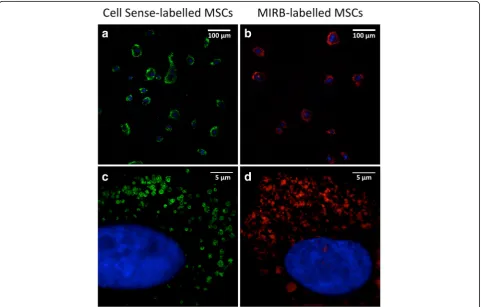
MIRB and Cell Sense uptake was assessed by confocal microscopy (Fig. 1a and b) to evaluate the labelling effi-ciency; cytoplasmic distribution in MSCs was observed through structured illumination microscopy (Fig. 1c and d), which provides a better resolved image and a more precise MIRB and Cell Sense localisation in MSC.
Cell viability was evaluated to exclude any toxic effect of SPIO or Cell Sense. MSC viability was not signifi-cantly different from the control group (86.8% versus 89.8%); 99.2% of MSCs were found to have fluorescent (red) signals from the rhodamine dye (MIRB) molecules distributed in the cytoplasm.
Similarly, after 24 h of incubation with Cell Sense, this fluorescently labelled tracer (detected in green) was found in 94.6% of MSCs, localised in vesicles diffusely distributed in the cytoplasm. We observed a slight de-crease in cell viability (83.6%) compared with control MSCs cultures (89.8%).
MRI of SPIO-labelled cells
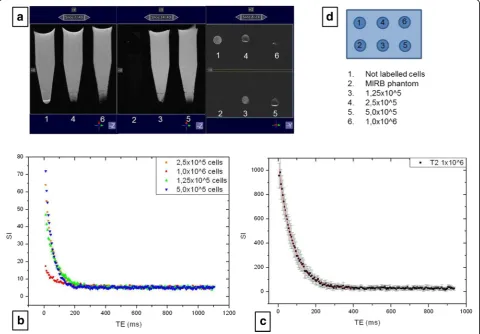
MIRB showed the following relaxation parameters: T1 = 559 ± 5 ms, T2 = 13.4 ± 0.8 ms, T2* = 3.6 ± 0.5 ms. T1, T2 and T2* values for unlabelled cells were 2041 ± 12 ms, 132 ± 0.4 ms and 4.6 ± 0.1 ms, respectively. Table 1 lists the relaxation times of the MIRB phan-tom, unlabelled and MIRB-labelled cells. Because the MIRB-labelled cells T2* decay times is too short and affected by agar susceptibility artefacts, meaningful curve fit cannot be obtained. T2-weighted images showed a hypointense signal corresponding to labelled cells pellet regions, visible in all samples with differ-ent numbers of cells (Fig. 2).
MRS of19F-labelled cells
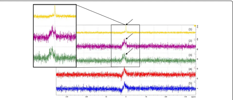
Only pellets containing 1 million and half a million of Cell Sense-labelled cells showed a detectable signal, although with a very low intensity in comparison with noise (Fig. 3). While negative control cells did not show any signal, the Cell Sense phantom showed its character-istic19F peak.
The two spectra acquired one week apart did not show any shift attributable to a pH variation.
Fig. 1Cell Sense-labelled MSCs and MIRB-labelled MSCs. MSCs were labelled with Cell Sense (10 mg/mL, green fluorescence;aandc) or with MIRB (50μg/mL, red fluorescence;bandd). Nuclei were counterstained with DAPI (blue). Images were acquired by confocal microscopy (aand
Discussion
Stem cells are being intensively studied for tissue infusion or transplantation into tissue for purposes of re-pair, revascularisation and other therapeutic measures [15–17]. However, the serial visualisation and tracking of transplanted stem cells, including their quantification at the site of injury and migration or retention in other sites, remain unresolved issues. Indeed, there is still
some uncertainty on the safety of these cells [18] and further pre-clinical validation is necessary before the clinical application of stem cell therapy.
A safe, non-invasive and repeatable imaging technique able to track the injected stem cells in vivo could solve some of the current issues, but no single imaging modal-ity can currently yield all the information required to monitor stem cell labelling and in vivo tracking.
As MRI is non-invasive and offers high spatial reso-lution (ranging from 50μm in animals and up to 300μm in whole body clinical scanners), it is a popular choice for in vivo cell tracking in pre-clinical and clinical studies [19]. For MRI tracking, stem cells need to be enriched with a contrast agent providing a sufficient positive or negative difference in SI to distinguish the cells from the image background. Contrast agents containing SPIO nanoparticles have been the preferred agent for short-term stem cell tracking, due to the pronounced signal decrease that even small amounts of these contrast media can create, owing to the so-called 'blooming artefact'.
Thanks to this negative contrast effect, the present study detected all the MIRB-labelled cell samples by a Table 1MIRB phantom cells, unlabelled cells and
MIRB-labelled cells relaxometry results. Reported errors are the fit’s
standard deviations
Cell number T1 relaxation time (ms)
T2 relaxation time (ms)
T2* relaxation time (ms)
0.125 E6 2128 ± 181 65.6 ± 0.8
0.25 E6 1493 ± 111 50.5 ± 0.5
0.5 E6 2000 ± 280 41.9 ± 0.4
1 E6 2500 ± 44 74.6 ± 0.5
Unlabelled cells 2041 ± 12 132 ± 0.4 4.6 ± 0.1
MIRB phantom 559 ± 5 13.4 ± 0.8 3.6 ± 0.5
Fig. 2aCoronal and axial T2-weighted images of six samples containing unlabelled cells (1), MIRB (2) and different amounts of MIRB labelled cells (3, 4, 5, 6).bT2 decays of the labelled cells samples.cT2 data fitting of the 1 × 10⁶cells pellet.dSamples positions
hypointense signal on T2-weighted images. The reso-lution of these images was compatible with in vivo ex-periments (acquisition time was 1 min 36 s, compatible with anaesthetised animal’s stay in the magnet) and can also depict the hypointensity related to pellets contain-ing the lowest number of cells (0.125 × 10⁶).
This study reports only mono-exponential relaxometry results. A bi-exponential model to fit the curves re-ported in Fig. 2b and c can be ruled out because previ-ous in vitro studies [20, 21], testing bi-exponential fitting functions in T2 decays, demonstrated a negligible com-ponent with a very short time constant and a predomin-ant component with a time constpredomin-ant comparable to the mono-exponential fitting relaxation time.
Furthermore, it can be assumed that our MIRB sample results were not affected by temperature dependence: in agreement with the literature [22] and as demonstrated by acquisitions repeated one week later, the proprieties of SPIO nanoparticles used as T2 contrast agents depend on the size of the nanoparticle core and are less influenced by temperature than T1 agents [22].
The SPIO negative contrast enhancement allows for detecting even very small numbers of labelled cells but can be confounded with other sources of magnetic sus-ceptibility effects, such as bleeds, blood vessels or air, de-pending on the anatomical structure under evaluation. As future pre-clinical applications may include lung evaluation for MSCs infusion in repair processes, such as broncho-pleural fistulae [23], and assessing cell mi-gration into the lungs, we also labelled MSCs with PFC nanoemulsions, which can be detected with19F MRI and MRS [9, 10].
Moreover, labelled MSCs stably express the hPAP gene, thus allowing cell detection by classical histochem-ical methods, making this gene a useful tracking tool for further in vivo MRI validation.
Our study labelled MSCs with a PFC emulsion conju-gated with BODIPY dye with a slight decrease in cell viability compared with the control (83.6% versus 89.8%), but only pellets containing 1 × 10⁶and 0.5 × 10⁶ of Cell Sense-labelled cells showed a detectable signal. Compared with labelling and tracking with metal-based contrast agents,19F labelling is less sensitive, therefore a relatively large amount of 19F, or a large number of labelled cells, must accumulate in order to generate a signal-to-noise ratio sufficient for tracking.
Our data support the use of both types of contrast media (SPIO and PFC) for MSCs labelling, although fur-ther efforts should be dedicated to improve the effi-ciency of PFC labelling.
Acknowledgements
The authors wish to thank Celsense, Inc. and Dr. Charles O’Hanlon for the supply of Cell Sense CS-ATM DM green. The paper has been edited by Anne Prudence Collins, Editor and Translator of Medical & Scientific Publications.
Authors’contributions
SR participated in the design of the study and contributed to the manuscript preparation. FPe, LS, MB, MGB participated in the design of the study. IZ, FPa, AB performed the MR experiments and contributed to the manuscript preparation. ER and FB participated in the design of the study and prepared and labelled the cells. All authors read and approved the final manuscript.
Competing interests
The authors declare that they have no competing interests.
Fig. 319F MRS of five samples containing Cell Sense (1) and different amounts of Cell Sense-labelled cells: 1 million (2), 5 × 10⁵(3), 2.5 × 10⁵(4),
Publisher’s Note
Springer Nature remains neutral with regard to jurisdictional claims in published maps and institutional affiliations.
Author details
1Department of Radiology, European Institute of Oncology, via Ripamonti
435, 20141 Milan, Italy.2Department of Thoracic Surgery, European Institute of Oncology, Milan, Italy.3Scientific Department, Neurological Institute IRCCS “Carlo Besta”, Milan, Italy.4Neuroimmunology and Neuromuscular Diseases Unit, Neurological Institute IRCCS“Carlo Besta”, Milan, Italy.5Department of Oncology and Hemato-Oncology, Università degli Studi di Milano, via Festa del Perdono 7, 20142 Milan, Italy.6Department of Neuroradiology, Neurological Institute IRCCS“Carlo Besta”, Milan, Italy.
Received: 24 December 2016 Accepted: 31 March 2017
References
1. Dai R, Wang Z, Samanipour R, Koo KI, Kim K (2016) Adipose-derived stem cells for tissue engineering and regenerative medicine applications. Stem Cells Int 2016:6737345
2. Ribot EJ, Gaudet JM, Chen Y, Gilbert KM, Foster PJ (2014) In vivo MR detection of fluorine-labelled human MSC using the bSSFP sequence. Int J Nanomedicine 9:1731–1739
3. Pittenger MF, Mackay AM, Beck SC et al (1999) Multilineage potential of adult human mesenchymal stem cells. Science 284(5411):143–147 4. Petrella F, Rizzo S, Borri A, Casiraghi M, Spaggiari L (2015) Current
perspectives in mesenchymal stromal cell therapies for airway tissue defects. Stem Cells Int 2015:746392
5. Helfer BM, Balducci A, Sadeghi Z et al (2013)19F MRI tracer preserves
in vitro and in vivo properties of hematopoietic stem cells. Cell Transplant 22:87–97
6. Heyn C, Bowen CV, Rutt BK, Foster PJ (2005) Detection threshold of single SPIO-labelled cells with FIESTA. Magn Reson Med 53:312–320
7. Stuckey DJ, Carr CA, Martin-Rendon E et al (2006) Iron particles for noninvasive monitoring of bone marrow stromal cell engraftment into, and isolation of viable engrafted donor cells from, the heart. Stem Cells 24:1968–1975
8. Bonetto F, Srinivas M, Heerschap A (2011) A novel (19)F agent for detection and quantification of human dendritic cells using magnetic resonance imaging. Int J Cancer 129:365–373
9. Canzi L, Castellaneta V, Navone S et al (2012) Human skeletal muscle stem cells anti-inflammatory activity ameliorate clinical outcome in amyotrophic lateral sclerosis model. Mol Med 18:401–411
10. Krafft MP (2001) Fluorocarbons and fluorinated amphiphiles in drug delivery and biomedical research. Adv Drug Deliv Rev 47:209–228
11. Ahrens ET, Helfer BM, O’Hanlon CF, Schirda C (2014) Clinical cell therapy imaging using a perfluorocarbon tracer and fluorine‐19 MRI. Magn Reson Med 72:1696–1701
12. Kok MB, de Vries A, Abdurrachim D et al (2011) Quantitative (1)H MRI, (19)F MRI, and (19)F MRS of cell-internalized perfluorocarbon paramagnetic nanoparticles. Contrast Media Mol Imaging 6:19–27 13. Tirotta I, Mastropietro A, Cordiglieri C et al (2014) A superfluorinated
molecular probe for highly sensitive in vivo(19)F-MRI. J Am Chem Soc 136:8524–8527
14. Schermelleh L, Heintzmann R, Leonhardt H (2010) A guide to super-resolution fluorescence microscopy. J Cell Biol 190:165–175 15. McKay R (2000) Stem cells: hype and hope. Nature 406:361–364
16. Deans RJ, Moseley AB (2000) Mesenchymal stem cells: biology and potential clinical uses. Exp Hematol 28:875–884
17. Kolios G, Moodley Y (2013) Introduction to stem cells and regenerative medicine. Respiration 85:3–10
18. Blum B, Benvenisty N (2009) The tumorigenicity of diploid and aneuploid human pluripotent stem cells. Cell Cycle 8:3822–3830
19. Rizzo S, Petrella F, Politi LS, Wang P (2017) Molecular imaging of stems cells: in vivo tracking and clinical translation. Stem Cells Int 2017:1783841 20. Lascialfari A, Zucca I, Asdente M et al (2005) Multiexponential T2-relaxation
analysis in cerebrally damaged rats in the absence and presence of a gadolinium contrast agent. Magn Reson Med 53:1326–1332
21. Jones CK, Whittall KP, MacKay AL (2003) Robust myelin water quantification: averaging vs. spatial filtering. Magn Reson Med 50:206–209
22. Estelrich J, Sánchez-Martín MJ, Busquets MA (2015) Nanoparticles in magnetic resonance imaging: from simple to dual contrast agents.
Int J Nanomedicine 10:1727–174123
23. Petrella F, Toffalorio F, Brizzola S et al (2014) Stem cell transplantation effectively occludes bronchopleural fistula in an animal model. Ann Thorac Surg 97:480–483
Submit your manuscript to a
journal and benefi t from:
7Convenient online submission 7Rigorous peer review
7Immediate publication on acceptance 7Open access: articles freely available online 7High visibility within the fi eld
7Retaining the copyright to your article