Electronic Thesis and Dissertation Repository
6-27-2013 12:00 AM
Pharmacogenetics of Oral Anticoagulants and Antiplatelets
Pharmacogenetics of Oral Anticoagulants and Antiplatelets
Inna Gong
The University of Western Ontario
Supervisor Richard Kim
The University of Western Ontario
Graduate Program in Pharmacology and Toxicology
A thesis submitted in partial fulfillment of the requirements for the degree in Doctor of Philosophy
© Inna Gong 2013
Follow this and additional works at: https://ir.lib.uwo.ca/etd
Part of the Medical Genetics Commons, and the Medical Pharmacology Commons
Recommended Citation Recommended Citation
Gong, Inna, "Pharmacogenetics of Oral Anticoagulants and Antiplatelets" (2013). Electronic Thesis and Dissertation Repository. 1335.
https://ir.lib.uwo.ca/etd/1335
This Dissertation/Thesis is brought to you for free and open access by Scholarship@Western. It has been accepted for inclusion in Electronic Thesis and Dissertation Repository by an authorized administrator of
(Thesis format: Integrated Article)
by
Inna Y. Gong
Graduate Program in Pharmacology and Toxicology
A thesis submitted in partial fulfillment of the requirements for the degree of
Doctor of Philosophy
The School of Graduate and Postdoctoral Studies The University of Western Ontario
London, Ontario, Canada
ii
Thromboembolic disorders are a major cause of morbidity and mortality. Therapeutic
intervention with anticoagulants and antiplatelets greatly reduces the risk of arterial and
venous thrombosis. However, the observed large interindividual variation in responsiveness
to these drugs indicates that subsets of patients are not attaining optimal therapy, resulting in
either lack of antithrombotic effect or elevated bleeding risk. Recently, single nucleotide
polymorphisms (SNPs) have been linked to the variation observed in efficacy and toxicity for
many cardiovascular drugs.
Warfarin has been the gold standard anticoagulant for prevention of stroke and
thromboembolism in atrial fibrillation (AF) and venous thromboembolism (VTE) patients.
SNPs in genes that affect warfarin metabolism (CYP2C9) and response (VKORC1) have an
important influence on response and dose, particularly during initiation. Accordingly, we
developed and evaluated the clinical utility of a pharmacogenetics-based initiation nomogram
in AF and VTE patients which provides safe and optimal anticoagulation therapy irrespective
of genetic variation.
The new oral anticoagulant (NOAC) rivaroxaban is highly dependent on the kidney for
elimination through glomerular filtration and active tubular secretion. Importantly,
interindividual variation in exposure and response to rivaroxaban has been reported. Using
iii
rivaroxaban clearance and brain accumulation. The contribution of interindividual variation
in transport and metabolism to the efficacy of rivaroxaban as well as other NOACs requires
to be addressed in patients.
Clopidogrel has been the gold standard antiplatelet for prevention of acute coronary
syndromes and stent thrombosis following percutaneous coronary intervention. Two
enzymes, CYP2C19 and PON1, have been proposed to affect clopidogrel bioactivation and
efficacy. We showed that CYP2C19 but not PON1 is capable of bioactivating clopidogrel to
its active metabolite. This is in line our finding that CYP2C19 genetic variation is a predictor
of clopidogrel pharmacokinetics and antiplatelet response while PON1 is not.
Taken together, these studies demonstrate the contribution of SNPs to the variation in
efficacy and toxicity of cardiovascular drugs, enabling personalized medicine for patients,
where an individual’s genetic makeup is used to guide drug selection and dosing.
Keywords
Oral anticoagulant therapy, antiplatelet therapy, warfarin, clopidogrel, rivaroxaban,
cytochrome P450 enzymes, single nucleotide polymorphisms, pharmacogenomics,
pharmacogenetics, metabolism, pharmacokinetics, pharmacodynamics, thromboembolic
iv
Chapter Four:
Gong IY, Tirona RG, Schwarz UI, Crown N, LaRue S, Langlois N, Dresser GK, Lazo-Langner A, Zou GY, Rodger M, Carrier M, Forgie M, Wells PS, Kim RB. (2011) Pharmacogenetics-guided warfarin loading and maintenance dosing regimen eliminates
VKORC1 and CYP2C9 associated variation in anticoagulation response. Blood,
118(11):3163-71.
IYG, RGT, UIS, NC, GKD, AL, PSW, and RBK designed the research study. IYG, NC, and SL participated in patient enrolment and data acquisition. IYG, RGT, UIS, NC, GKD, PSW, and RBK analyzed and interpreted of data. IYG and GZ conducted the statistical analysis. IYG and RBK wrote the manuscript. All authors provided feedback on the manuscript for important intellectual content. All authors approved the final version of the manuscript.
Chapter Five:
Gong IY, Schwarz UI, Crown N, Dresser GK, Lazo-Langner A, Wells PS, Kim RB, Tirona RG. (2011) Clinical and genetic determinants of warfarin pharmacokinetics and
pharmacodynamics during treatment initiation. PloS One, 6(11): e27808.
IYG, UIS, NC, GKD, AL, PSW, RBK, and RGT designed the research study. IYG measured warfarin and biomarker concentrations. IYG and RGT conducted the mathematical modeling. IYG, RBK, and RGT analyzed and interpreted of data. IYG and RGT wrote the manuscript. All authors provided feedback on the manuscript for important intellectual content. All authors approved the final version of the manuscript.
Chapter Six:
Gong IY, Mansell SE, Kim RB. (2013) Absence of both MDR1 (ABCB1) and BCRP
(ABCG2) transporters significantly alter rivaroxaban disposition and CNS entry. Basic Clin
Pharmacol Toxicol, 112(3):164-70.
IYG, and RBK designed the research study. IYG and SE conducted the experiments. IYG and RBK analyzed and interpreted the data. IYG and RBK wrote the manuscript. All authors approved the final version of the manuscript.
Chapter Seven:
Gong IY, Crown N, Suen CM, Schwarz UI, Dresser GK, Knauer MJ, Sugiyama D, DeGorter MK, Woolsey S, Tirona RG, Kim RB. (2012) Clarifying the importance of CYP2C19 and
PON1 in the mechanism of clopidogrel bioactivation and in vivo antiplatelet response. Eur
Heart J, 33(22):2856-2464a.
v
Dedication
vi
I would like to first express my deepest gratitude to my supervisor Dr Richard Kim. His expertise and vision has inspired me to recognize and embrace my own passion for knowledge and science. I could not have endured through the many challenges and hurdles faced as a PhD trainee without the strong support and encouragement from Richard. I am grateful for the wonderful opportunities he has given me that have made the past five years an invaluable experience.
Past and present members of my advisory committee have been helpful throughout my graduate program: Dr Rommel Tirona, Dr Ute Schwarz, Dr. Robert Gros, and Dr Ralf Rauch. In particular, I would like to especially thank Dr Tirona and Dr Schwarz for their guidance and support over the last five years. I have been fortunate to have their mentorship at each stage of my graduate program, as they have both generously shared their insight along this journey.
I would like to thank past and present members of the Kim and Tirona Lab for the engaging scientific and non-scientific discussions, fun, and laughter that have made my graduate experience that much more enjoyable and memorable: Dr Marianne DeGorter, Dr Michael Knauer, Dr Matilde Leon-Ponte, Sara LeMay, Colin Suen, Dr Wendy Teft, and Sarah Woolsey. I am fortunate to have colleagues that I call friends.
I have been privileged to have the opportunity for rewarding collaborations during my graduate training. I would like to thank my collaborators of the warfarin project for their
scientific input and contribution: Dr Natalie Crown, Dr George Dresser,Samantha LaRue,
Nicole Langlois, Dr Alejandro Lazo-Langner, Dr Guangyong Zou, Dr Dan Roden, Dr C. Michael Stein, Dr Marc Rodger, Dr Marc Carrier, Dr Melissa Forgie, and Dr Philip S Wells. I would like to thank my collaborators of the clopidogrel project for their scientific input and
contribution: Dr Natalie Crown, Dr George Dresser, Dr Daisuke Sugiyama, Dr Michael
Knauer, Dr Marianne DeGorter, Colin Suen, Sara LeMay, and Sarah Woolsey.
I would like to especially thank all the patients and healthy volunteers that have participated in our clinical studies. They have made a significant and valuable contribution to the area of pharmacogenomics research.
vii
Table of Contents
Abstract ... ii
Co-Authorship Statement... iv
Dedication ... v
Acknowledgments... vi
Table of Contents ... vii
List of Tables ... xiii
List of Figures ... xv
List of Appendices ... xvii
Abbreviations ... xviii
1 THROMBOEMBOLIC DISORDERS: PATHOGENESIS AND RATIONALE FOR ANTICOAGULANT AND ANTIPLATELET THERAPY ... 1
1.1 Introduction ... 2
1.2 Hemostasis ... 4
1.2.1 Platelet activation ... 4
1.2.2 Coagulation cascade... 7
1.3 Thrombosis ... 10
1.3.1 Arterial thrombosis ... 10
1.3.2 Atrial fibrillation ... 10
1.3.3 Percutaneous coronary intervention ... 15
1.3.4 Venous thrombosis... 16
1.4 Therapeutic interventions... 17
1.4.1 Oral anticoagulant therapy ... 17
1.4.2 Antiplatelet therapy ... 18
viii
2 PHARMACOGENETIC ADVANCES IN CARDIOVASCULAR MEDICINE:
RELEVANCE TO PERSONALIZED MEDICINE1 ... 24
2.1 Introduction ... 25
2.2 Oral Anticoagulants ... 29
2.2.1 Warfarin ... 29
2.2.2 New oral anticoagulants: dabigatran, rivaroxaban, and apixaban ... 33
2.3 Antiplatelets ... 34
2.3.1 Aspirin... 34
2.3.2 Clopidogrel ... 36
2.4 Conclusions ... 40
2.5 References ... 42
3 SPECIFIC AIMS AND HYPOTHESES ... 47
3.1 Specific aim 1 ... 48
3.2 Specific aim 2 ... 49
3.3 Specific aim 3 ... 51
3.4 Specific aim 4 ... 52
3.5 References ... 55
4 PROSPECTIVE EVALUATION OF A PHARMACOGENETICS-GUIDED WARFARIN LOADING AND MAINTENANCE DOSE REGIMEN FOR INITIATION OF THERAPY2 ... 57
4.1 Introduction ... 58
4.2 Experimental Section ... 60
4.2.1 Study sample and eligibility... 60
4.2.2 Clinical data collection and follow-up ... 63
ix
protocol ... 64
4.2.5 Loading doses ... 66
4.2.6 Maintenance doses ... 66
4.2.7 Refinement of loading and maintenance dose algorithm ... 72
4.2.8 Sample size ... 72
4.2.9 Primary and secondary outcomes ... 73
4.2.10 Statistical analysis ... 74
4.3 Results ... 77
4.3.1 Population characteristics ... 77
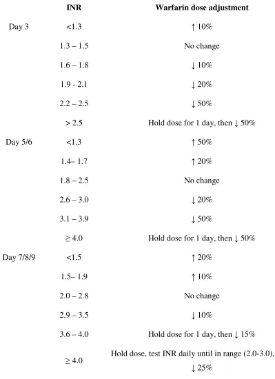
4.3.2 Time to first therapeutic INR (2.0-3.0) and overanticoagulation (INR ≥ 4) ... 77
4.3.3 Time to stable anticoagulation ... 78
4.3.4 Time spent within therapeutic range (INR 2.0-3.0) and above therapeutic range (INR > 3) ... 84
4.3.5 INR response time course during first 3 weeks of therapy ... 84
4.3.6 Secondary outcomes ... 86
4.3.7 Dosing algorithm assessment ... 86
4.4 Discussion ... 89
4.5 References ... 96
4.6 Supplemental Material ... 101
5 CLINICAL AND GENETIC DETERMINANT OF WARFARIN PHARMACOKINETICS AND PHARMACODYNAMICS DURING TREATMENT INITIATION3 ... 107
5.1 Introduction ... 108
5.2 Experimental Design ... 111
x
5.2.3 Warfarin drug level analysis ... 113
5.2.4 Proteins induced by vitamin K absence factor II (PIVKA-II) assay ... 113
5.2.5 Kidney function ... 114
5.2.6 PK-PD modeling ... 114
5.2.7 Vitamin K epoxide reductase protein expression in human liver ... 116
5.2.8 Determinants of warfarin kinetics and response ... 117
5.2.9 Statistical analysis ... 118
5.3 Results ... 119
5.3.1 PK-PD model performance ... 119
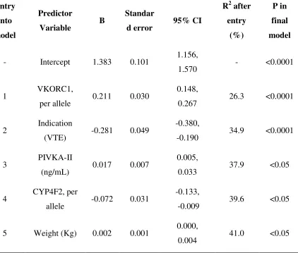
5.3.2 Determinants of S-warfarin clearance ... 123
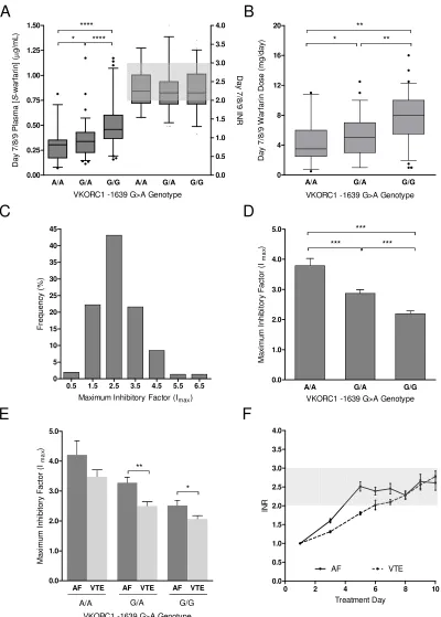
5.3.3 Therapeutic S-warfarin plasma concentration correlates with VKORC1 genotype ... 127
5.3.4 Determinants of S-warfarin PD ... 127
5.3.5 Correlation of VKORC1 genotype to hepatic VKOR protein levels ... 132
5.3.6 Simulated anticoagulation response with different warfarin initiation protocols ... 134
5.4 Discussion ... 138
5.5 References ... 144
6 ABSENCE OF BOTH MDR1 (ABCB1) AND BCRP (ABCG2) TRANSPORTERS SIGNIFICANTLY ALTER RIVAROXABAN DISPOSITION AND CNS ENTRY4 ... 150
6.1 Introduction ... 151
6.2 Methods... 153
6.2.1 Rivaroxaban permeability in polarized LLCPK, LMDR1 and Caco-2 monolayers ... 153
xi
6.2.4 Statistical analysis ... 155
6.3 Results ... 156
6.3.1 Permeability of rivaroxaban in LLCPK and LMDR1 cells ... 156
6.3.2 Permeability of rivaroxaban across intestinal Caco-2 cells ... 156
6.3.3 Rivaroxaban in vivo disposition in wildtype and knockout mice ... 160
6.4 Discussion ... 165
6.5 References ... 169
7 CLARIFYING THE IMPORTANCE OF CYP2C19 AND PON1 IN THE MECHANISM OF CLOPIDOGREL BIOACTIVATION AND IN VIVO ANTIPLATELET RESPONSE5 ... 172
7.1 Introduction ... 173
7.2 Methods... 175
7.2.1 Clinical study design ... 175
7.2.2 Clopidogrel bioactivation... 176
7.2.3 Liquid Chromatography Tandem Mass Spectrometry (LC-MS/MS) analysis ... 176
7.2.4 Data analysis ... 176
7.3 Results ... 177
7.3.1 Influence of CYP2C19, PON1, andCYP3A4 on clopidogrel kinetics and response... 177
7.3.2 Identification of other clopidogrel thiol metabolites in plasma ... 184
7.3.3 Biotransformation of clopidogrel to 2-oxo-clopidogrel ... 187
7.3.4 Biotransformation of 2-oxo-clopidogrel to H3 and H4 thiol metabolites187 7.3.5 Biotransformation of 2-oxo-clopidogrel to Endo thiol metabolite ... 188
7.4 Discussion ... 191
xii
7.6.1 Genotyping ... 204
7.6.2 Kinetics of clopidogrel metabolism ... 204
7.6.3 Kinetics of 2-oxo-clopidogrel metabolism ... 205
7.6.4 PON1-mediated hydrolysis of 2-oxo-clopidogrel ... 205
7.6.5 UHPLC-MS/MS analysis... 206
7.6.6 Midazolam LC-MS/MS analysis ... 207
7.6.7 Determination of paraoxonase activity ... 208
7.6.8 PON1 overexpression in an adenovirus system ... 208
7.6.9 Western blot analysis ... 209
7.6.10 Data analysis ... 209
8 DISCUSSION AND CONCLUSIONS ... 216
8.1 Summary and Discussion ... 217
8.1.1 Chapter Four ... 217
8.1.2 Chapter Five ... 218
8.1.3 Chapter Six... 219
8.1.4 Chapter Seven ... 220
8.2 Therapeutic Implications ... 221
8.3 Future Directions ... 225
8.4 Conclusions ... 228
8.5 References ... 232
Appendices ... 234
xiii
List of Tables
Table 1.3.1 Risk factor and characteristic differences between arterial thrombosis and venous
thrombosis. ... 13
Table 1.3.2 CHADS2 scoring system for determining stroke risk in atrial fibrillation patients. ... 14
Table 2.1.1 Summary of current evidence in cardiovascular pharmacogenetics. ... 28
Table 4.2.1 Patient characteristics (n=167)... 61
Table 4.2.2 Pharmacogenetics-based loading dose grid according to VKORC1 and CYP2C9 genotype. ... 68
Table 4.2.3 Final multiple linear regression for estimation of maintenance dose. ... 69
Table 4.2.4 Genetics-dependent dose grid for maintenance dose regression. ... 70
Table 4.2.5 Dose adjustment nomogram during initiation. ... 71
Table 4.3.1 Unadjusted and adjusted HRs for anticoagulation outcomes in patients with VKORC1 G/A or A/A and CYP2C9 variant genotype. ... 81
Table 4.3.2 Secondary outcomes following dosing with pharmacogenetics-based algorithm. ... 88
Table 4.6.1 Comparison of percent time spent within therapeutic range (2.0-3.0) and over range (>3) among VKORC1 and CYP2C9 genotype groups. ... 102
Table 4.6.2 Mean prescribed daily maintenance dose (mg) in relation to VKORC1 and CYP2C9 genotype. ... 103
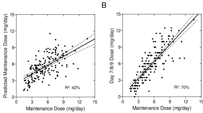
Table 5.3.1 Multiple linear regression analysis of independent predictors of S-warfarin clearance (L/day). ... 126
xiv
Table 6.3.2 Mean plasma tissue concentrations of rivaroxaban (ng/mL) after oral
administration of 2 mg/kg rivaroxaban (n = 6) in knockout and wild-type mice. ... 161
Table 6.3.3 Mean tissue concentrations of rivaroxaban (ng/mL) 4 hr after oral administration of 2 mg/kg rivaroxaban (n = 6) in knockout and wild-type mice. ... 162
Table 7.3.1 H4 active metabolite pharmacokinetic parameters following administration of a single 75 mg oral dose of clopidogrel. ... 180
Table 7.3.2 Platelet response pre- and 4 h post-clopidogrel administration. ... 182
Table 7.6.1 Healthy volunteer baseline demographics (n = 21). ... 212
Table 7.6.2 Kinetic parameters of clopidogrel metabolism determined in vitro. ... 213
xv
List of Figures
Figure 1.1 Schematic representation of arterial thrombosis. ... 6
Figure 1.2 Schematic representation of the coagulation cascade. ... 9
Figure 2.1 Pharmacogenetic determinants of interindividual variability in cardiovascular therapy... 27
Figure 4.1 The effect of pharmacogenetics-guided dosing on time to primary events. ... 80
Figure 4.2 The effect of pharmacogenetics-guided dosing on time to stability. ... 83
Figure 4.3 The effect of genotype-guided dosing on response time course during the first 3 weeks of warfarin therapy. ... 85
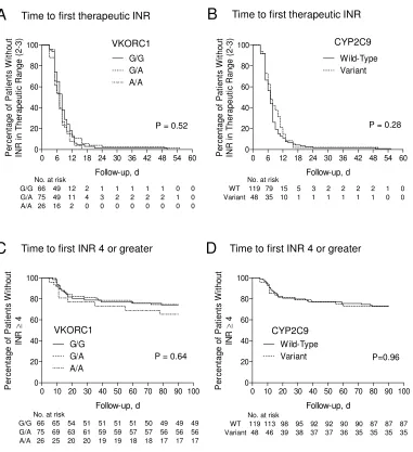
Figure 4.4 Association of predicted maintenance dose to observed maintenance dose. ... 87
Figure 4.5 A schematic representation of the pharmacokinetic-pharmacodynamic (PK-PD) model employed to determine loading doses and dose-adjustment nomogram. ... 104
Figure 4.6 Concentration response curves necessary for formulating loading doses. ... 105
Figure 4.7 An automated dose calculator that incorporates the WRAPID pharmacogenetics-based dosing algorithm and adjustment nomogram for warfarin initiation. ... 106
Figure 5.1 PK-PD model performance. ... 122
Figure 5.2 Determinants of S-warfarin clearance. ... 125
Figure 5.3 Determinants of maximal inhibitory factor, Imax. ... 130
Figure 5.4 The influence of VKORC1 -1639G>A promoter genotype on hepatic VKOR protein expression levels. ... 133
xvi
Figure 6.2 Rivaroxaban pharmacokinetics in mice. ... 163
Figure 6.3 Rivaroxaban liver, kidney and brain distribution in mice. ... 164
Figure 7.1 The role of CYP2C19 and PON1 genetic polymorphisms in clopidogrel pharmacokinetic and pharmacodynamic responses. ... 179
Figure 7.2 The role of CYP3A4 activity in clopidogrel pharmacokinetics and pharmacodynamics. ... 183
Figure 7.3 Representative chromatograms of derivatized H4 and Endo metabolite. ... 186
Figure 7.4 Clopidogrel bioactivation in vitro. ... 190
Figure 7.5 Schematic summary of clopidogrel bioactivation. ... 199
xvii
List of Appendices
Appendix A: Ethics Approval ... 235
Appendix B: Copyright Approval ... 241
Appendix C: Future of oral anticoagulation therapy: Importance of pharmacokinetic profile
and variability as determinants of dose and response to dabigatran, rivaroxaban, and
xviii
Abbreviations
ABCB1 ATP-binding cassette subfamily B member 1
ABCG2 ATP-binding cassette subfamily G member 2
ACS acute coronary syndromes
ADP adenosine diphosphate
ADR adverse drug reaction
AF atrial fibrillation
anti-Fxa anti factor Xa
API apixaban
ApoE apolipoprotein E
aPTT activated partial thromboplastin time
AUC area under the plasma concentration curve
BBB blood brain barrier
BCRP breast cancer resistant protein
BID twice daily
BMS bare-metal stent
CAD coronary artery disease
CALU calumenin
xix
CI confidence interval
CKD-EPI Chronic Kidney Disease Epidemiology Collaboration
CL clearance
CL/F apparent clearance
CLint intrinsic clearance
Cmax maximum plasma concentration
CNS central nervous system
COX cyclooxygenase
Cp plasma concentration
CrCl creatinine clearance
CYP cytochrome P450
CYP1A2 cytochrome P450 1A2
CYP2B6 cytochrome P450 2B6
CYP2C19 cytochrome P450 2C19
CYP2C9 cytochrome P450 2C9
CYP3A4 cytochrome P450 3A4
CYP4F2 cytochrome P450 4F2
DAB dabigatran
xx
DTI direct thrombin inhibitor
DVT deep vein thrombosis
ECG electrocardiogram
ECT ecarin clotting time
eGFR estimated glomerulus filtration rate
EM extensive metabolizer
ESRD end stage renal disease
FDA Food and Drug Administration
Fxa factor Xa
GGCX gamma-glutamyl carboxylase
GI gastrointestinal
GSH glutathione
GWAS genome-wide association study
Hemoclot diluted version of thrombin time
HR hazard ratio
IC50 drug affinity
Imax maximum inhibitory factor
xxi
INR international normalized ratio
K zero-order
ka absorption constant
ke elimination constant
keto ketoconazole
Km enzyme affinity
kout first-order
LC-MS/MS liquid chromatography tandem mass spectrometry
LHSC London Health Sciences Centre
LOWESS locally weighted scatterplot smoothing regression
MDR1 P-glycoprotein; multi drug resistance protein 1
Mdr1a multi drug resistance protein isoform 1a
Mdr1adef multie drug resistance protein isoform 1a deficient
Mdr1b multi drug resistance protein isoform 1b
MI myocardial infarction
MPB 2-bromo-3-methoxyacetophenone
NOAC new oral anticoagulant
NSAIDs nonsteroidal anti-inflammatory drugs
xxii
OD once daily
P-gp P-glycoprotein
PCI percutaneous coronary intervention
PD pharmacodynamics
PE pulmonary embolism
PGE1 prostaglandin E1
PIVKA-II proteins induced by vitamin K absence factor II
PK pharmacokinetics
PK-PD pharmacokinetics-pharmacodynamics
PON1 paraoxonase 1
PRU platelet reactive units
PT prothrombin time
QALY quality adjusted life years
RCT randomized clinical trial
rif rifampicin
RIV rivaroxaban
RM reduced metabolizer
xxiii
SEM standard error of the mean
SNP single nucleotide polymorphisms
SSE systemic emoblism
ST stent thrombosis
STEMI ST segment elevation myocardial infarction
t1/2 half-life
t1/2β terminal half-life
TF tissue factor
TIA transient ishemic attack
TOH The Ottawa Hospital
TT thrombin time
TTR time in therapeutic range
V volume of distribution
VASP vasodilator-stimulated phosphoprotein
Vd volume of distribution
VKOR vitamin K epoxide reductase
VKORC1 vitamin K epoxide reductase subunit 1
Vmax maximum rate achieved by enzymatic system
xxiv
vWF von Willebrand factor
1
THROMBOEMBOLIC DISORDERS: PATHOGENESIS
AND RATIONALE FOR ANTICOAGULANT AND
1.1
Introduction
Maintenance of blood fluidity within the vasculature is an important human physiological
process. Under normal conditions, there is a fine equilibrium between pathological states
of hypocoagulability and hypercoagulability. Hemostasis refers to a series of normal
physiological processes that confine blood to the vascular spaces, maintain blood fluidity,
and importantly, clot formation to limit hemorrhage following vascular injury.
Thrombosis is pathological clot formation when hemostasis is inappropriately activated
in the absence of a bleeding event. In the 1800s, Rudolf Virchow postulated a triad of
causes for thrombosis formation: changes in the composition of blood, alterations in the
vessel wall, and disruption of blood flow (1). Indeed, in the event of a vascular vessel
injury, a sequence of events is generated in response: vessel constriction to reduce blood
flow, hemostatic platelet plug formation at the trauma site following platelet adhesion,
activation, and aggregation, formation of a fibrin clot to stabilize the platelet plug by
activation of a series of proteins in the coagulation cascade. As such, a thrombus forms in
the presence of alterations in the hemostatic system leading to inappropriate platelet
aggregation and coagulation. A thrombus in a large blood vessel will decrease blood flow
through that vessel while a thrombus in a small blood vessel may completely cut-off
blood flow resulting in an occlusive thrombus. An embolism is the dislodging of the
thrombus from the site of formation that travels to a distal vessel leading to blood flow
Thromboembolic disorders are a significant source of mortality and morbidity. There are
two types of thrombosis, arterial and venous thrombosis, whereby the clinical and
therapeutic management differs, reflecting the distinct pathogenesis of the two
classifications. Arterial thrombosis usually occurs after the erosion or rupture of an
atherosclerotic plaque, potentially leading to ischemic events. Cardiac ischemia and
stroke are the most devastating clinical manifestations of atherothrombosis. Venous
thromboembolism (VTE) is represented by two main manifestations, deep venous
thrombosis (DVT) and pulmonary embolism (PE). The most devastating clinical
consequence of VTE is PE where majority of PEs result from DVT that have dislodged
from site of formation in the lower extremities and traveled to the pulmonary circulation.
Understanding the pathogenic processes leading to either arterial or venous thrombosis is
crucial for selecting effective and safe antithrombotic agents for management of patients
with thromboembolic disorders. While arterial thrombosis undoubtedly involves the
coagulation cascade, platelet activation and aggregation plays a more prominent role in
the rapidly flowing arteries. As such, arterial thrombosis is often referred to as white clot,
rich in platelets and little red blood cells. On the other hand, venous thrombosis is
associated with venous stasis and hypercoagulability. Thus, the coagulation cascade plays
a prominent role in formation of a venous thrombus, often referred to as red clot due to
1.2
Hemostasis
The endothelium of blood vessel walls plays an important role in maintaining vasculature
integrity. Vessel wall damage or disruption of the endothelium leads to exposing the
collagen present in the subendothelial matrix, resulting in platelet activation, aggregation,
and formation of a primary platelet plug. Concurrently, subendothelial tissue factor (TF)
is also exposed on the damaged endothelium leading to activation of the coagulation
cascade, resulting in formation of a fibrin mesh to stabilize the platelet plug (2).
1.2.1
Platelet activation
Platelets are small disk shaped colorless cells present in the blood that play a vital role in
hemostasis and are the key factor in the pathogenesis of arterial thrombosis. Vascular
injury may cause the rupture of an atherosclerotic plaque and denudation of the
endothelium (Figure 1.1). The exposure of subendothelial collagen bound to a protein
known as von Willebrand factor (vWF) facilitates platelet adhesion to the vessel wall. At
the site of injury, vWF binds platelets via the glycoprotein Ib/IX/V receptor complex on
the platelet membrane (3). At low shear rates, platelets can also bind to subendothelial
collagen through other receptors such as the glycoprotein IV, VI, and Ia/IIa. Upon
adhering to the vessel wall, platelets release the granules of platelet agonist thromboxane
A2 and adenosine diphosphate (ADP), initiating an autocrine/paracrine signaling cascade
of platelet activation. Activated platelets undergo a change in morphology from smooth
platelet aggregation. The formation of this hemostatic plug is essential for wound healing.
However, formation of a thrombotic occlusion in coronary arteries can cause tissue
ischemia leading to conditions known as acute coronary syndromes (ACS), in the form of
Figure 1.1 Schematic representation of arterial thrombosis.
An atherosclerosis plaque develops through the accumulation of lipid deposits. The
rupture of an atherosclerotic plaque is the primary trigger for arterial thrombosis by
promoting platelet activation and fibrin generation.
Endothelial Cells
Vessel wall
Collagen von Willebrand Factor
Tissue Factor
Activated Platelet Fibrin
1.2.2
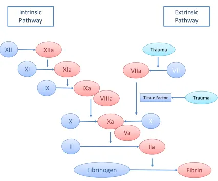
Coagulation cascade
The coagulation cascade plays a pivotal role in forming the fibrin mesh to reinforce the
hemostatic platelet plug. The ultimate formation of fibrin is dependent on two distinct
pathways: the extrinsic and intrinsic pathway. It was previously thought that both
pathways were equally crucial to hemostasis. However, it is evident now that the
extrinsic pathway is the predominant pathway for initiation and activation of the
coagulation cascade during hemostasis and response to vascular injury. The intrinsic and
extrinsic pathways are activated by distinct factors but merge into a common pathway to
activate thrombin and form fibrin. The ultimate generation of fibrin is governed by
initiation, amplification, and propagation of coagulation (4). The ability to form a fibrin
clot is dependent on a series of serine proteases that are clotting factors and/or cofactors
(Figure 1.2). These clotting factors are chronologically activated producing thrombin by
stepwise activation of a series of proenzymes.
During initiation phase, the extrinsic cascade is activated by tissue injury or trauma
triggering the exposure of TF on subendothelial cells, an essential cofactor for factor VII
(4). Activated factor VII, a serine protease will subsequently cleave factor X and factor V
to Xa and Va, respectively, to combine with phospholipid and calcium to form the
prothrombinase complex. The complex then cleaves a small amount of prothrombin
(factor II) to active thrombin (factor IIa). In addition to activating platelets, this small
amount of thrombin amplifies the coagulation reactions by positive feedback. Thrombin
factors ready to generate thrombin. On activated platelets, factor XIa activates IX to IXa,
which forms the tenase complex with factor VIIIa to activate factor X. Finally, factor Xa
and factor Va forms the prothrombinase complex in abundance to produce the thrombin
burst. The propagation of thrombin generation converts fibrinogen to fibrin for forming
the mesh.
The intrinsic pathway begins with the formation of a primary complex of coagulation
factors, including high molecular weight kininogen, prekallikrein, and factor XII, driven
by the collagen exposed on the damaged blood vessel wall (4). Prekallikrein is converted
to kallikrein and factor XII is activated to factor XIIa through a process called
autoactivation, resulting in the activation of factor XI to factor XIa. Subsequently, factor
XIa activates factor IX to factor IXa, which associates with factor VIIIa to form the
tenase complex to activate factor X to factor Xa. Lastly, the intrinsic pathway merges
with the extrinsic pathway in the common pathway where factor Xa activates
prothrombin to thrombin to form fibrin. The minor role that the intrinsic pathway plays in
hemostasis is evident in the fact that patients with deficiencies in primary proteins
prekallikrein, high molecular weight kininogen, and factor XII do not have
Figure 1.2 Schematic representation of the coagulation cascade.
The coagulation cascade can be activated by intrinsic and extrinsic factors to ultimately
generate thrombin and fibrin.
XII
XIIa
XI
XIa
IX
IXa
X
Xa
II
IIa
Fibrinogen
Fibrin
X
VII
VIIa
Tissue Factor Trauma
Trauma
Intrinsic
Pathway
Extrinsic
Pathway
VIIIa
1.3
Thrombosis
1.3.1
Arterial thrombosis
Arterial thrombosis (Table 1.1) typically occurs in conjunction with vascular
abnormalities that are the result of atherosclerosis (buildup of cholesterol and fatty
plaques on the inner walls of arteries), leading to coronary artery disease (CAD). ACS
reflects the clinical manifestations attributed to CAD, thrombus formation, and occlusion
of the coronary arteries. It has been suggested that thrombotic coronary occlusion
accounts for 50 - 70% of sudden deaths caused by ischemic heart disease (5). The three
types of ACS are unstable angina, non-ST segment elevation MI (NSTEMI), and ST
segment elevation MI (STEMI). An arterial thrombus that is fibrin-rich is often fully
occlusive, results in STEMI as characterized by clinically significant changes on an
electrocardiogram (ECG). Platelet-rich thrombus is likely partially occlusive, resulting in
unstable angina and NSTEMI, with little change on an ECG. Antiplatelet therapy is the
primary therapy for prevention of ACS recurrence.
1.3.2
Atrial fibrillation
Atrial fibrillation (AF) is the most common cardiac arrhythmia, affecting a significant
portion of the elderly population. Statistics show that there are currently 5.2 million
individuals with AF in North America and this number is expected to increase
increases from 2.3% among individuals over age 40 to 5.9% among individuals over 65.
The most devastating complication of AF is stroke, resulting from cardioembolization to
the central nervous system. In all age groups of AF patients, the incidence of stroke is
increased by four to five fold with a high mortality rate (7). Furthermore, stroke risk may
increase up to seven fold in presence of additional risk factors such as hypertension,
diabetes, and heart failure.
The pathophysiology of AF is such that the abnormal electrical charges from the atria
reduce the ability to pump blood into the ventricles resulting in stagnant blood flow
particularly in the left atrial appendage. The stasis in this area in individuals with
prolonged or insufficiently treated AF is a significant contributing factor to thrombus
formation. Indeed, more than 90% of the thrombi associated with nonvalvular AF
(absence of mitral valve disease) have been found in the left appendage (8). Emboli
carried away from thrombus formation site to the brain cerebral vessels may result in an
ischemic stroke or a transient ischemic attack (TIA).
AF has been associated with markers of coagulation and platelet activation that reflects
its hypercoagulable state. It has been shown that both vWF and TF are overexpressed in
the atrial endothelium of AF patients (9). Based on available evidence, pathogenesis of
thromboembolism in AF patients follows Virchow’s triad for thrombogenesis (9).
thrombus, the characteristics of an AF thrombus appear to be more similar to a venous
clot. Therefore, anticoagulation therapy is recommended as the primary therapy for
stroke risk reduction.
CHADS2 Score
The risk of stroke in AF patients is compounded with co-existing risk factors. The
CHADS2 score is a clinical prediction tool for estimating the risk of stroke in patients
with nonvalvular AF, where the annual stroke risk significantly increases in individuals
with higher CHADS2 scores (10). This system also allows for determining the
recommended treatment regimen, in terms of whether to initiate therapy with an
antiplatelet or oral anticoagulant. Components of the CHADS2 score include congestive
heart failure, hypertension, age ≥ 75, diabetes, and prior history of stroke or TIA (Table
Table 1.3.1 Risk factor and characteristic differences between arterial thrombosis
and venous thrombosis.
Arterial Thrombosis Venous Thrombosis
Artery Vein
Atherosclerosis Stasis
Underlying cardiovascular disease Irregular thrombin generation
Platelet rich Red blood cell rich
Table 1.3.2 CHADS2 scoring system for determining stroke risk in atrial fibrillation
patients.
Condition Points
C Congestive heart failure 1
H Hypertension 1
A Age ≥ 75 1
D Diabetes mellitus 1
1.3.3
Percutaneous coronary intervention
Percutaneous coronary intervention (PCI) is often part of the standard of care for ACS
patients, particularly those presented with STEMI or NSTEMI. The PCI procedure has
evolved dramatically since its introduction with bare-metal stents (BMS) to the
availability of drug-eluting stents (DES). The development of DES was to incorporate the
release of pharmacological agents in the stent design to inhibit responses to injury, a
primary contributor to restenosis after BMS implantation (11). However, although
restenosis rates are evidently reduced with DES, stent thrombosis (ST) rate has not
decreased. ST is the sudden occlusion of a stented coronary artery as a result of thrombus
formation, a severe complication after implantation owing to its high mortality (12). A
number of trials have observed occurrence of acute, subacute, and late ST following DES
implantation, and the rate is suspected to be substantially greater in the real-world
population (12). Moreover, ST as long as three years after stent implantation has been
noted with DES, which was rarely seen with BMS.
The pathogenesis of ST has not been completely delineated; however, factors increasing
risk of ST include the procedure itself, patient and lesion characteristics, and premature
cessfation of antiplatelet drugs (13). Stent implantation itself induces platelet adhesion
and activation of the coagulation cascade, as it is the introduction of a foreign object in
the vessel wall. DES were developed to prevent restenosis by reducing vascular smooth
muscle cell proliferation and migration (13). However, they also impair
induce TF expression and thereby thrombogenesis (14). Lastly, DES impair the
endothelial function of coronary arteries, promoting risk of ischemia and coronary
occlusion (13).
1.3.4
Venous thrombosis
In contrast to arterial thrombosis, venous thrombosis is typically not associated with
underlying vascular pathology (Table 1.3.1). VTE comprises of DVT and PE; DVT
occurs most often in the large veins of the lower extremities and when part of the
thrombus dislodges, the embolism can travel to the lungs to block blood flow to the
pulmonary artery resulting in PE. Venous stasis is an important risk factor for VTE by
promoting thrombus formation with reduced ability to clear activated coagulation factors
away from the site of injury (15). In fact, majority of venous thrombi is formed in regions
of slow blood flow (15). Stasis may be caused by immobility, orthopaedic surgery, and
increased venous pressure. Overall, prolonged impairment of venous function, sustained
hypercoagulability, and imbalance in the endogenous anticoagulant and fibrinolytic
systems all contribute to the risk of developing a clinically significant thrombus.
As previously noted, venous thrombi mostly consist of red blood cells and fibrin and
activation of the coagulation cascade is the primary contributor to venous thrombosis.
Thus, anticoagulant therapy is the primary therapy for prevention and management of
1.4
Therapeutic interventions
1.4.1
Oral anticoagulant therapy
For many decades, the vitamin K antagonist warfarin has been the gold standard of
therapy for prevention of ischemic stroke in AF patients. The mechanism of action of
warfarin is inhibition of vitamin K dependent clotting factor (II, VII, IX, X) activation in
the liver. In 1994, it was demonstrated in a group 3,692 patients that warfarin treatment
reduced stroke risk by 68% compared to individuals without treatment, along with
minimal bleeding risk (16). Indeed, pooled-analysis of six large randomized clinical trials
(RCTs) comparing aspirin with warfarin demonstrated superiority of warfarin in
reduction of ischemic stroke rate compared to aspirin (17). Furthermore, a meta-analysis
of 29 trials incorporating 28,044 AF patients demonstrated that warfarin improved the
incidence of stroke outcomes by 40% compared to antiplatelet therapy (18).
Anticoagulation therapy is also crucial for prevention of recurrent DVT and PE. Oral
anticoagulant therapy with warfarin has been the mainstay of long-term treatment and
prophylaxis of VTE (19). However, warfarin has an indirect pharmacological mode of
action whereby it inhibits the vitamin K epoxide reductase (VKOR) enzyme to prevent
the formation of coagulation factors in the liver (20). Thus, the onset of warfarin’s
setting of VTE, immediate anticoagulantion is necessary for minimizing risk of
recurrence. Accordingly, subcutaneous low-molecular-weight heparin (factor Xa
inhibitor) is the anticoagulant of choice to be used concurrently with warfarin during the
initial days of overlapping therapy.
We note here that although warfarin has been the gold standard of therapy for the past 60
years, its shortcomings (including the wide interindividual variation in responsiveness
and the need to titrate dose to therapeutic response using continuous monitoring) have led
to the development and market approval of several new oral anticoagulants. These new
agents exert therapeutic efficacy by directly inhibiting the coagulation cascade in the
systemic circulation. The new oral anticoagulants include direct thrombin inhibitors such
as dabigatran, and factor Xa inhibitors such as rivaroxaban and apixaban. The clinical
implications of the new agents to oral anticoagulation therapy will be discussed in the
forthcoming chapters.
1.4.2
Antiplatelet therapy
Antiplatelet therapy has emerged as a major success in reducing the risk for MI
associated with CAD. Currently, dual antiplatelet therapy with clopidogrel and aspirin is
the mainstay of therapy for prevention of recurrent cardiovascular events in patients with
ACS, and prevention of ST following PCI (21). The rationale for the dual therapy is to
thienopyridine drug, which inhibits ADP-mediated platelet activation by irreversibly
binding to P2Y12 G-protein coupled receptors on platelets (22). Aspirin exerts its
antiplatelet effect by inhibiting the activity of cyclooxygenase-1 (COX-1), preventing the
production of thromboxane A2 from arachidonic acid (22). Dual antiplatelet therapy has
been demonstrated to be effective in reducing arterial thrombosis and ischemic events in
large RCTs such as CURE and PCI-CURE (23, 24).
1.5
Conclusions
Thromboembolic disorders are major causes of morbidity and mortality. Failure to
provide optimal therapeutic intervention to reduce risks of stroke, ACS, and recurrent
VTE will undoubtedly be a significant health burden. As outlined earlier, the therapeutic
treatment of thromboembolic disorders heavily depends on the pathogenesis of the
disease. In conditions of high blood flow (arterial thrombosis), the dominant role of
platelets in the formation of a thrombus is the basis for use of antiplatelets in primary
treatment. On the other hand, the coagulation cascade plays a more prominent role in low
blood flow conditions (venous thrombosis) and anticoagulant therapy is more
appropriate. However, review of hemostasis demonstrates that platelet activation and
coagulation cascade play a synergistic role in thromboembolic events. Accordingly,
combination therapy with anticoagulants and antiplatelets cannot be ruled out for better
prevention of thromboembolic events compared to each therapy alone for high-risk
double-edged sword such that greater prevention of thrombosis is also associated with greater
risk of bleeding. Thus, delivering optimal antithrombotic therapy to individual patients at
risk for thrombosis will not only augment therapeutic prevention of thromboembolic
1.6
References
1. Bagot CN, Arya R. 2008. Virchow and his triad: a question of attribution. Br J
Haematol 143: 180-90
2. Furie B, Furie BC. 2008. Mechanisms of thrombus formation. N Engl J Med 359:
938-49
3. Davi G, Patrono C. 2007. Platelet activation and atherothrombosis. N Engl J Med
357: 2482-94
4. Monroe DM, Hoffman M. 2006. What does it take to make the perfect clot?
Arterioscler Thromb Vasc Biol 26: 41-8
5. Davies MJ. 2000. The pathophysiology of acute coronary syndromes. Heart 83:
361-6
6. Go AS, Hylek EM, Phillips KA, Chang Y, Henault LE, Selby JV, Singer DE.
2001. Prevalence of diagnosed atrial fibrillation in adults: national implications for rhythm management and stroke prevention: the AnTicoagulation and Risk
Factors in Atrial Fibrillation (ATRIA) Study. JAMA 285: 2370-5
7. Lip GY, Boos CJ. 2006. Antithrombotic treatment in atrial fibrillation. Heart 92:
155-61
8. Blackshear JL, Odell JA. 1996. Appendage obliteration to reduce stroke in cardiac
surgical patients with atrial fibrillation. Ann Thorac Surg 61: 755-9
9. Watson T, Shantsila E, Lip GY. 2009. Mechanisms of thrombogenesis in atrial
fibrillation: Virchow's triad revisited. Lancet 373: 155-66
10. Gage BF, Waterman AD, Shannon W, Boechler M, Rich MW, Radford MJ. 2001.
Validation of clinical classification schemes for predicting stroke: results from the
National Registry of Atrial Fibrillation. JAMA 285: 2864-70
11. Morice MC, Serruys PW, Sousa JE, Fajadet J, Ban Hayashi E, Perin M, Colombo
A, Schuler G, Barragan P, Guagliumi G, Molnar F, Falotico R, Lesions RSGRSwtS-CBVB-ESitToPwdNNCA. 2002. A randomized comparison of a
sirolimus-eluting stent with a standard stent for coronary revascularization. N
Engl J Med 346: 1773-80
12. Iakovou I, Schmidt T, Bonizzoni E, Ge L, Sangiorgi GM, Stankovic G, Airoldi F,
Chieffo A, Montorfano M, Carlino M, Michev I, Corvaja N, Briguori C, Gerckens U, Grube E, Colombo A. 2005. Incidence, predictors, and outcome of thrombosis
13. Luscher TF, Steffel J, Eberli FR, Joner M, Nakazawa G, Tanner FC, Virmani R. 2007. Drug-eluting stent and coronary thrombosis: biological mechanisms and
clinical implications. Circulation 115: 1051-8
14. Finn AV, Kolodgie FD, Harnek J, Guerrero LJ, Acampado E, Tefera K, Skorija
K, Weber DK, Gold HK, Virmani R. 2005. Differential response of delayed healing and persistent inflammation at sites of overlapping sirolimus- or
paclitaxel-eluting stents. Circulation 112: 270-8
15. Kroegel C, Reissig A. 2003. Principle mechanisms underlying venous
thromboembolism: epidemiology, risk factors, pathophysiology and pathogenesis.
Respiration 70: 7-30
16. Altman R, Vidal HO. 2011. Battle of oral anticoagulants in the field of atrial
fibrillation scrutinized from a clinical practice (the real world) perspective.
Thromb J 9: 12
17. van Walraven C, Hart RG, Singer DE, Laupacis A, Connolly S, Petersen P,
Koudstaal PJ, Chang Y, Hellemons B. 2002. Oral anticoagulants vs aspirin in
nonvalvular atrial fibrillation: an individual patient meta-analysis. JAMA 288:
2441-8
18. Hart RG, Pearce LA, Aguilar MI. 2007. Meta-analysis: antithrombotic therapy to
prevent stroke in patients who have nonvalvular atrial fibrillation. Ann Intern Med
146: 857-67
19. Geerts WH, Bergqvist D, Pineo GF, Heit JA, Samama CM, Lassen MR, Colwell
CW, American College of Chest P. 2008. Prevention of venous thromboembolism: American College of Chest Physicians Evidence-Based
Clinical Practice Guidelines (8th Edition). Chest 133: 381S-453S
20. Ansell J, Hirsh J, Hylek E, Jacobson A, Crowther M, Palareti G, American
College of Chest P. 2008. Pharmacology and management of the vitamin K antagonists: American College of Chest Physicians Evidence-Based Clinical
Practice Guidelines (8th Edition). Chest 133: 160S-98S
21. Braunwald E, Antman EM, Beasley JW, Califf RM, Cheitlin MD, Hochman JS,
Jones RH, Kereiakes D, Kupersmith J, Levin TN, Pepine CJ, Schaeffer JW, Smith EE, 3rd, Steward DE, Theroux P, Gibbons RJ, Alpert JS, Faxon DP, Fuster V, Gregoratos G, Hiratzka LF, Jacobs AK, Smith SC, Jr., American College of Cardiology/American Heart Association Task Force on Practice G. 2002. ACC/AHA guideline update for the management of patients with unstable angina and non-ST-segment elevation myocardial infarction--2002: summary article: a report of the American College of Cardiology/American Heart Association Task Force on Practice Guidelines (Committee on the Management of Patients With
22. Verstuyft C, Simon T, Kim RB. 2009. Personalized medicine and antiplatelet
therapy: ready for prime time? Eur Heart J 30: 1943-63
23. Yusuf S, Zhao F, Mehta SR, Chrolavicius S, Tognoni G, Fox KK, Clopidogrel in
Unstable Angina to Prevent Recurrent Events Trial I. 2001. Effects of clopidogrel in addition to aspirin in patients with acute coronary syndromes without
ST-segment elevation. N Engl J Med 345: 494-502
24. Mehta SR, Yusuf S, Peters RJ, Bertrand ME, Lewis BS, Natarajan MK,
Malmberg K, Rupprecht H, Zhao F, Chrolavicius S, Copland I, Fox KA, Clopidogrel in Unstable angina to prevent Recurrent Events trial I. 2001. Effects of pretreatment with clopidogrel and aspirin followed by long-term therapy in patients undergoing percutaneous coronary intervention: the PCI-CURE study.
____________________________
1
Portions of this chapter is reprinted with permission from Gong IY, Kim RB. 2013. Pharmacogenetic advances in cardiovascular medicine: Relevance to personalized
medicine. Current Genetic Medicine Reports 1(1) 1:14. Copyright 2013 Springer.
2
PHARMACOGENETIC ADVANCES IN
CARDIOVASCULAR MEDICINE: RELEVANCE TO
2.1
Introduction
Clinical trials have clearly demonstrated the therapeutic benefit for many cardiovascular
agents; however, some patients, even on the same dose, exhibit loss of efficacy or higher
risk of toxicity. We know that while an average dose of a medication can benefit a large
proportion of the patient population, the one-size-fits-all dosing regimen disregards the
importance of identified variation in drug metabolism, transport, and response pathways
known to exist in any given population. Not surprisingly, preventable adverse drug
reactions (ADRs) often occur in subsets of susceptible patients. Interindividual
differences in drug response are multifactorial and may be explained by an array of
factors including environmental, genetic, comorbidities, and drug-drug interactions.
Advances in the field of pharmacogenetics over the past decade have dramatically
improved our understanding of the impact of genetic variability to observed variation in
drug response and toxicity. Accordingly, we now have the capability to use an individual
genetic makeup in combination with clinical variables to choose appropriate drug therapy
and dosages in an a priori fashion. Therefore, the field of pharmacogenetics has
significant implication for optimizing cardiovascular drug therapy, considering the large
Associations of genetic variations with drug response largely fall within two categories
(Figure 2.1): and polymorphisms that affect drug pharmacokinetics by introducing
variability in systemic drug exposure; polymorphisms that affect pharmacodynamics by
affecting the drug’s ability to act at the target site. Not surprisingly, for the most part,
candidate gene approaches have been used to identify single nucleotide polymorphisms
(SNPs) in genes suspected to influence variation in response. On the other hand,
genome-wide association studies (GWAS) for drug response represent an unbiased screen across
the entire genome and may inform novel pathways of relevance to ADRs or therapeutic
response.
We now see a number of pharmacogenetic linkages for a number of medications, some of
which have resulted in FDA label changes to incorporate guidance on consideration of
genetic information during treatment. The present review highlights the genetic
determinants of commonly used cardiovascular drugs (Table 2.1), and how the use of
Figure 2.1 Pharmacogenetic determinants of interindividual variability in
cardiovascular therapy.
Genetic variability in drug transporter and metabolizing enzymes expressed at the level of
enterocytes and hepatocytes affect pharmacokinetics and overall disposition. Genetic
variability in the gene(s) encoding the drug’s pharmacological target affects
pharmacodynamics. Identified genetic variants confer altered drug sensitivity, response
and toxicity, allowing classification of patients as responders or nonresponders.
Gut Wall
Portal Vein
Enterocyte Hepatocyte
Drug Transporter
Drug Transporter
Metabolism
Target Site of Action
Enzyme
Responders
Adverse Drug Reactions: Decrease dose or alternative therapy Non-Responders:
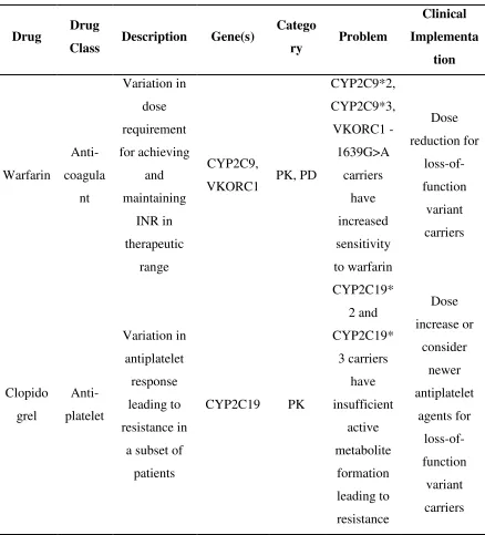
Table 2.1.1 Summary of current evidence in cardiovascular pharmacogenetics.
Drug Drug
Class Description Gene(s)
Catego
ry Problem
Clinical Implementa tion Warfarin Anti-coagula nt Variation in dose requirement for achieving and maintaining INR in therapeutic range CYP2C9,
VKORC1 PK, PD
CYP2C9*2, CYP2C9*3, VKORC1 -1639G>A carriers have increased sensitivity to warfarin Dose reduction for loss-of-function variant carriers Clopido grel Anti-platelet Variation in antiplatelet response leading to resistance in
a subset of
patients
CYP2C19 PK
CYP2C19* 2 and CYP2C19* 3 carriers have insufficient active metabolite formation leading to resistance Dose increase or consider newer antiplatelet agents for loss-of-function variant carriers
CYP, cytochrome P450; PD, pharmacodynamics; PK, pharmacokinetics; VKORC1,
2.2
Oral Anticoagulants
2.2.1
Warfarin
The vitamin K antagonist warfarin is a common oral anticoagulant prescribed in North
America for stroke prevention in atrial fibrillation (AF) patients and thromboembolism
prophylaxis in venous thromboembolism (VTE) (1). However, warfarin therapy is
particularly challenging due to marked and often unpredictable interindividual dosing
variation (up to 20-fold) to reach and maintain adequate anticoagulation. Not
surprisingly, its clinical use is associated with ADRs, mainly in the form of bleeding
events; in fact, warfarin was recently reported to account for one-third of hospitalizations
in the elderly (2). For most indications, optimal warfarin therapy is achieved by
maintaining the international normalized ratio (INR) within a narrow therapeutic range of
2-3. An insufficient warfarin dose leads to a lack of antithrombotic effect while
over-anticoagulation is associated with elevated bleeding risk. Aside from demographic (age,
gender, weight) and clinical variables (renal or hepatic disease, diet, drug-drug
interactions), pharmacogenetics explains a large portion of the observed variability in
warfarin dose requirement (3).
2.2.1.1
CYP2C9
Warfarin is administered as a racemic drug; S-warfarin is 3-5 times more potent than R
-warfarin. The clearance and thus pharmacokinetics of warfarin is largely dependent on
are enzymes that convert the R-warfarin to its inactive metabolite in varying extents,
CYP2C9 is the primary enzyme responsible for metabolism of S-warfarin. As such,
candidate gene studies have consistently shown that CYP2C9 polymorphisms
significantly affect warfarin sensitivity (4). In particular, common CYP2C9*2 and *3
variant alleles result in decreased enzymatic activity (30 and 90 % reduction,
respectively) compared to the wild-type allele (5). The clinical implication for these
SNPs is lower therapeutic dose requirement, increased time to stable anticoagulation and
increased bleeding risk due to greater rate of over-anticoagulation (6). A large
meta-analysis of 39 studies (n = 7,907) demonstrated that maintenance dose for CYP2C9*2 and
*3 homozygous patients were 36 and 78 % lower, respectively, as compared to wild-type
patients (7).
2.2.1.2
VKORC1
S-warfarin inhibits the vitamin K epoxide reductase, encoded by VKORC1, the enzyme
responsible for recycling oxidized vitamin K to the reduced form, an essential cofactor
for γ-glutamyl-carboxylase carboxylation (GGCX) in clotting factor II, VII, IX and X
activation (8). Common genetic variants in VKORC1 result in altered warfarin sensitivity
while rare polymorphisms result in warfarin resistance (9). Of note, the common
promoter SNP (VKORC1 -1639G>A, rs9923231) is thought to be the causative variation
responsible for the greater warfarin sensitivity, resulting in lowered dose requirement
2.2.1.3
Other SNPs
In addition to CYP2C9 and VKORC1 polymorphisms, SNPs in other genes have been
studied as potential contributors to warfarin response. These genes include GGCX,
calumenin (CALU), apolipoprotein E (ApoE) as well as multidrug resistance protein
(ABCB1) (11). However, the impact of these polymorphisms on warfarin response has
generally been minimal or none at all. An exception to this is the growing importance of
the CYP4F2 1297C>T genotype, whereby several retrospective studies have
demonstrated that variant T carriers (rs2108622) require 1 mg more than wild-type C
carriers (12). Moreover, CYP4F2 is the metabolizing enzyme of vitamin K accounting for
the pharmacological basis of the dose difference (13). However, CYP4F2 genotype only
accounts for a small portion of the observed maintenance dose variability (0-4%) (14).
These findings were confirmed by recent GWASs, where polymorphisms in CYP2C9 and
VKORC1 were the only genetic markers identified to influence warfarin dosing, while the
CYP4F2 genotype was only significant after adjusting for CYP2C9 and VKORC1 (15).
2.2.1.4
Clinical Applicability of SNPs
CYP2C9*2, CYP2C9*3 and VKORC1 -1639G>A variant carriers are at an increased risk
of over anticoagulation (INR > 4) and bleeding as well as delayed time to therapeutic
efficacy. Based on these findings, the FDA approved a new label for warfarin in 2007
advising physicians to consider pharmacogenetic testing for patients requiring warfarin.
CYP2C9, VKORC1, and CYP4F2 SNPs and clinical parameters (16). The majority of
these studies have focused on the effect of genetic variation on warfarin dose during
maintenance phase of anticoagulation. However, warfarin initiation is arguably the most
challenging therapeutic phase where risk of hemorrhage and recurrent VTE are greatest
(17). Moreover, we and others demonstrated that CYP2C9 and VKORC1 modulate
wafarin response even during early anticoagulation (18).
2.2.1.5
Clinical Implementation
There is a strong association between CYP2C9 and VKORC1 SNPs with warfarin
response and dose. However, the widespread use of pharmacogenetics-based warfarin
dosing remains has not yet been achieved, in part related to concerns regarding costs
associated with pharmacogenetic testing and additional clinical evidence to support
superiority of a pharmacogenetics-based approach. Recently, the COUMAGEN-II trial
showed clear superiority of pharmacogenetics-based warfarin dosing over standard care
with respective to time spent in therapeutic range and reduced occurrence of ADRs (19).
Additionally, a recent large scale community-based study found that genotyping during
warfarin therapy reduced the hospitalization rate for bleeding or thromboembolic event
by 30 % compared to a historical control group (20). A number of RCTs
(www.clinicaltrials.gov; COAG, GIFT, EU-PACT) involving larger sample sizes are
currently underway to more fully confirm such findings focusing on safety and efficacy
of pharmacogenetics-based dosing compared to standard dosing. Given the extent of
likely to be widely implemented, particularly given the rapid improvement in genotyping
technologies that has resulted in greater accuracy, turn-around time, and lower cost.
2.2.2
New oral anticoagulants: dabigatran, rivaroxaban, and
apixaban
Dabigatran, a direct thrombin inhibitor, and factor Xa inhibitors rivaroxaban and
apixaban, are new oral anticoagulants recently approved for AF and VTE. Dabigatran
etexilate is a pro-drug, requiring bioactivation to active dabigatran by esterases while
rivaroxaban undergoes metabolism predominantly by CYP2J2 and CYP3A4 (21).
Interestingly, all agents are substrates of P-glycoprotein (ABCB1) while rivaroxaban and
apixaban are also substrates of breast cancer resistance protein (BCRP) (22). Very
recently, a genome-wide subanalysis of the RE-LY trial demonstrated that a common
variant in the carboxylesterase 1 (CES1) gene (rs2244613) was associated with
dabigatran-related bleeding events. The polymorphism is thought to attenuate dabigatran
formation resulting in lowered systemic exposure and reduced bleeding risk, indicating
the potential for pharmacogenetics-based dosing. Further studies are needed to confirm
these findings in addition to identification of additional genetic variations in candidate
genes such as ABCB1 and BCRP to determine SNPs capable of modulating the efficacy
2.3
Antiplatelets
2.3.1
Aspirin
Aspirin is commonly prescribed for prevention of cardiovascular events. Aspirin
irreversibly binds and inactivates cyclooxygenase (COX) 1 and 2 in platelets, and thereby
reduce platelet aggregation (23). The term of aspirin resistance has been coined to note
the occurrence of cardiovascular events, presumably related to suboptimal antiplatelet
inhibitory effect in patients prescribed with therapeutic doses of aspirin. However,
classification of aspirin resistance has been highly controversial with prevalence of 5 – 60
% depending on the population and ex vivo response measurement used; a patient defined
as a non-responder in one test would be normal in another (24). Therefore, it has been
challenging to interpret the relevance of genetic markers associated with aspirin response.
2.3.1.1
PTGS1
Since COX1 is the direct pharmacological target of aspirin, considerable amount of focus
has been on the COX1 encoding gene, PTGS1. However, results of such studies have
been variable. Although genetic polymorphisms in this gene have been linked to greater
platelet aggregation in some studies, many others failed to replicate such an effect or find
2.3.1.2
Other SNPs
Several other genes encoding platelet activation pathway proteins have been linked to
aspirin antiplatelet response. The ITGB3 gene encodes GPIIIa protein and carriers of the
risk allele (rs5918) appear to confer elevated risk of MI, arterial and venous thrombosis.
However, other studies in this regard have been conflicting (25). A recent study found
carriers of an intronic SNP in the PEAR1 gene (rs12041331) corresponded to higher
aspirin response (26). Additionally, a SNP in the LPA gene (rs3798220) determined
plasma levels of apolipoprotein A and associated with differential aspirin efficacy in a
large placebo-controlled trial (27).
2.3.1.3
Clinical Implementation
The extent of clinically relevant genetic markers and their role to aspirin response remain
to be defined. The lack of consistent associations indicates it may be premature to include
genetic testing for aspirin therapy, at least based on currently published genetic markers.
It addition, a key impediment has been the lack of a standardized antiplatelet response