Abstract—Accumulation of reactive oxygen species (ROS) followed by an increase in oxidative stress is associated with cellular responses to nanoparticle induced cell damages. Finding the best method for assessing intracellular ROS production is the key step in the detection of oxidative stress induced injury. This study evaluates and compares four different methods for the measurement of intracellular ROS generation using fluorogenic probe, 2´,7´-dichlorofluorescein diacetate (DCFH-DA). Hydrogen peroxide (H2O2) was utilised as a positive control to assess the reactivity of the probe. Spherically shaped zinc oxide (ZnO) nanoparticles with an average particle size of 85.7 nm were used to determine the diverse roles of ROS in nanotoxicity in Hs888Lu and U937 cell lines. The results showed that different methods exhibit different patterns of ROS measurement. In conclusion this study found that the time point at which the DCFH-DA is added to the reaction, the incubation time and the oxidative species that is responsible for the oxidation of DCFH, have impact on the intracellular ROS measurement.
Index Terms—DCFH-DA, nanotoxicity, nanoparticle, ROS, ZnO.
I. INTRODUCTION
Reactive oxygen species are important intermediates constantly produced in vivo through a variety of normal metabolic processes as well as being common intermediates generated after exposure to drugs or ionizing radiation (Curtin, et al., 2002). The nanoparticles have small size and a large surface area that can lead to the sequential oxidation-reduction
_____________________________________________________________
ARO-The Scientific Journal of Koya University
Volume III, No 2(2015), Article ID: ARO.10080, 05 pages DOI: 10.14500/aro.10080
Received 23 April 2015; Accepted 01 September 2015
Regular research paper: Published 24 October 2015 Corresponding author’s e-mail: [email protected]
Copyright © 2015 Nigar A. Najim. This is an open access article distributed under the Creative Commons Attribution License.
reactions at nanoparticles surface to produce reactive species such as hydrogen peroxide (H2O2) and hydroxyl radial (Donaldson, et al., 2003; Lin, et al., 2009; Oberdorster, 2004; Xia, et al., 2008; Xia, et al., 2006).
Fluorogenic probes have been widely employed to monitor oxidative activity. Among these, 2´,7´-dichlorofluorescein diacetate (DCFH-DA) has been utilised as an assay to evaluate oxidative stress in cells (Wang and Joseph, 1999). This hydrophobic non-fluorescent molecule penetrates rapidly into the cell and is hydrolysed by intracellular esterase to give the dichlorofluorescin (DCFH), which is trapped inside the cells. In the presence of hydrogen peroxide (H2O2) and other ROS, DCFH molecule is oxidised to its highly fluorescent product 2´,7´-dichlorofluorescein (DCF) (Foucaud, et al., 2007; Loetchutinat, et al., 2005; Rota, et al., 1999).
The DCF fluorescence intensity can be easily measured and its fluorescence intensity is proportional to the amount of ROS. This probe has been used extensively for quantifying nanomaterial-induced ROS in a range of cell types, some examples of which include LNZ308 (glioma) cells exposed to ZnO nanoparticles (Ostrovsky, et al., 2009), ZnO induced ROS in Ana-1 (mouse macrophage) cells (Song, et al., 2010), ROS induced in A549 (human lung epithelial) cells after exposure to ZnO and silica nanoparticle (Lin, et al., 2006; Lin, 2009) and by ambient ultrafine particles, cationic polystyrene nanospheres, TiO2 and fullerol nanoparticles in RAW264.7 phagocytic cells (Xia, 2006).
In a previous study (Najim, et al., 2014) we demonstrated that the presence of zinc oxide (ZnO) nanoparticles with an average particle size of 85.7 nm induced cytotoxicity towards human histiocytic lymphoma (U937), neuron-phenotypic cells (SH-SY5Y), neuroblastoma (SH-SY5Y) and normal lung (Hs888Lu) cell lines.
Moreover, our data have also indicated that the cytotoxicity of ZnO was concentration dependent. The mechanism of nanoparticles cytotoxicity is not clear, however the generation of reactive oxygen species (ROS) was considered as one of the main causes of the nanotoxicity by the nanoparticles as shown
Comparative Study of Different Methods to
Determine the Role of Reactive Oxygen Species
Induced by Zinc Oxide Nanoparticles
Nigar A. Najim
1, 21
Department of Pharmacognosy and Pharmaceutical Chemistry, School of Pharmacy, Faculty of Medical Sciences, University of Sulaimani, Sulaimani, Kurdistan Region - F.R. Iraq
2
in previous studies (Li, et al., 2008; Oberdorster, 2004; Reeves, et al., 2008; Sharma, et al., 2009).
This study was designed to investigate the underlying mechanisms of nanoparticles induced cytotoxicity. While there is a number of different cellular injury responses to ROS generated from nanoparticles, we developed a rapid screening procedure for ZnO toxicity premised on oxidative stress injury. In this study four different methods for intracellular ROS detection were compared for their compatibility by using the same representative characterised probe, 2´,7´-dichlorofluorescein diacetate (DCFH-DA) (Grabinski, et al., 2007; Lin, 2009; Ostrovsky, 2009; Song, 2010; Wang and Joseph, 1999).
II. MATERIALS AND METHODS
A. Materials
ZnO Nanoparticles
The ZnO nanoparticles were synthesised using a method detailed elsewhere (Rusdi, et al., 2011). The ZnO nanoparticles were characterized using scanning electron microscopy (SEM), X-Ray diffraction (XRD) and transmission electron microscopy (TEM) techniques (Najim, 2014; Rusdi, 2011). ZnO average particle size used in these experiments was 85.7 nm.
The 100 mM stock solution of ZnO nanoparticles was prepared in 0.01 M phosphate buffer saline (PBS, Sigma, USA) and sonicated for 30 minutes. The stock solution was stored at 4ºC until required. ZnO nanoparticles stock concentration was vigorously vortexed and then diluted with complete medium prior to each experiment, to obtain concentrations of 100, 200, 500 and 1000 µM.
Cell lines and Culture Conditions
Lymphoma U937 cell line was provided by Dr. Mohamed Saifulaman (Faculty of Applied Sciences, UiTM, Malaysia). Normal lung Hs888Lu cell line was purchased from the American Type Culture Collection (ATCC, The Global Bioresource Centre, Manassas, USA). The U937 cells were cultured in Dulbecco’s Modified Eagle’s Medium (DMEM, Sigma, USA) with glucose (4500 mg/L), 1% non-essential amino acids (at a strength of 100 ×) (PAA Laboratory GmbH, Austria), 1% L-Glutamine (200 mM) (Sigma, USA), 1% Gentamicin (10 mg/mL) (PAA Laboratory GmbH, Austria) and supplemented with 10% fetal bovine serum (FBS, PAA Laboratory GmbH, Austria). Hs888Lu cells were adapted to grow in DMEM with glucose (4500 mg/L), 1% non-essential amino acids, 2% L-Glutamine (200mM), 1% of Penicillin/Streptomycin (10,000 units/mL of penicillin and 10 mg/mL of streptomycin) (PAA Laboratories GmbH, Austria), 1% sodium pyruvate (1 mM) (Sigma-Aldrich, USA) and supplemented with 10% FBS. All cell lines were maintained at 37ºC in a 5% CO2 atmosphere with 95% humidity (Incubator, Contherm Scientific Ltd, New Zealand).
Intracellular ROS Measurements
In order to establish the role of oxidative stress induced by ZnO nanoparticles, intracellular reactive oxygen species (ROS) generation was measured. In addition, 2´,7´-dichlorofluorescein diacetate (DCFH-DA, Sigma, USA) was used to detect and quantify the ROS level within the cells (Wang and Joseph, 1999). DCFH-DA stock solution (in methanol) of 10 mM was diluted in Hank’s balanced salt solution (HBSS, with Ca2+ and Mg2+, without phenol red, Gibco, USA) to yield a 20 µM working solution. Cells were plated in 96-well plate and incubated for 24 hours at 37°C before the experiment. Subsequently, various methods were applied to assess the ROS production.
Hydrogen peroxide (30% H2O2, Merck, Germany) was used as a positive control in these experiments to assess the reactivity of the probe (Ostrovsky, 2009; Winterbourn and Sutton, 1984) and also to validate the study. H2O2 final concentrations in media were 100 and 200 µM. Four different methods were used to determine their efficiency in the measurements of intracellular ROS, with variation in incubation time and termination of dye. The applied methods were a modification of that described by Wang and Joseph (1999 ), Grabinski (2007), Lin (2009), Song (2010), and Ostovsky (2009). Prior to each experiment for ROS measurements, cells (1×10⁵ cells/mL) were seeded in 96-well plates and left to grow overnight in humidified atmosphere containing 5% CO2 at 37 ºC incubator.
B. Methods
Method 1 (Coded as M1)
On the day of the experiments, after removing the medium, the cells were washed twice with PBS and then incubated with 50 µL/well of 20 µM of DCFH-DA (working solution) for 30 minutes in a dark environment at 37°C incubator. Immediately following incubation, DCFH-DA was removed and the cells were washed with PBS and treated with various concentrations of H2O2 (100 and 200 µM) or ZnO nanoparticles (ranging from 100 µM to 1 mM) for 24 hours. The fluorescence intensity from each well was measured with a 485 ± 10 nm excitation wavelength and a 520 ± 12.5 nm emission wavelength on a Glomax multi detection system (Promaga, USA). Results were representative of six independent experiments and were expressed as percentages of the value observed with control (no H2O2 or ZnO treatments) (Grabinski, 2007; Wang and Joseph, 1999 ).
Method 2 (Coded as M2)
Method 3 (Coded as M3)
For the treatments, the steps in method 2 were followed. After the treatment with H2O2 or ZnO nanoparticles, the cells were washed twice with PBS to eliminate DCFH-DA that did not enter the cells (Song, 2010). One hundred µL of media was added to each well and the fluorescence intensity was measured as described in method 1.
Method 4 (Coded as M4)
On the day of the experiments, after removing the medium, the cells were washed twice with PBS and then treated with various concentrations of H2O2 or ZnO nanoparticles. After 24 hours of exposure to ZnO nanoparticles, the cells were washed twice with PBS and then incubated with 50 µL/well of 20 µM of DCFH-DA (working solution) for 30 minutes in a dark environment at 37°C incubator. The fluorescence intensity from each well was measured as described in method 1 (Lin, 2006; Ostrovsky, 2009).
III. STATISTICAL ANALYSIS
GraphPad PRISM® version 5.0 program was used for statistical analysis. Means and standard deviations were determined
from six independent experiments.
All comparisons were made using two-tailed Student’s t-test and when positive indicated by asterisks (*p<0.05, **p<0.01 and ***p<0.001).IV. RESULTS AND DISCUSSION
A. Comparison of intracellular ROS detection methods
To obtain optimal results from intracellular ROS assay, two different cell lines such as normal lung (Hs888Lu) and lymphoma (U937) were used, because the quality of DCFH-DA relies on the cell types and culture conditions. In the presence of hydrogen peroxide (H2O2) and other ROS as well as heme protein catalysts such as peroxidases or cytochrome c, DCFH is oxidized to highly fluorescent DCF in cells (Burkitt and Wardman, 2001; Ischiropoulos, et al., 1999; LeBel, et al., 1992; Ohashi, et al., 2002). Thus, H2O2 was used as a positive control in this study to assess the reactivity of the probe and also to validate the method.
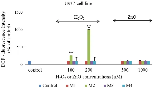
DCF- fluorescence intensity was measured (proportional to the intracellular ROS) in Hs888Lu and U937 cells exposed to H2O2 or ZnO according to the methods described earlier (Fig. 1 and Fig. 2). The results showed that different methods exhibit different pattern of ROS measurement. Higher amount of intracellular ROS was detected using method 2. H2O2 content increased the intensity of DCF- fluorescence in a dose-dependent manner. There were significant differences between intracellular ROS production in different cell lines. Using this method the percentages of fluorescence intensity were 164% and 1011% in Hs888Lu cell lines (p<0.05) and U937 cell lines (p<0.01) respectively after exposure to 200 µM H2O2 (Fig. 1 and Fig. 2).
Following methods 1, 3 and 4, the results did not indicate any change in DCF- fluorescence intensity thereafter in ROS measurements in selected cell lines exposed to H2O2 (Fig. 1 and Fig. 2).
Fig. 1. Comparison of four methods (M1, M2, M3 and M4) to determine the intracellular ROS in Hs888Lu cell line. DCF-fluorescence intensities in Hs888Lu cell lines are exposed to different concentrations of H2O2 or ZnO. The control represents ROS measurement in non-treated cells.
Fig. 2. Comparison of four methods (M1, M2, M3 and M4) to determine the intracellular ROS in U937 cell line. DCF-fluorescence intensities in U937 cell lines are exposed to different concentrations of H2O2 or ZnO. The control represents ROS measurement in non-treated cells.
After comparison of ROS-induction by H2O2 using the selected methods as a representative analysis of optimization experiments, ZnO nanoparticles – induced ROS in human cell lines was quantified according to the methods described earlier.
measurements in a nanoparticle dose-dependent study (Fig. 1 and Fig. 2). In addition, hydrogen peroxide produced significant increase in DCF- fluorescence compared to ZnO nanoparticles under the experimental conditions of method 2.
Following methods 1, 3 and 4 results indicated that the H2O2 and ZnO were weak inducer of DCF-fluorescence in both cell lines (Fig. 1 and Fig. 2). In contrast, previous studies (Grabinski, 2007; Ostrovsky, 2009; Song, 2010) were demonstrated different pattern of the ROS measurement with the selected methods. In previous studies, mouse keratinocyte (HEL-30) cells exposed to H2O2 in a dose-dependent manner showed an increase in the ROS production according to the method 1 by Grabinski (2007). According to method 3, Song and co-workers (2010) have illustrated an increase in intracellular levels of ROS after exposure to all particle size of ZnO nanoparticles (30 nm, 100 nm) in mouse macrophage (Ana-1) cell line. Ostrovesky and co-workers (2009) have reported an increase in the fluorescence intensity of DCF in the normal human astrocyte and human glioma (U87) cell lines exposed to H2O2 according to method 4. In addition, they found an increase in the fluorescence intensity of DCF after 24 hours exposure to 10 mM ZnO nanoparticles in the human glioma (U87) cells. The ZnO nanoparticles induced a smaller increase in the fluorescence intensity of DCF in the normal astrocytes cells as a compared to H2O2 (positive control) (Ostrovsky, 2009).
Researchers in previous works used dilution of DCFH-DA in HBSS free of Ca2+ and Mg2+ (Li, et al., 2002; Reinisch, et al., 2000). Thus, the above methods (M1, M2, M3 and M4) were repeated using HBSS without Ca2+ and Mg2+. The results (Data not shown) were very similar to the results shown in Fig. 1 and Fig. 2. This may imply that Ca2+ and Mg2+ in the medium of reaction were not contributing to the formation of DCF and thereafter to the intracellular ROS measurement.
It must be pointed out that the importance of the mechanisms and the possibilities that lead to the formation of highly fluorescent DCF must be carefully understood. Previous research works focused on the effects of endogenous and exogenous ROS in a variety of cell types, using 2´,7´-dichlorofluorescein diacetate (DCFH-DA) (Grabinski, 2007; Lin, 2009; Loetchutinat, 2005; Ohashi, 2002; Ostrovsky, 2009; Song, 2010; Xia, 2008; Xia, 2006).
Although there is clear evidence that DCFH-DA is de-acetylated to form DCFH by cellular esterase which is then oxidised to DCF by oxidising species in most cell lines. Some limitations of utilising this probe exist. Not all cell types possess sufficient esterase activity to produce the DCFH needed for accurate measurements of ROS. This may limit the availability of DCFH and result in an underestimation of intracellular ROS levels (Brubacher and Bols, 2001). In addition, DCF, oxidised fluorescent product of DCFH, is membrane permeable and can leak out of cells over time (Ubezio and Civoli, 1994). Incomplete DCF trapping may complicate interpretation of the data and hinder the precise evaluation of intracellular oxidation.
It remains unclear which reactive oxygen species are responsible for the oxidation of DCFH in cells, even though it is often assumed to be H2O2 (in the presence of cellular peroxidases and similar catalysts). It was reported that other biologically relevant ROS, including peroxynitrite and hydroxyl radicals (˙OH), can oxidize DCFH (Kooy, et al., 1997; Myhre, et al., 2003; Possel, et al., 1997). Others have found that DCFH showed rather low sensitivity towards oxidation by NO, O2˙ ¯ (Myhre, 2003; Rota, 1999). Rota and co-workers (1999) implied that DCFH cannot be used to measure superoxide free radical formation in cells because the oxidation of this compound leads to the formation of superoxide. Moreover, some oxidants require small quantities which rapidly increase DCF formation, whereas other oxidants may need higher concentrations and more time (Myhre, 2003). In addition, H2O2 cannot oxidise DCFH directly. The oxidation occurs as a result of the reaction of H2O2 with cellular peroxidase, cytochrom c, or Fe2+ (LeBel, 1992; Rota, 1999; Royall and Ischiropoulos, 1993).
An important question is which reactive oxygen species induced from ZnO nanoparticles responsible for the oxidation of DCFH in biological systems? One of the possible mechanisms of free radical induction by ZnO nanoparticles was proposed by Ai and co-workers (2003) which is that sequential oxidation-reduction reaction may occur at ZnO particle surface to produce reactive species such as H2O2 and hydroxyl radical (Ai, 2003). Some limitations of utilising this probe exist, even though it is often assumed that H2O2 (in presence of cellular peroxidases and similar catalysts) and hydroxyl radicals (˙OH) can oxidise DCFH (Ai, 2003).
V. CONCLUSION
This comparative analysis found different results for the measurements of ROS depending on which method was used, although same concentrations of H2O2 and DCFH-DA, same cells lines and cell culture conditions were used. The methods are varying in the step sequence of exposure to DCFH-DA and H2O2 or ZnO, elimination of DFH-DA and termination of DCF. There are several significant factors affecting the measurements such as the timing of loading DCFH-DA to the reaction, duration of DCFH-DA incubation and the oxidative species responsible for the oxidation of DCFH. All these will have an impact on the formation of DCF and thereafter on the intracellular ROS measurement. Our data show that the higher amounts of intracellular ROS were detected after exposure to H2O2 using method 2. In addition, there is no evidence of ROS production due to ZnO nanoparticle toxicity in lymphoma and normal lung cell lines.
ACKNOWLEDGMENT
REFERENCE
Ai, H., Bu, Y., and Han, K., 2003. Glycine-Zn+/Zn2+ and their hydrates: On the number of water molecules necessary to stabilize the switterionic glycine-Zn+/Zn2+ over the nonzwitterionic ones. Journal of Chemical Physics, 118 (24), pp.10973-10985 .
Brubacher, J.L., and Bols, N.C., 2001. Chemically de-acetylated 2',7'-dichlorodihydrofluorescein diacetate as a probe of respiratory burst activity in mononuclear phagocytes. Journal of Immunological Methods, 251(1-2), pp.81-91.
Burkitt, M.J., and Wardman, P., 2001. Cytochrome c is a potent catalyst of dichlorofluorescin oxidation: Implications for the role of reactive oxygen species in apoptosis. Biochemical and Biophysical Research, 282(1), pp.329-333.
Curtin, J.F., Donovan, M., and Cotter, T.G., 2002. Regulation and measurement of oxidative stress in apoptosis. Journal of Immunological
Methods, 265(1-2), pp.49-72.
Donaldson, K., Stone, V., Borm, P.J., Jimenez, L.A., Gilmour, P.S., Schins, R.P., Knaapen, A.M., Rahman, I., Faux, S.P., Brown, D.M., and MacNee, W., 2003. Oxidative stress and calcium signaling in the adverse effects of environmental particles (PM10). Free Radical Biology and Medicine, 34 (11), pp.1369-1382.
Foucaud, L., Wilson, M.R., Brown, D.M., and Stone, V., 2007. Measurement of reactive species production by nanoparticles prepared in biologically relevant media. Toxicology Letters, 174(1-3), pp.1-9.
Grabinski, C., Hussain, S., Lafdi, K., Braydich-Stolle, L., and Schlager, J., 2007. Effect of particle dimension on biocompatibility of carbon nanomaterials. Carbon, 45(14), pp.2828-2835.
Ischiropoulos, H., Gow, A., Thom, S.R., Kooy, N.W., Royall, J.A. and Crow, J.P., 1999. Detection of reactive nitrogen species using 2,7- dichlorodihydrofluorescein and dihydrorhodamine 123. Methods in
Enzymology, 301, pp.367-373.
Kooy, N., Royall, J. and Ischiropoulos, H., 1997. Oxidation of 2',7'-dichlorofluorescein by peroxynitrite. Free Radical Research, 27(3), pp.245-254.
LeBel, C.P., Ischiropoulos, H. and Bondy, S.C., 1992. Evaluation of the probe 2‘,7‘-dichiorofluorescin as an indicator of reactive oxygen species formation and oxidative stress. Chemical Research in Toxicology, 5(2), pp.227-231.
Li, N., Xia, T. and Nel, A.E., 2008. The role of oxidative stress in ambient particulate matter-induced lung diseases and its implications in the toxicity of engineered nanoparticles. Free Radical Biology and Medicine, 44(9), pp.1689-1699.
Li, Y., Nishimura, T., Teruya, K., Maki, T., Komatsu, T., Hamasaki, T., Kashiwagi, T., Kabayama, S., Shim, S.Y., Katakura, Y., Osada, K., Kawahara, T., Otsubo, K., Morisawa, S., Ishii, Y., Gadek, Z. and Shirahata, S., 2002. Protective mechanism of reduced water against alloxan-induced pancreatic β-cell damage: Scavenging effect against reactive oxygen species.
Cytotechnology, 40(1-3), pp.139-149.
Lin, W., Huang, Y., Zhou, X.D. and Ma, Y., 2006. In vitro toxicity of silica nanoparticles in human lung cancer cells. Toxicology and Applied
Pharmacology, 217(3), pp.252-259.
Lin, W., Xu, Y., Huang, C.C., Ma, Y., Shannon, K.B., Chen, D.-R. and Huang, Y.-W., 2009. Toxicity of nano- and micro-sized ZnO particles in human lung epithelial cells. Journal of Nanoparticle Research, 11(1), pp.25-39.
Loetchutinat, C., Kothan, S., Dechsup, S., Meesungnoen, J., Jay-Gerin, J.-P. and Mankhetkorn, S., 2005. Spectrofluorometric determination of intracellular levels of reactive oxygen species in sensitive and drug-resistant cancer cells using the 2',7'-dichlorofluorescein diacetate assay.
Radiation Physics and Chemistry, 72(2-3), pp.323-331.
Myhre, O., Andersen, J.M., Aarnes, H. and Fonnum, F., 2003. Evaluation of the probes 2',7'-dichlorofluorescin diacetate, luminol, and lucigenin as
indicators of reactive species formation. Biochemical Pharmacology, 65(10), pp.1575-1582.
Najim, N., Rusdi, R., Zain, M.M., Hamzah, A.S., Shaameri, Z. and Kamarulzaman, N., 2014. Effects of the absorption behaviour of ZnO nanoparticles on the optical measurements of cytotoxicity studies: In normal and cancer cell lines. Journal of Nanomaterials, 2014, pp.1-8.
Oberdorster, E., 2004. Manufactured nanomaterials (fullerenes, C60) induce oxidative stress in the brain of juvenile largemouth bass. Environmental
Health Perspectives, 112(10), pp.1058-1062.
Ohashi, T., Mizutani, A., Murakami, A., Kojo, S., Ishii, T. and Taketani, S., 2002. Rapid oxidation of dichlorodihydrofluorescin with heme and hemoproteins: formation of the fluorescein is independent of the generation of reactive oxygen species. FEBS Letters, 511(1-3), pp.21-27.
Ostrovsky, S., Kazimirsky, G., Gedanken, A. and Brodie, C., 2009. Selective cytotoxic effect of ZnO nanoparticles on glioma cells. Nano Research, 2(11), pp.882-890.
Possel, H., Noack, H., Augustin, W., Keilhoff, G. and Wolf, G., 1997. 2',7'-Dihydrodichlorofluorescein diacetate as a fluorescent marker for peroxynitrite formation. FEBS Letters, 416(2), pp.175-178.
Reeves, J.F., Davies, S.J., Dodd, N.J.F. and Jha, A.J., 2008. Hydroxyl radicals (•OH) are associated with titanium dioxide (TiO2) nanoparticle induced cytotoxicity and oxidative DNA damage in fish cells. Mutation
Research, 640(1-2), pp.113-122.
Reinisch, N., Wiedermann, C.J. and Ricevuti, G., 2000. Inhibition of human peripheral blood neutrophil respiratory burst by alcohol-based venipuncture site disinfection. Clinical and Diagnostic Laboratory Immunology, 7(6), pp.980-982.
Rota, C., Chignell, C.F. and Mason, R.P., 1999. Evidence for free Radical Formation during the oxidation of 2'-7'-dichlorofluorescin to the fluorescent dye 2'-7'-dichlorofluorescein by horseradish peroxidase: possible implications for oxidative stress measurements. Free Radical Biology and Medicine, 27(7), pp.873-881.
Royall, J. and Ischiropoulos, H., 1993. Evaluation of 2',7'-dichlorofluorescein and dihydrorhodamine 123 as fluorescent probes for intracellular H2O2 in cultured endothelial cells Archives of Biochemistry and Biophysics, 302(2), pp.348-355.
Rusdi, R., Rahman, A., Mohamed, N., Kamarudin, N. and Kamarulzaman, N., 2011. Preparation and band gap energies of ZnO nanotubes, nanorods and spherical nanostructures. Powder Technology, 210(1), pp.18-22.
Sharma, V., Shukla, R.K., Saxena, N., Parmar, D., Das, M. and Dhawan, A., 2009. DNA damaging potential of zinc oxide nanoparticles in human epidermal cells. Toxicology Letters, 185(3), pp.211-218.
Song, W., Zhang, J., Guo, J., Zhang, J., Ding, F., Li, L. and Sun, Z., 2010. Role of the dissolved zinc ion and reactive oxygen species in cytotoxicity of ZnO nanoparticles. Toxicology Letters, 199(3), pp.389-397.
Ubezio, P. and Civoli, F., 1994. Flow cytometric detection of hydrogen peroxide production induced by doxorubicin in cancer cells. Free Radical
Biology and Medicine, 16(4), pp.509-516.
Wang, H. and Joseph, J.A., 1999. Quantifying cellular oxidative stress by dichlorofluorescein assay using microplate reader. Free Radical Biology and
Medicine, 27(5-6), pp.612-616.
Winterbourn, C.C. and Sutton, H.C., 1984. Hydroxyl radical production from hydrogen peroxide and enzymatically generated paraquat radicals: Catalytic requirements and oxygen dependence. Archives of Biochemistry and
Biophysics, 235(1), pp.116-126.
Xia, T., Kovochich, M., Liong, M., Madler, L., Gilbert, B., Shi, H., Yeh, J.I., Zink, J.I. and Nel, A.E., 2008. Comparison of the mechanism of toxicity of zinc oxide and cerium oxide nanoparticles based on dissolution and oxidative stress properties. ACS nano, 2(10), pp.2121-2134.
Xia, T., Kovochich, M. and Nel, A., 2006. The role of reactive oxygen species and oxidative stress in mediating particulate matter injury.