R E S E A R C H
Open Access
Phenotypic and functional analysis of
SHANK3
stop mutations identified in individuals with ASD
and/or ID
Daniela M Cochoy
1, Alexander Kolevzon
2,3,4,5,6, Yuji Kajiwara
5, Michael Schoen
1, Maria Pascual-Lucas
1,7,
Stacey Lurie
2,5, Joseph D Buxbaum
2,3,4,5,8,9, Tobias M Boeckers
1and Michael J Schmeisser
1*Abstract
Background:SHANK proteins are crucial for the formation and plasticity of excitatory synapses. Although mutations in all threeSHANKgenes are associated with autism spectrum disorder (ASD),SHANK3appears to be the major ASD gene with a prevalence of approximately 0.5% forSHANK3mutations in ASD, with higher rates in individuals with ASD and intellectual disability (ID). Interestingly, the most relevant mutations are typicallyde novoand often are frameshift or nonsense mutations resulting in a premature stop and a truncation of SHANK3 protein.
Methods:We analyzed three differentSHANK3stop mutations that we identified in individuals with ASD and/or ID, one novel (c.5008A > T) and two that we recently described (c.1527G > A, c.2497delG). The mutations were inserted into the humanSHANK3asequence and analyzed for effects on subcellular localization and neuronal morphology when overexpressed in rat primary hippocampal neurons.
Results:Clinically, all three individuals harboring these mutations had global developmental delays and ID. In ourin vitro assay, c.1527G > A and c.2497delG both result in proteins that lack most of the SHANK3a C-terminus and accumulate in the nucleus of transfected cells. Cells expressing these mutants exhibit converging morphological phenotypes including reduced complexity of the dendritic tree, less spines, and less excitatory, but not inhibitory synapses. In contrast, the truncated protein based on c.5008A > T, which lacks only a short part of the sterile alpha motif (SAM) domain in the very SHANK3a C-terminus, does not accumulate in the nucleus and has minor effects on neuronal morphology.
Conclusions:In spite of the prevalence of SHANK3 disruptions in ASD and ID, only a few human mutations have been functionally characterized; here we characterize three additional mutations. Considering the transcriptional and functional complexity ofSHANK3in healthy neurons, we propose that any heterozygous stop mutation inSHANK3will lead to a dysequilibrium of SHANK3 isoform expression and alterations in the stoichiometry of SHANK3 protein complexes, resulting in a distinct perturbation of neuronal morphology. This could explain why the clinical phenotype in all three individuals included in this study remains quite severe - regardless of whether there are disruptions in one or more SHANK3 interaction domains.
Keywords:ASD, Autism, SHANK3, Intellectual disability, Nucleus, Dendrite, Spine, Synapse
* Correspondence:[email protected] 1
Institute for Anatomy and Cell Biology, Ulm University, Albert-Einstein-Allee 11, D-89081 Ulm, Germany
Full list of author information is available at the end of the article
Background
Autism spectrum disorder (ASD) is a neuropsychiatric condition manifesting in early development and is charac-terized by two core features: A) persistent deficits in social interaction and communication and B) the presence of restricted interests and/or repetitive behaviors [1]. The strong involvement of genetics in the development of ASD is supported by the identification of causative genetic ab-normalities in more than 20% of cases, with a significant number of the identified genes encoding proteins required for the correct formation, maturation, and maintenance of synaptic connections in the brain [2-9]. Among these, the
SHANK gene family plays a decisive role because diverse genetic variation inSHANK1,SHANK2, andSHANK3- all encoding large postsynaptic scaffold proteins - has been identified in individuals with ASD [10-16]. A crucial role of
SHANK3mutations in this context is supported by the following three facts: 1) SHANK3 haploinsufficiency is the critical factor for the development of neuropsychiatric symptoms in 22q13 deletion syndrome, also known as Phelan-McDermid syndrome, 2) the current prevalence forSHANK3mutations in individuals with ASD in general is between 0.5% and 0.7%, and 3) data indicate that a
SHANK3 mutation is present in approximately 2% of individuals with both ASD and intellectual disability (ID) [16-18].
Some individuals diagnosed with either ASD, ID, or both harbor frameshift or nonsense mutations in
SHANK3resulting in a premature stop codon and caus-ing a truncation of SHANK3 protein [15-17,19-22]. However, only a few studies have thus far addressed the impact of such mutations and their corresponding trun-cated proteins on neuronal function and morphology [15,19,22-25]. In this context, the de novo exon 21 frameshift mutation c.3679_3680insG - identified in two brothers diagnosed with both ASD and ID [15] - and thede novo exon 21 nonsense mutation c.3349C > T - identified in three brothers, all of them diagnosed with ID, with two having an additional diagnosis of schizophrenia (SCZ) [19] - have been most intensely studied up to date [15,19,23-25]. Insertion of either stop mutation into the rat Shank3a sequence at the corresponding sites results in the expression of truncated Shank3a variants lacking distinct parts of the C-terminus, a region crucial for ap-propriate synaptic targeting and assembly [26-29]. In con-trast to wild-type Shank3a, Shank3a harboring either c.3679_3680insG or c.3349C > T mutations no more clus-ter at synapses, but rather distribute in the somatodendritic compartment and localize to the nucleus when overex-pressed in primary hippocampal neurons [15,19,23-25]. Overexpression of Shank3a harboring the c.3679_3680insG mutation affects growth cone mobility and negatively inter-feres with synaptic transmission and transsynaptic signal-ing; the same mutation leads to reductions in the number
of excitatory synapses and dendritic spines [15,23,24]. Shank3a harboring the c.3349C > T mutation impairs the ability of Shank3a to promote the outgrowth of primary neurites, results in a less complex dendritic arbor, and leads to a specific reduction of excitatory, but not inhibitory synapses [19,25].In vitroexamination of ade novoexon 21 nonsense mutation c.2997C > G identified in a boy with ID demonstrated a reduction in neurite nodes, tips, and length, at early stages of neuronal differentiation [22]. Taken together, thesein vitrostudies show that trunca-tions of the distal C-terminus of Shank3a, caused by the c.3679_3680insG, c.3349C > T or c.2997C > G mu-tations are sufficient to disrupt neuronal morphology when the truncated variant is overexpressed in primary neuronal cultures.
However, the three stop mutations studied to date all affect exon 21 of SHANK3, but less is known about mutations identified in ASD and/or ID that affect other parts of the gene [15-17,19-22]. It is therefore of high interest to evaluate additional mutations, including those that disrupt other exons ofSHANK3 and to identify con-verging and/or distinct neuronal pathologies.
We inserted three mutations identified in subjects with ASD and/or ID into the human SHANK3a sequence: a nonsense mutation affecting exon 12 (c.1527G > A), a frameshift mutation affecting exon 21 (c.2497delG) - both recently described [17] - and one novel nonsense mutation affecting exon 22 (c.5008A > T). Clinical assessment of the corresponding subjects was followed by characterization of the impact of the three truncated SHANK3a variants with respect to subcellular localization, dendritic branch-ing, and spine and synapse formation, when overexpressed in rat primary hippocampal neurons with a wild-type Shank3 background.
Methods
Participant information
using the Mullen Scales of Early Learning; (5) The
Vineland Adaptive Behavior Scales-II, Survey Edition, to evaluate independence in daily life skills, including communication, socialization, and motor skills; (6)
Medical record review including analyzing any results from electroencephalographic and brain imaging stud-ies; and (7)Genetic testingfor confirming mutation and
de novoorigin, using Sanger sequencing. Vector constructs
Human SHANK3a complementary DNA (cDNA) based on NP_277052.1 was re-designed in collaboration with GeneArt® (Life Technologies, Carlsbad, CA, USA) for optimized GC content, and a Myc-tag was added immedi-ately after the initiation codon. Subcloning was performed using In-Fusion HD (Clontech Laboratories, Mountain View, CA, USA). The entireSHANK3acDNA was ampli-fied using the following set of primers: 5′-GTCCGGACTC AGATCTATGGAGCAGAAGCTGATCAG-3′ and 5′-GT
CGACTGCAGAATTCTCAGCTGCCGTCCAGCTGT-3′,
and further inserted into the pAcGFP1-C1 (Clontech Laboratories, Mountain View, CA, USA) vector using Bgl2 and EcoR1 sites. The sequence was confirmed by Sanger sequencing. The c.1527G > A variant was generated by using the primer set 5′-GCTTCTGaGAGGGCACCGT GAAG-3′ and 5′-TGCCCTCtCAGAAGCCGCCctcg-3′. The c.2497delG variant was generated by inserting a frag-ment containing the deletion followed by authentic human
SHANK3 cDNA corresponding to the sequence from immediately after the variation to the predicated premature termination codon. The c.5008A > T variant was generated by using the primer set 5′-GTGGTCCtAGTTCGACGTG GGCGACTGG-3′ and 5′-CGAACTaGGACCACAGCTG CAGGGGTTT-3′. The eGFP-Shank3a and DenMark con-structs have been described previously [29,30].
Antibodies
A novel polyclonal antibody directed against the rat Shank3a N-terminus (aa 333-470) was generated for this study according to the antibody production and purifica-tion protocol described in [31]. The anti-Shank3 PRC antibody has been described previously [31]. The follow-ing primary antibodies were purchased from commercial suppliers: anti-histone H3 (Cell Signaling Technology, Danvers, MA, USA), anti-green fluorescent protein (GFP) (Clontech, Laboratories, Mountain View, CA, USA), anti-c-Myc (Roche Applied Science, Mannheim, Germany), as well as anti-GAPDH, anti-VGLUT1, and anti-VGAT (all from Synaptic Systems, Goettingen, Germany).
Biochemistry
For whole culture extracts, transfected HEK293T cells were lysed in Triton X-100 Lysis Buffer (150 mM NaCl, 50 mM Tris HCl, 1% Triton X-100, pH 8,0, protease inhibitor mix,
Roche Applied Science, Mannheim, Germany). The NE-PER Nuclear and Cytoplasmic Extraction Reagents (Thermo Scientific, Bonn, Germany) were further used to obtain nuclear and cytoplasmic fractions from trans-fected HEK293T cells. Protein concentrations were deter-mined by Bradford protein assay, and the same amount of protein was loaded per lane for SDS-PAGE. Western blot analysis was conducted following standard protocols. HRP-conjugated secondary antibodies (Dako, Glostrup, Denmark) and the SuperSignal detection system (Thermo Scientific, Bonn, Germany) were used to visualize protein bands on X-ray films (GE Healthcare, Freiburg, Germany).
Animal experiments
All animal experiments in this study were performed based on the guidelines for the welfare of experimental animals issued by the Federal Government of Germany and by the local ethics committee (Ulm University), ID Number: 0.103.
Cell culture
HEK293T cells were maintained in DMEM at 37°C in 5% CO2. The preparation of hippocampal cultures
from rat was performed at embryonic stage 18 (E18) as described previously [32]. In brief, hippocampal neu-rons were seeded on poly-l-lysine (0.1 mg/ml, Sigma-Aldrich, Steinheim, Germany)-coated glass coverslips. Cells were grown in neurobasal medium, complemented with B27 supplement, 0.5 mM L-glutamine and penicillin/ streptomycin at 100 U/ml (all reagents from Life Tech-nologies, Darmstadt, Germany), and maintained at 37°C in 5% CO2.
Immunocytochemistry
Transfections
Vector constructs were transfected into HEK293T cells using PolyFect reagent (Qiagen, Hilden, Germany) as de-scribed previously [34] or into hippocampal neurons using Lipofectamine 2000 reagent (Life Technologies, Darmstadt, Germany).
Analysis of neuronal morphology
All analyses were done in a blinded fashion. Sholl ana-lysis was performed as described previously [35]. Con-centric circles (15, 30, 45, 60, 75, 90, 105, 120, 135, and 150 μm in diameter) were drawn around the soma of each neuron included in the analysis. The number of all dendrites crossing each circle was counted manually. For analysis of spines and filopodia, two secondary dendrites were randomly chosen per neuron and dendritic pro-trusions were counted manually among approximately 35-μm-long segments per dendrite. Dendritic protrusions shorter than 1μm with clearly visible head and neck were counted as spines, and dendritic protrusions longer than 1 μm and devoid of head and neck were counted as filo-podia. For analysis of synaptic contacts, four secondary dendrites were randomly chosen per neuron and signals positive for either VGLUT1 (excitatory contacts) or VGAT (inhibitory contacts) were manually counted among ap-proximately 50-μm-long segments per dendrite.
Statistical analysis
For all analyses in primary culture, five to eight neurons from three independent experiments were analyzed per condition; ‘n’ therefore ranged between 15 and 22. GraphPad Prism 5.01 (GraphPad Software, La Jolla, CA, USA) was used for all statistical analyses. Depending on the datasets, analysis was performed with unpaired Student’st -test or one-way ANOVA with Bonferroni post hoc test if data were normally distributed or with the Mann-Whitney
Utest or the Kruskal-Wallis test with Dunn’s multiple com-parisonpost hoctest if data were not normally distributed. Results
Clinical phenotype
Participant 1 (P1) (c.1527G > A) is a 5-year-old male that we have previously described [17] whose parents first became concerned about his development due to social and language delays. Prior to identifying aSHANK3 muta-tion using whole exome sequencing, a 17q12 microduplica-tion had been detected and previously described [36]. As such, caution is warranted in making direct phenotypic comparisons across the mutations described here and else-where. P1 has the use of approximately 5 to 10 words to identify objects when prompted, but he does not have any communicative language. While he is able to initiate inter-actions and make eye contact at times, he does not engage in reciprocal social interaction and is not interested in other
children. Behaviorally, he shows significant repetitive behav-ior, including forced expirations, pacing, and opening and closing doors. He tends to play in a patterned way and has pronounced deficits in imitation, pretend play, and symbolic play. There is also significant difficulty sus-taining focus, and he can be quite hyperactive. P1 met criteria for autistic disorder on the ADI-R, ADOS-G, and DSM-IV (Additional file 1: Table S1). With regard to his adaptive behavior, P1 demonstrates difficulties among all domains and overall functioning is low. He requires substantial support with the majority of self-care tasks, including feeding and dressing, and he is not toilet trained. He does eat and sleep well, however. Cognitively, P1 demonstrates abilities ranging from a 6-month-old (expressive language) to 15-6-month-old (fine motor) level on the Mullen, with significant variability in his profile. He shows relative strengths in fine and gross motor skills, understands basic instructions, and expresses himself with vocalizations and some gestures. He has difficulty labeling most objects and does not follow two-step instructions. P1 has no chronic medical problems. He has never had a seizure, and an electroencephalography (EEG) in the past was within normal limits. A past mag-netic resonance imaging (MRI) revealed diffuse ventricular enlargement and thinning of the parieto-occipital white matter and corpus callosum. P1 has no renal or cardiac ab-normalities but is reported to be allergic to penicillin. He has hypotonia and mild nonspecific gait abnormalities with toe walking. On dysmorphology exam, P1 has long eye-lashes, protruding ears, broad nasal bridge, full lips, and macrocephaly. Skin exam revealed two café au lait spots on his back (Additional file 2: Table S2).
features, P2 has a very limited attention span and is unable to focus for more than 45 to 60 s at a time. He is extremely active and restless and has the potential to be aggressive. Sleeping difficulties are prominent and characterized by early morning wakening. His adaptive functioning is low overall; P2 is unable to dress or feed himself, and he is not toilet trained. His gait is apraxic with left foot dragging and pronation of his feet bilaterally. However, he is able to ride a tricycle and to climb stairs with alternating feet. Cognitive testing shows that P2 is functioning at a 5-month-old (expressive language) to 20-month-old (gross motor) level on the Mullen. He is able to understand some simple commands and questions, in addition to identify common objects. However, he is not able to follow direc-tions or label body parts. In terms of medical features, P2 has been diagnosed with a seizure disorder and has local-ized sleep-potentiated epileptiform discharges mainly in the midline and central regions during slow wave sleep. There has never been a seizure observed on EEG, al-though clinically, they are accompanied by myoclonic twitching of his ankle and face. A previous MRI study showed evidence of leukodystrophy but was otherwise within normal limits. There was evidence of a significant regression in sign language and motor skills around 6 years old in the context of an increase of seizure activity. At one point during that period, P2 stopped walking for about 6 weeks and then slowly regained his ambulatory skills as the seizures were better controlled. A full meta-bolic disease workup was negative, but P2 has a short stat-ure, and a feeding tube is in place. He has gastrointestinal symptoms manifested by periods of fluctuating diarrhea and constipation and severe gastroesophageal reflux dis-ease (GERD) treated with famotidine. He has a history of recurring ear infections requiring myringotomy tube placement at 2 years old. He has no renal or cardiac prob-lems, and no allergies to food or medications. In terms of dysmorphic physical features, P2 has short stature and mild dolicocephaly (Additional file 2: Table S2).
Participant 3 (P3) (c.5008A > T) is a 12-year-old male whose parents first became concerned about his develop-ment at 2 ½ years old due to language regression and feed-ing problems. He had developed approximately 10 to 15 words by 18 months but subsequently lost all expressive languages. His language comprehension is also limited, but he is reportedly able to understand approximately 40 signs. He can express his needs through a Picture Ex-change Communication System (PECS), but not consist-ently. P3 interacts with family members but is generally uninterested in social interaction. He exhibits significant repetitive behaviors and will watch the same portion of a television show or movie repeatedly. He also engages in motor stereotypies, including hand flapping and rocking. He may grind his teeth, chew compulsively, and make re-peated stereotypic vocalizations. He is also sensory seeking
through tactile modalities, including pressure, and has a high pain threshold. P3 met full criteria for autistic dis-order on the ADI-R, ADOS-G, and DSM-IV (Additional file 1: Table S1). In terms of associated features, he may become aggressive and rarely bites others but is not self-injurious. There is no motoric hyperactivity, but he has significant sleep difficulties, including delays in sleep onset and early morning awakening. His adaptive functioning is low in all domains. He continues to have difficulty eating solid food and is unable to manipulate utensils. He has hypotonia but is able to ride a tricycle. P3 is not toilet trained. Cognitive functioning is also low in the 8-month (receptive language) to 30-month (gross motor) range on the Mullen. P3 was diagnosed with epilepsy at 7 years old after a 48-h video EEG showed clinical events but without electrographic correlation. A subsequent MRI that year revealed a venous angioma. During the same period of time, P3 experienced a regression in motor skills where he previously was able to hold a pencil and write his name but then lost this skill. At 8 years old, he experienced a 9-month period of significant muscle weakness and was unable to ambulate for 6 months. He continues to have hypotonia with mild nonspecific gait abnormalities, includ-ing intoeinclud-ing. He has no renal and no cardiac abnormalities. P3 is reportedly allergic to casein and gluten and on a re-stricted diet. He also has a history of chronic diarrhea (seven to eight times a day) and recurring ear infections, which have both improved. In terms of dysmorphic fea-tures, P3 is noted to have deep set eyes, fifth finger clino-dactyly, and second toes overlapping (Additional file 2: Table S2).
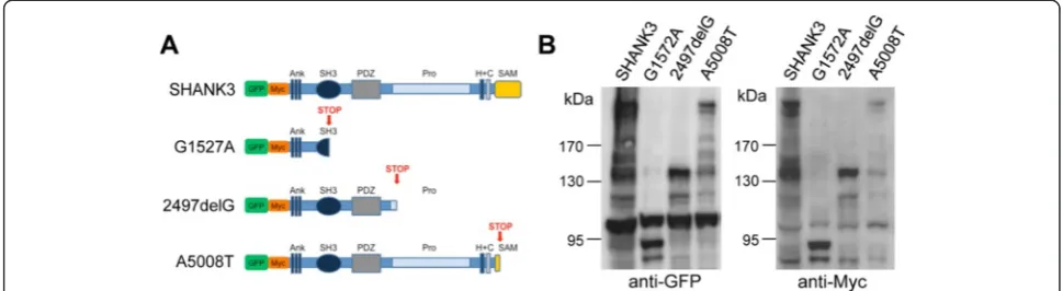
Domain composition and biochemical expression analysis of SHANK3 variants
the SAM domain (Figure 1A). To validate the functionality of our expression constructs, we transfected HEK293T cells and detected the corresponding fusion proteins by Western blot analysis of whole cell extracts using either anti-GFP (Figure 1B, left panel; Additional file 3: Figure S1A), anti-Myc (Figure 1B, right panel), or polyclonal anti-Shank3 antibodies (Additional file 3: Figure S1B,C). All four antibodies detected wild-type SHANK3 at the correct size (predicted molecular weight including tags ap-proximately 220 kD). Both GFP and Myc anti-bodies also detected all three truncated fusion proteins G1527A (approximately 90 kD), 2497delG (approximately 130 kD), and A5008T (approximately 215 kD) at the cor-rect size (Figure 1B). The anti-Shank3 PRC antibody recog-nized 2497delG and A5008T but failed to detect G1527A due to the lack of antigen sequence within this fusion pro-tein (Additional file 3: Figure S1B, for antigen sequence, see [31]). We therefore used a novel Shank3 N-term anti-body, which successfully detected G1527A (Additional file 3: Figure S1C).
Nuclear accumulation of truncated SHANK3 variants G1527A and 2497delG
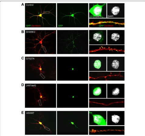
We transfected rat primary hippocampal neurons at DIV11 and analyzed subcellular localization of SHANK3, G1527A, 2497delG, and A5008T at DIV14. The den-dritic tree and denden-dritic spines were visualized by co-transfection of DenMark [30], with empty vector used as Control (Figure 2A,B,C,D,E). SHANK3 localized to the somatodendritic compartment where it mainly formed cluster-like structures within dendrites, spines, and at excitatory synapses (Figure 2B, Additional file 4: Figure S2A,B) -as previously shown for its homologues in mouse and rat
[23,26,37,38]. A5008T also localized to the somatodendri-tic compartment and appeared in cluster-like structures within dendrites and at excitatory synapses - albeit to a much lesser extent than SHANK3 (Figure 2E, Additional file 4: Figure S2A,B). In contrast, G1527A and 2497delG exclusively co-localized with DAPI-positive nuclei in transfected neurons, thus implicating nuclear or peri-nuclear enrichment of both fusion proteins (Figure 2C,D). To further investigate this phenomenon, overexpres-sion experiments were performed in HEK293T cells, which better allows biochemical detection of the fusion proteins in subcellular fractions due to higher transfec-tion rates. In line with our observatransfec-tions in primary neuronal cultures, both G1527A and 2497delG exclu-sively co-localized with DAPI-positive nuclei and were found biochemically enriched within the nuclear frac-tion (Addifrac-tional file 5: Figure S3A,B).
Overexpression of truncated SHANK3 variants results in distinct alterations of dendritic tree complexity
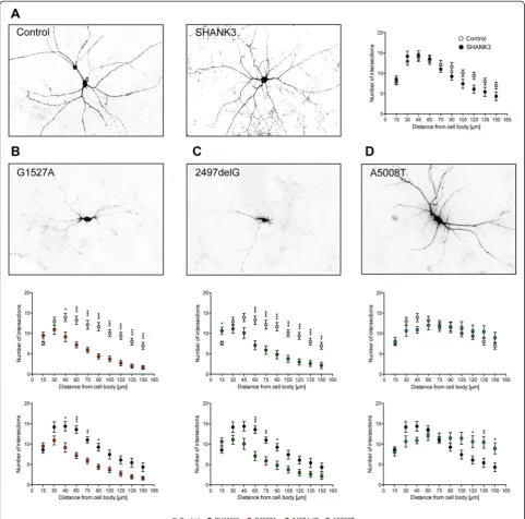
To assess neuronal morphology of transfected neurons, we first analyzed the dendritic tree. Primary dendrite number revealed no differences among all conditions (Additional file 6: Figure S4). Subsequent Sholl analysis showed that dendritic tree complexity was identical in Control and with overexpressed SHANK3 (Figure 3A). However, overexpres-sion of either G1527A or 2497delG resulted in a much lower complexity of the dendritic tree when compared to either Control or SHANK3 (Figure 3B,C) indicating severely impaired dendritic branching. Interestingly, overexpression of A5008T did not show any gross alter-ations of dendritic arborization when compared to
Control, but some enhanced branching of distal dendrites when compared to SHANK3 (Figure 3D).
Overexpression of truncated SHANK3 variants results in distinct alterations of dendritic spines and synaptic contacts
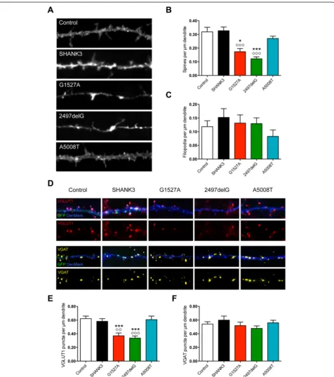
Further assessment of neuronal morphology included the evaluation of spines and filopodia among secondary
dendrites of transfected neurons (Figure 4A,B,C). Over-expression of either G1527A or 2497delG resulted in a strong reduction in spine density when compared to either Control or SHANK3 (Figure 4B), while filopodia density remained unchanged (Figure 4C). In contrast, A5008T overexpression did not affect spine or filopodia density at all (Figure 4B,C). These data show that overexpression of either G1527A or 2497delG results in a specific loss of
spines accompanied by a relative increase in the number of filopodia perμm dendrite (Figure 4B,C, for spine/filopodia and filopodia/spine ratios, see Table 1), while there was no significant effect of A5008T overexpression on spine or
filopodia density (Figure 4B,C, for spine/filopodia and filo-podia/spine ratios, see Table 1).
We next analyzed presynaptic specializations among sec-ondary dendrites of transfected neurons and discriminated
excitatory and inhibitory contacts by immunostaining for either VGLUT1 or VGAT (Figure 4D,E,F). In line with our findings on spines, the dendritic contact sites for excitatory synapses, overexpression of either G1527A or 2497delG, re-sulted in a major loss of VGLUT1-positive puncta perμm dendrite, while VGAT puncta density remained unaffected (Figure 4D,E,F). In contrast, there was no detectable change in the density of either VGLUT1- or VGAT-positive puncta when A5008T was compared to either Control or SHANK3 (Figure 4D,E,F). These findings demonstrate that overex-pression of both G1527A and 2497delG leads to a specific decrease in excitatory, but not inhibitory contacts, thus likely shifting the excitation/inhibition (E/I) balance to-wards inhibition. In contrast, no E/I imbalance phenotype can be implicated at this point when examining overexpres-sion of A5008T in primary hippocampal culture.
Discussion
In this study, we have described, for the first time, a novel truncating stop mutation in an individual with the diagnosis of both ASD and ID (c.5008A > T) (Table 2). In addition,in vitroanalyses with this and two other re-cently identified truncating stop mutations (c.1527G > A and c.2497delG, [17]) addressed the question if distinct mutations in SHANK3 result in mutation-specific or converging morphological phenotypes.
Interestingly, we found that SHANK3a variants harbor-ing either c.1527G > A or c.2497delG, thus lackharbor-ing large but distinct parts of the protein’s C-terminus (Figure 1A), exhibit overlapping phenotypes in all morphological pa-rameters investigated. When overexpressed, both trun-cated proteins exclusively accumulate in the nuclear compartment of transfected cells (Figures 2C,D, Additional file 5: Figure S3A,B) and this is accompanied by a severe reduction of dendritic tree complexity (Figure 3A,B,C). Moreover, we found a specific reduction of dendritic
spine and excitatory, but not inhibitory synapse density (Figure 4A,B,C,D,E,F). These findings are in line with previous in vitro studies showing similar phenotypes for rodent homologue Shank3a variants harboring ei-ther c.3679_3680insG or c.3349C > T [23,25].
In contrast, none of these phenotypes was observed in neurons overexpressing SHANK3a haboring the c.5008A > T mutation. However, some minor alterations in neuronal morphology were detected, and interestingly, they were op-posite from the ones related to c.1527G > A and c.2497delG as c.5008A > T produced slightly enhanced complexity of the distal dendritic tree (Figure 3D).
With respect to subcellular localization of truncated SHANK3, an important aspect to consider is domain composition. The SHANK3a c.5008A > T variant is only lacking a small part of the protein’s C-terminus, thereby exclusively disrupting the SAM domain (Figure 1A), a domain needed for the correct assembly of Shanks in the PSD [27]. In line with this, it still localizes to den-drites and excitatory synapses of transfected neurons in cluster-like structures, although with a much lower effi-ciency as compared to full-length SHANK3a (Figure 2E, Additional file 4: Figure S2A,B). The two other truncated SHANK3a variants analyzed here are lacking major parts of the C-terminus including distinct stretches of the proline-rich domain and - in all cases - the Homer binding site (a dendritic localization signal), a synaptic targeting element in between the Homer and cortactin binding sites and the SAM domain (Figure 1A). In line with this, each of these variants never forms cluster-like structures within dendrites or at spine synapses, but rather localizes to the nucleus of transfected neurons (Figure 2C,D).
A recent study on the transcriptional and functional complexity of the rodent Shank3 gene convincingly delineates all rodent Shank3 isoforms (Shank3a-f ) and reports distinct and/or overlapping phenotypes for each isoform with respect to subcellular distribution and neuronal morphology after overexpression in primary hippocampal neurons [37]. Summarizing what was ob-served, selected Shank3 isoforms either increase the number of spines and excitatory synapses or show the opposite effect, likely depending on their domain com-position and subcellular localization. Intriguingly, the Shank3 isoform Shank3b, which only contains the anky-rin repeats, the SH3 domain, and the PDZ domain [37], resembles two of the shorter truncated SHANK3a vari-ants described here. In primary hippocampal neurons, Table 1 Spine/filopodia and filopodia/spine ratios of rat
primary hippocampal neurons overexpressing different SHANK3 variants
Fusion protein Spines/filopodia Filopodia/spines
Control 2.70 0.37
SHANK3 2.15 0.47
G1527A 1.32 0.76
2497delG 0.94 1.07
A5008T 3.26 0.31
Table 2 Genetic information on the three participants included into this study
Sex Diagnosis Rearrangement Aa change Inheritance Reference
P1 M ASD/ID c.1527G > A p.W509X De novo Sooryaet al. 2013 [17]
P2 M ID c.2497delG p.P834RfsX58 De novo Sooryaet al. 2013 [17]
overexpressed Shank3b localizes to the nucleus accom-panied by a reduced number of mature spines and exci-tatory synapses [37] - just as we report in this study for overexpression of SHANK3a harboring either c.1527G > A or c.2497delG mutations (Figures 2, 4) and as others have reported in previous studies for the rodent Shank3a homologue harboring either c.3679_3680insG or c.3349C > T [23,25].
Although the exact function of each isoform is yet to be determined, it can already be hypothesized at this point that in a healthy neuron, the expression of SHANK3 iso-forms is fluid, adapting to the ever-changing needs of the cell. Hence, we can speculate that in neurons from indi-viduals with heterozygous stop mutation in SHANK3,
there will be disrupted functionality under specific condi-tions, due to expression of variant proteins at key stages.
Genotype-phenotype correlations can be complicated because of the potential for additional genetic variation contributing to more or less severe phenotypes. In fact, P1 has both an early truncating mutation inSHANK3, as well as a 17q duplication, and has one of the more severe presentations observed at the Seaver Autism Center. However, it is still of interest to note that the three individ-uals described here are all significantly affected, irrespective of the extent of the truncation. In fact, it is of particular interest that the A5008T variation is associated with a very mild cellular phenotype but with a severe behavioral phenotype in the participant. A better understanding of this association will require additional studies as noted below, although it is also possible that A5008T causes a different cellular phenotype, which has not been assessed in this study.
Transfection experiments as we have carried out here involve the modification of a single SHANK3 isoform (SHANK3a). In addition, we cannot assess other genetic variation that may alter severity and we cannot exclude the possibility that in patient cells, both c.1527G > A and c.2497delG - as being located in coding exons upstream the natural stop codon - lead to SHANK3 haploinsuffi-ciency due to degradation of the truncated proteins by the nonsense-mediated mRNA decay machinery [39]. It will therefore be essential to differentiate neurons from induced pluripotent stem cells (iPSCs) of participants af-fected by any of the aforementioned stop mutations and analyze neuronal morphology as well as domain com-position, subcellular localization, and mRNA and protein expression of endogenous SHANK3. However, neuronal culture will never completely reflect the impact of a given mutation on the network levelin vivo. Hence, an-other essential approach to study mutations inSHANK3
would be to generate animal models that carry the human mutation. With respect to stop mutations disrupting exon 21 of SHANK3, such as c.2497delG from this study, the recently publishedShank3ΔC/ΔC mouse, which is an exon
21 deletion model [40], may already provide some insights. Importantly, increases in C-terminally truncated Shank3 variants are detectable in hippocampal lysates from these animals. Compared to other Shankmutant mice [41,42], only minimal social deficits were observed in these ani-mals, and increased repetitive behavior was only evi-dent at a certain age (10 to 13 months). CA1 pyramidal neurons did not exhibit a morphological phenotype. The Shank3ΔC/ΔC mutants did clearly show impaired spatial learning and corresponding abnormalities in hippocampal CA3-CA1 physiology though, including decreased long-term potentiation (LTP) and decreased NDMA receptor-mediated synaptic transmission. How-ever, it has to be noted that only analyses of homozygous mutants have been reported so far. Studying heterozygous mutants - as the more ‘human-like’ model - would pos-sibly reveal different phenotypes not only in this but also in other models mimicking human mutations inSHANK3. Conclusions
Our in vitrooverexpression data show that the location of a stop mutation within the SHANK3a sequence de-termines both subcellular localization of the truncated protein and the morphological phenotype of the trans-fected neuron.
Considering domain composition of SHANK3, our data support previous studies [15,23-29,37] and strengthen the fact that only SHANK3 variants with an intact C-terminus including the Homer binding site, a synaptic targeting element in between the Homer and cortactin binding sites and the SAM domain, correctly and efficiently localize to synapses. Any disruption of these domains results in a dis-tinct phenotype of dendritic and synaptic morphology. Interestingly, our data imply that loss of the Homer and cortactin binding sites is sufficient to induce nuclear accu-mulation of the corresponding SHANK3 variants. This again supports previous studies proposing that SHANK3 might not only serve as a synaptic but also serve as a nu-clear protein [25,37] - although its exact nunu-clear function still remains an enigma.
Considering recent findings on the complexity of both subcellular localization and distinct morphological impact of different rodent Shank3 isoforms in healthy neurons [37], we propose that any heterozygous deleterious mu-tation inSHANK3will lead to altered SHANK3 isoform expression and thereby result in distinct spatial and temporal perturbations of SHANK3-dependent cellular processes. Although we cannot further investigate such a proposed patho-mechanistic model in the in vitro
individuals included in this study, with clear overlap in the symptom presentation, regardless of whether most or only part ofSHANK3is missing.
Additional files
Additional file 1: Table S1.Descriptive and diagnostic data by participant: Mullen Scales of Early Learning, Nonverbal IQ estimate, Vineland Adaptive Behavior Scales, ADI-R, and ADOS-2.
Additional file 2: Table S2.Medical features (a) and dysmorphic features (b) of the three participants.
Additional file 3: Figure S1.Additional biochemical expression analysis of SHANK3 variants in HEK293T cells. The anti-GFP antibody detects GFP at the right size after overexpression of the empty vector in HEK293T cells (A). Only the fusion proteins SHANK3, 2497delG and A5008T, but not G1527A, can be biochemically detected by the self-made anti-Shank3 PRC antibody (B). Successful detection of G1527A (as well as rat Shank3a, SHANK3, and 2497delG) was accomplished with a novel self-made anti-Shank3 N-term antibody (C). kDa, kilodalton.
Additional file 4: Figure S2.Additional subcellular distribution analysis of SHANK3 variants in rat primary hippocampal neurons. Quantitative analysis of GFP cluster density (A) and the density of GFP clusters overlapping with VGLUT1 signals (B) among secondary dendrites of rat primary hippocampal neurons filled with DenMark and overexpressing either SHANK3 (black bars) or A5008A > T (blue bars) as indicated (DIV11-14). ***P< 0.001 compared with SHANK3.
Additional file 5: Figure S3.Nuclear accumulation of the SHANK3 variants G1527A and 2497delG in HEK293T cells. (A, B) Subcellular distribution of SHANK3, G1527A, 2497delG and A5008T in HEK293T cells. (A) Merged pictures of the GFP signal, the phalloidin signal (visualizing the actin cytoskeleton) and the DAPI signal (upper row), the GFP signal alone (middle row) and the DAPI signal alone (lower row). Note strong overlap of both G1527A and 2497delG with the DAPI signal. (B) Biochemical detection of overexpressed G1527A and 2497delG in cytosolic (Cyt) and nuclear (Nuc) fractions isolated from HEK293T cells using an GFP antibody. Anti-GAPDH and Histone H3 anti-bodies were used to control fractionation as indicated. kDa, kilodalton.
Additional file 6: Figure S4.Primary dendrite number of rat primary hippocampal neurons overexpressing SHANK3 variants. Quantitative analysis of the number of primary dendrites of rat primary hippocampal neurons filled with DenMark and overexpressing either Control (white bar), SHANK3 (black bar), G1527A (red bar), 2497delG (green bar), or A5008A > T (blue bar) as indicated (DIV11-14). No statistical differences were observed.
Abbreviations
ADI-R:Autism Diagnostic Interview Revised; ADOS: Autism Diagnostic Observation Schedule; Ank: ankyrin repeats; ASD: autism spectrum disorder; cDNA: complementary DNA; DSM: Diagnostic and Statistical Manual for Mental Disorders; E18: embryonic stage 18; EEG: electroencephalography; GERD: gastroesophageal reflux disease; H + C: Homer and cortactin binding sites; ID: intellectual disability; iPSCs: induced pluripotent stem cells; IRB: Institutional Review Board; LTP: long-term potentiation; MRI: magnetic resonance imaging; N-term: N-terminus; P1: participant 1; P2: participant 2; P3: participant 3; PDZ: PSD-95/Dlg-1/ZO-1; PECS: Picture Exchange Communication System; PFA: paraformaldehylde; PRC: proline-rich clusters; Pro: proline-rich region; PSD: postsynaptic density; SAM: sterile alpha motif; SCZ: schizophrenia; SH3: Src-homology 3.
Competing interests
The Icahn School of Medicine at Mount Sinai and Dr Joseph Buxbaum hold a shared patent for Insulin-Like Growth Factor-1 in the treatment of Phelan-McDermid syndrome. The authors declare that they have no competing interests.
Authors’contributions
MJS conceived the outline of this study together with AK, JDB, and TMB. AK performed and supervised all of the clinical assessments, and SL coordinated the clinical assessments and descriptive data collection. YK subcloned the SHANK3variants. DMC performed all biochemistry and cell culture experiments together with MS and MPL. DMC and MJS analyzed allin vitro data. MJS drafted the figures and wrote the paper. All authors reviewed and approved the final version of the manuscript.
Acknowledgements
Patients gave their informed consent to publish individual details in this manuscript. The relevant consent forms are held by the Seaver Autism Center at Mount Sinai and are available upon request. The research leading to this paper has received support from the Innovative Medicines Initiative Joint Undertaking under grant agreement n° 115300, resources of which are composed of financial contribution from the European Union’s Seventh Framework Programme (FP7/2007-2013) and EFPIA companies’in kind contribution. In addition, the work was supported by the Seaver Foundation (to JDB and AK), by NIH grants MH093725, MH101584 (both to JDB), and MH100276 (to AK). MJS was supported by the Baustein 3.2 of Ulm University (L.SBN.0081 to MJS) and by the DAAD (PROCOPE 57049403 to MJS). DMC and MS were supported by an MD thesis grant from the Ulm University International Graduate School in Molecular Medicine and MPL was supported by an EMBO-Short-Term Fellowship. The study was further supported by grants from the Deutsche Forschungsgemeinschaft (DFG: BO 1718/4-1 to TMB). The authors gratefully acknowledge the professional technical assistance of M. Manz and S. Gerlach-Arbeiter and would like to thank Bassem A. Hassan, Leuven, for providing the DenMark construct and Andreas and Stefanie Grabrucker for the help with the data analysis.
Author details
1Institute for Anatomy and Cell Biology, Ulm University, Albert-Einstein-Allee
11, D-89081 Ulm, Germany.2Seaver Autism Center for Research and Treatment, Icahn School of Medicine at Mount Sinai, One Gustave L. Levy Place, New York, NY 10029, USA.3Friedman Brain Institute, Icahn School of Medicine at Mount Sinai, One Gustave L. Levy Place, New York, NY 10029, USA.4Mindich Child Health and Development Institute, Icahn School of Medicine at Mount Sinai, One Gustave L. Levy Place, New York, NY 10029, USA.5Department of Psychiatry, Icahn School of Medicine at Mount Sinai, One Gustave L. Levy Place, New York, NY 10029, USA.6Department of
Pediatrics, Icahn School of Medicine at Mount Sinai, One Gustave L. Levy Place, New York, NY 10029, USA.7Neuroscience Division, Center for Applied
Medical Research, CIMA, University of Navarra, Av. Pio XII 55, 31008 Pamplona, Spain.8Department of Neuroscience, Icahn School of Medicine at
Mount Sinai, One Gustave L. Levy Place, New York, NY 10029, USA.
9Department of Genetics and Genomic Sciences, Icahn School of Medicine at
Mount Sinai, One Gustave L. Levy Place, New York, NY 10029, USA.
Received: 11 January 2015 Accepted: 17 April 2015
References
1. Grzadzinski R, Huerta M, Lord C. DSM-5 and autism spectrum disorders (ASDs): an opportunity for identifying ASD subtypes. Mol Autism. 2013;4:12. 2. De Rubeis S, He X, Goldberg AP, Poultney CS, Samocha K, Cicek AE, et al.
Synaptic, transcriptional and chromatin genes disrupted in autism. Nature. 2014;515:209–15.
3. Buxbaum JD. Multiple rare variants in the etiology of autism spectrum disorders. Dialogues Clin Neurosci. 2009;11:35–43.
4. Huguet G, Ey E, Bourgeron T. The genetic landscapes of autism spectrum disorders. Annu Rev Genomics Hum Genet. 2013;14:191–213.
5. Devlin B, Scherer SW. Genetic architecture in autism spectrum disorder. Curr Opin Genet Dev. 2012;22:229–37.
6. Betancur C. Etiological heterogeneity in autism spectrum disorders: more than 100 genetic and genomic disorders and still counting. Brain Res. 2011;1380:42–77.
8. Betancur C, Sakurai T, Buxbaum JD. The emerging role of synaptic cell-adhesion pathways in the pathogenesis of autism spectrum disorders. Trends Neurosci. 2009;32:402–12.
9. Zoghbi HY, Bear MF, Synaptic dysfunction in neurodevelopmental disorders associated with autism and intellectual disabilites. Cold Spring Harb Perspect Biol; 2012, 4.
10. Verpelli C, Schmeisser MJ, Sala C, Boeckers TM. Scaffold proteins at the postsynaptic density. Adv Exp Med Biol. 2012;970:29–61.
11. Boeckers TM, Bockmann J, Kreutz MR, Gundelfinger ED. ProSAP/Shank proteins - a family of higher order organizing molecules of the postsynaptic density with an emerging role in human neurological disease. J Neurochem. 2002;81:903–10.
12. Grabrucker AM, Schmeisser MJ, Schoen M, Boeckers TM. Synaptic ProSAP/Shank scaffolds in the cross-hair of synaptopathies. Trends Cell Biol. 2009;21:594–603. 13. Sato D, Lionel AC, Leblond CS, Prasad A, Pinto D, Walker S, et al. SHANK1
deletions in males with autism spectrum disorder. Am J Hum Genet. 2012;90:879–87.
14. Berkel S, Marshall CR, Weiss B, Howe J, Roeth R, Moog U, et al. Mutations in the SHANK2 synaptic scaffolding gene in autism spectrum disorder and mental retardation. Nat Genet. 2010;42:489–91.
15. Durand CM, Betancur C, Boeckers TM, Bockmann J, Chaste P, Fauchereau F, et al. Mutations in the gene encoding the synaptic scaffolding protein SHANK3 are associated with autism spectrum disorders. Nat Genet. 2007;39:25–7. 16. Leblond CS, Nava C, Polge A, Gauthier J, Huguet G, Lumbroso S, et al.
Meta-analysis of SHANK mutations in autism spectrum disorders: a gradient of severity in cognitive impairments. PLoS Genet. 2014;10, e1004580.
17. Soorya L, Kolevzon A, Zweifach J, Lim T, Dobry Y, Schwartz L, et al. Prospective investigation of autism and genotype-phenotype correlations in 22q13 deletion syndrome and SHANK3 deficiency. Mol Autism. 2013;4:18. 18. Betancur C, Buxbaum JD. SHANK3 haploinsufficiency: a“common”but
underdiagnosed highly penetrant monogenic cause of autism spectrum disorders. Mol Autism. 2013;4:17.
19. Gauthier J, Champagne N, Lafrenière RG, Xiong L, Spiegelman D, Brustein E, et al. De novo mutations in the gene encoding the synaptic scaffolding protein SHANK3 in patients ascertained for schizophrenia. Proc Natl Acad Sci U S A. 2010;107:7863–8.
20. Hamdan FF, Gauthier J, Araki Y, Lin DT, Yoshizawa Y, Higashi K, et al. Excess of de novo deleterious mutations in genes associated with glutamatergic systems in nonsyndromic intellectual disability. Am J Hum Genet. 2011;88:306–16. 21. Boccuto L, Lauri M, Sarasua SM, Skinner CD, Buccella D, Dwivedi A, et al.
Prevalence of SHANK3 variants in patients with different subtypes of autism spectrum disorders. Eur J Hum Genet. 2013;21:310–6.
22. Gong X, Jiang YW, Zhang X, An Y, Zhang J, Wu Y, et al. High proportion of 22q13 deletions and SHANK3 mutations in Chinese patients with intellectual disability. PLoS One. 2012;7, e34739.
23. Durand CM, Perroy J, Loll F, Perrais D, Fagni L, Bourgeron T, et al. SHANK3 mutations identified in autism lead to modification of dendritic spine morphology via an actin-dependent mechanism. Mol Psychiatry. 2012;17:71–84. 24. Arons MH, Thynne CJ, Grabrucker AM, Li D, Schoen M, Cheyne JE, et al.
Autism-associated mutations in ProSAP2/Shank3 impair synaptic transmission and neurexin-neuroligin mediated transsynaptic signaling. J Neurosci. 2012;32:14966–78.
25. Grabrucker S, Proepper C, Mangus K, Eckert M, Chhabra R, Schmeisser MJ, et al. The PSD protein ProSAP2/Shank3 displays synapto-nuclear shuttling which is deregulated in a schizophrenia-associated mutation. Exp Neurol. 2014;253:126–37.
26. Roussignol G, Ango F, Romorini S, Tu JC, Sala C, Worley PF, et al. Shank expression is sufficient to induce functional dendritic spine synapses in aspiny neurons. J Neurosci. 2005;25:3560–70.
27. Baron MK, Boeckers TM, Vaida B, Faham S, Gingery M, Sawaya MR, et al. An architechtural framework that may lie at the core of the postsynaptic density. Science. 2006;311:531–5.
28. Boeckers TM, Liedtke T, Spilker C, Dresbach T, Bockmann J, Kreutz MR, et al. C-terminal synaptic targeting elements for postsynaptic density proteins ProSAP1/Shank2 and ProSAP2/Shank3. J Neurochem. 2006;92:519–24. 29. Grabrucker AM, Knight MJ, Proepper C, Bockmann J, Joubert M, Rowan M,
et al. Concerted action of zinc and ProSAP/Shank in synaptogenesis and synapse maturation. EMBO J. 2011;30:569–81.
30. Nicolaï LJ, Ramaekers A, Raemaekers T, Drozdezecki A, Mauss AS, Yan J, et al. Genetically encoded marker sheds light on neuronal connectivity in Drosophila. Proc Natl Acad Sci U S A. 2010;107:20553–8.
31. Schmeisser MJ, Ey E, Wegener S, Bockmann J, Stempel AV, Kuebler A, et al. Autistic-like behaviours and hyperactivity in mice lacking ProSAP1/Shank2. Nature. 2012;29:256–60.
32. Schmeisser MJ, Grabrucker AM, Bockmann J, Boeckers TM. Synaptic cross-talk between N-methyl-D-aspartate receptors and LAPSER1-beta-catenin at excitatory synapses. J Biol Chem. 2009;284:29146–57.
33. Grabrucker AM, Schmeisser MJ, Udvardi PT, Arons M, Schoen M, Woodling NS, et al. Amyloid beta protein-induced zinc sequestration leads to synaptic loss via dysregulation of the ProSAP2/Shank3 scaffold. Mol Neurodegener. 2011;6:65.
34. Gessert S, Schmeisser MJ, Tao S, Boeckers TM, Kühl M. The spatio-temporal expression of ProSAP/shank family members and their interaction partner LAPSER1 during Xenopus laevis development. Dev Dyn. 2011;240:1528–36. 35. Schmeisser MJ, Kühl SJ, Schoen M, Beth NH, Weis TM, Grabrucker AM, et al.
The Nedd4-binding protein 3 (N4BP3) is crucial for axonal and dendritic branching in developing neurons. Neural Dev. 2013;8:18.
36. Brandt T, Desai K, Grodberg D, Mehta L, Cohen N, Tryfon A, et al. Complex autism spectrum disorder in a patient with a 17q12 microduplication. Am J Med Genet A. 2012;158A:1170–7.
37. Wang X, Xu Q, Bey AL, Lee Y, Jiang YH. Transcriptional and functional complexity of Shank3 provides a molecular framework to understand the phenotypic heterogeneity of SHANK3 causing autism and Shank3 mutant mice. Mol Autism. 2014;5:30.
38. Mameza MG, Dvoretskova E, Bamann M, Hönck HH, Güler T, Boeckers TM, et al. SHANK3 gene mutations associated with autism facilitate ligand binding to the Shank3 ankyrin repeat region. J Biol Chem. 2013;288:26697–708. 39. Popp MW, Maquat LE. Organizing principles of mammalian
nonsense-mediated mRNA decay. Annu Rev Genet. 2013;47:139–65.
40. Kouser M, Speed HE, Dewey CM, Reimers JM, Widman AJ, Gupta N, et al. Loss of predominant Shank3 isoforms results in hippocampus-dependent impairments in behavior and synaptic transmission. J Neurosci. 2013;33:18448–68.
41. Jiang YH, Ehlers MD. Modeling autism by SHANK gene mutations in mice. Neuron. 2013;78:8–27.
42. Schmeisser MJ. Translational neurobiology inShankmutant mice–Model systems for neuropsychiatric disorders. Ann Anat. 2015;200:115–7.
Submit your next manuscript to BioMed Central and take full advantage of:
• Convenient online submission
• Thorough peer review
• No space constraints or color figure charges
• Immediate publication on acceptance
• Inclusion in PubMed, CAS, Scopus and Google Scholar
• Research which is freely available for redistribution