R E S E A R C H
Open Access
Cytokines and olfactory bulb microglia in
response to bacterial challenge in the
compromised primary olfactory pathway
Rosalind P Herbert
1, Julie Harris
1, Kim Pei Chong
1, Jamie Chapman
2, Adrian K West
1,2and Meng Inn Chuah
1,2*Abstract
Background:The primary olfactory pathway is a potential route through which microorganisms from the periphery could potentially access the central nervous system. Our previous studies demonstrated that if the olfactory
epithelium was damaged, bacteria administered into the nasal cavity induced nitric oxide production in olfactory ensheathing cells. This study investigates the cytokine profile of olfactory tissues as a consequence of bacterial challenge and establishes whether or not the bacteria are able to reach the olfactory bulb in the central nervous system.
Methods:The olfactory epithelium of C57BL/6 mice was damaged by unilateral Triton X-100 nasal washing, and
Staphylococcus aureuswas administered ipsilaterally 4 days later. Olfactory mucosa and bulb were harvested 6 h, 24 h and 5 days after inoculation and their cytokine profile compared to control tissues. The fate ofS. aureusand the response of bulbar microglia were examined using fluorescence microscopy and transmission electron microscopy.
Results:In the olfactory mucosa, administeredS. aureuswas present in supporting cells of the olfactory epithelium, and macrophages and olfactory nerve bundles in the lamina propria. Fluorescein isothiocyanate-conjugated
S. aureuswas observed within the olfactory mucosa and bulb 6 h after inoculation, but remained restricted to the peripheral layers up to 5 days later. At the 24-h time point, the level of interleukin-6 (IL-6) and tumour necrosis factor-αin the compromised olfactory tissues challenged with bacteria (12,466 ± 956 pg/ml and 552 ± 193 pg/ml, respectively) was significantly higher than that in compromised olfactory tissues alone (6,092 ± 1,403 pg/ml and 80 ± 2 pg/ml, respectively). Immunohistochemistry confirmed that IL-6 was present in several cell types including olfactory ensheathing cells and mitral cells of the olfactory bulb. Concurrently, there was a 4.4-, 4.5- and 2.8-fold increase in the density of iNOS-expressing cells in the olfactory mucosa, olfactory nerve and glomerular layers combined, and granule layer of the olfactory bulb, respectively.
Conclusions:Bacteria are able to penetrate the immunological defence of the compromised olfactory mucosa and infiltrate the olfactory bulb within 6 h even though a proinflammatory profile is mounted. Activated microglia may have a role in restricting bacteria to the outer layers of the olfactory bulb.
Keywords:Olfactory mucosa, Innate immunity, Cytokines, Bacterial infection, Microglia, Olfactory ensheathing cells
* Correspondence:[email protected]
1Menzies Research Institute Tasmania, 17 Liverpool Street, Hobart, TAS 7001,
Australia
2School of Medicine, The University of Tasmania, 17 Liverpool Street, Hobart,
TAS 7001, Australia
Background
The primary olfactory pathway represents a one-synapse route spanning the olfactory mucosa and the olfactory bulb. Normally, microbes are prevented from infecting the olfactory mucosa through sneezing, clearing of mu-cous, immune responses by cells in the nasal mucosa or apoptosis of infected cells [1,2]. Though generally suc-cessful, these protective strategies are known to fail under certain conditions. Viruses injected into the nasal cavity have been found to enter olfactory neurons, repli-cate, travel into the olfactory bulb and then into the cen-tral nervous system (CNS) [3,4]. In addition, viruses in circulation can also cross from capillaries in the lamina propria into olfactory neurons. Because of the close proximity and the synaptic connections between the nasal cavity and the CNS, the brain can potentially be infected in relatively short periods of time [3]. Ginkel and co-workers demonstrated that intranasal injection of a strain ofStreptococcus pneumonia, which is non-viable in the blood system, resulted in olfactory bulb and CNS infection via olfactory axonal transport after just 24 h [5]. The infection of olfactory neurons was thought to be through pneumococci interaction with gangliosides expressed on the cell membranes. The rarity of these infections suggests that a robust endogenous immuno-logical defence is likely to be present in olfactory tissues, but this immunological response has been poorly studied.
In a recent study, we showed that the intact olfactory epithelium was an effective barrier in preventing the in-filtration ofStaphylococcus aureus into the nasal lamina propria [6]. However if the olfactory epithelium was damaged by zinc sulphate or Triton-X irrigation, it was susceptible to bacterial infiltration, and this led to the appearance of inducible nitric oxide synthase (iNOS)-expressing cells in the lamina propria, some of which were the neuroglia of olfactory nerves, the olfactory en-sheathing cells (OECs) [6]. Consistent with this finding, nitric oxide production by OECS was demonstrated within 1 h of bacterial exposure in vitro [6]. Further-more, microarray analysis showed that OECs have enriched transcripts for the chemokines CXCL1 and MCP-1 [7], both of which increased when OECs were exposed to Escherichia coli and S. aureus in vitro [8]. CX3CL1, a cytokine known to influence the migration of
CX3CR1-positive innate immune cells into tissue [9], is
also expressed by olfactory neurons and OECs [10]. Thus, the evidence suggests the existence of an immune barrier in the nasal cavity that is contributed in part by OECs.
This study aims to build on these previous findings and obtain new knowledge about the immunological sta-tus of the olfactory mucosa and bulb. We investigated the cytokine and macrophage profile of damaged
olfactory tissues in response to bacterial challenge and tracked the infiltration of bacteria into the olfactory bulb.
Methods
All experiments involving animals in this project were approved by the Animal Experimentation Ethics Com-mittee of the University of Tasmania and are consistent with the Australian Code of Practice for the Care and Use of Animals for Scientific Purpose. mice aged 6– 7 weeks were maintained on a 12-h light:dark cycle in filtered, sterile air by the Menzies Research Institute Animal Services.
S. Aureus cultures and fluorescein isothiocyanate (FITC) conjugation
A loopful from slope cultures of S. aureus (ATCC 33862) (kindly donated by the Microbiology Department of the University of Tasmania) was streaked onto nutri-ent agar plates and incubated overnight at 370C. Single colonies were then picked and inoculated into 1 ml L-broth cultures (pH 7.2) and incubated overnight at 370 C, shaking at 300 rpm in an orbital shaker (Ratek Instruments, Australia). Bacteria from tubes that exhib-ited growth, as evidenced by turbidity of broth, were inoculated directly into the left nasal cavity of mice in the appropriate treatment group at the designated time points.
In order to visually localise bacteria under fluorescence microscopy, S. aureus was conjugated to FITC by incu-bating 10 μl FITC (1 mg/ml; Sigma Aldrich, Australia) in 1 ml L-broth and S. aureus at 370 C for 1.5 h. The culture was then washed with 500 μl filtered PBS and centrifuged at 12,000 rpm for 30 s. The supernatant was removed, and the pellet washed twice and resuspended in 200μl filtered PBS prior to nasal inoculation. Follow-ing FITC conjugation, random samples ofS. aureuswere checked on the fluorescence microscope to confirm that conjugation was successful (Figure 1A). In addition, some were plated on nutrient agar plates and left over-night at 370C. Growth was typically observed, indicating
viable bacteria as well as continued fluorescence (Figure 1B).
Cytokine assay of olfactory tissues
Animals were divided into four groups: (1) untreated mice (NT); (2) mice treated with 30μl 1% Triton X-100 alone (TX); (3) mice treated with 30μl 1% Triton X-100 and 30μl 0.9% sterile saline 4 days later (TXS); (4) mice treated with 30 μl Triton X-100 and 30 μlS. aureus 4 days later (TXB). The amount of S. aureusinoculated into the nasal cavity was equivalent to 3 x 10⁷.Bacteria were introduced 4 days after Triton X-100 washing be-cause at this time most of the damaged olfactory neu-rons have sloughed off while the basal cells have initiated proliferation to reconstitute the epithelium [11]. Nasal irrigation was performed unilaterally on the left side of the nasal cavity using 30-gauge plastic tubing attached to the end of a 29-G needle. Mice were lightly anaesthetised with 0.6% isoflurane in air prior to Triton X-100, saline orS. aureusinoculation.
Mice of the TX group were perfused 4 days after Tri-ton X-100 irrigation; mice of the TXS and TXB groups were perfused either 6 h (TXS6, TXB6 respectively) or 24 h (TXS24, TXB24 respectively) after saline or S. aur-eus inoculation. Mice were euthanised with sodium pentobarbitone (50 mg/kg i.p.) before transcardial perfu-sion with chilled phosphate-buffered saline (PBS) in an aseptic environment. The olfactory bulbs, olfactory tur-binates and septum were dissected. Tissue from each group was pooled, minced and homogenised in lysis buf-fer (catalogue no. 0103004-L; RayBioTech) with 3% pro-tease inhibitor. Samples were centrifuged at 13,000 g for 10 min at 4 °C. The supernatant and pellet were sepa-rated, snap frozen in liquid nitrogen and stored at−80 ° C until use in cytokine assay.
A modified Bradford assay [12] was carried out to es-tablish the total protein concentration of the pooled ol-factory tissue supernatant from each treatment group. This was done in order to estimate the correct sample amount to be used in the cytokine arrays. One part Bradford reagent was diluted for four parts distilled water. Standard protein samples at concentrations of 0.1, 0.15, 0.2, 0.25, 0.5 and 1.0 mg/ml were prepared using DNase free bovine serum albumin. Each standard was diluted 1:50 using Bradford reagent in a 96-well plate. Samples were diluted 1:100 in MilliQ water and then further diluted 1:100 in a 96-well plate using Bradford reagent. Replicates were made of each standard and sample. Absorbance at 595 nm was measured using a Tecan GENios microplate reader and analysed using the XFluor4 (v.4.5) software. From measured absorbance of standards, a standard curve was created and used to de-termine the concentration of the samples.
Cytokine assay was performed as three separate experiments for each time point. For each time point, tissue was pooled from three untreated mice and four mice from each of the treated groups (TX, TXS and TXB) and samples diluted to a concentration of 25 mg/ ml. Cytometric Bead Array Mouse Th1/Th2/Th17 Cyto-kine Kit (BD Biosciences) detects seven cytoCyto-kines (inter-feron-γ, interleukin-10, interleukin-17A, interleukin-2, interleukin-4, interleukin-6, tumour necrosis factor-α) using flow cytometry. The assay was run according to the manufacturer’s instructions. Replicates were run on a BD FACS Canto II flow cytometer with 2,100 events measured in the P1 gate, and total events were also recorded. FCAP array (v1.0.1) software was used to ana-lyse data collected from the flow cytometer. Raw data from the BD array were multiplied by the appropriate di-lution factor to calculate the true concentration of cyto-kines. Cytokine concentrations for the BD array were tabulated, and data analyses were conducted using single factor ANOVA and pairwise multiple comparison (Tukey test). A p value equal to or less than 0.05 was considered statistically significant.
Visualisation of FITC-conjugated S. aureus and immunohistochemical staining for interleukin-6 (IL-6) A total of 30μl ofS. aureus was inoculated into the left nasal cavity of eight 6–7-week-old C57/BL6 male mice that had previously undergone unilateral nasal washing with 1% Triton-X 100 solution. Mice were killed at 6 h (n= 3), 24 h (n= 2) and 5 days (n= 3) after inoculation. Transcardial perfusion was conducted with chilled PBS followed by Zamboni’s fixative (ZF; 15% picric acid + 0.85% paraformaldehyde). Mice were decapitated and excess tissue trimmed away, leaving the olfactory bulbs and nasal cavity en bloc; they were then stored in ZF at 4 °C. Tissue was washed with PBS several times after 2– 3 days to remove ZF before being transferred to 0.01% azide in PBS. The nasal cavity and olfactory bulb en bloc were decalcified in clinical fast decal fluid (Lomb Scien-tific) for 1–1.5 h. Specimens were washed with distilled water, trimmed and cryoprotected by transferring to 10% sucrose overnight, followed by 30% sucrose. They were then frozen in cryomatrix, sectioned at 10 μm in the horizontal plane using a cryostat (Leica DM-1850), placed onto SuperFrost Plus slides (Menzel Glaser, Ger-many) and stored at -200C. Unstained sections from all animals were examined on an Olympus BX50 fluores-cent microscope to localise FITC-conjugatedS. aureus.
were then incubated with Mouse on Mouse diluent from the M.O.M Kit for 5 min. Sections were incubated with primary antibody rat anti- IL-6 (1:100; Invitrogen) over-night at 40C. Control sections were incubated overnight with diluent alone or isotype control. Next, sections were incubated with biotinylated goat anti-rat antibody (1:1;000, Molecular Probes) for 1 h followed by incuba-tion with streptavidin conjugated to HRP for 30 min, and DAB-chromogen solution for 5 to 6 min. Sections were counterstained with Nuclear Red in 0.03% azide for 1.5 min, coverslipped and viewed on a Leica DM-2500 light microscope. Control sections did not show positive immunoreactivity.
Olfactory ensheathing cell culture and immunostaining for IL-6
Olfactory ensheathing cells were isolated from the olfac-tory nerve layer of the olfacolfac-tory bulb and the olfacolfac-tory mucosa of 3-day-old mouse pups as described in a previ-ous study [13]. Briefly, the olfactory bulbs and mucosa were dissected and digested for 20 min using 0.25% tryp-sin in MEM-H (Gibco) at 37 °C. Dulbecco’s modified Eagle’s medium with HEPES (DMEM-H; Gibco) supple-mented with 10% foetal calf serum (DMEM-10FCS) was added to terminate digestion, and the suspension was cen-trifuged at 500 g for 10 min. The supernatant was removed and the pellet resuspended in DMEM-H. Cells were dissociated and plated onto acid-washed 13-mm coverslips coated with 4% poly-L-lysine. Following incu-bation for 24 h at 37 °C, cytosine-β-D-arabinofuranoside (100μM) was added and cells were incubated for another 2 days at 37 °C until cultures were sufficiently free of contaminating cells. The medium was then replaced with fresh DMEM- 10FCS supplemented with bovine pituitary extract (50 μg/ml; Sigma) to allow cellular expansion. Cultures yielded 95% OECs based on positive immunor-eactivity with anti-p75NTR.
The immunostaining procedure for IL-6 was similar to that used on tissues sections, except that cultures were treated with a shorter incubation time (1 h) with the pri-mary antibody. Cells were counterstained with Nuclear Red in 0.03% azide for 1 min, dehydrated and mounted with DPX.
Immunofluorescent staining with Iba1, anti-iNOS and tomato lectin, and quantitative analysis
The left nasal cavity of six 6–7-week-old C57/BL6 male mice that had previously undergone unilateral nasal washing with 1% Triton-X 100 solution was inoculated with 30 μl of S. aureus(3 mice) or saline (3 mice). The mice were euthanised with sodium pentobarbitone (50 mg/kg i.p.) at 24 h after bacterial inoculation and transcardially perfused with chilled PBS followed by 4% paraformaldehyde. For comparison, three untreated 6–
7-week-old C57/BL6 male mice were also killed and trans-cardially perfused. Mice were decapitated, and the heads trimmed and processed for cryosectioning, as described for immunohistochemistry in the preceding section. Ten-micron-thick sections were post-fixed with 4% par-aformaldehyde for 15 min, washed gently three times with PBS and blocked using Dako Protein Block Serum-free (DakoCytomation) for 5 min prior to incubation with primary antibodies. Microglia were identified by positive immunoreactivity for Iba1 or binding to tomato lectin [14,15]. In single labelling, sections were incubated with rabbit-anti iNOS/NOS type II (1:400; BD Bios-ciences), Iba1 (1:200; Wako) or biotinylated tomato lec-tin (10 μg/ml diluted 1:200; Vector Labs) for 2 h. This was followed by incubation with goat anti-rabbit Alexa-Fluor 594 (1:1,000; Molecular Probes), goat anti-mouse AlexaFluor 488 (1:1000; Molecular Probes) or streptavi-din AlexaFluor 488 (1:1000; Molecular Probes) respect-ively for 1 h at room temperature in the dark, followed by 0.0001% Nuclear Yellow (Sigma) staining for 5 min prior to mounting and storage at 40C. In some sections, double labelling with Iba1 and tomato lectin was done to ascertain colocalisation. Double labelling with anti-iNOS and tomato lectin was also done to identify anti- iNOS-producing microglia.
Nine separate frames, taken with 40× objective, cap-turing the olfactory nerve and glomerular layers of the rostral part of the olfactory bulb from each animal were used in the cell counts of iNOS- and Iba1-positive cells. Cell counts and epithelial distance, as represented by the basal lamina, were measured using Image J. The data were analysed by two-way ANOVA and pairwise mul-tiple comparison (Tukey test). Apvalue equal to or less than 0.05 was considered statistically significant.
Transmission electron microscopy
600 C oven for 48 h. Thick (1 μm) and ultrathin (70– 90 nm) sections were cut using a Leica ultramicrotome and placed on formvar-coated copper palladium grids (ProSciTech, Qld). Thin sections were stained with satu-rated uranyl acetate in 5% ethanol (30 min) followed by lead citrate (5 min) prior to being viewed on a Philips CM100 transmission electron microscope.
Results
Olfactory tissues damaged by Triton-X 100 significantly upregulate the expression of IL-6 and TNF-αwhen exposed to bacteria
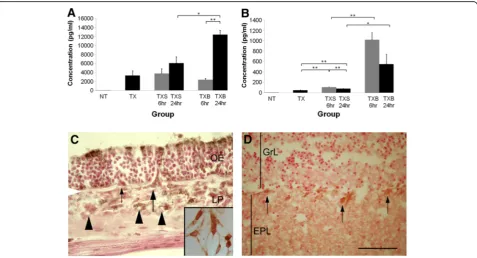
Triton X-100 was used in this study because its cellular effects are well characterised [11,17]. It selectively destroys mature olfactory neurons and leaves the rest of the olfactory mucosa intact with no direct damage to the olfactory bulb. In the normal olfactory mucosa and bulb there was negligible expression of IL-6 (26 ± 7 pg/ml)
(Figure 2A). Olfactory epithelial ablation by Triton-X 100 induced expression of IL-6, which was measured 4 days later (3,309 ± 1,052 pg/ml; p<0.01). When this damaged olfactory mucosa was exposed to S. aureus, a further sig-nificant upregulation of IL-6 was observed 24 h later (12,466 ± 956 pg/ml). This was also significantly higher than that of similarly ablated olfactory tissues subjected to saline wash at the 24-h time point (6,092 ± 1,403 pg/ml; p<0.05). No significant difference between these two treatment groups (p= 0.26) was detected at the 6-h time point. There was no significant difference in IL-6 con-centration between the TX (3,309 ± 1,052 pg/ml) and TXS groups (6 h = 3,748 ± 1,086 pg/ml, p= 0.80; 24 h = 6,092 ± 1,403 pg/ml,p= 0.15), suggesting that nasal wash-ingper sedid not induce IL-6 upregulation.
Immunohistochemical staining showed that IL-6 was present in the apical region of sustentacular cells in the olfactory epithelium, as well as in cells throughout the
Figure 2Measurement of interleukin-6 (IL-6; A) and tumour necrosis factor-α(TNF-α; B) in groups of mice: untreated (NT); unilateral nasal wash with Triton X-100 alone (TX); unilateral nasal wash with Triton X-100 followed by saline wash (TXS); unilateral nasal wash with Triton X-100 followed byS. aureusinoculation (TXB).Undamaged normal olfactory tissues express negligible amounts of IL-6 and TNF-α. At 4 days post-Triton X-100 damage (TX), olfactory tissues had an initial expression of IL-6 and TNF-αof 3,309 ± 1,052 pg/ml and 49 ± 6 pg/ml respectively. The level of IL-6 is significantly increased by the presence of bacteria at the 24-h time point (12,466 ± 956 pg/ml). The upregulation of IL-6 appears to follow that of TNF-α, which occurs at the 6-h time point, albeit at a lower concentration (1,024 ± 137 pg/ml). Thereafter, at the 24-h time point, a decreasing trend in TNF-αproduction is observed.Error barsrepresent SEM. *p<0.05; **p<0.01. IL-6 expression in the olfactory mucosa (C) and olfactory bulb (D) 24 h post-inoculation withS. aureus.IL-6 immunoreactivity is present in the cytoplasm of select groups of supporting cells in the apical part of the olfactory epithelium (OE). Some cells straddling the OE and lamina propria (LP), possibly OECs or macrophages, express IL-6 (large arrow), while others do not (small arrow). Several cells in the LP are immunopositive for IL-6 including some that surround the small lumen (arrowheads), which are most likely endothelial cells or inflammatory cells.Insetshows immunoreactivity for IL-6 in cultured OECs.White arrowheadat the edge of the micrograph indicates location of basal lamina. In the olfactory bulb mitral cell soma (arrows) show positive immunostaining for IL-6 with more diffuse staining in the external plexiform layer (EPL).GrL= granule cell layer;scale bar= 40μm
lamina propria (Figure 2C), some of which could be OECs or infiltrating immune cells. IL-6-positive cells that straddled the olfactory epithelium and the lamina propria are consistent with OECs as they emerge from the olfactory epithelium to accompany differentiating axons [18]. Alternatively they could be macrophages that reside in the olfactory epithelium [10]. Other IL-6-positive cells within the lamina propria appeared to sur-round blood vessels, and these cells could either be endothelial cells or infiltrating inflammatory cells. Subse-quent isolation of OECs from the olfactory mucosa and bulb, and cell culture confirmed that OECs expressed IL-6 (inset in Figure 2C). In the olfactory bulb, immu-nostaining for IL-6 was observed in the external plexi-form layer and mitral cells (Figure 2D) and less intensely in the glomerular and olfactory nerve layer.
At 4 days post-injury with Triton X-100, olfactory tis-sues showed an initial level of 49 ± 6 pg/ml TNF-α, which increased significantly to 1,024 1 ± 37 pg/ml 6 h afterS. aureus exposure (p<0.01) (Figure 2B). In com-parison, Triton X-100 damaged olfactory tissues exposed to saline alone yielded 108 ± 4 pg/ml TNF-α. There was no significant difference (p= 0.10) in the TNF-α re-sponse to bacteria between 6 and 24 h (552 ± 193 pg/ ml), although there appeared to be a downward trend. TNF-αconcentration at both 6 and 24 h after S. aureus inoculation remained significantly higher when com-pared to the saline control (6 h = 108 ± 4 pg/ml,p<0.01; 24 h = 80 ± 2 pg/ml,p<0.05).
Significant differences among treatment groups were only observed in IL-6 and TNF-αexpression. Concentra-tions of IFN-γ, IL-10, IL-17A, IL-2 and IL-4 were all at or below the theoretical limit of detection of the BD array in all groups (data not shown).
FITC-conjugated S. aureus are localised in the olfactory mucosa and periphery of the olfactory bulb
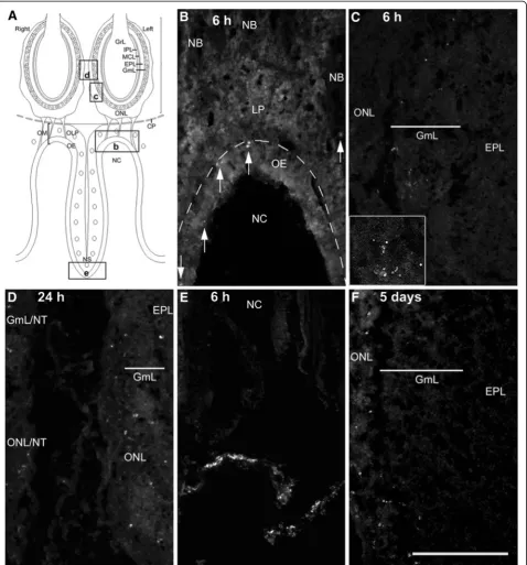
At 6 and 24 h after bacterial inoculation into the Triton-X 100 washed nasal cavity, FITC-labelledS. aureuscould be localised to both the olfactory mucosa and bulb (Figure 3A-C). In the olfactory mucosa, FITC labelling was observed mainly in the olfactory epithelium and in rare instances in nerve bundles within the lamina pro-pria. In the olfactory bulb, fluorescence was observed in the glomerular and olfactory nerve layers. The aggre-gates of punctate fluorescent label observed in the olfac-tory bulb (inset in Figure 3C) suggested that the S. aureus was most likely to be intracellular. The majority of fluorescent labels were found on the ipsilateral side of bacterial inoculation, although some could be found in the glomerular and olfactory nerve layer of the contralat-eral bulb (Figure 3D). On the whole, the number of FITC-labelledS. aureuslocalised to olfactory tissues was relatively low. Numerous FITC-labelled S. aureus were
located near the nares at 6 h (Figure 3E), suggesting that a large number of bacteria were being expelled. Five days after bacterial inoculation, some FITC- labelling remained in the olfactory mucosa, the olfactory nerve and glomerular layers of the ipsilateral olfactory bulb (Figure 3F). At no time was FITC labelling observed in the deeper layers of the olfactory bulb.
Transmission electron microscopy of S. aureus in olfactory mucosa
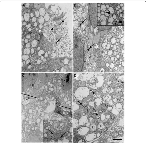
Ultrastructural observations at the 6- and 24-h time points yielded similar results. Up to 24 h after inocula-tion of S. aureusinto the nasal cavity previously washed with Triton-X 100, some bacteria remained on the sur-face of the epithelium (Figure 4A). The junctional com-plex between supporting cells remained intact and no bacteria were observed extracellularly in the olfactory epithelium. This suggested thatS. aureuswas unlikely to have accessed the lamina propria via spaces between epithelial cells. Instead, endocytosed S. aureuswas loca-lized either in vesicles or in the cytosol of supporting cells (Figure 4B). Highly vesiculated cells resembling macrophages were observed straddling the olfactory epi-thelium and lamina propria (Figure 4C), some of which in the lamina propria were shown to have endocytosed bacteria (inset in Figure 4C). S. aureuswas present in a small number of axon bundles (Figure 4D), most likely olfactory rather than trigeminal nerves given that the axons were all unmyelinated.
Number of iNOS-positive cells in the olfactory mucosa and bulb is increased significantly following exposure to S. aureus
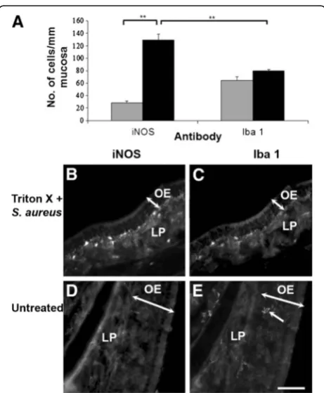
epithelium (Figure 5B). About 62% of these cells were positively immunolabeled with Iba1 (Figure 5A, 5C), indi-cating that they were macrophages present either in the
lamina propria or olfactory epithelium (Figure 5), while the rest were likely to be OECs, as demonstrated in our previous study [6]. Although the density of activated cells,
as represented by iNOS-immunoreactivity, was signifi-cantly increased in theS. aureus-treated tissue, the density of macrophages was not significantly increased compared to untreated tissue (Figure 5A).
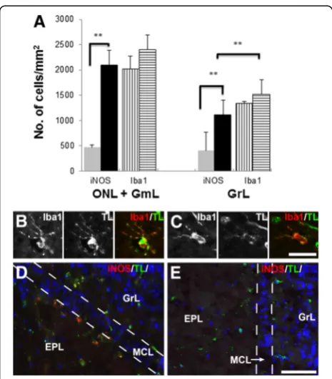
In the olfactory bulb, there was also a general increase in the density of iNOS-expressing cells in theS. aureus -treated mice, as shown by the quantitative analysis done on the olfactory nerve, glomerular and granule cell
layers (Figure 6A). Although no significant increase in Iba1-positive cells was observed in the olfactory bulb, they had an activated morphology, as evidenced by the hypertrophied cell bodies and ramified processes. Fur-ther confirmation that the Iba1-positive cells were microglia was provided by their positive binding to to-mato lectin with variable intensities (Figure 6B, C). In the S. aureus-exposed olfactory bulbs, iNOS-expressing
microglia were present not just in the peripheral layers, but also in the vicinity of the mitral cell layer (Figure 6D). In comparison, saline-treated olfactory bulbs generally showed sparse or no immunoreactivity for iNOS (Figure 6E), similar to untreated olfactory bulbs (data not shown).
Discussion
This study demonstrates that damage to the olfactory epithelium compromises the protective barrier and allows bacteria to gain access to the outer layers of the olfactory bulb as rapidly as 6 h following exposure. Infil-tration ofS. aureusinto the nasal tissues is accompanied by upregulation of IL-6, TNFα and iNOS expression. In the olfactory mucosa the cellular immune defence is provided by macrophages and olfactory ensheathing cells, while microglia constitute the major defensive cell in the olfactory bulb. S. aureus is restricted to the
olfactory nerve and glomerular layers of the olfactory bulb up to 5 days after bacterial exposure.
Biological significance of cytokines in the immune response of olfactory tissue
Cytokines are humoral components in innate immunity that are vital in the initiation and regulation of an appro-priate immune response [19]. In this study expression of IL-6 and TNF-α was found to significantly increase at both 6 and 24 h after bacterial inoculation in mice whose olfactory epithelium had been previously damaged. The results are consistent with an earlier study in which elevated IL-6 and TNF-α mRNA levels in the murine olfactory mucosa and bulb were demonstrated at 24 h following intranasal inoculation with Satratoxin G, a mycotoxin that damages the olfactory epithelium [20]. Signalling via cytokines such as IL-6 and TNF-α, pro-duced by macrophages as a result of bacterial detection, has been shown to be vital in the initiation of immune responses to directly remove the infecting agent [21,22].
TNF-α, being a pro-inflammatory cytokine, induces a number of different responses that can potentially ex-acerbate tissue damage. However, without it, brain abscesses induced by S. aureus persist, as evidenced by increased bacterial burdens [23]. Therefore, tight regula-tion of TNF-α expression needs to occur to maintain a balance between destruction of normal tissue and re-moval of potentially devastating bacteria. Pituitary ad-enylate cyclase-activating peptide (PACAP) is a protein that is produced by OECs and supporting cells of the ol-factory epithelium [24] that has been shown to protect against TNF-α induced cell death in olfactory neurons [25]. It is possible that PACAP is produced locally by ol-factory ensheathing cells and sustentacular cells in order to mitigate the damaging effects of TNF-αin the process of bacterial elimination. Furthermore the IL-6 receptor is known to be expressed transiently by OECs following neuronal cell death in response to bulbectomy [26], sug-gesting that it may be a primary target of IL-6, including that produced by OECs themselves. IL-6/IL-6R binding in OECs may be protective by inducing nuclear trans-location of STAT transcription pathways, thereby acti-vating anti-apoptotic pathways [27]. The survival of OECs would have particular importance in compro-mised olfactory tissue as these cells have a vital contri-bution to olfactory neuronal regeneration and innate immunity [28,29].
Studies on macrophages and OECs have shown that NFκB nuclear translocation occurs in response to bac-teria and pathogen-associated molecular patterns, result-ing in iNOS transcription and nitric oxide production [6,30]. Though this may be a direct effect of bacteria on innate immune cells, nitric oxide may also be induced by TNF-α binding to mTNFR1, which then activates a
Figure 5Density of iNOS- and Iba1-positive cells in the normal (grey bars) and bacteria-challenged (black bars) olfactory mucosa (A).The density of iNOS-positive cells following bacterial exposure is significantly more than that in the unperturbed olfactory mucosa, increasing from 28 ± 2 to 129 ± 10 cells/mm mucosa. This resulting high density of iNOS-positive cells is greater than the density of Iba1-positive cells, indicating that a proportion of the iNOS-positive cells are not macrophages.Error barsrepresent SEM. **p<0.01. The iNOS- and Iba1-positive cells are predominantly located in the lamina propria (LP), close to the olfactory epithelium (OE;B,C). In the normal, intact olfactory mucosa (D,E), Iba1-positive cells can occasionally be observed in the olfactory epithelium (arrow
number of cellular pathways, including the activation and nuclear translocation of NFκB [31,32]. NFκB activa-tion also induces IL-6 transcripactiva-tion [33,34]. In our study, a significant upregulation of TFN-α in olfactory tissues at the 6-h time point preceded a significant upregulation of IL-6 at the 24-h time point. A possible explanation is that while TNF-α is produced to recruit factors that re-move S. aureus, it also induces the production of IL-6. In this study, at 24 h after bacterial infection, IL-6 immunoreactivity was present in supporting cells, olfac-tory ensheathing cells, fibroblasts, endothelial cells and innate immune cells. IL-6 has been previously shown to be expressed by macrophages and olfactory ensheathing cells [26,35] as well as endothelial cells and fibroblasts in
inflammatory conditions [36,37]. IL-6 mediates neutro-phil responses (Dalrymple et al., 1995; Dalrympleet al., 1996) and is also known to enhance neuron survival (Toulmondet al., 1992; Loddicket al., 1998), and there-fore may be neuroprotective with regard to olfactory neuronal survival. Therefore, cytokines such as TNF-α and IL-6 could work in synergy to maintain olfactory tis-sue homeostasis while removing the bacterial infection.
Infiltration of bacteria in the primary olfactory pathway This is the first study to show that bacteria in the nasal cavity were able to reach the olfactory bulbs in as little as 6 h. Previous studies had used longer time points to assess bacterial invasion in the nasal connective tissue and olfactory bulb [5,6]. In our study, S. aureus was localised ultrastructurally to supporting cells of the ol-factory epithelium, macrophages and a small number of olfactory nerve bundles. Although previous studies have indicated that olfactory ensheathing cells have anti-bacterial properties by their expression of Toll-like receptors (TLRs), nuclear translocation of nuclear factor kappa B and production of nitric oxide in the presence of bacteria [6,16,29], these mechanisms in combination with macrophages of the lamina propria may not be en-tirely effective in preventing infiltrating bacteria from accessing the olfactory bulb. Therefore, under such cir-cumstances, olfactory ensheathing cells could act as a host for invading bacteria [38].
Mitral cells were the major population of cells in the olfactory bulb showing immunoreactivity for IL-6, with more diffuse staining in the external plexiform layer. The immunostaining pattern suggests that the mitral cell dendrites extending through the external plexiform layer and terminating in the glomerular layer were stimulated by S. aureus, which then induced upregulation of IL-6 expression. Previous studies involving intranasal inocula-tion of mouse hepatitis virus revealed that viral RNA could be detected in mitral cells 5 days after, eventually resulting in a significant decrease in the mitral cell population [39]. This suggested that mitral cells are vul-nerable to infection from peripheral sources. Therefore, expression of IL-6 may be upregulated in response to bacteria as a protective mechanism to inhibit infection of the cells.
Activation of microglia in the olfactory bulb
In this study the presence of S. aureus in the olfactory nerve and glomerular layers was accompanied by the ap-pearance of activated microglia throughout the olfactory bulb, suggesting that although not in direct contact with the pathogens, an intercellular sensing mechanism induces rapid activation. A recent study has suggested that olfactory bulb microglia may be distinct from microglia from other parts of the CNS by having a lower
Figure 6Density of iNOS- and Iba1-positive cells in the olfactory nerve (ONL), glomerular (GmL) and granule layers (GrL) in the unperturbed and bacterial challenged olfactory bulbs (A).These layers show a significant increase in the number of iNOS-positive cells in response to bacterial challenge (black bars) compared to those in the untreated control olfactory bulb (grey bars), 4.5 fold increase in the ONL + GmL and 2.8 fold increase in the GrL. However there is no significant increase in the number of Iba1-positive cells between the untreated olfactory bulb (barswith
vertical lines) andS. aureus-exposed olfactory bulb (barswith
horizontal lines).Error barsrepresent SEM. **p<0.01. Fluorescence
activation threshold [40]. Lalancette-Hébert and co-workers showed that following induction of focal cere-bral ischemia by occlusion of the left middle cerecere-bral artery, a significant induction of TLR2 signals in micro-glia of the olfactory bulbs was detected 6 h later, which preceded any increase in TLR2 signals in and around the ischaemic site. Furthermore, when lipopolysacchar-ide, a component of gram-negative bacterial cell wall, was injected into the nasal cavity, upregulation of TLR2 signals in the olfactory bulb, particularly in the glomeru-lar layer, was simiglomeru-larly detected at 6 h, and this spread to other parts of the brain by 24 h [40]. These observations suggest that microglia of the olfactory bulbs have a more alert, activated phenotype than microglia in the rest of the CNS. Thus, they play a key role in the regulation of inflammatory processes in the brain. In this regard, the finding in our study thatS. aureuswas restricted to the outer nerve and glomerular layers at 5 days after inocu-lation is further support that the activated microglia of the olfactory bulb have a role in neutralising bacterial pathogens and are likely to be the key player that pre-vents pathogens in the periphery of the olfactory bulb from further migration into the CNS. In this regard, ol-factory microglia clearly have a beneficial effect in safe-guarding the CNS from harmful pathogens.
However, the immunomodulating property of olfactory bulb microglia could act as a modifier of therapeutic agents administered intranasally. Several studies have shown the efficacy of the primary olfactory pathway as a route of entry for growth factors and hormones into the CNS [41-43]. Given their higher basal activity, it is pos-sible that intranasally administered compounds, both natural and synthetic, could trigger activation of olfac-tory bulb microglia, thus releasing a host of molecules including pro-inflammatory cytokines that have wide-ranging effects not only on surrounding cells, but also on the administered compound [44,45]. For example, elevated secretory response from the microglia could change the internalisation and transport, biodistribution and perhaps ultimately the therapeutic potential of the administered compound. Thus, the activity of olfactory bulb microglia warrants further investigation, in terms of their physiological role in protecting the brain from pathogenic insults as well as their interaction with nas-ally administered exogenous compounds.
Conclusions
Bacteria are able to penetrate the immunological defence of the compromised olfactory mucosa and infiltrate the olfactory bulb within 6 h even though a pro-inflammatory profile is mounted. Activated microglia may have a role in restricting bacteria to the outer layers of the olfactory bulb. These findings have implications with regard to
susceptibility to CNS infections in the aftermath of dam-age to the nasal lining.
Competing interests
The authors declare that they have no competing interests.
Acknowledgments
This work was supported by grants from the Royal Hobart Hospital Research Foundation to MIC and the Institutional Research Grant Scheme to JC and MIC.
Authors’contributions
RH carried out the olfactory epithelial ablation, cytokine array, fluorescence imaging and cell culture, and assisted in the preparation of figures. JH carried out the olfactory epithelial ablation and immunofluorescence, and assisted in the preparation of figures. KPC participated in olfactory epithelial ablation and transmission electron microscopy. JC participated in transmission electron microscopy. AW participated in the design and data analysis of the study. MIC conceived of the study, participated in the design, assisted in some experiments and drafted the manuscript. All authors read and approved the final manuscript.
Received: 16 January 2012 Accepted: 29 May 2012 Published: 29 May 2012
References
1. Mori I, Goshima F, Imai Y, Kohsaka S, Sugiyama T, Yoshida T, Yokochi T, Nishiyama Y, Kimura Y:Olfactory receptor neurons prevent dissemination of neurovirulent influenza A virus into the brain by undergoing virus-induced apoptosis.J Gen Virol2002,83:2109–2116.
2. Claeys S, de Belder T, Holtappels G, Gevaert P, Verhasselt B, van
Cauwenberge P, Bachert C:Human beta-defensins and toll-like receptors in the upper airway.Allergy2003,58:748–753.
3. Charles PC, Walters E, Margolis F, Johnston RE:Mechanism of neuroinvasion of Venezuelan equine encephalitis virus in the mouse.
Virology1995,208:662–671.
4. Bi Z, Barna M, Komatsu T, Reiss CS:Vesicular stomatitis virus infection of the central nervous system activates both innate and acquired immunity.J Virol1995,69:6466–6472.
5. van Ginkel FW, McGhee JR, Watt JM, Campos-Torres A, Parish LA, Briles DE: Pneumococcal carriage results in ganglioside-mediated olfactory tissue infection.PNAS2003,100:14363–14367.
6. Harris JA, West AK, Chuah MI:Olfactory ensheathing cells: Nitric oxide production and innate immunity.Glia2009,57:1848–1857. 7. Vincent AJ, Taylor JM, Choi-Lundberg DL, West AK, Chuah MI:Genetic
expression profile of olfactory ensheathing cells is distinct from that of Schwann cells and astrocytes.Glia2005,51:132–147.
8. Vincent AJ, West AK, Chuah MI:Glial modulation of the innate immune response: Olfactory ensheathing cells join the melee?InIn New Research
on Innate Immunity. Edited by Durand MMCV. New York: Nova Science
Publishers, Inc; 2008:339–349.
9. Imai T, Hieshima K, Haskell C, Baba M, Nagira M, Nishimura M, Kakizaki M, Takagi S, Nomiyama H, Schall TJ, Yoshie O:Identification and molecular characterization of fractalkine receptor CX3CR1, which mediates both leukocyte migration and adhesion.Cell1997,91:521–530.
10. Ruitenberg MJ, Vukovic J, Blomster L, Hall JM, Jung S, Filgueira L, McMenamin PG, Plant GW:CX3CL1/fractalkine regulates branching and migration of monocyte-derived cells in the mouse olfactory epithelium.
J Neuroimmunol2008,205:80–85.
11. Verhaagen J, Oestreicher AB, Grillo M, Khew-Goodall YS, Gispen WH, Margolis FL:Neuroplasticity in the olfactory system: differential effects of central and peripheral lesions of the primary olfactory pathway on the expression of B-50/GAP43 and the olfactory marker protein.J Neurosci Res1990,26:31–44.
12. Bradford MM:A rapid and sensitive method for the quantitation of microgram quantities of protein utilizing the principle of protein-dye binding.Anal Biochem1976,72:248–254.
14. Montero Domínguez M, González B, Zimmer J:Neuroprotective effects of the anti-inflammatory compound triflusal on ischemia-like
neurodegeneration in mouse hippocampal slice cultures occur independent of microglia.Exp Neurol2009,218:11–23.
15. Seifert S, Pannell M, Uckert W, Färber K, Kettenmann H:Transmitter- and hormone-activated Ca2+ responses in adult microglia/brain
macrophages in situ recorded after viral transduction of a recombinant Ca2+ sensor.Cell Calcium2011,49:365–375.
16. Leung JY, Chapman JA, Harris JA, Hale D, Chung RS, West AK, Chuah MI: Olfactory ensheathing cells are attracted to, and can endocytose, bacteria.Cell Mol Life Sci2008,65:2732–2739.
17. Cummings DM, Emge DK, Small SL, Margolis FL:Pattern of olfactory bulb innervation returns after recovery from reversible peripheral deafferentation.J Comp Neurol2000,421:362–373.
18. Tennent R, Chuah MI:Ultrastructural study of ensheathing cells in early development of olfactory axons.Dev Brain Res1996,95:135–139. 19. Bauer S, Kerr BJ, Patterson PH:The neuropoietic cytokine family in
development, plasticity, disease and injury.Nat Rev Neurosci2007,8:221–232. 20. Islam Z, Harkema JR, Pestka JJ:Satratoxin G from the black mold
Stachybotrys chartarum evokes olfactory sensory neuron loss and inflammation in the murine nose and brain.Environ Health Perspect2006, 114:1099–1107.
21. Wang JE, Jorgensen PF, Almlof M, Thiemermann C, Foster SJ, Aasen AO, Solberg R:Peptidoglycan and lipoteichoic acid from Staphylococcus aureus induce tumor necrosis factor alpha, interleukin 6 (IL-6), and IL-10 production in both T cells and monocytes in a human whole blood model.Infect Immun2000,68:3965–3970.
22. Nau GJ, Richmond JF, Schlesinger A, Jennings EG, Lander ES, Young RA: Human macrophage activation programs induced by bacterial pathogens.Proc Natl Acad Sci USA2002,99:1503–1508.
23. Kielian T, Bearden ED, Baldwin AC, Esen N:IL-1 and TNF-alpha play a pivotal role in the host immune response in a mouse model of Staphylococcus aureus-induced experimental brain abscess.
J Neuropathol Exp Neurol2004,63:381–396.
24. Hegg CC, Au E, Roskams AJ, Lucero MT:PACAP is present in the olfactory system and evokes calcium transients in olfactory receptor neurons.
J Neurophysiol2003,90:2711–2719.
25. Kanekar S, Gandham M, Lucero MT:PACAP protects against TNF[alpha]-induced cell death in olfactory epithelium and olfactory placodal cell lines.Mol Cell Neurosci2010,45:345–354.
26. Nan B, Getchell ML, Partin JV, Getchell TV:Leukemia inhibitory factor, interleukin-6, and their receptors are expressed transiently in the olfactory mucosa after target ablation.J Comp Neurol2001,435:60–77. 27. Yamashita T, Sawamoto K, Suzuki S, Suzuki N, Adachi K, Kawase T, Mihara M,
Ohsugi Y, Abe K, Okano H:Blockade of interleukin-6 signaling aggravates ischemic cerebral damage in mice: possible involvement of Stat3 activation in the protection of neurons.J Neurochem2005,94:459–468. 28. Vincent AJ, West AK, Chuah MI:Morphological and functional plasticity of
olfactory ensheathing cells.J Neurocytol2005,34:65–80.
29. Vincent AJ, Choi-Lundberg DL, Harris JA, West AK, Chuah MI:Bacteria and PAMPs activate NFκB and Gro production in a subset of olfactory ensheathing cells and astrocytes but not in Schwann cells.Glia2007, 55:905–916.
30. Xie QW, Kashiwabara Y, Nathan C:Role of transcription factor NF-kappa B/ Rel in induction of nitric oxide synthase.J Biol Chem1994,269:4705–4708. 31. Liu Z, Hsu H, Goeddel DV, Karin M:Dissection of TNF receptor 1 effector
functions: JNK activation is not linked to apoptosis while NF-[kappa] B activation prevents cell death.Cell1996,87:565–576.
32. Ozes ON, Mayo LD, Gustin JA, Pfeffer SR, Pfeffer LM, Donner DB:NF-κB activation by tumour necrosis factor requires the Akt serine-threonine kinase.Nature1999,401:82–85.
33. Matsusaka T, Fujikawa K, Nishio Y, Mukaida N, Matsushima K, Kishimoto T, Akira S:Transcription factors NF-IL6 and NF-kappa B synergistically activate transcription of the inflammatory cytokines, interleukin 6 and interleukin 8.Proc Natl Acad Sci1993,90:10193–10197.
34. Libermann TA, Baltimore D:Activation of interleukin-6 gene expression through the NF-kappa B transcription factor.Mol Cell Biol1990,10:2327–2334. 35. Roet KC, Bossers K, Franssen EH, Ruitenberg MJ, Verhaagen J:A
meta-analysis of microarray-based gene expression studies of olfactory bulb-derived olfactory ensheathing cells.Exp Neurol2011,229:10–45.
36. Gauldie J, Richards C, Harnish D, Lansdorp P, Baumann H:Interferon beta 2/B-cell stimulatory factor type 2 shares identity with monocyte-derived hepatocyte-stimulating factor and regulates the major acute phase protein response in liver cells.Proc Natl Acad Sci USA1987,84:7251–7255. 37. Lotz M, Jirik F, Kabouridis P, Tsoukas C, Hirano T, Kishimoto T, Carson DA:B
cell stimulating factor 2/interleukin 6 is a costimulant for human thymocytes and T lymphocytes.J Exp Med1988,167:1253–1258. 38. Macedo-Ramos H, Campos FSO, Carvalho LA, Ramos IB, Teixeira LM, De
Souza W, Cavalcante LA, Baetas-da-Cruz W:Olfactory ensheathing cells as putative host cells for Streptococcus pneumoniae: evidence of bacterial invasion via mannose receptor-mediated endocytosis.Neurosci Res2011, 69:308–313.
39. Pearce BD, Hobbs MV, McGraw TS, Buchmeier MJ:Cytokine induction during T-cell-mediated clearance of mouse hepatitis virus from neurons in vivo.J Virol1994,68:5483–5495.
40. Lalancette-Hébert M, Phaneuf D, Soucy G, Weng YC, Kriz J:Live imaging of Toll-like receptor 2 response in cerebral ischaemia reveals a role of olfactory bulb microglia as modulators of inflammation.Brain2009, 132:940–954.
41. Wang ZL, Cheng SM, Ma MM, Ma YP, Yang JP, Xu GL, Liu XF:Intranasally delivered bFGF enhances neurogenesis in adult rats following cerebral ischemia.Neurosci Lett2008,446:30–35.
42. Yang JP, Liu HJ, Cheng SM, Wang ZL, Cheng X, Yu HX, Liu XF:Direct transport of VEGF from the nasal cavity to brain.Neurosci Lett2009, 449:108–111.
43. Marks DR, Tucker K, Cavallin MA, Mast TG, Fadool DA:Awake intranasal insulin delivery modifies protein complexes and alters memory, anxiety, and olfactory behaviors.J Neurosci2009,29:6734–6751.
44. Hanisch U-K:Microglia as a source and target of cytokines.Glia2002, 40:140–155.
45. Lalancette-Hébert Ml, Moquin A, Choi AO, Kriz J, Maysinger D:
Lipopolysaccharide-QD micelles induce marked induction of TLR2 and lipid droplet accumulation in olfactory bulb microglia.Mol Pharm2010, 7:1183–1194.
doi:10.1186/1742-2094-9-109
Cite this article as:Herbertet al.:Cytokines and olfactory bulb microglia in response to bacterial challenge in the compromised primary olfactory pathway.Journal of Neuroinflammation20129:109.
Submit your next manuscript to BioMed Central and take full advantage of:
• Convenient online submission • Thorough peer review
• No space constraints or color figure charges • Immediate publication on acceptance
• Inclusion in PubMed, CAS, Scopus and Google Scholar
• Research which is freely available for redistribution