RESEARCH
Gut colonization
with vancomycin-resistant
Enterococcus
and risk
for subsequent enteric infection
Jordan E. Axelrad
1,2*, Benjamin Lebwohl
2, Edward Cuaresma
2, Ken Cadwell
3,4, Peter H. R. Green
2and Daniel E. Freedberg
2Abstract
Background: Gut colonization with vancomycin-resistant Enterococcus (VRE) is associated with poor outcomes. This study evaluated the impact of VRE colonization on subsequent acquisition of enteric pathogens.
Methods: We performed a retrospective cohort study of adults admitted to an ICU from 2012 to 2017 who were screened for VRE colonization and subsequently underwent stool testing with a gastrointestinal pathogen PCR panel (GI PCR) with or without PCR testing for Clostridium difficile. Our primary outcome was the presence of any enteric pathogen. Cox proportional hazards modeling was used to adjust for factors associated with enteric infection. Results: Of 761 patients who underwent VRE screening and subsequent GI PCR, 131 (17%) were colonized with VRE. Patients with VRE colonization were less likely to test positive on GI PCR compared to patients without VRE (9.2% vs 18%, p = 0.01); specifically for E. coli species (p = 0.03) and viral (p = 0.04) enteric infections. In 716 patients who underwent C. difficile testing, there was a trend towards more C. difficile infections in patients colonized with VRE (15% vs 10%, p = 0.11). On multivariable analysis, patients with VRE had a decreased risk of a positive GI PCR (aHR 0.47, 95% CI 0.25–0.88, p = 0.02) but not C. difficile infection, effects which persisted during 5 years of follow-up. Among positive tests, there was a greater proportion of C. difficile with VRE (57% vs 28%, p < 0.01).
Conclusions: VRE colonization was associated with a decreased risk of subsequent non-C. difficile enteric infection. VRE domination of the gut microbiome may protect against acquisition of common enteric pathogens.
Keywords: Vancomycin-resistant enterococci (VRE), Multiplex PCR testing, Enteric infection, Clostridium difficile
© The Author(s) 2018. This article is distributed under the terms of the Creative Commons Attribution 4.0 International License (http://creat iveco mmons .org/licen ses/by/4.0/), which permits unrestricted use, distribution, and reproduction in any medium, provided you give appropriate credit to the original author(s) and the source, provide a link to the Creative Commons license, and indicate if changes were made. The Creative Commons Public Domain Dedication waiver (http://creat iveco mmons .org/ publi cdoma in/zero/1.0/) applies to the data made available in this article, unless otherwise stated.
Background
Vancomycin-resistant enterococci (VRE) have emerged as one of the most important nosocomial pathogens and may result in severe bloodstream, intra-abdominal, and urinary tract infections [1–3]. Typically, asymptomatic VRE gut colonization precedes infection with susceptible hosts comprising patients who are severely ill, exposed to multiple and prolonged courses of antimicrobial agents, hospitalized for long lengths of stay (LOS), residing in a long-term care facility, located in close proximity to
another colonized or infected patient, or hospitalized in a room previously occupied by a patient colonized with VRE [1, 4, 5]. Colonization may persist for weeks to years [6–8]. Intensive Care Units (ICUs) are common reser-voirs for antibiotic resistant organisms including VRE with rates of colonization via rectal swab ranging from 9.7 to 51.9% [9, 10].
The clinical implications of VRE infection include the limited availability of antimicrobial therapies against VRE and the ability of VRE to transfer the genetic determinant for vancomycin resistance to other pathogens [6, 11]. VRE colonization is also associated with worse clinical outcomes. In a recent propensity score matched cohort study comparing the outcomes of patients with VRE col-onization to those without colcol-onization at the time they
Open Access
*Correspondence: [email protected]
1 Division of Gastroenterology, Department of Medicine, Inflammatory Bowel Disease Center, NYU Langone Health, 240 East 38th Street, 23rd Floor, New York, NY, USA
were admitted to the ICU, VRE colonization was associ-ated with increased mortality, LOS, and costs [9].
Enteric infection is a major cause of morbidity and mortality, and infection in compromised patients may result in more severe illness [12, 13]. In recent years, highly sensitive and specific molecular multiplex PCR assays have started to replace conventional microbio-logical tests as a rapid and accurate means of diagnosing enteric infection [14, 15]. Despite the recognition of VRE as a dangerous, health care-associated infection, little is known regarding the impact of gut VRE colonization on the acquisition of other enteric pathogens. When present in hospitalized patients, VRE often dominates the gut microbiome. In bone marrow transplant patients who are exposed to multiple antibiotics or ICU patients with pro-longed stays, VRE frequently constitutes over 30% of all gut bacteria [16]. In these or similar at-risk patients, VRE appears to displace commensal anaerobes but whether it also displaces enteric pathogens has not previously been studied. The objectives of the present study were to eval-uate the risk, risk factors, and pathogenic distribution of enteric infection in patients colonized with VRE using PCR-based stool tests.
Results
Of 3330 patients screened for VRE on admission to an ICU, 371 (11%) were positive for VRE and 761 (23%) underwent subsequent stool PCR testing. Patients with VRE were more likely to undergo stool testing compared to patients without VRE (35% vs 21%, p < 0.01). Of 761 patients who underwent subsequent stool PCR testing, 131 (17%) were positive for VRE (Table 1). Patients col-onized with VRE were less likely to be Hispanic (10.7% vs 18%, p = 0.03) and more likely to be exposed to any
antibiotic after VRE screening (100% vs 77%, p < 0.01). Such patients had a longer median hospital LOS and ICU LOS, and a higher in-hospital mortality. There were no other statistically significant demographic or clini-cal differences between patients with and without VRE colonization.
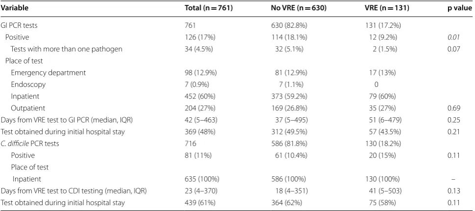
Over 5 years of data collection, patients colonized with VRE were less likely to subsequently test positive on the GI PCR compared to patients without VRE colo-nization (9.2% vs 18%, p = 0.01; Table 2) during an
epi-sode of diarrhea. Specifically, patients colonized with VRE had a lower proportion of GI PCR tests positive for E. coli species (3.8% vs 9.5%, p = 0.03) or viral (2.3%
vs 7.0%, p = 0.04) enteric infections (Fig. 1). Of patients
who underwent VRE screening, a subset of 716 were sub-sequently tested for C. difficile. Among these patients, 130 (18%) were positive for C. difficile. Patients colonized with VRE were more likely to test positive for C. difficile
although this was not statistically significant (15% vs 10%, p = 0.11; Table 2, Fig. 1).
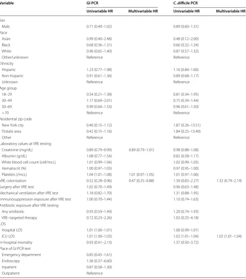
On multivariable Cox regression analysis, patients with VRE colonization had a decreased risk of a positive GI PCR (aHR 0.47, 95% CI 0.25–0.88, p = 0.02, Table 3). This effect persisted over 5 years of follow up (log-rank 0.03, Fig. 2a). On excluding 325 patients who underwent stool testing within 30 days of VRE screening, on logistic regression, there was no change in this result (aOR 0.48, 95% CI 0.23–1.19, p = 0.04). Patients with a longer ICU LOS had an increased risk of C. difficile (aHR 1.02, 95% CI 1.01–1.04, p = 0.01; Table 3). There was a trend toward an increased risk of C. difficile in patients with VRE colo-nization (aHR 1.32, 95% CI 0.79–2.19, p = 0.21; log-rank 0.29, Fig. 2b).
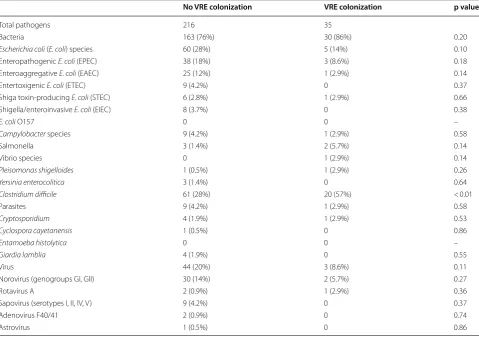
In examining the distribution of pathogens detected among those with positive stool PCR tests, 216 total pathogens were detected in patients without VRE colonization and 35 total pathogens were detected in patients with VRE colonization (Table 4). Of positive tests, there was a greater proportion of C. difficile among patients with VRE (57% vs 28%, p < 0.01) and a non-sig-nificant trend toward more bacterial infections (86% vs 76%, p = 0.20) and fewer viral infections (8.6% vs 20%, p = 0.11).
Discussion
In the present study, gut VRE colonization status pre-dicted the risk of subsequent enteric infection as detected by broad, multiplex PCR stool testing. VRE colonization decreased the risk of a subsequent non-C. difficile enteric infection, a finding that persisted over 5 years of follow up. Although VRE colonization did not clearly impact subsequent C. difficile, it did modify the distribution of enteric infections such that C. difficile was the most com-mon pathogen detected in VRE colonized patients with positive stool testing. These data have significant clini-cal implications in assessing the risk and management of enteric infection in patients colonized with VRE.
Recently, we have examined utilization of the GI PCR test in the general population and in specific populations, such as those with celiac disease and inflammatory bowel disease (IBD) [17–19]. This is the first analysis to focus on VRE and enteric infections utilizing multiplex stool PCR testing. Gut VRE colonization has a dramatic effect on the intestinal microbiome which may in turn protect against infection with non-C. difficile enteric pathogens. As such, the utility of broad stool PCR testing in patients with gut VRE colonization may be limited, and perhaps, should be restricted to C. difficile testing alone.
pressure from vancomycin [20]. In addition, selec-tive eradication of Gram-negaselec-tive bacteria by antibiot-ics reduces RegIII-γ levels and allows for the expansion of Gram-positive bacteria including Enterococcus [21]. Consequently, under certain conditions, Enterococ-cus exhibits a densely dominating phenotype in the gut [2]. In the setting of other ecological conditions, a more diverse set of pathogens may appear including multiple
Gram negatives [22, 23]. These findings may explain why
Enterococcus, while not a particularly virulent gut patho-gen in isolation, is associated with all-cause infection and mortality [2, 9, 20]. VRE may dominate the gut microbi-ome, crowding out both healthy commensals and
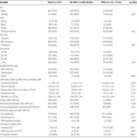
non-C. difficile enteric bacteria and viruses. Moreover, SagA, a secreted peptidoglycan hydrolase from E. faecium has demonstrated in vivo to trigger a protective intestinal Table 1 Baseline characteristics of patients with and without vancomycin resistant Enterococcus colonization who underwent testing with a gastrointestinal pathogen PCR panel or Clostridium difficile test
Variable Total (n = 761) No VRE (n = 630, 82.8%) VRE (n = 131, 17.2%) p value
Sex
Male 387 (51%) 330 (52%) 57 (43.5%)
Female 374 (49%) 300 (48%) 74 (56.5%) 0.07
Race
Asian 31 (4.1%) 27 (4.3%) 4 (3.1%)
Black 89 (11%) 77 (12%) 12 (9.2%)
White 286 (38%) 231 (37%) 55 (42.0%)
Other/unknown 355 (47%) 295 (47%) 60 (45.8%) 0.55
Ethnicity
Hispanic 128 (17%) 114 (18%) 14 (10.7%)
Non-hispanic 280 (37%) 220 (35%) 60 (45.8%)
Unknown 353 (46%) 296 (47%) 57 (43.5%) 0.03
Age group
18–29 58 (7.6%) 45 (7.1%) 13 (9.9%)
30–49 137 (18%) 115 (18%) 22 (16.8%)
50–69 366 (48%) 304 (48%) 62 (47.3%)
> 70 200 (26%) 166 (26%) 34 (26.0%) 0.74
Residential zip code
New York city 414 (54%) 342 (54%) 72 (55.0%)
Tristate area 328 (43%) 273 (43%) 55 (42.0%)
Other 19 (2.5%) 15 (2.4%) 4 (3.1%) 0.88
Laboratory values at VRE testing (median, IQR)
Creatinine (mg/dL) 1.1 (0.8–2.1) 1.1 (0.7–1.1) 1.03 (0.8–2.0) 0.58
Albumin (g/dL) 2.8 (2.4–3.2) 2.8 (2.4–3.2) 2.7 (2.3–3.1) 0.11
White blood cell count (cells × 103/µL) 10 (6.5–14) 10 (6.5–14) 9.6 (6.3–14) 0.74
Hematocrit (%) 33 (32–34) 33 (32–34) 33 (31–34) 0.19
Platelets (× 103/µL) 138 (135–141) 138 (135–141) 137 (135–141) 0.11
Surgery after VRE test 246 (32%) 209 (33%) 37 (28%) 0.27
Mechanical ventilation after VRE test 450 (59%) 371 (59%) 79 (60%) 0.76
Immunosuppression exposure after VRE test 430 (57%) 348 (55%) 82 (63%) 0.12
Antibiotic exposure after VRE test
Any antibiotic 719 (95%) 483 (77%) 131 (100%) < 0.01
Vancomycin IV 571 (75%) 467 (74%) 104 (79%) 0.21
VRE-targeted therapy 15 (2.6%) 13 (2.7%) 2 (1.9%) 0.64
LOS (days; median, IQR)
Hospital LOS 15 (8–31) 14 (7–29) 19 (10–44) < 0.01
Intensive care unit LOS 4 (2–8) 4 (2–8) 5 (3–9) 0.01
epithelial cell program that limits the pathogenesis of enteric infections [1, 2]. In the present study, the protec-tive effect of VRE persisted over time and is consistent with previous literature demonstrating low rates of VRE eradication, even after ICU and hospital discharge, and withdrawal of known risk factors [6–8].
Clostridium difficile is unique in that the relative abun-dance of the pathogen in the gut does not directly pre-dict C. difficile carriage versus overt infection [24]. As a virulent, spore-forming and toxin-producing pathogen, gut microbiome abundance is not necessarily required to produce clinically significant symptoms [24, 25]. Among Table 2 Gastrointestinal pathogen PCR panel and Clostridium difficile test outcomes in patients with and without vancomycin resistant Enterococcus colonization
Variable Total (n = 761) No VRE (n = 630) VRE (n = 131) p value
GI PCR tests 761 630 (82.8%) 131 (17.2%)
Positive 126 (17%) 114 (18.1%) 12 (9.2%) 0.01
Tests with more than one pathogen 34 (4.5%) 32 (5.1%) 2 (1.5%) 0.07
Place of test
Emergency department 98 (12.9%) 81 (12.9%) 17 (13%)
Endoscopy 7 (0.9%) 7 (1.1%) 0
Inpatient 452 (60%) 373 (59.2%) 79 (60%)
Outpatient 204 (27%) 169 (26.8%) 35 (27%) 0.69
Days from VRE test to GI PCR (median, IQR) 42 (5–463) 37 (5–495) 51 (6–479) 0.25
Test obtained during initial hospital stay 369 (48%) 312 (49.5%) 57 (43.5%) 0.21
C. difficile PCR tests 716 586 (81.8%) 130 (18.2%)
Positive 81 (11%) 61 (10.4%) 20 (15%) 0.11
Place of test
Inpatient 635 (100%) 586 (100%) 130 (100%) –
Days from VRE test to CDI testing (median, IQR) 23 (4–370) 18 (4–351) 41 (5–503) 0.13
Test obtained during initial hospital stay 439 (61%) 364 (62%) 75 (58%) 0.11
positive tests in patients with VRE colonization, C. diffi-cile was the most common pathogen, consistent with the concept of VRE as a marker for a loss of gastrointestinal colonization resistance. Non-C. difficile enteric infections
as detected by the GI PCR may not necessarily depend on loss of gastrointestinal colonization resistance but rather intrinsic pathogenicity and abundance under specific gut microbiome conditions. Several studies have suggested a Table 3 Predictors of a positive gastrointestinal pathogen PCR panel or Clostridium difficile test
Variable GI PCR C. difficile PCR
Univariable HR Multivariable HR Univariable HR Multivariable HR
Sex
Male 0.71 (0.49–1.02) 0.89 (0.60–1.31)
Race
Asian 0.99 (0.40–2.48) 0.48 (0.12–2.00)
Black 0.68 (0.36–1.31) 0.66 (0.32–1.34)
White 0.96 (0.65–1.40) 0.87 (0.57–1.32)
Other/unknown Reference Reference
Ethnicity
Hispanic 1.23 (0.77–1.98) 1.16 (0.84–1.60)
Non-hispanic 0.91 (0.61–1.36) 0.89 (0.68–1.17)
Unknown Reference Reference
Age group
18–29 0.54 (0.21–1.38) 0.81 (0.34–1.95)
30–49 1.17 (0.69–2.01) 0.75 (0.39–1.44)
50–69 0.99 (0.64–1.53) 0.96 (0.61–1.50)
> 70 Reference Reference
Residential zip code
New York city 0.40 (0.15–1.12) 1.87 (0.26–13.51)
Tristate area 0.42 (0.15–1.16) 1.84 (0.25–13.40)
Other Reference Reference
Laboratory values at VRE testing
Creatinine (mg/dL) 0.89 (0.79–0.99) 0.89 (0.79–1.01) 0.98 (0.88–1.08)
Albumin (g/dL) 1.08 (0.77–1.54) 0.82 (0.58–1.17)
White blood cell count (cell/mcL) 1.01 (0.99–1.04) 1.02 (0.99–1.05)
Hematocrit (%) 1.00 (0.97–1.03) 0.97 (0.95–1.00)
Platelets (/mcL) 1.04 (1.01–1.08) 1.01 (0.97–1.05) 1.01 (0.97–1.06)
VRE colonization 0.52 (0.28–0.96) 0.47 (0.25–0.88) 1.39 (0.83–2.27) 1.32 (0.79–2.19)
Surgery after VRE test 1.02 (0.70–1.49) 0.96 (0.63–1.48)
Mechanical ventilation after VRE test 1.18 (0.82–1.70) 1.31 (0.88–1.95)
Immunosuppression exposure after VRE test 1.00 (0.70–1.44) 1.10 (0.74–1.63)
Antibiotic exposure after VRE testing
Any antibiotic 0.93 (0.59–1.49) 1.20 (0.74–1.93)
VRE–targeted therapy 0.72 (0.23–2.26) 1.02 (0.25–4.18)
LOS
Hospital LOS 1.01 (1.00–1.01) 1.00 (0.99–1.01)
ICU LOS 1.01 (1.00–1.03) 1.02 (1.01–1.04) 1.02 (1.01–1.04)
In-hospital mortality 0.93 (0.41–2.13) 1.37 (0.50–3.72)
Place of GI PCR test
Emergency department 0.85 (0.45–1.61)
Endoscopy 1.56 (0.37–6.60)
Inpatient 0.87 (0.58–1.30)
Fig. 2 Time to positive gastrointestinal pathogen PCR panel (a) or positive Clostridium difficile PCR (b) stratified by VRE colonization status
Table 4 Distribution of pathogens among those patients with a positive stool PCR result
No VRE colonization VRE colonization p value
Total pathogens 216 35
Bacteria 163 (76%) 30 (86%) 0.20
Escherichia coli (E. coli) species 60 (28%) 5 (14%) 0.10
Enteropathogenic E. coli (EPEC) 38 (18%) 3 (8.6%) 0.18
Enteroaggregative E. coli (EAEC) 25 (12%) 1 (2.9%) 0.14
Entertoxigenic E. coli (ETEC) 9 (4.2%) 0 0.37
Shiga toxin-producing E. coli (STEC) 6 (2.8%) 1 (2.9%) 0.66
Shigella/enteroinvasive E. coli (EIEC) 8 (3.7%) 0 0.38
E. coli O157 0 0 –
Campylobacter species 9 (4.2%) 1 (2.9%) 0.58
Salmonella 3 (1.4%) 2 (5.7%) 0.14
Vibrio species 0 1 (2.9%) 0.14
Pleisomonas shigelloides 1 (0.5%) 1 (2.9%) 0.26
Yersinia enterocolitica 3 (1.4%) 0 0.64
Clostridium difficile 61 (28%) 20 (57%) < 0.01
Parasites 9 (4.2%) 1 (2.9%) 0.58
Cryptosporidium 4 (1.9%) 1 (2.9%) 0.53
Cyclospora cayetanensis 1 (0.5%) 0 0.86
Entamoeba histolytica 0 0 –
Giardia lamblia 4 (1.9%) 0 0.55
Virus 44 (20%) 3 (8.6%) 0.11
Norovirus (genogroups GI, GII) 30 (14%) 2 (5.7%) 0.27
Rotavirus A 2 (0.9%) 1 (2.9%) 0.36
Sapovirus (serotypes I, II, IV, V) 9 (4.2%) 0 0.37
Adenovirus F40/41 2 (0.9%) 0 0.74
possible link between Enterococcus and C. difficile, and our data may also suggest that VRE colonization and
C. difficile share common gut ecological preferences, although this hypothesis requires further study [24, 26].
There are several limitations to this study. Patients colonized with VRE may receive more intense or longer duration courses of antibiotics, and it may be the anti-biotics rather than VRE itself that confers and increased risk for C. difficile infection and a lower risk of other bac-terial enteric infection. Although this study adjusted for antibiotics and nearly all patients were exposed, we had limited ability to adjust for differences in antibiotic dura-tion or anti-anaerobic potency. Previous studies suggest that patients with VRE colonization have higher acute severity of illness at the time of ICU admission compared to patients who are not colonized. Such patients are also at increased risk for C. difficile and therefore, residual confounding, if present, would likely lead to an underes-timate of the true protective effect of VRE colonization on enteric infections. While all patients screened for VRE were admitted to an ICU, there may be selection bias in patients who underwent stool PCR tests for diarrhea as patients with VRE were more likely to undergo stool test-ing. However, as stool testing generally leads to further findings, we believe these data likely underestimate the effect of VRE colonization on subsequent enteric infec-tion. Moreover, although VRE colonization may persist for years, VRE status was not confirmed at same time as GI PCR or C. difficile PCR testing. In addition, PCR testing fails to discriminate between active infection and asymptomatic colonization, and there is consider-able uncertainty regarding clinical interpretation and cost-effectiveness of such multiplex assays [27]. The GI PCR panel does not assess for the presence of Cytomeg-alovirus (CMV), a pathogen of increasing importance. Lastly, our sample size gave us limited ability to specify differences between enteropathogen types on the GI PCR panel.
In summary, VRE colonization was associated with a reduction in the risk of subsequent non-C. difficile
enteric infection. Although changes in the gut micro-biome likely underlie these findings, further study is required to specifically assess for the impact of VRE colo-nization on the gut microbiome and how these changes may directly impact acquisition of enteric pathogens.
Methods
Study population
We performed a retrospective cohort study using the electronic medical records of patients at New York Pres-byterian-Columbia University Medical Center, a quater-nary care institution in New York City. We identified all adult patients (≥ 18 years) admitted to any 1 of 9 distinct
ICUs within our hospital network comprised of 2 large hospitals between 2012 and 2017 who were screened for VRE colonization via rectal swab and culture, and subse-quently stool testing for diarrhea using a multiplex PCR assay. Patients were excluded if they had an ICU LOS less than 2 days or if they were diagnosed with an enteric infection by a GI PCR test before their VRE screen. GI or C. difficile PCR stool testing for diarrhea may have occurred during the index hospitalization or at any time point after discharge.
Vancomycin‑resistant Enterococcus screening
The primary exposure was VRE colonization, determined by the result of routine surveillance swabs for VRE which are performed on every patient within 1 h of ICU admis-sion in our hospital network. Flocked rectal swabs were gathered by nurses with the patient in the left lateral decubitus position with the swab inserted deeply into the rectal canal and rotated 5 times. Swabs were trans-ported in 1 mL of liquid Amies media for direct culture onto chromogenic differential media impregnated with 6 µg/mL of vancomycin (Remel). Plates were incubated at 33–37 ºC under aerobic conditions for 24 h and inter-preted categorically according to the manufacturer’s instructions.
Gastrointestinal pathogen PCR panel stool test
Gastrointestinal pathogens were identified using the Fil-mArray gastrointestinal pathogen PCR panel (GI PCR; BioFire Diagnostics, Salt Lake City, UT). This assay tests for 22 analytes in spontaneously voided stool including 13 bacteria, 5 viruses, and 4 parasites including Campy-lobacter (jejuni, coli, and upsaliensis), Plesiomonas shigelloides, Salmonella, Yersinia enterocolitica, Vibrio (parahaemolyticus, vulnificus, and cholerae), enteroag-gregative Escherichia coli (E. coli; EAEC), enteropatho-genic E. coli (EPEC), enterotoxigenic E. coli (ETEC), Shiga-like toxin-producing E. coli (STEC), E. coli O157, Shigella/enteroinvasive E. coli (EIEC), Cryptosporidium
spp., Cyclospora cayetanensis, Entamoeba histolytica, Giardia lamblia, adenovirus (AdV) F40/41, astrovirus, norovirus GI/GII, rotavirus A, and sapovirus (I, II, IV, and V). The GI PCR is capable of the simultaneous detec-tion and identificadetec-tion of nucleic acids from multiple bac-teria, viruses, and parasites directly from stool samples in Cary Blair transport media. The multiplex PCR process takes approximately 1 h. The clinical sensitivity and spec-ificity is 94.5–100% for all targets [14, 28].
Clostridium difficile testing
toxins A and B at some institutions, this is not the case at our institution. C. difficile testing needed to be ordered separately, which was done at the discretion of providers. Therefore, some patients in this cohort did not receive testing for C. difficile. All patients, however, underwent testing with a GI PCR test.
Co‑variables
Automated queries were used to extract the following values from the medical record: dates of PCR tests, PCR results, date of birth, zip code, place of GI PCR test (e.g. emergency department, outpatient visit, inpatient hos-pitalization, endoscopy), sex, race, ethnicity, laboratory values at the time of VRE screening, history of surgery, mechanical ventilation, immunosuppression exposure, antibiotic exposure, hospital and ICU LOS after VRE screening. Exposure to linezolid or daptomycin was sub-classified as VRE-directed antimicrobials.
Outcomes and statistical analyses
Chi square tests were used to compare categorical vari-ables and the t test or Mann–Whitney test for continu-ous variables. Linearity of the continucontinu-ous variables was assessed visually by plotting residuals against predicted values and examining for a bowed pattern. When cell counts were less than 5, Fisher’s exact test was used. To account for time from VRE screening to enteric infection testing, we evaluated outcomes using a Cox proportional hazards model, with observation time beginning at the time of VRE screening and ending upon a positive GI PCR test, patient death, or their last clinical encounter at our medical center. Survival curves were constructed to illustrate differences in time to outcome. To build the final model, a full model was constructed including all possible predictors of enteric infections, and co-variables were removed in a stepwise manner if they had no inde-pendent relationship with the outcome (p > 0.05) or if they did not alter the relationship between VRE coloniza-tion status and GI PCR result (< 10% change in the beta-coefficient representing VRE colonization), and adjusted hazard ratios (aHR) were reported. Logistic regres-sion was also performed to assess predictors of enteric infection to confirm our above analyses. We then con-structed a logistic regression model limited to GI PCR tests obtained within 30 days of a VRE test and report an adjusted odds ratio (aOR). All tests were considered sig-nificant at a 2-sided p value less than 0.05. SPSS software (IBM) was used to perform all statistical analyses.
Authors’ contributions
JA designed the study, collected the data, performed the analysis, and drafted and revised the manuscript. BL conceptualized, drafted and revised the manu-script. EC revised the manumanu-script. KC revised the manumanu-script. PG revised the manuscript. DF designed and supervised the overall study, conceptualized,
drafted and revised the manuscript. All authors ensured the accuracy and integrity of this article. All authors read and approved the final manuscript.
Author details
1 Division of Gastroenterology, Department of Medicine, Inflammatory Bowel Disease Center, NYU Langone Health, 240 East 38th Street, 23rd Floor, New York, NY, USA. 2 Division of Digestive and Liver Disease, Department of Medi-cine, Columbia University Medical Center, New York, NY, USA. 3 Kimmel Center for Biology and Medicine at the Skirball Institute, New York University School of Medicine, New York, NY, USA. 4 Department of Microbiology, New York University School of Medicine, New York, NY, USA.
Acknowledgements None.
Disclosures
The authors disclose no potential conflicts relevant to this manuscript.
Competing interests
The authors declare that they have no competing interests.
Availability of data and materials
The dataset used and analyzed during the current study is available from the corresponding author on reasonable request.
Consent for publication Not applicable.
Ethics approval and consent to participate
The study was approved by the Columbia University Medical Center Institu-tional Review Board.
Funding
Dr. Freedberg was supported in part by a Research Scholar Award from the American Gastroenterological Association and in part by NIH K23-DK111847.
Publisher’s Note
Springer Nature remains neutral with regard to jurisdictional claims in pub-lished maps and institutional affiliations.
Received: 9 June 2018 Accepted: 3 July 2018
References
1. Bonten MJ, Willems R, Weinstein RA. Vancomycin-resistant enterococci: why are they here, and where do they come from? Lancet Infect Dis. 2001;1:314–25.
2. Arias CA, Murray BE. The rise of the Enterococcus: beyond vancomycin resistance. Nat Rev Microbiol. 2012;10:266–78.
3. Murray BE. Vancomycin-resistant enterococcal infections. N Engl J Med. 2000;342:710–21.
4. Papadimitriou-Olivgeris M, Drougka E, Fligou F, et al. Risk factors for ente-rococcal infection and colonization by vancomycin-resistant enterococci in critically ill patients. Infection. 2014;42:1013–22.
5. Amberpet R, Sistla S, Parija SC, et al. Risk factors for intestinal colonization with vancomycin resistant enterococci’ A prospective study in a level III pediatric intensive care unit. J Lab Phys. 2018;10:89–94.
6. Shenoy ES, Paras ML, Noubary F, et al. Natural history of colonization with methicillin-resistant Staphylococcus aureus (MRSA) and vancomy-cin-resistant Enterococcus (VRE): a systematic review. BMC Infect Dis. 2014;14:177.
7. Haverkate MR, Derde LPG, Brun-Buisson C, et al. Duration of colonization with antimicrobial-resistant bacteria after ICU discharge. Intensive Care Med. 2014;40:564–71.
•fast, convenient online submission
•
thorough peer review by experienced researchers in your field
• rapid publication on acceptance
• support for research data, including large and complex data types
•
gold Open Access which fosters wider collaboration and increased citations maximum visibility for your research: over 100M website views per year
•
At BMC, research is always in progress.
Learn more biomedcentral.com/submissions
Ready to submit your research? Choose BMC and benefit from:
9. Jung E, Byun S, Lee H, et al. Vancomycin-resistant Enterococcus coloniza-tion in the intensive care unit: clinical outcomes and attributable costs of hospitalization. Am J Infect Control. 2014;42:1062–6.
10. Amberpet R, Sistla S, Parija SC, et al. Screening for intestinal colonization with vancomycin resistant enterococci and associated risk factors among patients admitted to an adult intensive care unit of a large teaching hospital. J Clin Diagn Res. 2016;10:DC06–9.
11. Chang S, Sievert DM, Hageman JC, et al. Infection with vancomycin-resistant Staphylococcus aureus containing the vanA resistance gene. N Engl J Med. 2003;348:1342–7.
12. Riddle MS, DuPont HL, Connor BA. ACG clinical guideline: diagnosis, treatment, and prevention of acute diarrheal infections in adults. Am J Gastroenterol. 2016;111:602–22.
13. Scallan E, Griffin PM, Angulo FJ, et al. Foodborne illness acquired in the United States—unspecified agents. Emerg Infect Dis. 2011;17:16–22. 14. Piralla A, Lunghi G, Ardissino G, et al. FilmArray™ GI panel performance
for the diagnosis of acute gastroenteritis or hemorragic diarrhea. BMC Microbiol. 2017;17:111.
15. Reddington K, Tuite N, Minogue E, et al. A current overview of commer-cially available nucleic acid diagnostics approaches to detect and identify human gastroenteritis pathogens. Biomol Detect Quantif. 2014;1:3–7. 16. Taur Y, Xavier JB, Lipuma L, et al. Intestinal domination and the risk of
bacteremia in patients undergoing allogeneic hematopoietic stem cell transplantation. Clin Infect Dis. 2012;55:905–14.
17. Axelrad JE, Joelson A, Nobel YR, et al. Enteric infection in relapse of inflammatory bowel disease: the utility of stool microbial PCR testing. Inflamm Bowel Dis. 2017;23:1034–9.
18. Nobel YR, Axelrad J, Lewis SK, et al. Stool PCR for gastrointestinal patho-gens in patients with and without immune-mediated intestinal diseases. Dig Dis Sci. 2018;63:996–1002.
19. Axelrad JE, Joelson A, Nobel Y, et al. The distribution of enteric infections utilizing stool microbial polymerase chain reaction testing in clinical practice. Dig Dis Sci. 2018;63:1900–9.
20. Gao W, Howden BP, Stinear TP. Evolution of virulence in Enterococcus faecium, a hospital-adapted opportunistic pathogen. Curr Opin Microbiol. 2017;41:76–82.
21. Brandl K, Plitas G, Mihu CN, et al. Vancomycin-resistant entero-cocci exploit antibiotic-induced innate immune deficits. Nature. 2008;455:804–7.
22. Byndloss MX, Olsan EE, Rivera-Chávez F, et al. Microbiota-activated PPAR-γ signaling inhibits dysbiotic Enterobacteriaceae expansion. Science. 2017;357:570–5.
23. Pedicord VA, Lockhart AAK, Rangan KJ, et al. Exploiting a host-commensal interaction to promote intestinal barrier function and enteric pathogen tolerance. Sci Immunol. 2016;1:eaai7732.
24. Vincent C, Miller MA, Edens TJ, et al. Bloom and bust: intestinal microbiota dynamics in response to hospital exposures and Clostridium difficile colonization or infection. Microbiome. 2016;4:12.
25. Rupnik M, Wilcox MH, Gerding DN. Clostridium difficile infection: new developments in epidemiology and pathogenesis. Nat Rev Microbiol. 2009;7:526–36.
26. Kamada N, Chen GY, Inohara N, et al. Control of pathogens and pathobi-onts by the gut microbiota. Nat Immunol. 2013;14:685–90.
27. Freeman K, Mistry H, Tsertsvadze A, et al. Multiplex tests to identify gastrointestinal bacteria, viruses and parasites in people with suspected infectious gastroenteritis: a systematic review and economic analysis. Health Technol Assess. 2017;21:1–188.