Standards in Genomic Sciences (2014) 9:1221-1235 DOI:10.4056/sigs.4698398
Non-contiguous finished genome sequence and description of
Alistipes ihumii
sp. nov.
Anne Pfleiderer1†, Ajay Kumar Mishra1†, Jean-Christophe Lagier1, Catherine Robert1, Aurelia Caputo1, Didier Raoult1,2 and Pierre-Edouard Fournier1*
1Aix-Marseille Université, URMITE, UM63, Faculté de médecine, Marseille, France 2 King Fahd Medical Research Center, King Abdul Aziz University, Jeddah, Saudi Arabia * Correspondence: ([email protected])
†These two authors contributed equally to this work
Keywords:Alistipes ihumii, genome, culturomics, taxono-genomics
Alistipes ihumii strain AP11T sp. nov. is the type strain of A. ihumii sp. nov., a new species
within the genus Alistipes. This strain, whose genome is described here, was isolated from the fecal flora of a 21-year-old French Caucasian female, suffering from a severe restrictive form of anorexia nervosa since the age of 12 years. A. ihumii is a Gram-negative anaerobic bacil-lus. Here we describe the features of this organism, together with the complete genome se-quence and annotation. The 2,753,264 bp long genome (one chromosome but no plasmid) contains 2,254 protein-coding and 47 RNA genes, including 3 rRNA genes.
Introduction
Alistipes ihumii
strain AP11
T(= CSUR P204 = DSM
26107) is the type strain of
A. ihumii
sp. nov. This
bacterium is a Gram-negative, non-spore-forming,
anaerobic and non-motile bacillus that was
isolat-ed from the stool of a 21-year-old French female
suffering from anorexia nervosa, and is part of a
“culturomics” study aiming at cultivating
individ-ually all species within human feces [1-3].
Prokaryotic taxonomy is episodically confronted
with the advancement of methodological and
con-ceptual innovations. The current classification
methodology for prokaryotes is known as
polyphasic taxonomy, and relies on a combination
of phenotypic and genotypic characteristics [4].
The number of completely sequenced genomes is
geometrically increasing with time, concurrently
with the decrease in cost of such techniques. To
date, more than 6,000 bacterial genomes have
been published and approximately 25,000
ge-nome sequencing projects have been announced
[5]. We recently proposed to integrate genomic
in-formation in the taxonomic framework for the
de-scription of new bacterial species [6-27].
The genus
Alistipes
(Rautio
et al
. 2003) was
creat-ed in 2003 [28] and is composcreat-ed of strictly
anaer-obic Gram-negative rods that resemble the
Bacteroides fragilis
group in that most species are
bile-resistant and indole-positive [29]. This genus
is currently comprised of five species with validly
published names, including
A. finegoldii
,
A.
putredinis
[28],
A. indistinctus
[30],
A. onderdonkii
and
A. shahii
[31], to which we added three
pro-posed new species,
A. senegalensis
[8],
A.
timonensis
[9] and
A. obesi
[22].
Here we present a summary classification and a
set of features for a new
Alistipes
species,
A. ihumii
sp. nov. strain AP11
T(= CSUR P204 = DSM 26107),
together with the description of the complete
ge-nomic sequence and its annotation.
Classification and features
A stool sample was collected from a 21-year-old
French Caucasian female suffering from severe
re-strictive form of anorexia nervosa since the age of
12 years. At the time of sample collection, she was
hospitalized in our hospital for recent aggravation
of her medical condition (BMI: 10.4 kg/m
2). The
bacterial species from other stool specimens
[6-27]. The fecal specimen was stored at -80°C
im-mediately after collection. Strain AP11
Twas
iso-lated in November 2011 after 2 days of
inocula-tion in anaerobic blood culture bottle with the
ad-dition of 5mL of thioglycolate and further
inocula-tion on Columbia agar (BioMerieux, Marcy l’Etoile,
France).
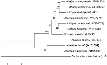
This strain exhibited a 95% 16S rRNA sequence
similarity with
A. indistinctus
[30], the
phylogenetically closest
Alistipes
species with a
validly published name (Table 1, Figure 1), and
92% with
A. onderdonkii
[28] and
A. putredinis
[31]. This value was in the range of 16S rRNA
se-quence identities among species within the genus
Alistipes
that range from 90 to 95%, and lower
than the 98.7% 16S rRNA gene sequence
thresh-old recommended by Stackebrandt and Ebers to
delineate a new species without carrying out
DNA-DNA hybridization [41].
Table 1. Classification and general features of Alistipes ihumii strain AP11T according to the MIGS
rec-ommendations [32]
MIGS ID Property Term Evidence codea
Current classification
Domain Bacteria
Phylum Bacteroidetes Class Bacteroidia Order Bacteroidales Family Rikenellaceae Genus Alistipes Species Alistipes ihumii Type strain AP11T
TAS [33] TAS [34,35] TAS [34,36] TAS [34,37] TAS [34,38] TAS [28,39]
IDA IDA
Gram stain Negative IDA
Cell shape Rod IDA
Motility nonmotile IDA
Sporulation nonsporulating IDA
Temperature range mesophile IDA
Optimum temperature 37°C IDA
MIGS-6.3 Salinity unknown IDA
MIGS-22 Oxygen requirement anaerobic IDA
Carbon source unknown Energy source unknown
MIGS-6 Habitat human gut IDA
MIGS-15 Biotic relationship free living IDA
MIGS-14
Pathogenicity Biosafety level Isolation
unknown 2
human feces
MIGS-4 Geographic location France IDA
MIGS-5 Sample collection time November 2011 IDA
MIGS-4.1 Latitude & Longitude 43.296482 & 5.36978 IDA
MIGS-4.3 Depth surface IDA
MIGS-4.4 Altitude 0 m above sea level IDA
aEvidence codes - IDA: Inferred from Direct Assay; TAS: Traceable Author Statement (i.e., a direct report
Figure 1. Phylogenetic tree highlighting the position of Alistipes ihumii strain AP11T relative to other type
strains within the genus Alistipes. GenBank accession numbers are indicated in parentheses. Sequences were aligned using CLUSTALW, and phylogenetic inferences obtained using the maximum-likelihood method within the MEGA software. Numbers at the nodes are percentages of bootstrap values obtained by repeating the analysis 500 times to generate a majority consensus tree. Bacteroides splanchnicus was used as the outgroup. The scale bar represents a 2% nucleotide sequence divergence.
Different growth temperatures (25, 30, 37, 45°C)
were tested. Growth was observed between 25
and 45°C, with optimal growth at 37°C after 24
hours of inoculation. Colonies were about 0.2 mm
in diameter, transparent, and exhibited a
ß-hemolytic activity on blood-enriched Columbia
agar. Growth of the strain was tested on 5% sheep
blood agar, under anaerobic and microaerophilic
conditions using the GENbag anaer and GENbag
microaer systems, respectively (BioMerieux), and
under aerobic conditions with or without 5% CO
2.
Optimal growth of this strain was obtained
anaer-obically, weak growth was observed under
microaerophilic conditions, and no growth was
observed under aerobic atmosphere. The motility
test was negative. Cells grown on agar are
Gram-negative rods (Figure 2) and have mean diameter
and length of 0.72 and 1.69 µm, respectively, as
determined using electron microscopy (Figure 3).
Strain AP11
Texhibited oxidase but no catalase
ac-tivities. Using API 50CH (BioMérieux), we
ob-served that strain AP11
Twas asaccharolytic. Using
API 32A (BioMérieux), positive reactions were
ob-tained for α-glucosidase, β-glucosidase,
N-acetyl-β-glucosaminidase, mannose and raffinose
fer-mentation, alkaline phosphatase, leucyl glycine
arylamidase, alanine arylamidase, and glutamyl
glutamic acid arylamidase. Weak reactions were
observed for α-galactosidase and glutamic acid
decarboxilase. Negative reactions were obtained
for urease, arginine dihydrolase, β-galactosidase,
6 phospho-galactosidase, α-arabinosidase,
β-glucuronidase, α-fucosidase, nitrate reduction,
indole production, arginine arylamidase, proline
arylamidase, phenylalanine arylamidase, leucine
arylamidase, pyroglutamic acid arylamidase,
tyro-sine arylamidase, glycine arylamidase, histidine
arylamidase, and serine arylamidase.
A. ihumii
is
susceptible to amoxicillin, imipenem, and
clindamycin, but resistant to vancomycin. When
compared with representative species from the
genus
Alistipes
, strain AP11
Texhibited the
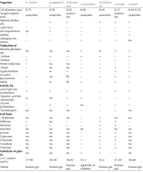
pheno-typic differences detailed in Table 2.
Figure 2. Gram stain of A. ihumii strain AP11T
Figure 3. Transmission electron microscopy of A. ihumii strain AP11T, using a Morgani
Table 2. Differential characteristics of Alistipes strains†
Properties A. ihumii A. senegalensi s
A.timone
nsis A.putredinis
A.indistinc
tus A.shahii A.obesi
Cell diameter (µm) 0.72 0.56 0.62 0.40 0.60 0.15 0.44-0.76
Oxygen
require-ment anaerobic anaerobic anaero-bic anaerobic anaerobic anaero-bic anaerobic Pigment
produc-tion – + + – + + +
Gram stain – – – – – – –
Salt requirement na + – – – – –
Motility – – – – – – +
Endospore
for-mation – – – – – – na
Production of Alkaline
phospha-tase na na na + w + +
Catalase – + + + + – +
Oxidase + – – – – – –
Nitrate reductase – na na – – – –
Urease – na na – – + –
β-galactosidase – w + – – + +
N-acetyl-glucosamine + na W – + + +
Indole – w W + – + –
Activity for Leucyl glycine
arylamidase + + + + – + +
Glutamic acid
de-carboxylase w na + + – – –
Glycine
arylamidase – + + na – – –
Chymotrypsin na na na – – – na
Acid from
L-Arabinose na na na – + na na
Raffinose + na – – + + –
Mannose + + – – + + –
Mannitol na na na na + na na
Sucrose na na na – + + na
D-glucose na na na – + + na
D-fructose na na na – + + na
D-maltose na na na – + + na
D-lactose na na na – + + na
Hydrolysis of
gela-tin na na na + + – na
G+C content
(mol%) 57.90 58.40 58.82 55.3 55.2 57.20 58.60
Habitat human gut human gut human gut appendix of children human gut human gut human gut na = data not available; w = weak
†Alistipes ihumii strain AP11T, A. senegalensis strain JC50T, A. timonensis strain JC136T, A. putredinis strain
ATCC29800 T, A. indistinctus strain YIT12060T, A. shahii strain WAL 8301T, A. obesi strain ph8T and A. finegoldii
Matrix-assisted laser-desorption/ionization
time-of-flight (MALDI-TOF) MS protein analysis was
carried out as previously described [42] using a
Microflex spectrometer (Bruker Daltonics, Leipzig,
Germany). Twelve individual colonies were
depos-ited on a MTP 384 MALDI-TOF target plate
(Bruker). The twelve AP11
Tspectra were
import-ed into the MALDI BioTyper software (version 2.0,
Bruker) and analyzed by standard pattern
match-ing (with default parameter settmatch-ings) against the
main spectra of 4,706 bacteria, including spectra
from
A. finegoldii
,
A. onderdonkii
,
A. shahii
,
A.
senegalensis
,
A. obesi
and
A. timonensis
, used as
reference data in the BioTyper database. The
out-put score enabled the presumptive identification
and discrimination of the tested species from
those in the database: a score > 2 with a validated
species identifies a strain at the species level; and
a score < 1.7 indicates a species-level match was
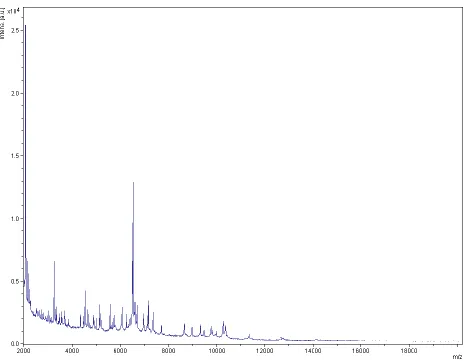
not made. For strain AP11
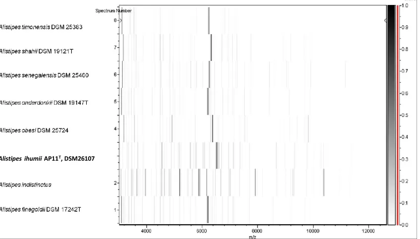
T, no significant score
was obtained, suggesting that our isolate was not
a member of any known species (Figures 4 and 5).
We added the spectrum from strain AP11
Tto our
database.
Figure 4. Reference mass spectrum from A. ihumii strain AP11T. Spectra from 12 individual colonies were
compared and a reference spectrum was generated.
Genome sequencing information
Genome project history
The organism was selected for sequencing on the
basis of its phylogenetic position and 16S rRNA
similarity to other members of the
Alistipes
genus,
and is part of a “culturomics” study of the human
digestive flora aiming at isolating all bacterial
spe-cies within human feces. It was the eighth
Figure 5. Gel view comparing spectra from Alistipes ihumii strain AP11T and other members of the genus
Alistipes (A. obesi, A. timonensis, A. senegalensis, A. shahii, A. onderdonkii and A. finegoldii). The Gel View displays the raw spectra of all loaded spectrum files arranged in a pseudo-gel like look, with each peak dis-played as a band or bar. The peak intensity is reflected by the intensity of the gray color. The right y-axis shows the relationship between the shades of gray and the peak intensity in arbitrary units. The x-axis records the m/z value. The left y-axis displays the running spectrum number originating from subsequent spectra loading.
Table 3. Project information
MIGS ID Property Term
MIGS-31 Finishing quality High-quality draft
MIGS-28 Libraries used One 454 paired end 3-kb library MIGS-29 Sequencing platforms 454 GS FLX Titanium
MIGS-31.2 Fold coverage 35×
MIGS-30 Assemblers Newbler version 2.5.3
MIGS-32 Gene calling method Prodigal
Genbank ID CAPH00000000
Genbank Date of Release November 28, 2012
Gold ID Gi20720
MIGS-13 Project relevance Study of the human gut microbiome
Growth conditions and DNA isolation
A. ihumii
sp. nov. strain AP11
T, (= CSURP204 =
DSM 26107), was grown aerobically on 5% sheep
blood agar medium at 37°C. Five Petri dishes were
spread and resuspended in 3x100µl of G2 buffer
(EZ1 DNA Tissue kit, Qiagen). A first mechanical
lysis was performed by glass powder on the
Fastprep-24 device (Sample Preparation system,
MP Biomedicals, USA) for 2×20 seconds. DNA was
treated with 2.5 µg/µL of lysozyme (30 minutes at
37°C) and extracted using the BioRobot EZ 1
Ad-and purified on a Qiamp kit (Qiagen). The yield
and the concentration of DNA was 70.7 ng/µl as
measured by using Quant-it Picogreen kit
(Invi-trogen) on the Genios Tecan fluorometer.
Genome sequencing and assembly
A 3kb paired-end sequencing strategy (Roche,
Meylan, France) was used. DNA (5 µg) was
me-chanically fragmented for the paired-end
sequenc-ing, using a Covaris device (Covaris Inc., Woburn,
MA,USA) with an enrichment size of 3-4 kb. The
Alistipes massilioanorexius DSM 26107
ilent 2100 BioAnalyzer on a DNA Labchip 7500
which yielded an optimal size of 2.3 kb. The
li-brary was constructed using the 454 GS FLX
Tita-nium paired-end rapid library protocol.
Circulari-zation and nebuliCirculari-zation were performed which
generated a pattern of optimal size of 457 bp. PCR
amplification was performed for 17 cycles
fol-lowed by double size selection. The
single-stranded paired-end library was quantified using
a Quant-it Ribogreen Kit (Invitrogen) and the
Genios Tecan fluorometer. The library
concentra-tion equivalence was calculated as 1.94× 10
10molecules/µL. The library was stored at -20°C
un-til further use.
The paired-end library was clonally amplified
with 0.5 and 1 cpb in 2 emPCR reactions with the
GS Titanium SV emPCR Kit (Lib-L) v2 (Roche). The
yield of the shotgun emPCR reactions was 6.24
and 16.24% respectively for the two kinds of
paired- end emPCR reactions according to the
quality expected (range of 5 to 20%) from the
Roche procedure. Two libraries were loaded on
the GS Titanium PicoTiterPlates (PTP Kit 70x75,
Roche) and pyrosequenced with the GS Titanium
Sequencing Kit XLR70 and the GS FLX Titanium
sequencer (Roche). The run was performed
over-night and then analyzed on the cluster through the
gsRunBrowser and Newbler assembler (Roche). A
total of 260,838 passed filter wells were obtained
and generated 96.3 Mb with an average length of
369 bp. The passed filter sequences were
assem-bled using Newbler with 90% identity and 40 bp
as overlap. The final assembly identified 9
scaf-folds and 60 contigs (> 1,500 bp) and generated a
genome size of 2.75 Mb which corresponds to a
coverage of 35× genome equivalent.
Genome annotation
Open Reading Frames (ORFs) were predicted
us-ing Prodigal [44] with default parameters but the
predicted ORFs were excluded if they were
span-ning a sequencing gap region. The predicted
bac-terial protein sequences were searched against
the GenBank database [45] and the Clusters of
Orthologous Groups (COG) databases using
BLASTP. The tRNAScan-SE tool [46] was used to
find tRNA genes, whereas ribosomal RNAs were
found by using RNAmmer [47] and BLASTn
against the GenBank database. Lipoprotein signal
peptides and numbers of transmembrane helices
were predicted using SignalP [48] and TMHMM
[49] respectively. ORFans were identified if their
BLASTP
E
-value was lower than 1e
-03for
ment length greater than 80 amino acids. If
align-ment lengths were smaller than 80 amino acids,
we used an
E
-value of 1e
-05. Such parameter
thresholds have already been used in previous
works to define ORFans.
Orthologous gene sets composed of one gene from
A. ihumii
compared to each of
A. obesi
strain ph8
T(GenBank accession number CAHA00000000),
A.
finegoldii
strain AHN 2437 (CP003274),
A.
indistinctus
strain YIT 12060 (ADLD00000000),
A.
putredinis
strain DSM 17216 (ABFK00000000),
A.
senegalensis strain
JC50
T(CAHI00000000),
A.
shahii
strain WAL 8301 (FP929032), and
A.
timonensis
strain JC136
T(CAEG00000000) were
identified using the Proteinortho software
(ver-sion 1.4) [50] using a 30% protein identity and an
E
-value of 1e
-05. The average percentage of
nucleo-tide sequence identity of each orthologous set was
determined using the Needleman-Wunsch
algo-rithm global alignment technique. Artemis [51]
was used for data management and DNA Plotter
[52] was used for visualization of genomic
fea-tures. The Mauve alignment tool was used for
multiple genomic sequence alignment and
visuali-zation [53].
Genome properties
The genome of
A. ihumii
strain AP11
Tis 2,753,264
Figure 6. Graphical circular map of the chromosome. From the outside in, the outer two circles show open reading frames oriented in the forward and reverse (colored by COG categories) directions, respectively. The third circle marks the rRNA gene operon (red) and tRNA genes (green). The fourth circle shows the G+C% content plot. The innermost circle shows GC skew, purple and olive indicating negative and posi-tive values, respecposi-tively.
Genome comparison with other
Alistipes
species
Here, we compared the genome of
A. ihumii
strain
AP11
Tto those of
A. obesi
strain ph8
T(GenBank
accession number CAHA00000000),
A. finegoldii
strain AHN 2437 (CP003274),
A. indistinctus
strain
YIT 12060 (ADLD00000000),
A. putredinis
strain
DSM 17216 (ABFK00000000),
A. senegalensis
strain
JC50
T(CAHI00000000),
A. shahii
strain
WAL 8301 (FP929032), and
A. timonensis
strain
JC136
T(CAEG00000000). The draft genome of
A.
ihumii
is larger than that of
A. putredinis
(2.75 and
2.55 Mb, respectively) but smaller than those of
A.
indistinctus
,
A. obesi
,
A. timonensis
,
A. finegoldii
,
A.
shahii
and
A. senegalensis
(2.85, 3.16, 3.49, 3.73,
3.76, and 4.01 Mb, respectively). The G+C content
of
A. ihumii
is comparable to that of
A. shahii
(57.90 and 57.60%, respectively), lower than
those of
A. timonensis
and
A. senegalensis
(58.8
and 58.4%, respectively) and higher than those of
A. putredinis
,
A. indistinctus
and
A. finegoldii
(53.30, 54.80 and 56.60%, respectively).
A. ihumii
Table 4. Nucleotide content and gene count levels of the genome
Attribute Value % of totala
Genome size (bp) 2,753,264
DNA coding region (bp) 2,320,878 84.29
DNA G+C content (bp) 1,594,140 57.90
Number of replicons 1
Extrachromosomal elements 0
Total genes 2,301 100
RNA genes 47 2.04
rRNA operons 1
Protein-coding genes 2,254 97.95
Genes with function prediction 1,540 66.92
Genes assigned to COGs 1,465 63.66
Protein coding genes assigned Pfam domains 1,834 79.70
Genes with peptide signals 296 12.86
Genes with transmembrane helices 457 19.86
CRISPR repeats 1
aThe total is based on either the size of the genome in base pairs or the total
num-ber of protein coding genes in the annotated genome
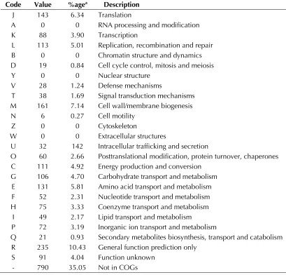
Table 5. Number of genes associated with the 25 general COG functional categories Code Value %agea Description
J 143 6.34 Translation
A 0 0 RNA processing and modification K 88 3.90 Transcription
L 113 5.01 Replication, recombination and repair B 0 0 Chromatin structure and dynamics D 19 0.84 Cell cycle control, mitosis and meiosis
Y 0 0 Nuclear structure
V 28 1.24 Defense mechanisms
T 38 1.69 Signal transduction mechanisms M 161 7.14 Cell wall/membrane biogenesis N 6 0.27 Cell motility
Z 0 0 Cytoskeleton
W 0 0 Extracellular structures
U 32 142 Intracellular trafficking and secretion
O 60 2.66 Posttranslational modification, protein turnover, chaperones C 111 4.92 Energy production and conversion
G 106 4.70 Carbohydrate transport and metabolism E 131 5.81 Amino acid transport and metabolism F 52 2.31 Nucleotide transport and metabolism H 75 3.33 Coenzyme transport and metabolism
I 49 2.17 Lipid transport and metabolism
P 72 3.19 Inorganic ion transport and metabolism
Q 21 0.93 Secondary metabolites biosynthesis, transport and catabolism R 235 10.43 General function prediction only
S 91 4.04 Function unknown
- 790 35.05 Not in COGs
788, 821, and 828, respectively), comparable to
that of
A. shahii
(833) and smaller than those of
A.
finegoldii
and
A. putredinis
(866 and 915,
respec-tively).
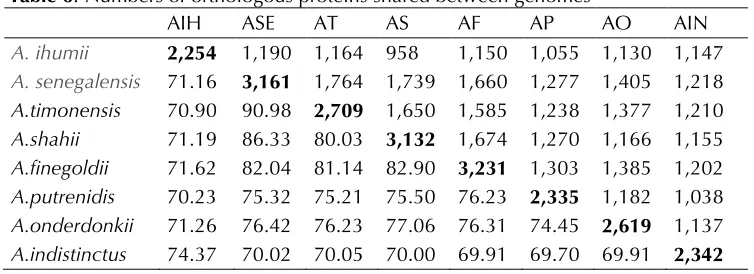
The average genomic nucleotide sequence identity
between
A. ihumii
and other
Alistipes
species
ranged from 70.23 to 74.37%, whereas values
ranged from 69.70 to 90.98% among other
Alistipes
species (Table 6).
However, the distribution of genes into COG
cate-gories was not entirely similar in all eight
com-pared genomes (Figure 7).
Conclusion
On the basis of phenotypic, phylogenetic and
ge-nomic analyses, we formally propose the creation
of
Alistipes ihumii
sp. nov. that contains strain
AP11
T. This bacterial strain has been isolated from
the fecal flora of a patient suffering from anorexia
nervosa living in Marseille, France. Several other
new bacterial species were also cultivated from
this patient as well as fecal samples from other
pa-tients using microbial culturomics [6-27], thus
suggesting that the human fecal flora from human
remains partially unknown.
Table 6. Numbers of orthologous proteins shared between genomes
AIH ASE AT AS AF AP AO AIN
A. ihumii 2,254 1,190 1,164 958 1,150 1,055 1,130 1,147
A. senegalensis 71.16 3,161 1,764 1,739 1,660 1,277 1,405 1,218
A.timonensis 70.90 90.98 2,709 1,650 1,585 1,238 1,377 1,210
A.shahii 71.19 86.33 80.03 3,132 1,674 1,270 1,166 1,155
A.finegoldii 71.62 82.04 81.14 82.90 3,231 1,303 1,385 1,202
A.putrenidis 70.23 75.32 75.21 75.50 76.23 2,335 1,182 1,038
A.onderdonkii 71.26 76.42 76.23 77.06 76.31 74.45 2,619 1,137
A.indistinctus 74.37 70.02 70.05 70.00 69.91 69.70 69.91 2,342 Upper right triangle- numbers of orthologous proteins shared between genomes; low-er left triangle- avlow-erage plow-ercentage of nucleotide identity between orthologous gene sets shared between genomes; bold- numbers of proteins per genome. Abbreviations: AIH- A. ihumii, ASE- A. senegalensis, AT- A. timonensis, AS- A. shahii, AF- A. finegoldii, AP- A. putredinis, AO- A. obesi, AIN- A. indistinctus
Description of
Alistipes ihumii
sp. nov.
Alistipes ihumii
(i.hum.i’i. N.L. gen. n. ihumii, based
on the acronym IHUMI, the Institut
Hospitalo-Universitaire Méditerranée-Infection, where the
type strain was isolated).
Colonies are 0.2 mm in diameter and are
translu-cent on blood-enriched Columbia agar. Cells are
rod-shaped with a mean diameter of 0.72 µm and
a mean length of 1.69 µm. Optimal growth is
achieved anaerobically. No growth is obtained
aerobically but weak growth is observed in
microaerophilic conditions. Growth occurs
be-tween 25°C and 45°C, with an optimal growth
ob-served at 37°C.
Cells stain Gram-negative, are non motile and are
asaccharolytic.
Activities
present
are
α-glucosidase, β-α-glucosidase,
N-acetyl-β-glucos-aminidase, mannose and rafinnose fermentation,
alkaline phosphatase, leucyl glycine arylamidase,
alanine arylamidase, and glutamyl glutamic acid
arylamidase. Cells are negative for urease,
argi-nine dihydrolase, β-galactosidase,
6-phospho-β-galactosidase, α-arabinosidase, β-glucuronidase,
α-fucosidase, nitrate reduction, indole production,
arginine arylamidase, proline arylamidase,
phe-nylalanine arylamidase, leucine arylamidase,
pyroglutamic acid arylamidase, tyrosine
amidase, glycine arylamidase, histidine
aryl-amidase, and serine arylamidase. Cells are
suscep-tible to amoxicillin, imipenem, and clindamycin,
but resistant to vancomycin. The G+C content of
the genome is 57.90%. The 16S rRNA and genome
sequences are deposited in Genbank under
acces-sion numbers JX101692 and CAPH00000000,
re-spectively.
The type strain AP11
T(= CSUR P204 = DSM
26107) was isolated from the fecal flora of a
21-year-old French Caucasian female suffering from
severe anorexia nervosa.
Acknowledgments
The authors thank the Xegen Company for
auto-mating the genomic annotation process. This
study was funded by the Mediterranee-Infection
Foundation.
References
1. Lagier JC, Armougom F, Million M, Hugon P, Pagnier I, Robert C, Bittar F, Fournous G, Gimenez G, Maraninchi M, et al. Microbial culturomics: paradigm shift in the human gut microbiome study. Clin Microbiol Infect 2012; 18:1185-1193. PubMed
2. Dubourg G, Lagier JC, Armougom F, Robert C, Hamad I, Brouqui P. The gut microbiota of a pa-tient with resistant tuberculosis is more compre-hensively studied by culturomics than by
metagenomics. Eur J Clin Microbiol Infect Dis 2013; 2013:637-645.
3. Pfleiderer A, Lagier JC, Armougom F, Robert C, Vialettes B, Raoult D. Culturomics identified 11 new bacterial species from a single anorexia ner-vosa stool sample. [Epub ahead of print]. Eur J Clin Microbiol Infect Dis 2013.
pro-karyote strains for taxonomic purposes. Int J Syst Evol Microbiol 2010; 60:249-266. PubMed http://dx.doi.org/10.1099/ijs.0.016949-0
5. Genome Online Database.
http://www.genomesonline.org/cgi-bin/GOLD/index.cgi
6. Kokcha S, Mishra AK, Lagier JC, Million M, Leroy Q, Raoult D, Fournier PE. Non-contiguous fin-ished genome sequence and description
ofBacillus timonensis sp. nov. Stand Genomic Sci
2012; 6:346-355. PubMed
http://dx.doi.org/10.4056/sigs.2776064
7. Lagier JC, El Karkouri K, Nguyen TT, Armougom F, Raoult D, Fournier PE. Non-contiguous finished genome sequence and description
ofAnaerococcus senegalensis sp. nov. Stand Ge-nomic Sci 2012; 6:116-125. PubMed
http://dx.doi.org/10.4056/sigs.2415480
8. Mishra AK, Gimenez G, Lagier JC, Robert C, Raoult D, Fournier PE. Non-contiguous finished genome sequence and description ofAlistipes senegalensis sp. nov. Stand Genomic Sci 2012; 6:304-314.
http://dx.doi.org/10.4056/sigs.2625821
9. Lagier JC, Armougom F, Mishra AK, Ngyuen TT, Raoult D, Fournier PE. Non-contiguous finished genome sequence and description of Alistipes timonensis sp. nov. Stand Genomic Sci 2012; 6:315-324. PubMed
http://dx.doi.org/10.4056/sigs.2685971
10. Mishra AK, Lagier JC, Robert C, Raoult D, Four-nier PE. Non-contiguous finished genome se-quence and description ofClostridium senegalense sp. nov. Stand Genomic Sci 2012; 6:386-395.
PubMed
11. Mishra AK, Lagier JC, Robert C, Raoult D, Four-nier PE. Non-contiguous finished genome se-quence and description ofPeptoniphilus
timonensis sp. nov. Stand Genomic Sci 2012; 7 :1-11. PubMed
http://dx.doi.org/10.4056/sigs.2956294
12. Mishra AK, Lagier JC, Rivet R, Raoult D, Fournier PE. Non-contiguous finished genome sequence and description of Paenibacillus senegalensis sp. nov. Stand Genomic Sci 2012; 7:70-81. PubMed http://dx.doi.org/10.4056/sigs.3056450
13. Lagier JC, Gimenez G, Robert C, Raoult D, Four-nier PE. Non-contiguous finished genome se-quence and description ofHerbaspirillum massiliense sp. nov. Stand Genomic Sci 2012; 7:200-209. PubMed
14. Roux V, El Karkouri K, Lagier JC, Robert C, Raoult D. Non-contiguous finished genome sequence and description of Kurthia massiliensis sp. nov. Stand Genomic Sci 2012; 7:221-232. Pub-Medhttp://dx.doi.org/10.4056/sigs.3206554
15. Kokcha S, Ramasamy D, Lagier JC, Robert C, Raoult D, Fournier PE. Non-contiguous finished genome sequence and description
ofBrevibacterium senegalense sp. nov. Stand Ge-nomic Sci 2012; 7:233-245. PubMed
http://dx.doi.org/10.4056/sigs.3256677
16. Ramasamy D, Kokcha S, Lagier JC, N’Guyen TT,
Raoult D, Fournier PE. Non-contiguous finished genome sequence and description
ofAeromicrobium massilense sp. nov. Stand Ge-nomic Sci 2012; 7:246-257. PubMed
http://dx.doi.org/10.4056/sigs.3306717
17. Lagier JC, Ramasamy D, Rivet R, Raoult D, Four-nier PE. Non-contiguous finished genome se-quence and description ofCellulomonas massiliensis sp. nov. Stand Genomic Sci 2012; 7:258-270. PubMed
http://dx.doi.org/10.4056/sigs.3316719
18. Lagier JC, El Karkouri K, Rivet R, Couderc C, Raoult D, Fournier PE. Non-contiguous finished genome sequence and description
ofSenegalemassilia anaerobia sp. nov. Stand Ge-nomic Sci 2013; 7:343-356. PubMed
http://dx.doi.org/10.4056/sigs.3246665
19. Mishra AK, Hugon P, Lagier JC, Nguyen TT, Rob-ert C, Couderc C, Raoult D, Fournier PE. Non-contiguous finished genome sequence and de-scription of Peptoniphilus obesi sp. nov. Stand Genomic Sci 2013; 7:357-369. PubMed http://dx.doi.org/10.4056/sigs.32766871
20. Mishra AK, Lagier JC, Nguyen TT, Raoult D, Fournier PE. Non-contiguous finished genome se-quence and description ofPeptoniphilus
senegalensis sp. nov. Stand Genomic Sci 2013; 7:370-381. PubMed
http://dx.doi.org/10.4056/sigs.3366764
21. Lagier JC, El Karkouri K, Mishra AK, Robert C, Raoult D, Fournier PE. Non-contiguous finished genome sequence and description of Enterobacter massiliensis sp. nov. Stand Genomic Sci 2013; 7:399-412. PubMed
http://dx.doi.org/10.4056/sigs.3396830
Ge-nomic Sci 2013; 7:427-439. PubMed http://dx.doi.org/10.4056/sigs.3336746
23. Mishra AK, Hugon P, Robert C, Couderc C, Raoult D, Fournier PE. Non-contiguous finished genome sequence and description of
Peptoniphilus grossensis sp. nov. Stand Genomic Sci 2012; 7:320-330. PubMed
24. Mishra AK, Hugon P, Nguyen TT, Raoult D, Fournier PE. Non contiguous-finished genome se-quence and description of Enorma massiliensis gen. nov., sp. nov., a new member of the Family Coriobacteriaceae. Stand Genomic Sci 2013; 8:290-305. PubMed
http://dx.doi.org/10.4056/sigs.3426906
25. Ramasamy D, Lagier JC, Gorlas A, Raoult D, Fournier PE. Non contiguous-finished genome se-quence and description ofBacillus
massiliosenegalensis sp. nov. Stand Genomic Sci
2013; 8:264-278. PubMed
http://dx.doi.org/10.4056/sigs.3496989
26. Ramasamy D, Lagier JC, Nguyen TT, Raoult D, Fournier PE. Non contiguous-finished genome se-quence and description of of Dielma fastidiosa gen. nov., sp. nov., a new member of the Family Erysipelotrichaceae. Stand Genomic Sci 2013; 8:336-351. PubMed
http://dx.doi.org/10.4056/sigs.3567059
27. Mishra AK, Lagier JC, Robert C, Raoult D, Four-nier PE. Genome sequence and description of Timonella senegalensis gen. nov., sp. nov., a new member of the suborder Micrococcinae. Stand Genomic Sci 2013; 8:318-335. PubMed http://dx.doi.org/10.4056/sigs.3476977
28. Rautio M, Eerola E, Väisänen-Tunkelrott ML, Molitoris D, Lawson P, Collins MD, Jousimies-Somer H. Reclassification ofBacteroides
putredinis (Weinberg et al., 1937) in a new genus Alistipes gen. nov., as Alistipes putredinis comb. nov., and description of Alistipes finegoldii sp. nov., from human sources. Syst Appl Microbiol
2003; 26:182-188. PubMed
http://dx.doi.org/10.1078/072320203322346029
29. Tyrrell KL, Warren YA, Citron DM, Goldstein EJC. Re-assessment of phenotypic identifications of Bacteroides putredinis to Alistipes species using molecular methods. Anaerobe
2011; 17:130-134. PubMed
http://dx.doi.org/10.1016/j.anaerobe.2011.04.002
30. Nagai F, Morotomi M, Watanabe Y, Sakon H, Tanaka R. Alistipes indistinctus sp. nov. and Odoribacter laneus sp. nov., common members of the human intestinal microbiota isolated from
faeces. Int J Syst Evol Microbiol 2009; 60 :1296-1302. PubMed
http://dx.doi.org/10.1099/ijs.0.014571-0
31. Song Y, Könönen E, Rautio M, Liu C, Bryk A, Eerola E, Finegold SM. Alistipes onderdonkii sp. nov. and Alistipes shahii sp. nov., of human origin. Int J Syst Evol Microbiol 2006; 56 :1985-1990. PubMed
http://dx.doi.org/10.1099/ijs.0.64318-0
32. 12. Field D, Garrity G, Gray T, Morrison N, Selengut J, Sterk P, Tatusova T, Thomson N, Allen MJ, Angiuoli SV, et al. The minimum information about a genome sequence (MIGS) specification. Nat Biotechnol 2008; 26:541-547.
33. Woese CR, Kandler O, Wheelis ML. Towards a natural system of organisms: proposal for the do-mains Archaea, Bacteria, and Eucarya. Proc Natl Acad Sci USA 1990; 87:4576-4579. PubMed http://dx.doi.org/10.1073/pnas.87.12.4576
34. Validation List No. 143. Int J Syst Evol Microbiol
2012; 62:1-4.
http://dx.doi.org/10.1099/ijs.0.039487-0
35. Krieg NR, Ludwig W, Euzéby J, Whitman WB. Phylum XIV. Bacteroidetes phyl. nov. In: Krieg NR, Staley JT, Brown DR, Hedlund BP, Paster BJ, Ward NL, Ludwig W, Whitman WB (eds), Bergey's Man-ual of Systematic Bacteriology, Second Edition, Volume 4, Springer, New York, 2011, p. 25. 36. Krieg NR. Class I. Bacteroidia class. nov. In: Krieg
NR, Staley JT, Brown DR, Hedlund BP, Paster BJ, Ward NL, Ludwig W, Whitman WB (eds), Bergey's Manual of Systematic Bacteriology, Second Edi-tion, Volume 4, Springer, New York, 2011, p. 25. 37. Krieg NR. Order I. Bacteroidales ord. nov. In:
Krieg NR, Staley JT, Brown DR, Hedlund BP, Pas-ter BJ, Ward NL, Ludwig W, Whitman WB (eds), Bergey's Manual of Systematic Bacteriology, Se-cond Edition, Volume 4, Springer, New York, 2011, p. 25.
38. Krieg NR, Staley JT, Brown DR, Hedlund BP, Pas-ter BJ, Ward NL, Ludwig W, Whitman WB. Fami-ly III. Rikenellaceae fam. nov. In: Krieg NR, Staley JT, Brown DR, Hedlund BP, Paster BJ, Ward NL, Ludwig W, Whitman WB (eds), Bergey's Manual of Systematic Bacteriology, Second Edition, Vol-ume 4, Springer, New York, 2011, p. 54. 39. Validation List no. 94. Validation of publication
40. Ashburner M, Ball CA, Blake JA, Botstein D, But-ler H, Cherry JM, et al. Gene ontology: tool for the unification of biology. The Gene Ontology Consortium. Nat Genet 2000; 25:25-29. PubMed http://dx.doi.org/10.1038/75556
41. Stackebrandt E, Ebers J. Taxonomic parameters revisited: tarnished gold standards. Microbiol To-day 2006; 33:152-155.
42. Seng P, Drancourt M, Gouriet F, La Scola B, Fournier PE, Rolain JM, Raoult D. Ongoing revo-lution in bacteriology: routine identification of bacteria by matrix-assisted laser desorption ioni-zation time-of-flight mass spectrometry. Clin In-fect Dis 2009; 49:543-551. PubMed
http://dx.doi.org/10.1086/600885
43. Field D, Garrity G, Gray T, Morrison N, Selengut J, Sterk P, Tatusova T, Thomson N, Allen MJ, Angiuoli SV, et al. The minimum information about a genome sequence (MIGS) specification.
Nat Biotechnol 2008; 26:541-547. PubMed http://dx.doi.org/10.1038/nbt1360
44. Prodigal. http://prodigal.ornl.gov 45. GenBank database.
http://www.ncbi.nlm.nih.gov/genbank
46. Lowe TM, Eddy SR. tRNAscan-SE: a program for improved detection of transfer RNA genes in ge-nomic sequence. Nucleic Acids Res 1997; 25:955-964. PubMed
http://dx.doi.org/10.1093/nar/25.5.0955
47. Lagesen K, Hallin P, Rodland EA, Staerfeldt HH, Rognes T, Ussery DW. RNAmmer: consistent and rapid annotation of ribosomal RNA genes.
Nucle-ic Acids Res 2007; 35:3100-3108. PubMed http://dx.doi.org/10.1093/nar/gkm160
48. Bendtsen JD, Nielsen H, von Heijne G, Brunak S. Improved prediction of signal peptides: SignalP 3.0. J Mol Biol 2004; 340:783-795. PubMed http://dx.doi.org/10.1016/j.jmb.2004.05.028
49. Krogh A, Larsson B, von Heijne G, Sonnhammer EL. Predicting transmembrane protein topology with a hidden Markov model: application to complete genomes. J Mol Biol 2001; 305 :567-580. PubMed
http://dx.doi.org/10.1006/jmbi.2000.4315
50. Lechner M, Findeib S, Steiner L, Marz M, Stadler PF, Prohaska SJ. Proteinortho: Detection of (Co-)orthologs in large-scale analysis. BMC Bioinfor-matics 2011; 12:124. PubMed
http://dx.doi.org/10.1186/1471-2105-12-124
51. Rutherford K, Parkhill J, Crook J, Horsnell T, Rice P, Rajandream MA, Barrell B. Artemis: sequence visualization and annotation. Bioinformatics
2000; 16:944-945. PubMed
http://dx.doi.org/10.1093/bioinformatics/16.10.94 4
52. Carver T, Thomson N, Bleasby A, Berriman M, Parkhill J. DNAPlotter: circular and linear interac-tive genome visualization. Bioinformatics 2009; 25:119-120. PubMed
http://dx.doi.org/10.1093/bioinformatics/btn578
53. Darling AC, Mau B, Blattner FR, Perna NT. Mauve: multiple alignment of conserved genomic sequence with rearrangements. Genome Res
![Table 1. Classification and general features of Alistipes ihumii strain AP11T according to the MIGS rec-ommendations [32]](https://thumb-us.123doks.com/thumbv2/123dok_us/699992.2067855/2.612.77.541.220.651/table-classification-general-features-alistipes-ihumii-according-ommendations.webp)