E X T E N D E D G E N O M E R E P O R T
Open Access
Non contiguous-finished genome sequence
and description of
Microbacterium gorillae
sp. nov.
Linda Hadjadj
1, Jaishriram Rathored
1, Mamadou Bhoye Keita
1, Caroline Michelle
1, Anthony Levasseur
1,
Didier Raoult
1,2, Pierre-Edouard Fournier
1, Jean-Marc Rolain
1and Fadi Bittar
1*Abstract
Strain G3T(CSUR P207 = DSM 26203) was isolated from the fecal sample of a wild gorilla (Gorilla gorillasubsp gorilla) from Cameroon. It is a Gram-positive, facultative anaerobic short rod. This strain exhibits a 16S rRNA sequence similarity of 98.2 % withMicrobacterium thalassium,the closest validly publishedMicrobacteriumspecies and member of the familyMicrobacteriaceae. Moreover, it shows a low MALDI-TOF-MS score (1.1 to 1.3) that does not allow any identification. Thus, it is likely that this strain represents a new species. Here we describe the phenotypic features of this organism, the complete genome sequence and annotation. The 3,692,770 bp long genome (one chromosome but no plasmid) contains 3,505 protein-coding and 61 RNA genes, including 4 rRNA genes. In addition, digital DNA-DNA hybridization values for the genome of the strain G3Tagainst the closest Microbacteriumgenomes range between 19.7 to 20.5, once again confirming its new status as a new species. On the basis of these polyphasic data, consisting of phenotypic and genomic analyses, we propose the creation of Microbacterium gorillaesp. nov.that contains the strain G3T.
Keywords:Microbacterium gorillae, Genome, Culturomics, Taxonomo-genomics, Gorilla stool sample
Introduction
Strain G3T (= CSUR P207 = DSM 26203) is the type strain ofMicrobacterium gorillaesp. nov. This bacterium is a Gram-positive, non-spore-forming, indole-negative, facultative anaerobic rod shaped bacillus. It was isolated from the feces of western lowland gorilla in Cameroon as part of a culturomics study to describe the bacterial communities of the gorilla gut [1]. By applying a large variety of culture conditions, culturomics allowed previ-ously the isolation of numerous new bacterial species from gorilla fecal samples [1].
Furthermore, since the creation of the genus Microbac-terium by Orla-Jensenin (1919) [2] to date, 91 bacterial species belonging to this genus have been validly pub-lished [3]. These species are Gram-positive and non-endospore-forming bacteria. Many studies have described
Microbactertium species in diverse origins including human clinical specimens, soil, sea sediments, plants and hairspray [4–7].
In this report, we present a summary classification, phenotypic features for M. gorillae sp. nov. strain G3T, together with the description of the complete genome sequence and annotation. These characteristics support the circumscription of the speciesM. gorillae[8].
Organism information
Classification and features
Information about the fecal sample collection and con-servation are described previously [1]. Strain G3T (Table 1) was isolated in January 2012 as part of a cul-turomics study [1] by cultivation on Columbia agar sup-plemented with sheep blood (BioMérieux, Craponne, France).
When compared to sequences available in GenBank, the 16S rRNA gene sequence of M. gorillae strain G3T (GenBank accession number JX650056) exhibited an identity of 98.2 % with Microbacterium thalassium, the * Correspondence:[email protected]
1Unité de recherche sur les maladies infectieuses et tropicales émergentes (URMITE), UM63, CNRS7278, IRD 198, Inserm 1095, IHU Méditerranée Infection, Faculté de Médecine et de Pharmacie, Aix-Marseille Université, Marseille, France
Full list of author information is available at the end of the article
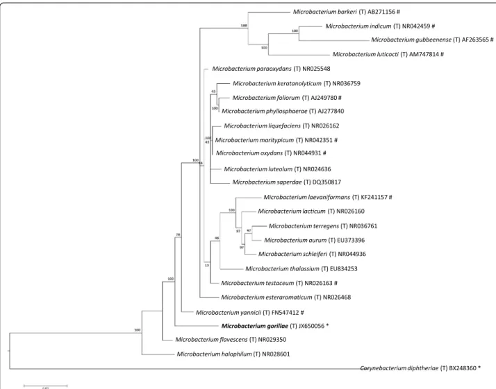
closest validly published Microbacterium species. This value was equal to the percentage of 16S rRNA gene se-quence threshold recommended by Meier-Kolthoff et al. for classActinobacteriato delineate a new species without carrying out DNA-DNA hybridization with maximum error probability of 0.1 % [9]. Figure 1 presents the 16S rRNA based tree for the strain G3Tand other Microbac-teriumspecies.
Different growth temperatures (20, 25, 30, 37, 45 °C) were tested. Growth occurred between 25 °C and 37 °C, but the optimal growth was observed at 25 °C, 24 h after inoculation. No growth occurred at 20 and 45 °C. Col-onies were 0.8 mm in diameter, appear as gray color on Columbia agar supplemented with sheep blood. Growth of the strain was tested under anaerobic and microaero-philic conditions using GENbag anaer and GENbag microaer systems, respectively (BioMérieux), and under aerobic conditions, with or without 5 % CO2. Growth was achieved under aerobic (with and without CO2), microaerophilic and anaerobic conditions. Gram staining showed Gram positive short bacilli (Fig. 2, left panel). A motility test with API M medium (BioMérieux) pro-duced a negative result. Cells grown on agar do not sporulate and the rods have a mean length of 1μm and a mean width of 0.5μm. Both the length and the diam-eter were ddiam-etermined by negative staining transmission electron microscopy (Fig. 2, right panel).
Strain G3T exhibited catalase activity but not oxidase activity using ID color catalase and oxidase reagent, re-spectively (BioMérieux). In assays with API 50CH sys-tem (BioMérieux), strain G3T produced acid from esculin, D-cellobiose, D-maltose, D-lactose, D-mannose, D-mannitol, D-saccharose, D-trehalose and gentiobiose. By contrast, acid production was not observed for glycerol, erythritol, D-arabinose, arabinose, D-ribose, D-xylose, L-xylose, D-adonitol, methyl-αD-xylopyranoside, D-galactose, D-glucose, L-fructose, L-sorbose, L-rhamnose, dulcitol, inositol, D-sorbitol, methyl-αD-mannopyranoside, Methyl-αD-glucopyranoside, xylitol, D-tagatose, D-turanose, D-lyxose, D-fucose, fucose, D-arabitol, L-arabitol, potassium gluconate, potassium 2-cetogluconate, potassium 5-cetogluconate, D-melezitose, D-raffinose, Glycogen, N-acetylglucosamin, amygdalin, arbutin, salicin and hydrolysis of starch. Using APIZYM, positive enzyme activities were observed for esterase (C4), esterase lipase (C8), leucine aramidase, phosphatase acid, naphtol-AS-BI-phosphohydrolase, α-mannosidase, α- glucosidase and N-acetyl-β-glucosaminidase. Negative results for lipase (C14), phosphatase alcalin, valine arylamidase, cystine arylamidase, trypsin, α-chymotrypsin, α-galactosidase, β – galactosidase, β-glucosidase, β-glucuronidase, β -glucosidase, and α-fucosidase.
M. gorillae is susceptible to amoxicillin (25 μg), erythromycin (15UI), doxycyclin (30UI), rifampicin (30μg), vancomycin (50μg), amoxicillin-clavulanic acid (20 μg + 10 μg), trimethoprim-sulfamethoxazole (1.25μg / 23.75μg) and imipenem (10μg) but resistant to ciprofloxacin (5μg) and gentamycin (15μg).
When compared to other Microbacterium species [10–16], M. gorillae sp. nov. strain G3T
exhibited the phenotypic differences detailed in Additional file 1: Table S1.
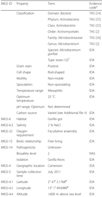
Table 1Classification and general features ofMicrobacterium gorillaestrain G3T
MIGS ID Property Term Evidence
codea
Classification Domain:Bacteria TAS [34]
Phylum:Actinobacteria TAS [35]
Class:Actinobacteria TAS [35]
Order:Actinomycetales TAS [2]
Family:Microbacteriaceae TAS [36]
Genus:Microbacterium TAS [2]
Species:Microbacterium gorillae
IDA
Type strain: G3T IDA
Gram stain Positive IDA
Cell shape Rod-shaped IDA
Motility Non-motile IDA
Sporulation Non-sporulating IDA
Temperature range Mesophilic IDA
Optimum temperature
25 °C IDA
pH range; Optimum Not determined
Carbon source Varied (see Additional file4) IDA
MIGS-6 Habitat Gorilla gut IDA
MIGS-6.3 Salinity 2 % NaCl IDA
MIGS-22 Oxygen requirement
Facultative anaerobic IDA
MIGS-15 Biotic relationship Free living IDA
MIGS-14 Pathogenicity Unknown
Biosafety level 2 NAS
Isolation Gorilla feces IDA
MIGS-4 Geographic location Cameroon IDA
MIGS-5 Sample collection time
July 2011 IDA
MIGS-4.1 Latitude 2° 47′2.1768″ IDA
MIGS-4.2 Longitude 13° 1′49.6986″ IDA
MIGS-4.4 Altitude >600 m above sea level IDA
a
Evidence codes - IDA: Inferred from Direct Assay; TAS: Traceable Author Statement
Extended feature descriptions
Matrix-assisted laser-desorption/ionization time-of-flight (MALDI-TOF) MS protein analysis was carried out as previously described [17] using a Microflex spectrometer (Bruker Daltonics, Leipzig, Germany). Twelve distinct deposits were done for strain G3Tfrom 12 isolated col-onies. Two microliters of matrix solution (saturated so-lution of alpha-cyano-4-hydroxycinnamic acid) in 50 % acetronitrile and 2.5 % trifluoroacetic-acid were distrib-uted on each smear and submitted at air drying for five minutes. Then, the spectra from the 12 different col-onies were imported into the MALDI BioTyper software (version 2.0, Bruker) and analyzed by standard pattern
matching (with default parameter settings) against 5,626 bacterial spectra including 43 spectra from 33 Microbac-terium species, used as reference data, in the BioTyper database. Briefly, a score≥2 with a species with a validly published name provided allows the identification at the species level, a score≥1.7 but < 2 allows the identification at the genus level; and a score < 1.7 does not allow any identification. For strain G3T, no good score was obtained, suggesting that our isolate was not a member of any known species. We incremented our database with the spectrum from strain G3T (Additional file 2: Figure S1). The gel view highlighted spectrum differences with other Microbacteriumspecies (Additional file 3: Figure S2).
Fig. 1Phylogenetic tree highlighting the position ofMicrobacterium gorillaestrain G3Trelative to other type strains within theMicrobacterium
Genome sequencing information
Genome project history
According to phenotypic characteristics of this strain and MALDI-TOF result and because of the low16S rRNA similarity to other members of the genus Micro-bacterium, it is likely that the strain represents a new species and thus it was chosen for genome sequencing. It was the 20th genome of a Microbacterium species (Genomes Online Database) and the first genome of Microbacterium gorillae sp. nov. A summary of the project information is shown in Table 2. The GenBank accession number is CDAR00000000 and consists of 14 contigs. Table 2 shows the project information and its association with MIGS version 2.0 compliance [18].
Growth conditions and genomic DNA preparation
Microbacterium gorillae sp.nov strain G3T (= CSUR P207 = DSM 26203) was grown aerobically on 5 % sheep blood-enriched Columbia agar (BioMérieux) at 25 °C.
Bacteria grown on four Petri dishes were resuspended in 3x500μl of TE buffer and stored at 80 °C. Then, 500μl of this suspension were thawed, centrifuged 3 min at 10,000 rpm and resuspended in 3x100μL of G2 buffer (EZ1 DNA Tissue kit, Qiagen). A first mechanical lysis was performed by glass powder on the Fastprep-24 de-vice (Sample Preparation system, MP Biomedicals, USA) using 2x20 s cycles. DNA was then treated with 2.5μg/ μL lysozyme (30 min at 37 °C) and extracted using the BioRobot EZ1 Advanced XL (Qiagen). The DNA was then concentrated and purified using the Qiamp kit (Qiagen). The yield and the concentration was measured by the Quant-it Picogreen kit (Invitrogen) on the Genios Tecan fluorometer at 50 ng/μl.
Genome sequencing and assembly
Genomic DNA of M. gorillae was sequenced on the MiSeq Technology (Illumina Inc, San Diego, CA, USA) with the 2 applications: paired end and mate paired. The gDNA was barcoded in order to be mixed with 11 others projects with the Nextera Mate Pair sample prep kit (Illumina) and with 17 others projects with the Nextera XT DNA sample prep kit (Illumina).
gDNA was quantified by a Qubit assay with the high sensitivity kit (Life technologies, Carlsbad, CA, USA) to 46.7 ng/μlTo prepare the paired end library, dilution was performed to require 1 ng of each genome as input. The « tagmentation » step fragmented and tagged the DNA. Then limited cycle PCR amplification (12 cycles) completed the tag adapters and introduced dual-index barcodes. After purification on AMPure XP beads (Beckman Coulter Inc, Fullerton, CA, USA), the librar-ies were then normalized on specific beads according to the Nextera XT protocol (Illumina). Normalized librar-ies were pooled for sequencing on the MiSeq. The pooled single strand library was loaded onto the re-agent cartridge and then onto the instrument along with the flow cell. Automated cluster generation and
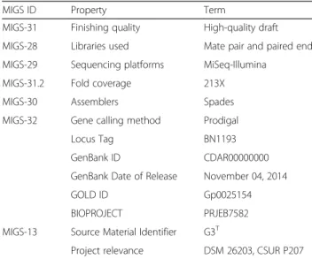
Table 2Project information
MIGS ID Property Term
MIGS-31 Finishing quality High-quality draft
MIGS-28 Libraries used Mate pair and paired end
MIGS-29 Sequencing platforms MiSeq-Illumina
MIGS-31.2 Fold coverage 213X
MIGS-30 Assemblers Spades
MIGS-32 Gene calling method Prodigal
Locus Tag BN1193
GenBank ID CDAR00000000
GenBank Date of Release November 04, 2014
GOLD ID Gp0025154
BIOPROJECT PRJEB7582
MIGS-13 Source Material Identifier G3T
Project relevance DSM 26203, CSUR P207
Fig. 2Gram staining (left panel) and Transmission electron microscopy using a Morgani 268D (Philips) at an operating voltage of 60 kV (right panel) of
paired end sequencing with dual index reads were per-formed in a single 39-h run in 2x250-bp.
Total information of 7.6 Gb was obtained from a 931 K/ mm2cluster density with a cluster passing quality control filters of 82.8 % (17,658,000 clusters). Within this run, the index representation for M. gorillae was determined to 5.11 %. The 732,922 paired end reads were trimmed and filtered by Trimmomatic tool using the recommended pa-rameters for Illumina sequence data [19].
Two mate pair libraries were prepared with 1 and 1.5 μg of genomic DNA using the Nextera mate pair Illumina guide. The genomic DNA sample was simultan-eously fragmented and tagged with a mate pair junction adapter. The pattern of the fragmentation was validated on an Agilent 2100 BioAnalyzer (Agilent Technologies Inc, Santa Clara, CA, USA) with a DNA 7500 labchip. The DNA fragments ranged from 1 kb to 11 kb in size with the majority of fragments at 8.8 and 9.4 kb of size. No size selection was performed and 45 ng for the 1st li-brary and 600 ng for the second lili-brary of tagmented fragments were circularized. The circularized DNA was mechanically sheared to small fragments with the major-ity at 400 and 380 bp on the Covaris device S2 in micro-tubes (Covaris, Woburn, MA, USA). The library profile was visualized on a High Sensitivity Bioanalyzer LabChip (Agilent Technologies Inc, Santa Clara, CA, USA) and the final concentration library was measured at 0.65 and 0.59 nmol/l respectively. The libraries were normalized at 2nM and pooled. After a denaturation step and dilu-tion at 15 pM, the pool of libraries was loaded onto the reagent cartridge and then onto the instrument along with the flow cell. Automated cluster generation and se-quencing run were performed in a single 39-h run in a 2x251-bp. The first libray was loaded three times on a flowcell and the second once. Within these runs, the index representation for M. gorillae was determined as an average at 3.51 %. The 1,881,286 paired reads were filtered according to the read qualities. The global paired end and mate pair libraries lead to 2,614,208 paired reads which were trimmed by Trimmomatic [19] then assembled by Spades software using the recommended options “–careful” and “-k 127” to fix the kmer size to 127 [20]. The final assembly identified 14 scaffolds gen-erating a genome size of 3.69 Mb which corresponds to genome coverage of 213X.
Genome annotation
Open Reading Frames (ORFs) were predicted using Prodigal [21] with default parameters but the predicted ORFs were excluded if they spanned a sequencing gap region. The predicted bacterial protein sequences were searched against the GenBank database [22] and the Clusters of Orthologous Groups (COG) databases using BLASTP. The tRNAScanSE tool [23] was used to find
tRNA genes, whereas ribosomal RNAs were found using RNAmmer [24] and BLASTn against the GenBank data-base. Lipoprotein signal peptides and the number of transmembrane helices were predicted using SignalP [25] and TMHMM [26] respectively. ORFans were iden-tified if their BLASTP E-value was lower than 1e-03 for alignment length greater than 80 amino acids. If align-ment lengths were smaller than 80 amino acids, we used an E-value of 1e-05. Such parameter thresholds have already been used in previous works to define ORFans. Artemis [27] was used for data management and DNA Plotter [28] for visualization of genomic features. The Mauve alignment tool (version 2.3.1) was used for mul-tiple genomic sequence alignment [29]. To estimate the mean level of nucleotide sequence similarity at the gen-ome level between M. gorillae sp. nov. strain G3T and other members of the genus Microbacterium, we used the MAGI home-made software to calculate the average genomic identity of gene sequences (AGIOS) among compared genomes [30]. Briefly, this software combines the Proteinortho software [31] for detecting orthologous proteins in pairwise genomic comparisons, then re-trieves the corresponding genes and determines the mean percentage of nucleotide sequence identity among orthologous ORFs using the Needleman-Wunsch global alignment algorithm. Finally, we used Genome-to-Genome Distance Calculator (GGDC) web server avail-able at (http://ggdc.dsmz.de) to estimate of the overall similarity among the compared genomes and to replace the wet-lab DNA-DNA hybridization (DDH) by a digital DDH (dDDH) [32, 33]. GGDC 2.0 BLAST+ was chosen as alignment method and the recommended formula 2 was taken into account to interpret the results.
Genome properties
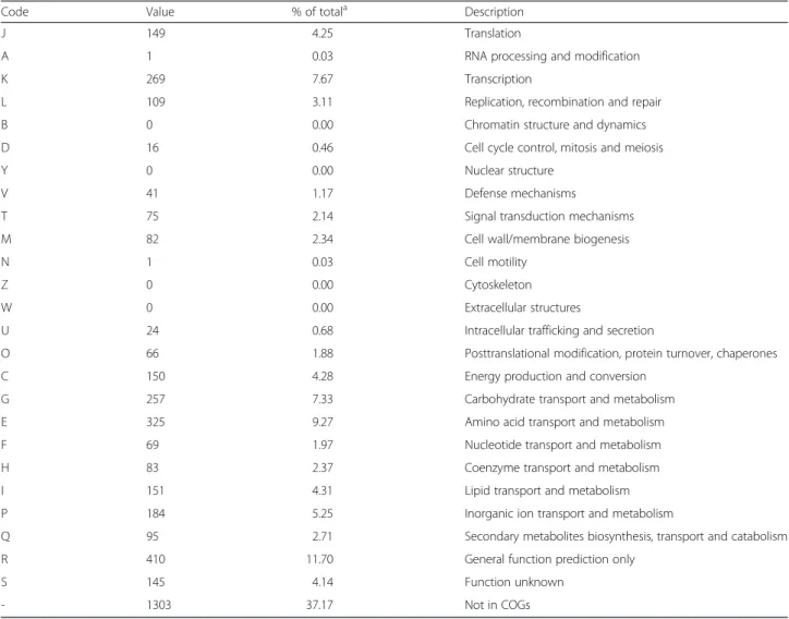
The genome of M. gorillaestrain G3T is 3,692,770 bp-long with a 69.3 % G+C content (Table 3, Fig. 3). Of the 3,566 predicted genes, 3,505 were protein-coding genes and 61 were RNA genes, including 4 complete rRNA operons (Additional file 4). A total of 2,412 genes (68.82 %) were assigned a putative function. A total of 6.33 % were identified as Pseudo-genes. The remaining genes were annotated as hypothetical pro-teins. The properties and the statistics of the genome are summarized in Table 3. The distribution of genes into COGs functional categories is presented in Table 4 and Additional file 4.
Insights from the genome sequence
Microbacterium laevaniformans strain OR221 (AJGR00 000000), Microbacterium luticocti strain DSM 19459 (AULS00000000), Microbacterium paraoxydans strain 77MFTsu3.2 (AQYI00000000), Microbacterium testaceum strain StLB037 (AP012052) and Microbacterium yannicii strain PS01 (CAJF00000000). The draft genome ofM. gor-illaehas a larger size than those ofM. indicum, M. luti-cocti, M. laevaniformans, M. paraoxydansandM. barkeri, (3.69 vs 2.81, 3.11, 3.43, 3.48 and 3.64 Mb respectively) but is smaller than those ofM. maritypicum, M. testaceumand M. yannicii (3.69 vs 4.0, 3.98 and 3.95 Mb respectively). The G+C content ofM. gorillaeis higher than those ofM. laevaniformans and M. maritypicum (69.3 vs 68.0 and 68.2 % respectively) but lower than those of M. indicum, M. luticocti, M. testaceum, M. yannicii, M. paraoxydans andM. barkeri(69.3 vs 71.4, 70.7, 70.3, 69.5, 69.5, 69.2 %, respectively). The gene content ofM. gorillaeis lower than those of M. maritypicum and M. testaceum, (3,505 vs 3,856 and 3,676 genes respectively) but higher than those of, M. paraoxydens, M. yannicii, M. laevaniformans, M. barkeri, M. luticocti and M. indicum (3,312, 3,279. 3,249, 3,099, 2,355, 2,183 genes respectively) (Table 5). However the distribution of genes into COG categories was similar
Table 3Nucleotide content and gene count levels of the genome
Attribute Value % of totala
Genome size (bp) 3,692,770 100
DNA coding (bp) 3,396,745 92
DNA G + C (bp) 2,558,287 69.3
DNA scaffolds 14
Total genes 3,566 100
Protein coding genes 3,505 98.3
RNA genes 61 1.71
Pseudo genes 226 6.33
Genes in internal clusters ND ND
Genes with function prediction 2,412 68.8
Genes assigned to COGs 2,202 62.8
Genes with Pfam domains 0 0
Genes with signals peptides 365 10.4
Genes with transmembrane helices 843 24.1
CRISPR repeats 0 0
a
The total is based on either the size of the genome in base pairs or the total number of protein coding genes in the annotated genome
ND: Not determined
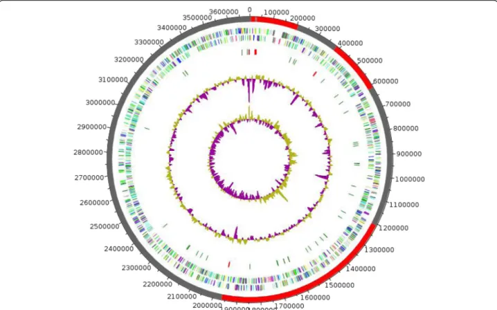
Fig. 3Graphical circular map of theMicrobacterium gorillaestrain G3Tchromosome. The outer two circles show open reading frames oriented in
in all compared genomes (Additional file 5: Figure S3). In addition, M. gorillae shares 1,593, 1,658, 1,269, 1,396, 1,390, 1,416, 1,498 and 1,497 orthologous genes with M. barkeri,M. maritypicum,M. indicum,M. laevaniformans, M. luticocti, M. paraoxydans, M. testaceum and M.
yanniciirespectively (Table 5). Among compared genomes except M. gorillae, AGIOS values range from 75.51 % between M. indicum and M. maritypicum to 85.33 % betweenM. maritypicumandM. barkeri. WhenM. goril-lae was compared to other species, AGIOS values range
Table 4Number of genes associated with the 25 general COG functional categories
Code Value % of totala Description
J 149 4.25 Translation
A 1 0.03 RNA processing and modification
K 269 7.67 Transcription
L 109 3.11 Replication, recombination and repair
B 0 0.00 Chromatin structure and dynamics
D 16 0.46 Cell cycle control, mitosis and meiosis
Y 0 0.00 Nuclear structure
V 41 1.17 Defense mechanisms
T 75 2.14 Signal transduction mechanisms
M 82 2.34 Cell wall/membrane biogenesis
N 1 0.03 Cell motility
Z 0 0.00 Cytoskeleton
W 0 0.00 Extracellular structures
U 24 0.68 Intracellular trafficking and secretion
O 66 1.88 Posttranslational modification, protein turnover, chaperones
C 150 4.28 Energy production and conversion
G 257 7.33 Carbohydrate transport and metabolism
E 325 9.27 Amino acid transport and metabolism
F 69 1.97 Nucleotide transport and metabolism
H 83 2.37 Coenzyme transport and metabolism
I 151 4.31 Lipid transport and metabolism
P 184 5.25 Inorganic ion transport and metabolism
Q 95 2.71 Secondary metabolites biosynthesis, transport and catabolism
R 410 11.70 General function prediction only
S 145 4.14 Function unknown
- 1303 37.17 Not in COGs
a
The total is based on the total number of protein coding genes in the annotated genome
Table 5Genomic comparison ofM. gorillaesp. nov., strain G3Twith otherMicrobacteriumspecies.
Species M. gorillae M. barkeri M. maritypicum M. indicum M. laevaniformans M. luticocti M. paraoxydans M. testaceum M. yannicii
M. gorillae 3,505 1,593 1,658 1,269 1,396 1,390 1,416 1,498 1,497
M. barkeri 75.91 3,099 2,111 1,390 1,511 1,461 1,595 1,685 1,684
M. maritypicum 75.22 85.33 3,856 1,429 1,581 1,549 1,634 1,755 1,734
M. indicum 75.39 76.16 75.51 2,183 1,296 1,191 1,324 1446 1,349
M. laevaniformans 75.80 76.59 76.07 76.05 3,249 1414 1,602 1,638 1,580
M. luticocti 76.41 76.99 76.50 76.34 77.94 2,355 1,395 1,433 1,512
M. paraoxydans 75.66 76.36 75.90 76.43 78.49 77.34 3,312 1,710 1,632
M. testaceum 75.64 76.48 75.84 76.30 77.64 77.64 77.52 3,676 1,723
M. yannicii 75.85 76.89 76.34 76.53 78.06 78.60 77.82 78.10 3,279
from 75.22 % with M. maritypicum to 76.41 % with M. luticocti (Table 5). dDDH estimation of the strain G3T against the compared genomes ranged between 19.70 to 20.50. These values are very low and below the cutoff of 70 %, thus confirming again the new species status of the strain G3T.
Conclusions
On the basis of phenotypic characteristics, phylogenetic position, genomic analyses (taxonogenomics) and GGDC results, we formally propose the creation of Microbacter-ium gorillae sp. nov. that contains the strain G3T. This strain has been isolated from a gorilla stool sample col-lected from Cameroon.
Taxonomic and nomenclatural proposals
Description ofMicrobacterium gorillaesp. nov.
Microbacterium gorillae (go.ril’lae. NL neut. gen gorilla, pertaining to a gorilla from which the stool sample was obtained).
Cells stain Gram-positive, are small rod, non-endospore-forming, non-motile and have a diameter of 0.5μm and a length of 1μm. Colonies are gray and 2 mm in diameter on blood-enriched Columbia agar. Growth oc-curs between 25 and 37 °C, with optimal growth observed at 25 °C.
Strain G3Texhibited catalase activity but not oxidase ac-tivity. Strain produces acid from esculin, cellobiose, D-maltose, D-lactose, D-mannose, D-mannitol, D-saccharose, D-trehalose and gentiobiose but not from glycerol, erythri-tol, D-arabinose, L-arabinose, D-ribose, D-xylose, L-xylose, D-adonitol, methyl-αxylopyranoside, galactose, D-glucose, L-fructose, L-sorbose, L-rhamnose, dulcitol, inositol, D-sorbitol, methyl-αD-mannopyranoside, Methyl-αglucopyranoside, xylitol, tagatose, turanose, D-lyxose, D-fucose, L-fucose, D-arabitol, L-arabitol, potassium gluconate, potassium 2-cetogluconate, potassium 5-cetogluconate, D-melezitose, D-raffinose, Glycogen, N-acetylglucosamin, amygdalin, arbutin, salicin and hydrolysis of starch.
Positive enzyme activities were observed for esterase (C4), esterase lipase (C8), leucine aramidase, phosphatase acid, naphtol-AS-BI-phosphohydrolase, α-mannosidase, α- glucosidase and N-acetyl-β-glucosaminidase. Negative results for lipase (C14), phosphatase alcalin, valine ary-lamidase, cystine aryary-lamidase, trypsin, α-chymotrypsin, α-galactosidase, β – galactosidase, β-glucosidase, β -glucuronidase,β-glucosidase, andα-fucosidase.
M. gorillae is susceptible to amoxicillin, erythromycin, doxycyclin, rifampicin, vancomycin, amoxicillin-clavulanic acid, trimethoprim-sulfamethoxazole and imipenem but resistant to ciprofloxacin and gentamycin.
The G+C content of the genome is 69.3 %. The 16S rRNA and genome sequences are deposited in GenBank
under accession numbers JX650056 and CDAR00000000, respectively. The type strain G3T (= CSUR P207 = DSM 26203) was isolated from the fecal sample of a western lowland gorilla from Cameroon.
Additional files
Additional file 1: Table S1.Differential phenotypic characteristics
betweenMicrobacterium gorillaesp. nov. strain G3Tand others
Microbacteriumstrains. (DOCX 14 kb)
Additional file 2: Figure S1.Reference mass spectrum fromM. gorillae
strain G3T. Spectra from 12 individual colonies were compared and a
reference spectrum was generated. (PPTX 44 kb)
Additional file 3: Figure S2.Gel view comparingMicrobacterium gorillae
strain G3Tspectra with other members of the genusMicrobacterium. The
gel view displays the raw spectra of all loaded spectrum files arranged in a pseudo-gel like look. The x-axis records the m/z value. The left y-axis displays the running spectrum number originating from subsequent spectra loading. The peak intensity is expressed by a gray-scale scheme code. The color bar and the right y-axis indicate the relation between the color a peak is displayed with and the peak intensity in arbitrary units. Displayed species are indicated on the right. (PPTX 76 kb)
Additional file 4: Folder S1.Annotation results. (RAR 1566 kb)
Additional file 5: Figure S3.Distribution of functional classes of
predicted genes ofM. gorillaestrain G3Twith 8 members ofMicrobacterium
genus. (PPTX 63 kb)
Abbreviations
CSUR:Collection de souches de l’Unité des Rickettsies; URMITE: Unité de Recherche sur les Maladies Infectieuses et Tropicales Emergentes; DSM: Deutsche Sammlung von Mikroorganismen; MALDI-TOF MS: Matrix-assisted laser-desorption/ionization time-of-flight mass spectrometry; TE buffer: Tris-EDTA buffer; GGDC: Genome-to-Genome Distance Calculator; dDDH: digital DNA-DNA hybridization.
Competing interests
The authors declare that they have no competing interests.
Authors’contributions
LH wrote the manuscript and analyzed the data. MBK performed laboratory experiments and helped to draft the manuscript. CM performed the sequencing and helped to draft the manuscript. JR and AL performed bioinformatics analysis and helped to draft the manuscript. DR, PF, JMR and FB conceived the study, participated in its design and coordination and helped to draft the manuscript. All authors read and approved the final manuscript.
Acknowledgements
Fadi Bittar was supported by a Chair of Excellence IRD provided by the Institut de Recherche pour le Développement / Méditerranée-Infection foundation. Mamadou Bhoye Keita was funded by the Méditerranée-Infection foundation. The authors thank Xegen company for automating the genome annotation process.
Author details
1
Unité de recherche sur les maladies infectieuses et tropicales émergentes (URMITE), UM63, CNRS7278, IRD 198, Inserm 1095, IHU Méditerranée Infection, Faculté de Médecine et de Pharmacie, Aix-Marseille Université, Marseille, France.2King Fahad Medical Research Center, King Abdul Aziz University, Jeddah, Saudi Arabia.
References
1. Bittar F, Keita MB, Lagier JC, Peeters M, Delaporte E, Raoult D.Gorilla gorilla gorillagut: a potential reservoir of pathogenic bacteria as revealed using culturomics and molecular tools. Sci Rep. 2014;4:7174.
2. Skerman VBD, McGowan V, Sneath PHA. Approved Lists of Bacterial Names. Int J Syst Bacteriol. 1980;30:225–420.
3. Abstract for the genusMicrobacterium. NamesforLife, LLC. Retrieved June 26, 2015. (doi:10.1601/tx.6034).
4. Gneiding K, Frodl R, Funke G. Identities ofMicrobacterium spp.Encountered in Human Clinical Specimens. J Clin Microbiol. 2008;46:3646.
5. Anand S, Bala K, Saxena A, Schumann P, Lal R.Microbacterium amylolyticum
sp. nov., isolated from soil from an industrial waste site. Stand Int J Syst Evol Microbiol. 2012;62:2114–20.
6. Bakir MA, Kudo T, Benno Y.Microbacterium hatanonissp. nov., isolated as a contaminant of hairspray. Int J Syst Evol Microbiol. 2008;58(Pt 3):654–8. 7. Alves A, Riesco R, Correia A, Trujillo ME.Microbacterium proteolyticumsp.
nov. isolated from roots of Halimione portulacoides. Int J Syst Evol Microbiol. 2015;65(Pt 6):1794–8.
8. Sentausa E, Fournier PE. Advantages and limitations of genomics in prokaryotic taxonomy. Clin Microbiol Infect. 2013;19(9):790–5.
9. Meier-Kolthoff JP, Göker M, Spröer C, Klenk HP. When should a DDH experiment be mandatory in microbial taxonomy? Arch Microbiol. 2013;6:413–8. 10. Zhang Y, Ren H, Zhang G.Microbacterium hydrothermalesp. nov., an
actinobacterium isolated from hydrothermal sediment. Int J Syst Evol Microbiol. 2014;64:3508–12.
11. Shivaji S, Bhadra B, Rao RS, Chaturvedi P, Pindi PK, Raghukumar C.
Microbacterium indicumsp. nov., isolated from a deep-sea sediment sample from the Chagos Trench, Indian Ocean. Int J Syst Evol Microbiol. 2007;57: 1819–22.
12. Vaz-Moreira I, Lopes AR, Falsen E, Schumann P, Nunes OC, Manaia CM.
Microbacterium luticoctisp. nov., isolated from sewage sludge compost. Int J Syst Evol Microbiol. 2008;58:1700–4.
13. Laffineur K, Avesani V, Cornu G, Charlier J, Janssens M, Wauters G, et al. Bacteremia due to a novelMicrobacteriumspecies in a patient with leukemia and description ofMicrobacterium paraoxydanssp. nov. J Clin Microbiol. 2003;41(5):2242–6.
14. Schippers A, Bosecker K, Sproer C, Schumann P.Microbacterium oleivoranssp. nov. andMicrobacterium hydrocarbonoxydanssp. nov., novel crude-oil-degrading Gram-positive bacteria. Int J Syst Evol Microbiol. 2005;55(Pt 2):655–60. 15. Sharma P, Diene SM, Thibeaut S, Bittar F, Roux V, Gomez C, et al. Phenotypic
and genotypic properties ofMicrobacterium yannicii, a recently described multidrug resistant bacterium isolated from a lung transplanted patient with cystic fibrosis in France. BMC Microbiol. 2013;13:97.
16. Takeuchi M, Hatano K. Proposal of six new species in the genus
Microbacteriumand transfer of“Flavobacterium marinotypicum”ZoBell and Upham to the genusMicrobacteriumasMicrobacterium maritypicumcomb. nov. Int J Syst Bacteriol. 1998;48 Pt 3:973–82.
17. Seng P, Drancourt M, Gouriet F, et al. Ongoing revolution in bacteriology: routine identification of bacteria by matrix-assisted laser desorption ionization time-of-flight mass spectrometry. Clin Infect Dis. 2009;49:543–51. 18. Field D, Garrity G, Gray T, et al. The minimum information about a genome
sequence (MIGS) specification. Nat Biotechnol. 2008;26:541–7.
19. Bolger AM, Lohse M, Usadel B. Trimmomatic: a flexible trimmer for Illumina sequence data. Bioinformatics. 2014;30:2114–20.
20. Bankevich A, Nurk S, Antipov D, Gurevich AA, Dvorkin M, Kulikov AS, et al. SPAdes: a new genome assembly algorithm and its applications to single-cell sequencing. J Comput Biol. 2012;19:455–77.
21. Prodigal. [http://prodigal.ornl.gov].
22. GenBank database. http://www.ncbi.nlm.nih.gov/genbank.
23. Lowe TM, Eddy SR. tRNAscan-SE: a program for improved detection of transfer RNA genes in genomic sequence. Nucleic Acids Res. 1997;25:955–64. 24. Lagesen K, Hallin P, Rodland EA, Staerfeldt HH, Rognes T, Ussery DW.
RNAmmer: consistent and rapid annotation of ribosomal RNA genes. Nucleic Acids Res. 2007;35:3100–8.
25. Bendtsen JD, Nielsen H, von Heijne G, Brunak S. Improved prediction of signal peptides: SignalP 3.0. J Mol Biol. 2004;340:783–95.
26. Krogh A, Larsson B, von Heijne G, Sonnhammer EL. Predicting
transmembrane protein topology with a hidden Markov model: application to complete genomes. J Mol Biol. 2001;305:567–80.
27. Rutherford K, Parkhill J, Crook J, Horsnell T, Rice P, Rajandream MA, Barrell B. Artemis: sequence visualization and annotation. Bioinformatics. 2000;16:944–5.
28. Carver T, Thomson N, Bleasby A, Berriman M, Parkhill J. DNAPlotter: circular and linear interactive genome visualization. Bioinformatics. 2009;25:119–20. 29. Darling AC, Mau B, Blattner FR, Perna NT. Mauve: multiple alignment of
conserved genomic sequence with rearrangements. Genome Res. 2004; 14:1394–403.
30. Ramasamy D, Mishra AK, Lagier JC, et al. A polyphasic strategy incorporating genomic data for the taxonomic description of novel bacterial species. Int J Syst Evol Microbiol. 2014;64:384–91.
31. Lechner M, Findeiss S, Steiner L, Marz M, Stadler PF, Prohaska SJ. Proteinortho: detection of (co-)orthologs in large-scale analysis. BMC Bioinformatics. 2011;12:124.
32. Auch AF, von Jan M, Klenk HP, Göker M. Digital DNA-DNA hybridization for microbial species delineation by means of genome-to-genome sequence comparison. Stand Genomic Sci. 2010;2:117–34.
33. Meier-Kolthoff JP, Auch AF, Klenk HP, Göker M. Genome sequence-based species delimitation with confidence intervals and improved distance functions. BMC Bioinformatics. 2013;14:60.
34. Woese CR, Kandler O, Wheelis ML. Towards a natural system of organisms: proposal for the domainsArchae, Bacteria, andEukarya. Proc Natl Acad Sci U S A. 1990;87:4576–9.
35. Stackebrandt E, Rainey FA, Ward-Rainey NL. Proposal for a new hierarchic classification system,Actinobacteriaclassis nov. Int J Syst Bacteriol. 1997;47:479–91. 36. Validation List no. 53 in IJSEM. Validation of the Publication of New Names
and New Combinations Previously Effectively Published Outside the IJSB. Int J Syst Evol Microbiol. 1995;25:418–9.
37. Ashburner M, Ball CA, Blake JA, Botstein D, Butler H, Cherry JM, et al. Gene ontology: tool for the unification of biology. The Gene Ontology Consortium Nat Genet. 2000;25:25–9.
38. Castresana J. Selection of conserved blocks from multiple alignments for their use in phylogenetic analysis. Mol Biol Evol. 2000;17:540–52. 39. Stamatakis A. RAxML-VI-HPC: maximum likelihood-based phylogenetic
analyses with thousands of taxa and mixed models. Bioinformatics. 2006;22:2688–90.
• We accept pre-submission inquiries
• Our selector tool helps you to find the most relevant journal • We provide round the clock customer support
• Convenient online submission • Thorough peer review
• Inclusion in PubMed and all major indexing services • Maximum visibility for your research
Submit your manuscript at www.biomedcentral.com/submit