PRIMARY RESEARCH
Comprehensive analysis of DNA
methylation and gene expression profiles
in cholangiocarcinoma
Cheng Zhang
1, Bingye Zhang
1, Di Meng
2and Chunlin Ge
1*Abstract
Background: The incidence of cholangiocarcinoma (CCA) has risen in recent years, and it has become a significant health burden worldwide. However, the mechanisms underlying tumorigenesis and progression of this disease remain largely unknown. An increasing number of studies have demonstrated crucial biological functions of epige-netic modifications, especially DNA methylation, in CCA. The present study aimed to identify and analyze methylation-regulated differentially expressed genes (MeDEGs) involved in CCA tumorigenesis and progression by bioinformatics analysis.
Methods: The gene expression profiling dataset (GSE119336) and gene methylation profiling dataset (GSE38860) were obtained from the Gene Expression Omnibus (GEO) database. Differentially expressed genes (DEGs) and differentially methylated genes (DMGs) were identified using the limma packages of R and GEO2R, respectively. The MeDEGs were obtained by overlapping the DEGs and DMGs. Functional enrichment analyses of these genes were then carried out. Protein–protein interaction (PPI) networks were constructed using STRING and visualized in Cytoscape to determine hub genes. Finally, the results were verified based on The Cancer Genome Atlas (TCGA) database.
Results: We identified 98 hypermethylated, downregulated genes and 93 hypomethylated, upregulated genes after overlapping the DEGs and DMGs. These genes were mainly enriched in the biological processes of the cell cycle, nuclear division, xenobiotic metabolism, drug catabolism, and negative regulation of proteolysis. The top nine hub genes of the PPI network were F2, AHSG, RRM2, AURKB, CCNA2, TOP2A, BIRC5, PLK1, and ASPM. Moreover, the expres-sion and methylation status of the hub genes were significantly altered in TCGA.
Conclusions: Our study identified novel methylation-regulated differentially expressed genes (MeDEGs) and explored their related pathways and functions in CCA, which may provide novel insights into a further understanding of methylation-mediated regulatory mechanisms in CCA.
Keywords: Cholangiocarcinoma, Methylation, Bioinformatics
© The Author(s) 2019. This article is licensed under a Creative Commons Attribution 4.0 International License, which permits use, sharing, adaptation, distribution and reproduction in any medium or format, as long as you give appropriate credit to the original author(s) and the source, provide a link to the Creative Commons licence, and indicate if changes were made. The images or other third party material in this article are included in the article’s Creative Commons licence, unless indicated otherwise in a credit line to the material. If material is not included in the article’s Creative Commons licence and your intended use is not permitted by statutory regulation or exceeds the permitted use, you will need to obtain permission directly from the copyright holder. To view a copy of this licence, visit http://crea-tivecommons.org/licenses/by/4.0/. The Creative Commons Public Domain Dedication waiver ( http://creativecommons.org/publicdo-main/zero/1.0/) applies to the data made available in this article, unless otherwise stated in a credit line to the data.
Background
Cholangiocarcinoma (CCA) is a fatal malignancy origi-nating from the epithelial cells of the biliary tree [1]. According to its anatomical location, the most contem-porary classification of CCA includes intrahepatic, peri-hilar, and distal subtypes [2]. Surgical resection remains the only potentially curative treatment for all subtypes [1]. Unfortunately, early diagnosis is rare due to a lack of
Open Access
*Correspondence: [email protected]
1 Department of Pancreatic and Biliary Surgery, The First Hospital of China Medical University, Shenyang 110001, Liaoning, China
specific clinical symptoms [3]. Only about one-third of CCA patients are eligible for surgery; most patients are often diagnosed with unresectable or metastatic disease [4]. Even so, the recurrence and progression to distant metastasis are common within 2 years of resection [5]. Overall survival for patients with unresectable CCA does not exceed 14 months, even with systemic chemotherapy [6]. In addition, the incidence of CCA seems to be on the rise, especially for intrahepatic tumors [6]. Therefore, cumulative cholangiocarcinoma mortality rates have also increased by 39% [2]. Thus, exploring the molecular mechanisms underlying the pathogenesis and develop-ment of CCA is urgently needed.
DNA methylation is a central epigenetic modification that plays a key role in cellular processes, such as genome regulation, organism development, and disease [7]. Importantly, the dysregulation of DNA methylation pat-terns has been increasingly recognized as an important cellular event during both the initiation and late stages of oncogenesis [8, 9]. To date, numerous studies have demonstrated a crucial role for both hypermethylation of tumor suppressor genes and global hypomethylation of oncogenes in cancer development and progression, including in CCA [8, 10]. For instance, Chen et al. [11] found that the O6-methylguanine-DNA methyltrans-ferase (MGMT) promoter was highly methylated, and the expression level of MGMT was positively corre-lated with overall survival rates and histological grade in CCA. In addition, the aberrant methylation of GATA-5, ANGPTL4, and DLEC1 has been shown to participate in the initiation and progression of this disease [12–14]. Despite the identification of several individual genes with specific hypomethylation or hypermethylation in CCA, comprehensive network studies based on methylation profiles and related pathways for these genes have been largely inadequate.
Over the past decade, gene profiling and next-gen-eration sequencing technology have emerged as indis-pensable tools for cancer studies because they allow the detection of cancer-related genetic and epigenetic alterations, such as mutations, copy number variations, and DNA methylation changes across more extensive genomic regions [15, 16]. The bioinformatics analysis of these data can provide valuable information for CCA research. For example, Kong et al. identified three dif-ferentially expressed genes (DEGs), UCA1, miR-122, and CLIC1, based on next-generation sequencing data in CCA [17]. Analysis of these dysregulated genes showed that they could promote CCA progression through the regulation of miR-122/CLIC1 and activation of the ERK/MAPK signaling pathway [17]. Furthermore, both Farshidfar et al. and Jusakul et al. identified many dif-ferentially methylated genes (DMGs) by methylation
profiling or whole-genome sequencing that contributed to cholangiocarcinogenesis and might serve as meth-ylation biomarkers in CCA [18, 19]. However, separate DEG and DMG analysis from a single study are limited, and multiple overlapping available datasets may provide more accurate and reliable clues through comprehensive bioinformatics analysis [20]. Thus far, there is still a lack of conjoint analysis involving both gene expression and methylation profiling microarray datasets in CCA.
In the present study, we performed an integrated bio-informatics analysis based on gene expression profil-ing by high-throughput sequencprofil-ing (GSE119336) and gene methylation profiling microarray (GSE38860). The methylation-regulated differentially expressed genes (MeDEGs) were identified and then subjected to enrich-ment analysis. Moreover, we constructed a protein–pro-tein interaction (PPI) network to identify hub genes to screen novel biomarkers and therapeutic targets in CCA for future research.
Methods
RNA‑Seq and microarray data
We obtained the gene expression profiling dataset gener-ated by high-throughput sequencing (GSE119336) and the microarray-based gene methylation profiling dataset (GSE38860) from the publicly available Gene Expression Omnibus database (GEO, https ://www.ncbi.nlm.nih.gov/ geo/). The expression dataset (GSE119336) contained 15 pairs of CCA tumors and matched non-tumor tissues, which was based on the GPL11154 platform (Illumina HiSeq 2000). The DNA methylation dataset (GSE38860), which was generated on the GPL8490 platform (Illumina HumanMethylation27 BeadChip), included a total of 28 primary CCA tissues and six matched adjacent normal tissues.
Identification of MeDEGs
genes. The hypomethylation-high expression genes and hypermethylation-low expression genes were identified as methylation-regulated differentially expressed genes (MeDEGs).
Functional enrichment analysis
Gene Ontology (GO) annotation and pathway analysis of the MeDEGs were conducted using The Search Tool for the Retrieval of Interacting Genes (STRING, https :// strin g-db.org/) [21], Kyoto Encyclopedia of Genes and Genomes (KEGG, https ://www.kegg.jp/), and Reactome (https ://react ome.org/). Statistically significant thresh-olds were set at a P < 0.05.
PPI network construction, module analysis, and identification of hub genes
The PPI network of hypomethylation-high expression genes and hypermethylation-low expression genes was constructed using the STRING online database. An interaction score of > 0.4 and P < 0.05 were regarded as statistically significant. Cytoscape (version 3.6.1; http:// www.cytos cape.org/) software was utilized to visualize the network. Molecular Complex Detection (MCODE), an app in Cytoscape, was used to screen modules within the PPI network with a standard of combined scores > 3 and nodes ≥ 5. Hub genes were identified using the Cyto-Hubba in Degree-ranked method with a selection crite-rion for hub genes set at a degree score of greater than 16.
Validation of hub genes
MEXPRESS (http://mexpr ess.be) is a straightforward and easy-to-use web tool for the integration and visualization of expression and DNA methylation based on The Can-cer Genome Atlas (TCGA) database at a single-gene level [22, 23]. We used MEXPRESS to validate hypomethyla-tion-high expression genes and hypermethylation-low expression genes. The cBioPortal (http://cbiop ortal .org) is an open-access resource for interactive exploration of multidimensional cancer genomics datasets that provides access to data for more than 5000 tumor samples from 20 cancer studies [24]. The cBioPortal was used to explore the genetic alterations of hub genes.
Results
Identification of MeDEGs in CCA
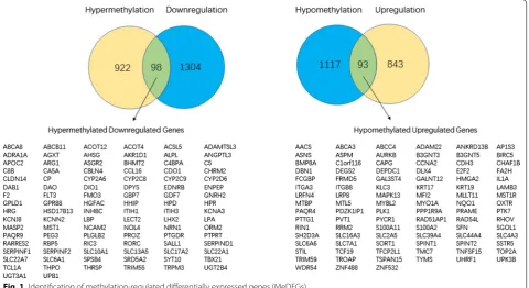
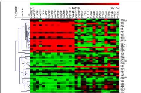
The limma packages of R and GEO2R were used to screen for DEGs and DMGs, respectively. For the gene expression dataset GSE119336, a total of 2338 DEGs were identified, comprising 936 upregulated genes and 1402 downregulated genes. For the DMGs in the gene methylation dataset (GSE38860), 1020 hypermethylated genes and 1210 hypomethylated genes were obtained. As shown in Fig. 1, we identified 98 hypermethylated, down-regulated genes and 93 hypomethylated, updown-regulated genes after the overlapping of the DEGs and DMGs. The heat map of the top 50 MeDEGs is shown in Fig. 2.
Functional enrichment analysis of MeDEGs
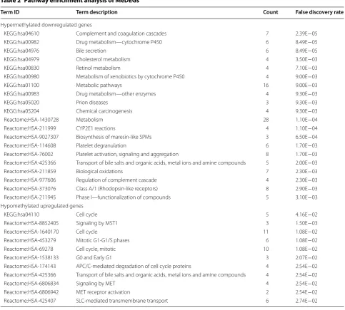
The results of the gene ontology enrichment analysis for the MeDEGs are shown in Table 1. In the biological process group, hypomethylated, upregulated genes were mainly enriched in the cell cycle, nuclear division, nega-tive regulation of cell cycle process, cell cycle process, and negative regulation of mitotic nuclear division. The hypermethylated, downregulated genes were mainly enriched in xenobiotic metabolic process, drug catabolic process, negative regulation of proteolysis, acute-phase response, and monocarboxylic acid metabolic process. We also found that the hypermethylated, downregulated genes were related to endopeptidase inhibitor activity, enzyme inhibitor activity, and serine-type endopepti-dase inhibitor activity in the molecular function group and extracellular region in the cellular component group. Pathway enrichment was also performed using KEGG and Reactome, and the results are presented in Table 2. We found that hypermethylated genes predominantly participated in metabolism-related pathways, including general metabolic pathways, drug or xenobiotic metab-olism by cytochrome P450, and cholesterol metabo-lism. For hypomethylated genes, the most significantly
enriched pathways involved the cell cycle (mitotic G1-G1/S phases; G0 and early G1), SLC-mediated trans-membrane transport, and signaling by MST1.
PPI network construction, module analysis, and identification of hub genes
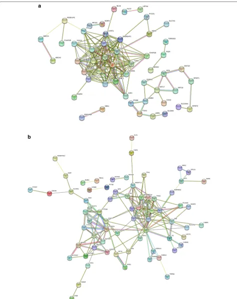
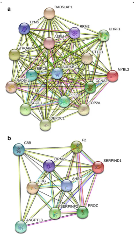
The PPI network of hypomethylation-high expression genes and hypermethylation-low expression genes was constructed using the STRING online database. An interaction score of > 0.4 and P < 0.05 indicated statisti-cal significance. The results are presented in Fig. 3. Using MCODE in Cytoscape, we identified two modules in the PPI network with a standard of combined scores > 3 and nodes ≥ 5 (Fig. 4). Nine hub genes were identified using CytoHubba with a degree score greater than 16, includ-ing F2, AHSG, RRM2, AURKB, CCNA2, TOP2A, BIRC5, PLK1, and ASPM.
Verification of hub genes
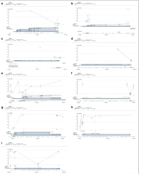
seven hypomethylated upregulated genes and two hyper-methylated downregulated genes were significantly dif-ferent in the TCGA database, which were consistent with our results (Table 3). Except for ASPM, the meth-ylation levels of these hub genes were consistent with
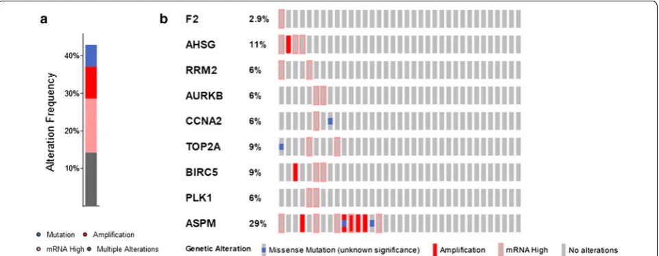
our findings (Fig. 5a–i: AURKB, PLK1, CCNA2, ASPM, RRM2, TOP2A, BIRC5, F2, and AHSG). In addition, the presence of genetic alterations in the hub genes was examined using the cBioPortal tool. More than 40% of the patient tumors had at least one hub gene alteration
Table 1 Gene ontology enrichment analysis of MeDEGs
Category Term ID Term description Count False discovery rate
Hypermethylated downregulated genes
BP_FAT GO:0019752 Carboxylic acid metabolic process 22 5.98E−07
BP_FAT GO:0071466 Cellular response to xenobiotic stimulus 9 8.39E−05
BP_FAT GO:0006805 Xenobiotic metabolic process 7 8.00E−04
BP_FAT GO:0042737 Drug catabolic process 7 8.00E−04
BP_FAT GO:0045861 Negative regulation of proteolysis 11 8.00E−04
BP_FAT GO:0006953 Acute-phase response 5 1.30E−03
BP_FAT GO:0010951 Negative regulation of endopeptidase activity 9 1.30E−03
BP_FAT GO:0032787 Monocarboxylic acid metabolic process 12 1.30E−03
BP_FAT GO:0001676 Long-chain fatty acid metabolic process 6 3.30E−03
BP_FAT GO:0044281 Small molecule metabolic process 23 3.30E−03
CC_FAT GO:0005576 Extracellular region 38 2.40E−08
CC_FAT GO:0005615 Extracellular space 25 2.40E−08
CC_FAT GO:0044421 Extracellular region part 28 2.40E−08
CC_FAT GO:0044432 Endoplasmic reticulum part 19 1.30E−03
MF_FAT GO:0004866 Endopeptidase inhibitor activity 9 1.20E−04
MF_FAT GO:0004857 Enzyme inhibitor activity 11 4.20E−04
MF_FAT GO:0004867 Serine-type endopeptidase inhibitor activity 6 7.80E−04
MF_FAT GO:0008395 Steroid hydroxylase activity 4 1.90E−03
MF_FAT GO:0015370 Solute:sodium symporter activity 5 3.60E−03
MF_FAT GO:0016491 Oxidoreductase activity 13 3.60E−03
Hypomethylated upregulated genes
BP_FAT GO:0007049 Cell cycle 20 3.20E−03
BP_FAT GO:0000280 Nuclear division 8 1.61E−02
BP_FAT GO:0010948 Negative regulation of cell cycle process 8 1.61E−02
BP_FAT GO:0022402 Cell cycle process 15 1.61E−02
BP_FAT GO:0045839 Negative regulation of mitotic nuclear division 4 1.61E−02
BP_FAT GO:0045930 Negative regulation of mitotic cell cycle 8 1.61E−02
BP_FAT GO:0048519 Negative regulation of biological process 41 1.61E−02
BP_FAT GO:0051726 Regulation of cell cycle 17 1.61E−02
BP_FAT GO:1903047 Mitotic cell cycle process 12 1.61E−02
BP_FAT GO:0007093 Mitotic cell cycle checkpoint 6 1.66E−02
BP_FAT GO:0051716 Cellular response to stimulus 46 3.22E−02
BP_FAT GO:0007568 Aging 7 3.23E−02
BP_FAT GO:0031577 Spindle checkpoint 3 3.23E−02
BP_FAT GO:0051302 Regulation of cell division 6 3.23E−02
BP_FAT GO:2000816 Negative regulation of mitotic sister chromatid separation 3 3.23E−02
BP_FAT GO:0007010 Cytoskeleton organization 13 3.36E−02
BP_FAT GO:0007051 Spindle organization 5 3.36E−02
BP_FAT GO:0007052 Mitotic spindle organization 4 3.36E−02
BP_FAT GO:0007346 Regulation of mitotic cell cycle 10 3.36E−02
and ASPM (29%) was the most frequently altered gene of the nine queried genes (Fig. 6).
Discussion
Cholangiocarcinoma (CCA) is a fatal malignancy that arises from cholangiocytes. In recent years, the inci-dence of CCA has risen, and CCA has become a major health burden worldwide [25]. The majority of patients with CCA present with unresectable or metastatic dis-ease at the time of diagnosis because of the lack of spe-cific symptoms or sensitive indicators [26]. Conventional radiotherapy and current chemotherapy treatments are not very effective [2]. Given the diagnostic difficulty and limited treatment options, the prognosis for patients with
CCA is extremely poor [27]. Therefore, identifying novel molecular biomarkers and understanding the underlying mechanisms of carcinogenesis and progression in CCA are critically needed.
DNA methylation is a central epigenetic modification that plays a key role in cellular processes and generally occurs on cytosines that precede a guanine nucleotide [28, 29]. Typically, these cytosine–phosphate–guanine (CpG) sites are concentrated in large clusters (i.e., CpG islands) that are enriched mostly in the promoter region of genes [30]. Because the binding sites for many tran-scription factors are GC-rich, CpG islands are likely to enhance binding to transcriptional start sites [31]. CpG islands may also enhance the accessibility of DNA
Table 2 Pathway enrichment analysis of MeDEGs
Term ID Term description Count False discovery rate
Hypermethylated downregulated genes
KEGG:hsa04610 Complement and coagulation cascades 7 2.39E−05
KEGG:hsa00982 Drug metabolism—cytochrome P450 6 8.49E−05
KEGG:hsa04976 Bile secretion 6 8.49E−05
KEGG:hsa04979 Cholesterol metabolism 4 3.50E−03
KEGG:hsa00830 Retinol metabolism 4 7.10E−03
KEGG:hsa00980 Metabolism of xenobiotics by cytochrome P450 4 9.00E−03
KEGG:hsa01100 Metabolic pathways 16 9.00E−03
KEGG:hsa00983 Drug metabolism—other enzymes 4 9.30E−03
KEGG:hsa05020 Prion diseases 3 9.30E−03
KEGG:hsa05204 Chemical carcinogenesis 4 9.30E−03
Reactome:HSA-1430728 Metabolism 28 1.10E−04
Reactome:HSA-211999 CYP2E1 reactions 4 1.10E−04
Reactome:HSA-9027307 Biosynthesis of maresin-like SPMs 3 6.50E−04
Reactome:HSA-114608 Platelet degranulation 6 1.70E−03
Reactome:HSA-76002 Platelet activation, signaling and aggregation 8 1.70E−03
Reactome:HSA-425366 Transport of bile salts and organic acids, metal ions and amine compounds 5 2.00E−03
Reactome:HSA-211859 Biological oxidations 7 2.30E−03
Reactome:HSA-977606 Regulation of complement cascade 4 2.30E−03
Reactome:HSA-373076 Class A/1 (Rhodopsin-like receptors) 8 2.90E−03
Reactome:HSA-211945 Phase I—functionalization of compounds 5 3.10E−03
Hypomethylated upregulated genes
KEGG:hsa04110 Cell cycle 5 4.16E−02
Reactome:HSA-8852405 Signaling by MST1 3 1.50E−03
Reactome:HSA-1640170 Cell cycle 11 1.08E−02
Reactome:HSA-453279 Mitotic G1-G1/S phases 6 1.08E−02
Reactome:HSA-69278 Cell cycle, mitotic 10 1.08E−02
Reactome:HSA-1538133 G0 and Early G1 3 2.07E−02
Reactome:HSA-174143 APC/C-mediated degradation of cell cycle proteins 4 2.54E−02
Reactome:HSA-425366 Transport of bile salts and organic acids, metal ions and amine compounds 4 2.54E−02
Reactome:HSA-6806834 Signaling by MET 4 2.54E−02
Reactome:HSA-6806942 MET receptor activation 2 2.54E−02
by regulating the chromatin structure. Methylation of CpG islands can impair transcription factor binding, recruit repressive methyl-binding proteins, and stably silence gene expression [31]. Thus, CpG islands are nor-mally unmethylated in transcriptionally active genes and methylated in the promoters of silenced genes. To date, numerous studies have demonstrated a crucial role for both hypermethylation of tumor suppressor genes and hypomethylation of oncogenes in cancer development and progression [30]. For example, Wu et al. [32] found that JMJD2C could enhance the metastasis of colorec-tal cancer by decreasing the histone methylation of the MALAT1 promoter, thereby upregulating MALAT1 expression and enhancing the activity of the β-catenin signaling pathway. Furthermore, Liang et al. [33] iden-tified many novel methylation-regulated differentially expressed genes involved in colon cancer tumorigenesis and progression by identifying hypermethylated down-regulated genes and hypomethylated updown-regulated genes.
The initiation and progression of CCA is a complex and multistage process regulated by both genetic and epige-netic alterations [34]. An increasing number of studies have demonstrated the crucial biological functions of epigenetic modifications, especially DNA methylation, in CCA. For example, significant differences in the meth-ylation levels of OPCML and HOXD9 were observed in serum cell-free DNA from CCA patients, and these two genes can be used for the differential diagnosis between cholangiocarcinoma and other biliary diseases [35]. Moreover, Wang et al. found that DANCR could bind EZH2 and modulate the histone methylation of the FBP1 promoter, thereby regulating the proliferation and migra-tion of CCA cells [25]. However, previous studies mainly focused on specific genes with aberrant DNA methyla-tion or individual gene methylamethyla-tion profiling microar-rays. However, a systematic conjoint analysis involving both gene expression and methylation profiling in CCA may yield more accurate and reliable results. Thus, we performed an integrated bioinformatics analysis based on both gene expression and gene methylation profil-ing (GSE119336 and GSE38860, respectively) to screen for novel biomarkers and therapeutic targets in CCA for future research.
In the present study, we identified a total of 93 hypo-methylated, upregulated genes and 98 hypermethyl-ated, downregulated genes by integrating the DEGs and DMGs. Functional and pathway enrichment analysis indicated that the hypomethylated, upregulated genes were mainly enriched in the biological processes of the cell cycle, mitotic cell cycle checkpoint, mitotic sister chromatid separation, spindle assembly, nuclear divi-sion, and cell division. These findings are reasonable because it is well known that dysregulation of cell cycle Fig. 4 Module analysis of methylation-regulated differentially
expressed genes. a Hypomethylated upregulated genes. b Hypermethylated downregulated genes
Table 3 Verification of the expression levels of 7 hypomethylated upregulated genes and 2 hypermethylated downregulated genes based on TCGA databases
Category Gene ID logFC P value
Upregulated TOP2A 4.343206 2.81E−27
Upregulated PLK1 4.137076 2.59E−24
Upregulated BIRC5 3.999543 5.92E−23
Upregulated RRM2 3.512025 2.17E−20
Upregulated AURKB 4.000693 2.92E−20
Upregulated CCNA2 3.299934 3.11E−19
Upregulated ASPM 2.892425 1.85E−12
Downregulated AHSG − 6.23907 1.1E−09
progression, including nuclear division, sister chroma-tid separation, and spindle assembly, is closely related to cancer 1 [36–39]. Specifically, we identified many upregulated MeDEGs that participate in cell cycle pro-gression, including BIRC5, PLK1, SGOL1, and ASPM. Some of these MeDEGs are involved in tumorigenesis. For instance, SGOL1, a member of the shugoshin family of proteins, is thought to protect centromeric cohesion during mitosis, and its dysregulation at centromeres can lead to chromosome missegregation and miotic arrest [40, 41]. Mu et al. found that SGOL1 expression levels are higher in prostate cancer tissues, and SGLO1 knock-down results in the inhibition of tumor cell proliferation, migration, and invasion [42]. In addition, SGOL1 has been found in other cancers (e.g., breast cancer and glio-blastoma) [43–45]. However, few studies have described the methylation state of SGOL1, and research on SGOL1 in CCA has been rarely reported. Therefore, further research on the relationship between these MeDEGs and the cell cycle is warranted.
Pathway analysis also revealed that the hypomethyl-ated, upregulated genes were enriched in MST1 sign-aling, which has demonstrated significant effects in multiple types of human cancer. MST1 can bind to its specific receptor MST1R, and MST1/MST1R signal-ing plays an important role in regulatsignal-ing inflammation and stimulating chemotaxis and phagocytosis [46]. The upregulation of MST1R promotes the progression of many epithelial cancers, including pancreatic, lung, and breast cancer [46–48]. We previously demonstrated that MST1R is upregulated and associated with overall sur-vival in CCA [49]. However, the underlying mechanisms
regulating MST1R remain largely unknown. In the cur-rent study, we found that MST1R was hypomethylated in CCA, which provides a foundation for future research.
[56, 57]. Therefore, further investigation is needed. The cytochrome P450 (CYP) enzymes are membrane-bound hemoproteins that play a pivotal role in the natural prod-uct biosynthesis, drug metabolism, cellular metabo-lism and homeostasis [58, 59]. Anti-cancer drugs and herbs undergo metabolism mediated by CYP enzymes in the body to form numerous stable metabolites and this metabolism may significantly alter their therapeutic potential [60]. Besides, several studies have demonstrated the important role CYP enzymes played in CCA progres-sion. For example, Zhang et al. found that 1,2-dichloro-propane, a carcinogenic paint remover, could influence the proliferation and apoptosis of cholangiocytes and this effect is mediated through CYP450 [61]. Moreover, Khenjanta et al. found that CYP39A1 was down regulated in 70% of CCA patients and low expression of CYP39A1 demonstrated a significant correlation with metastasis [62]. However, the mechanisms of methylation regulation mechanism for these CYP enzymes are still unknown.
In the PPI networks generated for hypomethylated, upregulated and hypermethylated, downregulated genes, significantly more interactions than expected were observed with a PPI enrichment P value < 1.0e−16, which indicates that these genes are biologically con-nected. Additional analysis identified nine hub genes (F2, AHSG, RRM2, AURKB, CCNA2, TOP2A, BIRC5, PLK1, and ASPM), which were validated using the TCGA data-base. Many of these genes are associated with CCA. For example, Shen et al. found that AURKB is overexpressed in CCA and correlates with overall survival and tumor grade [63]. AURKB is also upregulated and hypometh-ylated in hepatocellular carcinoma [64, 65]. However, aberrant methylation of AURKB in CCA has not been previously reported.
We explored potential genetic alterations of the identi-fied hub genes using cBioPortal. We observed that more than 40% of the patient tumors analyzed had at least one hub gene alteration. Of the nine evaluated genes, ASPM (Abnormal Spindle Microtubule Assembly) was the most frequently altered (29%). The ASPM protein is involved in mitotic spindle regulation and coordination of mitotic processes [66]. Previous studies have shown that ASPM plays an essential role in tumorigenesis and the develop-ment of numerous types of cancers, including pancreatic and breast cancer and clear cell renal cell carcinoma [67, 68]. However, the mechanisms of methylation regulation for ASPM and the role of ASPM in CCA are not known.
Several limitations to the present study should be men-tioned. First, our research only focused on upregulated, hypomethylated genes and downregulated, hypermeth-ylated genes. However, contra-regulated genes were not included and need to be studied further. Second, our study was limited to the data of bioinformatics arrays
and tools. We did not investigate clinical parameters and prognosis, which may reduce the reliability of our find-ings. Third, although we validated the identified hub genes using the TCGA database, biological experiments were not performed. Our future research will focus on experimental verification of these results. Finally, we obtained the MeDEGs by overlapping the DEGs and DMGs from two different datasets (expression dataset GSE119336 and DNA methylation dataset GSE38860) but did not include a dataset that included both expres-sion and methylation data for CCA. In addition, the numbers of samples and datasets for the DEG and DMGs were small, which could introduce false positives and reduce the reliability of our findings. Therefore, an inde-pendent expression and methylation study for CCA that includes large-scale multicenter clinical samples should be carried out.
Conclusions
In conclusion, using an integrated bioinformatics analy-sis, our study identified methylation-regulated differen-tially expressed genes and explored their related pathways and functions in CCA. In addition, we constructed a PPI network that identified nine hub genes. Our findings may deepen the understanding of the methylation-mediated regulatory mechanisms underlying CCA and provide some novel therapeutic targets for further research.
Abbreviations
CCA : cholangiocarcinoma; MeDEGs: methylation-regulated differentially expressed genes; GEO: Gene Expression Omnibus; DEGs: differentially expressed genes; DMGs: differentially methylated genes; PPI: protein–protein interaction; TCGA : The Cancer Genome Atlas; MCODE: Molecular Complex Detection; GO: Gene Ontology; KEGG: Kyoto Encyclopedia of Genes and Genomes; STRING: The Search Tool for the Retrieval of Interacting Genes; CpG: cytosine–phosphate–guanine; MGMT: O6-methylguanine-DNA methyltrans-ferase; ASPM: Abnormal Spindle Microtubule Assembly.
Acknowledgements
We thank BioMed Proofreading (http://www.biome dproo fread ing.com/) for its linguistic assistance during the preparation of this manuscript.
Authors’ contributions
The authors contributed to this study and manuscript in the following man-ner: data collection, CZ and BZ; statistical analysis, CZ and DM; writing and editing, CZ and DM; supervision, CG; funding acquisition, CG. All authors read and approved the final manuscript.
Funding
The study was supported by the Educational Commission of Liaoning Prov-ince, People’s Republic of China (Grant Number L2014294).
Availability of data and materials
The data used to support the findings of this study are included in the article.
Ethics approval and consent to participate
Not applicable.
Consent for publication
Competing interests
The authors declare that they have no competing interests.
Author details
1 Department of Pancreatic and Biliary Surgery, The First Hospital of China Medical University, Shenyang 110001, Liaoning, China. 2 Department of Ger-ontology, The First Hospital of China Medical University, Shenyang 110001, Liaoning, China.
Received: 30 September 2019 Accepted: 17 December 2019
References
1. Guo SS, Wang Y, Fan QX. Raddeanin A promotes apoptosis and ame-liorates 5-fluorouracil resistance in cholangiocarcinoma cells. World J Gastroenterol. 2019;25(26):3380–91.
2. Razumilava N, Gores GJ. Cholangiocarcinoma. Lancet. 2014;383(9935):2168–79.
3. Bagante F, Spolverato G, Merath K, et al. Intrahepatic cholangiocarcinoma tumor burden: a classification and regression tree model to define prog-nostic groups after resection. Surgery. 2019;166:983–90.
4. Bergquist A, von Seth E. Epidemiology of cholangiocarcinoma. Best Pract Res Clin Gastroenterol. 2015;29(2):221–32.
5. Belkouz A, Labeur TA, Dierks J, et al. Prognostic immunohistochemical biomarkers of chemotherapy efficacy in biliary tract cancer: a systematic review and meta-analysis. Crit Rev Oncol Hematol. 2019;141:82–94. 6. Jung C, Lavole J, Barret M, et al. Local therapy in advanced
cholangiocar-cinoma: a review of current endoscopic, medical, and oncologic treat-ment options. Oncology. 2019;97:191–201.
7. Malpeli G, Innamorati G, Decimo I, et al. Methylation dynamics of RASSF1A and its impact on cancer. Cancers (Basel). 2019;11(7):E959. 8. Jasek K, Kubatka P, Samec M, et al. DNA methylation status in cancer
disease: modulations by plant-derived natural compounds and dietary interventions. Biomolecules. 2019;9(7):289.
9. Magzoub MM, Prunello M, Brennan K, Gevaert O. The impact of DNA methylation on the cancer proteome. PLoS Comput Biol. 2019;15(7):e1007245.
10. Tang B. Inference of crosstalk effects between DNA methylation and lncRNA regulation in NSCLC. Biomed Res Int. 2018;2018:7602794. 11. Chen J, Li Z, Chen J, et al. Downregulation of MGMT promotes
prolifera-tion of intrahepatic cholangiocarcinoma by regulating p21. Clin Transl Oncol. 2019. https ://doi.org/10.1007/s1209 4-019-02140 -9.
12. Liu P, Zhou TF, Qiu BA, et al. Methylation-mediated silencing of GATA5 gene suppresses cholangiocarcinoma cell proliferation and metastasis. Transl Oncol. 2018;11(3):585–92.
13. Yu Y, Zhang M, Liu J, et al. Long non-coding RNA PVT1 promotes cell proliferation and migration by silencing ANGPTL4 expression in cholan-giocarcinoma. Mol Ther Nucleic Acids. 2018;13:503–13.
14. Kim Y, Lee K, Jeong S, Wen X, Cho NY, Kang GH. DLEC1 methylation is associated with a better clinical outcome in patients with intrahe-patic cholangiocarcinoma of the small duct subtype. Virchows Arch. 2019;475(1):49–58.
15. Huang CC, Du M, Wang L. Bioinformatics analysis for circulating cell-free DNA in cancer. Cancers (Basel). 2019;11(6):E805.
16. Stark R, Grzelak M, Hadfield J. RNA sequencing: the teenage years. Nat Rev Genet. 2019;20:631–56.
17. Kong L, Wu Q, Zhao L, Ye J, Li N, Yang H. Upregulated lncRNA-UCA1 contributes to metastasis of bile duct carcinoma through regulation of miR-122/CLIC1 and activation of the ERK/MAPK signaling pathway. Cell Cycle. 2019;18(11):1212–28.
18. Farshidfar F, Zheng S, Gingras MC, et al. Integrative genomic analysis of cholangiocarcinoma identifies distinct IDH-mutant molecular profiles. Cell Rep. 2017;18(11):2780–94.
19. Jusakul A, Cutcutache I, Yong CH, et al. Whole-genome and epigenomic landscapes of etiologically distinct subtypes of cholangiocarcinoma. Cancer Discov. 2017;7(10):1116–35.
20. Tong Y, Song Y, Deng S. Combined analysis and validation for DNA methylation and gene expression profiles associated with prostate cancer. Cancer Cell Int. 2019;19:50.
21. Szklarczyk D, Gable AL, Lyon D, et al. STRING v11: protein-protein association networks with increased coverage, supporting functional discovery in genome-wide experimental datasets. Nucleic Acids Res. 2019;47(D1):D607–13.
22. Koch A, Jeschke J, Van Criekinge W, van Engeland M, De Meyer T. MEX-PRESS update 2019. Nucleic Acids Res. 2019;47(W1):W561–5. 23. Koch A, De Meyer T, Jeschke J, Van Criekinge W. MEXPRESS: visualizing
expression, DNA methylation and clinical TCGA data. BMC Genomics. 2015;16:636.
24. Cerami E, Gao J, Dogrusoz U, et al. The cBio cancer genomics portal: an open platform for exploring multidimensional cancer genomics data. Cancer Discov. 2012;2(5):401–4.
25. Wang N, Zhang C, Wang W, et al. Long noncoding RNA DANCR regu-lates proliferation and migration by epigenetically silencing FBP1 in tumorigenesis of cholangiocarcinoma. Cell Death Dis. 2019;10(8):585. 26. Hamaoka M, Kozaka K, Matsui O, et al. Early detection of intrahepatic
cholangiocarcinoma. Jpn J Radiol. 2019;10:1131–42.
27. Pang Q, Zhou L, Hu XS, et al. Biliary stenting alone versus biliary stenting combined with 125I particles intracavitary irradiation for the treatment of advanced cholangiocarcinoma. Sci Rep. 2019;9(1):11348. 28. Győrffy B, Bottai G, Fleischer T, et al. Aberrant DNA methylation impacts
gene expression and prognosis in breast cancer subtypes. Int J Cancer. 2016;138(1):87–97.
29. Edwards JR, Yarychkivska O, Boulard M, Bestor TH. DNA methylation and DNA methyltransferases. Epigenetics Chromatin. 2017;10:23. 30. Kulis M, Esteller M. DNA methylation and cancer. Adv Genet.
2010;70:27–56.
31. Moore LD, Le T, Fan G. DNA methylation and its basic function. Neu-ropsychopharmacology. 2013;38(1):23–38.
32. Wu X, Li R, Song Q, et al. JMJD2C promotes colorectal cancer metastasis via regulating histone methylation of MALAT1 promoter and enhancing β-catenin signaling pathway. J Exp Clin Cancer Res. 2019;38(1):435.
33. Liang Y, Zhang C, Dai DQ. Identification of differentially expressed genes regulated by methylation in colon cancer based on bioinformat-ics analysis. World J Gastroenterol. 2019;25(26):3392–407.
34. Wu K, Yin X, Jin Y, Liu F, Gao J. Identification of aberrantly methylated differentially expressed genes in prostate carcinoma using integrated bioinformatics. Cancer Cell Int. 2019;19:51.
35. Wasenang W, Chaiyarit P, Proungvitaya S, Limpaiboon T. Serum cell-free DNA methylation of OPCML and HOXD9 as a biomarker that may aid in differential diagnosis between cholangiocarcinoma and other biliary diseases. Clin Epigenetics. 2019;11(1):39.
36. Sun J, Shi R, Zhao S, et al. Cell division cycle 45 promotes papillary thyroid cancer progression via regulating cell cycle. Tumour Biol. 2017;39(5):1010428317705342.
37. Li W, Wang HY, Zhao X, et al. A methylation-phosphorylation switch determines Plk1 kinase activity and function in DNA damage repair. Sci Adv. 2019;5(3):7566.
38. Guo L, Mohd KS, Ren H, et al. Phosphorylation of importin-α1 by CDK1-cyclin B controls mitotic spindle assembly. J Cell Sci. 2019. https ://doi. org/10.1242/jcs.23231 4.
39. Koch LB, Opoku KN, Deng Y, et al. Autophosphorylation is sufficient to release Mps1 kinase from native kinetochores. Proc Natl Acad Sci USA. 2019;116:17355–60.
40. Lee HS, Lin Z, Chae S, et al. The chromatin remodeler RSF1 controls centromeric histone modifications to coordinate chromosome segre-gation. Nat Commun. 2018;9(1):3848.
41. Lang F, Sun Z, Pei Y, Singh RK, Jha HC, Robertson ES. Correction: shu-goshin 1 is dislocated by KSHV-encoded LANA inducing aneuploidy. PLoS Pathog. 2019;15(4):e1007732.
42. Mu J, Fan L, Liu D, Zhu D. Overexpression of shugoshin1 predicts a poor prognosis for prostate cancer and promotes metastasis by affecting epithelial-mesenchymal transition. Oncol Targets Ther. 2019;12:1111–8.
•fast, convenient online submission
•
thorough peer review by experienced researchers in your field
• rapid publication on acceptance
• support for research data, including large and complex data types
•
gold Open Access which fosters wider collaboration and increased citations maximum visibility for your research: over 100M website views per year
•
At BMC, research is always in progress.
Learn more biomedcentral.com/submissions
Ready to submit your research? Choose BMC and benefit from: 44. Rahane CS, Kutzner A, Heese K. A cancer tissue-specific FAM72 expression
profile defines a novel glioblastoma multiform (GBM) gene-mutation signature. J Neurooncol. 2019;141(1):57–70.
45. Lang F, Sun Z, Pei Y, Singh RK, Jha HC, Robertson ES. Shugoshin 1 is dislocated by KSHV-encoded LANA inducing aneuploidy. PLoS Pathog. 2018;14(9):e1007253.
46. Babicky ML, Harper MM, Chakedis J, et al. MST1R kinase accelerates pancreatic cancer progression via effects on both epithelial cells and macrophages. Oncogene. 2019;38(28):5599–611.
47. Krishnaswamy S, Bukhari I, Mohammed AK, et al. Identification of the splice variants of Recepteur d’Origine nantais (RON) in lung cancer cell lines. Gene. 2018;679:335–40.
48. Torrezan GT, de Almeida F, Figueiredo M, et al. Complex landscape of germline variants in Brazilian patients with hereditary and early onset breast cancer. Front Genet. 2018;9:161.
49. Zhang C, Ge C. A simple competing endogenous RNA network identifies novel mRNA, miRNA, and lncRNA markers in human cholangiocarcinoma. Biomed Res Int. 2019;2019:3526407.
50. Liu T, Wang RX, Han J, et al. Comprehensive bile acid profiling in heredi-tary intrahepatic cholestasis: genetic and clinical correlations. Liver Int. 2018;38(9):1676–85.
51. Sohail MI, Schmid D, Wlcek K, et al. Molecular mechanism of taurocholate transport by the bile salt export pump, an ABC transporter associated with intrahepatic cholestasis. Mol Pharmacol. 2017;92(4):401–13. 52. Strautnieks SS, Byrne JA, Pawlikowska L, et al. Severe bile salt export
pump deficiency: 82 different ABCB11 mutations in 109 families. Gastro-enterology. 2008;134(4):1203–14.
53. Scheimann AO, Strautnieks SS, Knisely AS, Byrne JA, Thompson RJ, Finegold MJ. Mutations in bile salt export pump (ABCB11) in two children with progressive familial intrahepatic cholestasis and cholangiocarci-noma. J Pediatr. 2007;150(5):556–9.
54. Sissung TM, Rajan A, Blumenthal GM, et al. Reproducibility of pharmaco-genetics findings for paclitaxel in a heterogeneous population of patients with lung cancer. PLoS ONE. 2019;14(2):e0212097.
55. Seborova K, Vaclavikova R, Soucek P, et al. Association of ABC gene pro-files with time to progression and resistance in ovarian cancer revealed by bioinformatics analyses. Cancer Med. 2019;8(2):606–16.
56. Srimunta U, Sawanyawisuth K, Kraiklang R, et al. High expression of ABCC1 indicates poor prognosis in intrahepatic cholangiocarcinoma. Asian Pac J Cancer Prev. 2012;13(Suppl):125–30.
57. Fujikura K, Yamasaki T, Otani K, et al. BSEP and MDR3: useful immuno-histochemical markers to discriminate hepatocellular carcinomas from
intrahepatic cholangiocarcinomas and hepatoid carcinomas. Am J Surg Pathol. 2016;40(5):689–96.
58. Manikandan P, Nagini S. Cytochrome P450 structure, function and clinical significance: a review. Curr Drug Targets. 2018;19(1):38–54.
59. Li Z, Jiang Y, Guengerich FP, Ma L, Li S, Zhang W. Engineering cytochrome P450 enzyme systems for biomedical and biotechnological applications. J Biol Chem. 2019. https ://doi.org/10.1074/jbc.REV11 9.00875 8. 60. Sumsakul W, Mahavorasirikul W, Na-Bangchang K. Inhibitory
activi-ties of thai medicinal plants with promising activiactivi-ties against malaria and cholangiocarcinoma on human cytochrome P450. Phytother Res. 2015;29(12):1926–33.
61. Zhang X, Zong C, Zhang L, et al. Exposure of mice to 1,2-dichloropropane induces CYP450-dependent proliferation and apoptosis of cholangio-cytes. Toxicol Sci. 2018;162(2):559–69.
62. Khenjanta C, Thanan R, Jusakul A, et al. Association of CYP39A1, RUNX2 and oxidized alpha-1 antitrypsin expression in relation to cholangiocarci-noma progression. Asian Pac J Cancer Prev. 2014;15(23):10187–92. 63. Shen YC, Hu FC, Jeng YM, et al. Nuclear overexpression of mitotic
regulatory proteins in biliary tract cancer: correlation with clinicopatho-logic features and patient survival. Cancer Epidemiol Biomarkers Prev. 2009;18(2):417–23.
64. Cai C, Wang W, Tu Z. Aberrantly DNA methylated-differentially expressed genes and pathways in hepatocellular carcinoma. J Cancer. 2019;10(2):355–66.
65. Sang L, Wang XM, Xu DY, Zhao WJ. Bioinformatics analysis of aberrantly methylated-differentially expressed genes and pathways in hepatocel-lular carcinoma. World J Gastroenterol. 2018;24(24):2605–16. 66. Xu Z, Zhang Q, Luh F, Jin B, Liu X. Overexpression of the ASPM gene is
associated with aggressiveness and poor outcome in bladder cancer. Oncol Lett. 2019;17(2):1865–76.
67. Hsu CC, Liao WY, Chan TS, et al. The differential distributions of ASPM isoforms and their roles in Wnt signaling, cell cycle progression, and pancreatic cancer prognosis. J Pathol. 2019. https ://doi.org/10.1002/ path.5341.
68. Tang J, Lu M, Cui Q, et al. Overexpression of ASPM, CDC20, and TTK confer a poorer prognosis in breast cancer identified by gene co-expression network analysis. Front Oncol. 2019;9:310.
Publisher’s Note