Biology
Review
Hedgehog Signalling in the Embryonic
Mouse Thymus
Alessandro Barbarulo†, Ching-In Lau†, Konstantinos Mengrelis†, Susan Ross†, Anisha Solanki†, José Ignacio Saldaña and Tessa Crompton *
Immunobiology Section, UCL Institute of Child Health, London WC1N 1EH, UK; [email protected] (A.B.); [email protected] (C.-I.L.); [email protected] (K.M.); [email protected] (S.R.); [email protected] (A.S.); [email protected] (J.I.S.)
* Correspondence: [email protected]; Tel.: +44-207-905-2893 † These authors contributed equally to this work.
Academic Editors: Henk Roelink and Simon J. Conway
Received: 14 June 2016; Accepted: 14 July 2016; Published: 16 July 2016
Abstract: T cells develop in the thymus, which provides an essential environment for T cell fate specification, and for the differentiation of multipotent progenitor cells into major histocompatibility complex (MHC)-restricted, non-autoreactive T cells. Here we review the role of the Hedgehog signalling pathway in T cell development, thymic epithelial cell (TEC) development, and thymocyte–TEC cross-talk in the embryonic mouse thymus during the last week of gestation.
Keywords: thymus; Hedgehog; Shh; Ihh; T cell development; thymic epithelial cell (TEC); Gli3; Gli2; Gli1
1. Introduction
T cells develop in the thymus, which provides an essential environment for T cell fate specification, and for the differentiation of multipotent progenitor cells into major histocompatibility complex (MHC)-restricted, non-autoreactive T cells. Here we review the role of the hedgehog (Hh) signalling pathway in T cell development, thymic epithelial cell (TEC) development, and thymocyte–TEC cross-talk in the embryonic thymus.
The thymus is an epithelial organ surrounded by a mesenchymal capsule. In the mouse embryo, it develops from the third pharyngeal pouches, which also gives rise to the parathyroids [1]. By embryonic day (E)12.5, the thymus and parathyroid have become distinct organs, and the thymus first starts to be seeded by haematopoietic progenitor cells that migrate from the foetal liver. Progenitor cells enter the thymus from the outside through the capsule, and migrate towards the center of the thymus as they differentiate (illustrated in Figure1). This is in contrast to the adult thymus, which is seeded by progenitors that enter through blood vessels at the corticomedullary junction.
During embryonic life, mature T cells are first produced on ~E18.5, which then start to leave the thymus and move to peripheral lymphoid organs by birth. In addition, TECs develop into two distinct populations—medullary(m) TEC and cortical(c) TEC—with different functions and locations within the thymus [2]. Haematopoietic progenitor cells are dependent on cTEC for T cell fate specification and positive selection of the T cell receptor repertoire (TCR), while mTEC are necessary for tolerance induction (negative selection of the TCR repertoire and differentiation of regulatory T cells) [3]. At the same time, TECs require signals from thymocytes for their development, and thymocyte–TEC cross talk in the embryonic thymus has been shown to be essential for establishment of the mature mTEC population and the architecture of the mature thymus, while its role in cTEC differentiation is less well-defined [2,4,5].
Figure 1.Hedgehog (Hh) expression in different microenvironments as thymocytes migrate through the embryonic thymus. The cartoon illustrates the stages of T cell development in the embryonic thymus, as developing thymocytes move through different thymus microenvironments and receive different amounts of Hh signal. Molecules of Indian hedgehog (Ihh, expressed by double positive (DP) thymocytes) are shown in green, and molecules of Sonic hedgehog (Shh, expressed by TEC in the sucbcapsular region and medulla) are shown in yellow. Progenitor cells first enter the embryonic thymus through the capsule on ~E12.5, and migrate towards the centre of the thymus as they differentiate. DP cells first appear on E16.5 and are located in the cortex. Mature single positive (SP) T cells and mature medullary TEC (mTEC) and cortical TEC (cTEC) populations are present by birth.
During T cell development, thymocytes pass through a series of stages that have been defined by the expression of cell surface markers as they migrate through the thymus [6] (see Figure1). In brief, the earliest thymocytes—termed CD4´CD8´double negative (DN)—rearrange the TCRβchain locus and express the pre-TCR in order to differentiate to the CD4+CD8+ double positive (DP) stage. They then rearrange the TCRαlocus and must signal through theαβTCR to differentiate to mature CD4+ single positive (SP) or CD8+ SP T cell. The DN population has been further subdivided by cell surface expression of CD44 and CD25. The earliest DN1 populations are CD44+CD25´; these then acquire CD25 expression (DN2), lose CD44 expression (DN3), and finally become CD44´CD25´(DN4) cells before differentiating to DP cell, often via a CD8+ immature single positive (ISP) intermediate.
The development of TEC in the embryonic thymus is less well understood, but both lineages of TEC originate from a common CD45´Epcam-1+CD40lowCD205lowprogenitor cell population [7,8]. As development proceeds, these cells gain greater intensity of CD40 and CD205 expression, resulting in transitional progenitors which have the potential to differentiate into two populations—cTEC (CD40intCD205highLy51+) or mTEC (CD40highCD205lowUEA-1+) [9,10].
2. Expression of Hedgehog (Hh) Proteins and Pathway Components in the Embryonic Thymus
repressors of transcription in the absence of Hh and activators of transcription when the Hh signal is transduced [12,13]. Gli2 functions predominantly as a transcriptional activator in vivo and is required to initiate the Hh signal [14]. In general, Gli3 functions as a transcriptional repressor in vivo, and can act to repress expression ofShhby repression of an intermediate transcriptional activator [15]. Hh proteins are required for patterning, organogenesis, and cell fate determination in many tissues during development, including the foetal thymus [16–20].
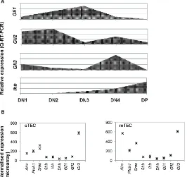
Reverse transcription (RT)-PCR has shown that developing foetal thymocytes express the Gli transcription factors, Ptch1, Smo, and Ihh, but not Shh; however, TEC express Shh [21–25]. Quantitative (Q)RT-PCR from sorted thymocyte subsets on E16.5 showed that the Gli genes are differentially expressed during T cell development, with highest expression of Gli2 in the DN1 and DN2 populations, whereas Gli1 expression is highest in the DN3 population, and Gli3 in the DN4 population [21,23–25]. In contrast, Ihh expression is undetectable in the earliest DN thymocyte subsets, and is most highly expressed in the DP population (Figures1and2). Flow cytometry, RT-PCR and QRT-PCR have demonstrated that Smo is highly expressed in DN thymocytes, and down-regulated following pre-TCR signalling, whereas Ptch1 expression is maintained in DP cells [22]. Expression levels of Smo reduce with maturity in the DP population, but DP cells can transduce Hh signals [26].
[image:3.595.167.433.399.650.2]Hh pathway components are also expressed by TEC in the embryonic thymus [21,22,27–29]. We carried out microarray analysis of gene expression in fluorescence activated cell sorting (Facs)-sorted cTEC and mTEC purified from E14.5 foetal thymus organ culture (FTOC) after 7 days in culture to allow maturation of both cTEC and mTEC populations (GSE81433). This analysis showed expression of Smo, Ptch1 and Hh genes, in addition to high expression of Gli3 (Figure2B). Thus, in the foetal thymus, TECs express both the Hh proteins and the machinery to transduce the Hh signal.
3. Regulation of T Cell Development by Hh Signalling
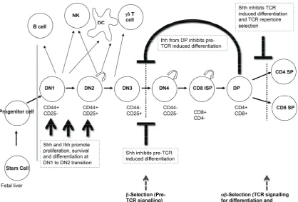
[image:4.595.90.509.195.478.2]Analysis of T cell development in the foetal thymus of multiple Hh pathway mutants has shown that Hh signalling is an important regulator of T cell development in the embryo, which affects several stages of thymocyte development (Figure3). We will review the experimental evidence showing that Hh signalling influences three key check-points in embryonicαβT cell development: the transition from DN1 to DN2 cell; pre-TCR induced differentiation to DP cell; and the transition from DP to mature SP cell.
Figure 3.Hh signalling regulates multiple stages of thymocyte development. The cartoon illustrates the different stages of thymocyte differentiation that are regulated by Hh signalling in the embryonic thymus. (TCR: T cell receptor.)
3.1. Hedgehog Signalling in DN1 and DN2 Cells
As developing thymocytes differentiate from the earliest DN1 progenitors to express cell surface CD25 and become DN2 cells, they specify to the T cell fate, and initiate recombination of theirTCRβ
gene locus [6]. At this stage, Shh, Gli2 and Gli3 are required for differentiation and expansion of the DN2 population. Thymi from E13.5Shh´/´embryos are much smaller than those of wild type (WT) littermates and have ~10 times fewer thymocytes and a reduction in the DN2 population [19]. Reduced differentiation to DN2 cell has also been observed in E13.5Gli2´/´andGli3´/´thymus [23,25]. In contrast, analysis of Gli1-deficient foetal thymus has shown that Gli1 is dispensable for the DN1 to DN2 transition [24]. In many tissues, Gli3 functions to repress Hh pathway activation, and Gli3-deficiency has the opposite phenotype to Shh-deficiency, so the fact that mutants of bothShhandGli3show reduced early thymocyte differentiation might seem surprising. However, the expression pattern of the Gli transcription factors in the DN populations is consistent with the findings from the Gli-mutants in early DN cell development.Gli2andGli3are both highly expressed in DN1 and DN2 cells, while expression ofGli1in early DN subsets is relatively low (Figure2).
The role of Ihh in early thymocyte development has also been studied [21]. Analysis of E13.5
transition. However, analysis of the double-mutantShh+/´Ihh´/´foetal thymus showed a reduction in the DN2 population, indicating that Shh and Ihh have redundant functions at the DN1 to DN2 transition [21].
3.2. The Transition from DN to DP Cell and Pre-TCR Signalling
Differentiation from DN3 cell through to DP cell requires successful rearrangement of theTCRβ
gene locus and formation of the pre-TCR complex [6]. Signalling through the pre-TCR complex results in a complex transcriptional programme, leading to expansion, survival, allelic exclusion of theTCRβ
gene locus, and differentiation to DP cell [30,31]. In foetal thymocyte development, differentiation from DN3 to DN4 cell seems to be somewhat uncoupled from pre-TCR signal transduction and TCRβ chain expression [32], and thymocytes that have failed to rearrange theirTCRβgene locus die at the DN4 stage [32,33].
Hedgehog signalling to DN thymocytes negatively regulates pre-TCR-induced differentiation to DP cell in both mouse and human [22,25,34]. The first evidence for this came from in vitro studies which demonstrated that Hh signalling negatively regulates DN to DP differentiation in foetal thymus organ cultures (FTOCs) that were treated with recombinant (r) Shh [22]. Thymocyte development was arrested at the DN3 stage, whereas FTOCs treated with a neutralizing monoclonal antibody (mAb) against Shh showed increased differentiation from DN to DP cell. Thymocytes from
Recombination-activating gene 1(Rag1)´/´FTOCs can be induced to differentiate from DN3 to DP cell by treatment with anti-CD3 mAb, which mimics the pre-TCR signal [31,35]. Addition of the anti-Shh mAb to anti-CD3 treatedRag1´/´FTOC enhanced thymocyte differentiation to the DP stage, whereas treatment with rShh arrested it, confirming the negative regulatory role of Shh at this developmental stage.
In the embryo, DP cells first appear on E16.5, allowing observation of the transition from DN to DP population in a synchronized manner. Analysis of the E16.5Shh´/´foetal thymus unexpectedly showed that the transition from DN to DP cell was reduced compared to WT, in contrast to the in vitro studies [19]. However, the E16.5Shh´/´thymus also had increased apoptosis in the DN4 population compared to WT, consistent with increased death of cells that had failed to rearrange the TCRβlocus. This suggested that the requirement for Shh before pre-TCR signalling might impact the production of DP cells on E16.5 [19].
In vivo evidence for the negative regulatory role of Hh signalling at this developmental stage came from analysis of foetal thymocyte development in embryos mutant forGli2andGli3[23,25]. From E13.5 to E15.5Gli2´/´thymi were smaller than that of WT littermates, whereas the E17.5Gli2´/´
thymus contained over twice as many thymocytes as WT. The expansion of theGli2´/´thymus on the embryonic day after DP cells first appear, together with the expression pattern of Gli2 in the foetal thymus which shows three-fold upregulation in DN4 compared to DN3 cells, suggested that Gli2 plays a negative role in thymocyte expansion and progression to DP after pre-TCR signal transduction during foetal development. This was confirmed in anti-CD3-treatedRag1´/´Gli2´/´FTOCs, which expanded and differentiated to DP faster than their anti-CD3 treatedRag1´/´Gli2+/+ counterparts. Interestingly, anti-CD3-treatedRag1´/´Shh´/´FTOC also differentiated faster than anti-CD3-treated
Rag1´/´Shh+/+ FTOC, confirming that Gli2 is downstream of Shh as a negative regulator of differentiation after pre-TCR signalling [25]. In contrast, analysis of anti-CD3-treated Rag1´/´ Gli3´/´FTOCs revealed a partial arrest at the DN-to-DP stage after pre-TCR signalling [23]. The same result was observed in E16.5Gli3´/´thymus, indicating that Gli3 functions as a transcriptional repressor of the Hh pathway at this developmental stage, and that Gli3 activity is required for normal differentiation from DN to DP cell.
another stromal cell population. Therefore, the impact of Hh-mediated transcription in developing thymocytes on their differentiation and function was investigated by the production of transgenic (tg) mice which over-expressed activator (Gli2A-tg (Gli2ΔN2-tg)), or repressor (Gli2R-tg (Gli2ΔC2-tg)) forms of Gli2 in T lineage cells only (driven by thelckpromoter) [36,37]. Anti-CD3-treatedRag1´/´
Gli2A-tg FTOC differentiated more slowly to DP than their non-transgenic counterparts, whereas anti-CD3-treatedRag1´/´Gli2R-tg FTOC—in which physiological Hh-mediated transcription is
repressed—differentiated more quickly [25]. Thus, Hh pathway activation in developing thymocytes negatively regulates pre-TCR-mediated differentiation to DP cell.
3.3. Ihh at the Transition from DN to DP Cell and Its Role in Homeostasis and the Control of DP Cell Number
Ihh is produced by both the foetal thymus stroma and developing thymocytes with a six-fold higher expression in the DP population compared to DN cells and stroma, which show similarIhh
expression levels [21] (Figure2). The DP population expresses low levels of the target geneGli1, suggesting that Ihh is not signalling to the DP population in an autocrine manner, but feeding back to signal to the DN3 population, which express the highest levels ofGli1.
In the E16.5Ihh´/´embryo, both thymocyte numbers and the proportion of DP cells were reduced, indicating that Ihh promotes T cell development. Interestingly, however, the E16.5Ihh+/´
thymus contained 2.4 times more DP cells than WT, implying that Ihh also negatively regulates thymocyte development.Ihh+/+Rag1´/´FTOCs contained more cells than theirIhh+/´Rag1´/´
counterparts, but after anti-CD3 treatment for 5 days, theIhh+/´Rag1´/´FTOCs differentiated faster than theIhh+/+Rag1´/´FTOC, confirming that Ihh promotes early DN thymocyte development before the pre-TCR signal, but negatively regulates the pre-TCR-induced transition to the DP stage.
In theIhh+/´E16.5 thymus, the sorted CD25+DN population contained significantly more cells in cell cycle (S/G2+M) than its WT counterpart. Taken together, these data indicated that Ihh produced by the DP population feeds back to negatively regulate the differentiation and expansion of the DN3 population after pre-TCR signal transduction. Thus, as the total amount of Ihh protein present in the thymus largely depends on the size of the DP population, this negative feedback loop may be thought of as a counting system to regulate thymocyte number and maintain thymocyte homeostasis [21].
3.4. Hh Signalling in TCR Repertoire Selection and the Transition from DP to SP Cell
Analysis of embryonic thymus fromShh´/´,Gli1´/´, Gli2´/´, Gli2A-transgenic (tg) and
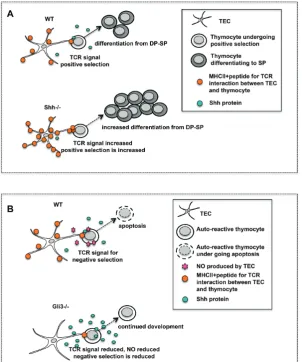
[image:7.595.148.451.140.503.2]Gli2R-tg, and in vitro FTOC experiments have all shown that Shh negatively regulates the DP to SP transition, most likely by lowering TCR signal strength (see Figure4) [24,26,36].
Figure 4. Hh signalling at the transition from DP to SP thymocyte. (A) The cartoon illustrates the influence of Shh on positive selection. Positive selection is increased in theShh´/´thymus and the proportion of CD4SP cells is increased. Cell surface expression of major histocompatibility complex class II (MHCII) is increased onShh´/´TEC compared to wild type (WT) TEC. (B) The influence of Gli3 on negative selection. In theGli3´/´thymus MHCII expression is decreased in TEC and nitric oxide (NO) activity is decreased. This may allow SP thymocytes to escape from negative selection.
TheShh´/´embryonic thymus is smaller than WT but has a higher SP:DP ratio, reflecting an accelerated rate of differentiation from DP to SP [36]. Shh´/´FTOCs had a greater proportion of mature CD4SP cells and an increased CD4SP:CD8SP ratio than WT (Figure4A). Likewise,Gli2´/´and
Gli1´/´FTOCs showed increased differentiation from DP to SP cells and increased CD4SP:CD8SP ratio [24]. The increased differentiation towards the CD4SP lineage might reflect an increase in TCR signal strength, and both positive and negative selection of transgenic TCR were increased inShh´/´, Gli2´/´, andGli1´/´FTOC compared to control [24,36]. Analysis of the Gli2A-tg and Gli2R-tg embryonic thymus confirmed that physiological Hh-mediated transcription in thymocytes negatively regulates the DP-to-SP transition and TCR repertoire selection [26].
higher CD5 expression on the CD4SP population, and a higher CD4SP:CD8SP ratio than WT [26]. Doubling the Gli2R-transgene copy number further increased the proportion of CD4SP cells, their CD5 cell surface expression, and the CD4SP:CD8SP ratio [26]. Interestingly, these dose-dependent phenotypic changes were reversed with rShh treatment of WT FTOC. Decreased cell surface CD5 expression and a lower CD4SP:CD8SP ratio were observed when WT FTOC were treated with rShh [36]. Therefore, an increased level of Shh signalling appeared to give rise to weaker TCR signal strength, leading to reduced selection from DP to CD4SP; whereas selection to the CD8 lineage, which is believed to require lower strength and duration of TCR signal transduction, was less affected.
Gata3 is a key regulator of CD4 lineage commitment, and strong TCR signals increase expression of Gata3, which drives differentiation towards the CD4 lineage [41]. Interestingly, treatment of WT FTOCs with rShh decreased the proportion of Gata3-expressing DP and SP cells, while attenuating Shh signalling by treatment with the neutralising anti-Shh mAb increased the intensity and expression of Gata3 on the DP and SP4 populations [26]. Thus, the influence of modulation of Shh signalling on Gata3 expression is consistent with the effect of Shh on CD4/CD8 lineage commitment, and with its influence on TCR signal strength [36,42].
TCR signal strength is a crucial determinant of the TCR repertoire and CD4/8 lineage commitment and the influence of Shh on TCR signal strength may thus alter both of these: Since Shh-expressing TECs are scattered around the medulla and the cortico-medullary junction, their influence on TCR signal strength and the outcome of TCR ligation (i.e., positive or negative selection and CD4/8 lineage decision) for each cell is dependent on the location of the cell relative to the source of Shh. It is therefore possible that Shh-expressing TEC may have specialised functions in the thymus, such as induction of positive selection or commitment to the CD8 lineage.
3.5. Gli3 Regulates Nos2 Expression during Negative Selection
Analysis of theGli3mutant thymus showed that Gli3 acts as a repressor of Hh signalling in the foetal thymus and that Gli3 mutation may also influence TCR repertoire selection in developing thymocytes [23,27]. These experiments were carried out using constitutive Gli3 knock-outs, so that the lineage-specific requirements for Gli3 are not known, and both theGli3´/´foetal thymus stroma and foetal thymocytes had increased expression of the transcriptional target of Hh-signalling,
Gli1[24,27]. Treatment ofGli3´/´FTOC with a neutralising anti-Shh mAb reducedGli1expression in the stroma [27], demonstrating that the increase in Gli1 expression was a result of increased Hh signalling.
PCR array analysis of gene expression in theGli3´/´foetal thymus stroma compared to WT identified Gli3-target genes in the stroma, and revealed thatNos2is a Gli3 target gene, which was significantly down regulated in theGli3´/´andGli3+/´stroma [27].Nos2expression recovered to WT levels whenGli3+/´FTOC was treated with an anti-Hh neutralising mAb, but was not affected whenGli3´/´FTOC were treated with an anti-Hh mAb, suggesting thatNos2required Gli3 for its physiological levels of expression (most likely through Gli3R’s repression of an intermediate transcriptional repressor ofNos2) [27].
Nos2 is induced in the thymic stroma during negative selection. It causes the production of nitric oxide (NO), a potent pro-apoptotic agent, which promotes apoptosis of autoreactive thymocytes [43]. Treatment of WT andGli3´/´FTOC with anti-CD3 mAb to mimic negative selection, showed that the
4. Hedgehog Signalling in Foetal Thymic Epithelial Cell (TEC) Development and Function
The role of the Hh pathway in embryonic TEC development has been less extensively investigated than its function in T cell development, although Shh is involved early in thymus organogenesis during patterning of the pharyngeal pouches [44]. In this section, we will briefly discuss the early role of Shh in pharyngeal patterning and thymus organogenesis before reviewing recent experiments demonstrating that Hh signalling is required for normal TEC development in the embryonic thymus during the last week of gestation.
Shh is involved in the formation and patterning of the entire pharyngeal apparatus and affects the third pouch and the thymic anlage [44]. InShh´/´embryos, the development of the third pharyngeal pouch is defective and the thymic rudiment fails to bud off entirely from the pharyngeal endoderm [45]. However, once theShh´/´thymus is formed, although it is small, it is capable of supporting T cell development; however, there are defects in normal TEC development [19,21].
There are two types of TEC, which are derived from a common progenitor, and which have distinct functions, localisations within the thymus, and cell surface markers [2,4,5,46]. Cortical(c) TECs provide DL4 for T cell fate specification, and present MHC+peptide ligands for positive selection. These cTECs are defined as EpCam1+, CD205+, Ly51+ and MHCII+, and express genes for antigen presentation, includingCathepsin-L,Prss16,andβ5t. Medullary(m) TECs are specialised for induction of negative selection, are defined as cell surface EpCam1+, CD40+, CD205´, Ly51´and MHCII+, and bind the lectin UEA-1. They express theAiregene andCathepsin-S, facilitating the expression and presentation of Tissue Restricted Antigens (TRA) for induction of tolerance. During embryonic development, the cell surface markers CD40 and CD205 can be used to map TEC development. CD40lowCD205lowprogenitor cells gain cell surface CD40 and CD205 expression, resulting in transitional progenitors which have the potential to differentiate into the two mature populations: cTEC (CD40intCD205highLy51+) and mTEC (CD40highCD205lowUEA-1+) [9,10].
Embryonic TECs express components of the Hh signalling pathway as well as the genes encoding the Hh ligands (Figure2). Therefore, to investigate if TECs are transducing Hh signals in vivo in the embryonic thymus, Gli Binding Site (GBS)-Green Fluorescent Protein (GFP) transgenic embryos—which report Hh-mediated transcription by expression of GFP—were analysed [27]. Hh pathway activation was observed in the TEC populations of embryos from E14.5 through to birth (neonate). The greatest intensity of GFP-fluorescence was observed on E14.5, and fluorescence was seen in the CD40lowCD205lowprogenitor cells, which made up about one third of TEC, and in the emerging CD40intCD205highand CD40highCD205lowpopulations. On E18.5, higher expression was observed in cells that had specified to the mTEC lineage. At birth, the CD40lowCD205lowprogenitor population made up less than 1% of all TEC, but both the mature cTEC and mTEC populations contained about 20% of cells that expressed GFP [29].
Analysis of theShh´/´foetal thymus has recently shown that Shh is required for normal TEC development and the regulation of mTEC/cTEC lineage choice [29]. In the Shh´/´foetal thymus, there was a reduction in thymus size, overall cell number, and number of thymocytes and of TECs (CD45´EpCam1+) compared to WT littermates [19,21,29]. The numbers of both mature cTECs (CD45´EpCam1+Ly51+UEA1´) and mTECs (CD45´EpCam1+Ly51´UEA1+) were reduced in
available to developing thymocytes, and so it is possible that this may affect the outcome of TCR repertoire selection to the CD4 T cell lineage by changing MHCII-restricted TCR antigen dwell time or avidity [47] (see Figures4and5).
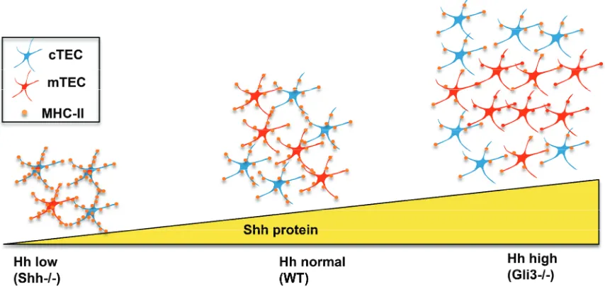
Figure 5.Impact of Shh-deficiency and Gli3-deficiency on TEC differentiation and function. In the Shh´/´embryonic thymus, Hh protein levels are low, there are fewer TEC, and the ratio of mTEC (shown in red) to cTEC (shown in blue) is decreased, but expression of MHCII is increased on individual TEC, compared to WT. In theGli3´/´embryonic thymus—which has increased levels of Hh pathway activation—the opposite phenotype is seen. TEC numbers are increased, but MHCII expression on individual TEC is reduced compared to WT.
The influence of Shh on foetal TEC development was confirmed when WT FTOC were treated with recombinant Hhip, a Hh-binding protein that neutralizes endogenous Hh proteins in the cultures. Hhip-treatment led to an overall decrease in TEC cell numbers and a significant decrease in the proportion of mature mTECs. The proportion of mTECs that expressed Aire protein was also reduced, but there was an increase in cell surface MHC-II on the mTEC population [27].
The opposite was observed inGli3´/´foetal thymus (Figure 5). There was an increase in overall TEC numbers but a decrease in cell surface expression of MHC-II in both cTEC and mTEC populations (Figures4B and5) [29]. This is consistent with Gli3 acting as a suppressor ofShhin the foetal thymus [27].
5. Conclusions and Future Directions
Hedgehog signalling influences multiple stages of T cell development, and also regulates foetal TEC differentiation (Figures3and5). Very few pathways or secreted signalling molecules have been identified that regulate foetal TEC development or cTEC/mTEC lineage choice, so it will also be important to identify the mechanisms of Shh’s regulation of TEC differentiation in the embryonic thymus. It will be interesting to identify Hh target genes in different subsets of TEC, and test the impact of mutation of these genes on TEC development and function, and on the ability of TEC to support T cell development.
In the future, it will also be important to investigate the interactions between Hh signalling and other pathways that interact with Hh signalling in other tissues, and also regulate foetal thymocyte development—such as BMP2/4 signalling [48–50]. BMP2/4 signalling is also involved in early thymus organogenesis and is required for early TEC development, so it will also be interesting to assess interactions between BMP and Shh signalling in TEC development [51,52].
Acknowledgments: This work was funded by grants from the MRC (G0900161/1), BBSRC (BB/I026324/1), Wellcome Trust (090233/Z/09/Z) and supported by the National Institute for Health Research Biomedical Research Centre at Great Ormond Street Hospital for Children NHS Foundation Trust and University College London. AB was funded by a fellowship from the Istituto Pasteur/Cenci Bolognetti Foundation. AS and TC were funded by Great Ormond Street Hospital Children’s Charity.
Author Contributions:A.B., C.-I.L., K.M., S.R. and A.S. contributed equally to the preparation of this review. J.I.S. carried out the microarray analysis, which was analysed by A.S.. T.C. Contributed to preparation of this review.
Conflicts of Interest:The authors declare no conflict of interest.
References
1. Gordon, J.; Manley, N.R. Mechanisms of thymus organogenesis and morphogenesis.Development2011,138, 3865–3878. [CrossRef] [PubMed]
2. Anderson, G.; Takahama, Y. Thymic epithelial cells: Working class heroes for T cell development and repertoire selection.Trends Immunol.2012,33, 256–263. [CrossRef] [PubMed]
3. Klein, L.; Kyewski, B.; Allen, P.M.; Hogquist, K.A. Positive and negative selection of the T cell repertoire: What thymocytes see (and don’t see).Nat. Rev. Immunol.2014,14, 377–391. [CrossRef] [PubMed]
4. Anderson, G.; Jenkinson, W.E. Border control: Anatomical origins of the thymus medulla.Eur. J. Immunol. 2015,45, 2203–2207. [CrossRef] [PubMed]
5. Alves, N.L.; Takahama, Y.; Ohigashi, I.; Ribeiro, A.R.; Baik, S.; Anderson, G.; Jenkinson, W.E. Serial progression of cortical and medullary thymic epithelial microenvironments. Eur. J. Immunol. 2014, 44, 16–22. [CrossRef] [PubMed]
6. Shah, D.K.; Zuniga-Pflucker, J.C. An Overview of the Intrathymic Intricacies of T Cell Development. J. Immunol.2014,192, 4017–4023. [CrossRef] [PubMed]
7. Bleul, C.C.; Corbeaux, T.; Reuter, A.; Fisch, P.; Monting, J.S.; Boehm, T. Formation of a functional thymus initiated by a postnatal epithelial progenitor cell.Nature2006,441, 992–996. [CrossRef] [PubMed]
8. Bennett, A.R.; Farley, A.; Blair, N.F.; Gordon, J.; Sharp, L.; Blackburn, C.C. Identification and characterization of thymic epithelial progenitor cells.Immunity2002,16, 803–814. [CrossRef]
9. Baik, S.; Jenkinson, E.J.; Lane, P.J.L.; Anderson, G.; Jenkinson, W.E. Generation of both cortical and Aire(+) medullary thymic epithelial compartments from CD205(+) progenitors.Eur. J. Immunol.2013,43, 589–594. [CrossRef] [PubMed]
10. Shakib, S.; Desanti, G.E.; Jenkinson, W.E.; Parnell, S.M.; Jenkinson, E.J.; Anderson, G. Checkpoints in the development of thymic cortical epithelial cells.J. Immunol.2009,182, 4488. [CrossRef]
11. Ingham, P.W.; Nakano, Y.; Seger, C. Mechanisms and functions of Hedgehog signalling across the metazoa. Nat. Rev. Genet.2011,12, 393–406. [CrossRef] [PubMed]
12. Park, H.L.; Bai, C.; Platt, K.A.; Matise, M.P.; Beeghly, A.; Hui, C.C.; Nakashima, M.; Joyner, A.L. Mouse Gli1 mutants are viable but have defects in SHH signaling in combination with a Gli2 mutation.Development 2000,127, 1593–1605. [PubMed]
13. Sasaki, H.; Nishizaki, Y.; Hui, C.; Nakafuku, M.; Kondoh, H. Regulation of Gli2 and Gli3 activities by an amino-terminal repression domain: Implication of Gli2 and Gli3 as primary mediators of Shh signaling. Development1999,126, 3915–3924. [PubMed]
14. Bai, C.B.; Auerbach, W.; Lee, J.S.; Stephen, D.; Joyner, A.L. Gli2, but not Gli1, is required for initial Shh signaling and ectopic activation of the Shh pathway.Development2002,129, 4753–4761. [PubMed]
16. Chiang, C.; Litingtung, Y.; Lee, E.; Young, K.E.; Corden, J.L.; Westphal, H.; Beachy, P.A. Cyclopia and defective axial patterning in mice lacking Sonic hedgehog gene function. Nature 1996, 383, 407–413. [CrossRef] [PubMed]
17. St-Jacques, B.; Hammerschmidt, M.; McMahon, A.P. Indian hedgehog signaling regulates proliferation and differentiation of chondrocytes and is essential for bone formation. Genes Dev. 1999,13, 2072–2086. [CrossRef] [PubMed]
18. Razzaque, M.S.; Soegiarto, D.W.; Chang, D.; Long, F.; Lanske, B. Conditional deletion of Indian hedgehog from collagen type 2alpha1-expressing cells results in abnormal endochondral bone formation. J. Pathol. 2005,207, 453–461. [CrossRef] [PubMed]
19. Shah, D.K.; Hager-Theodorides, A.L.; Outram, S.V.; Ross, S.E.; Varas, A.; Crompton, T. Reduced thymocyte development in sonic hedgehog knockout embryos.J. Immunol.2004,172, 2296–2306. [CrossRef] [PubMed] 20. Ohba, S. Hedgehog signaling in Endochondral Ossification.J. Dev. Biol.2016,4. [CrossRef]
21. Outram, S.V.; Hager-Theodorides, A.L.; Shah, D.K.; Rowbotham, N.J.; Drakopoulou, E.; Ross, S.E.; Lanske, B.; Dessens, J.T.; Crompton, T. Indian hedgehog (Ihh) both promotes and restricts thymocyte differentiation. Blood2009,113, 2217–2228. [CrossRef] [PubMed]
22. Outram, S.V.; Varas, A.; Pepicelli, C.V.; Crompton, T. Hedgehog signaling regulates differentiation from double-negative to double-positive thymocyte.Immunity2000,13, 187–197. [CrossRef]
23. Hager-Theodorides, A.L.; Dessens, J.T.; Outram, S.V.; Crompton, T. The transcription factor Gli3 regulates differentiation of foetal CD4(´)CD8(´) double-negative thymocytes.Blood2005,106, 1296–1304. [CrossRef] [PubMed]
24. Drakopoulou, E.; Outram, S.V.; Rowbotham, N.J.; Ross, S.E.; Furmanski, A.L.; Saldana, J.I.; Hager-Theodorides, A.L.; Crompton, T. Non-redundant role for the transcription factor Gli1 at multiple stages of thymocyte development.Cell Cycle2010,9, 4144–4152. [CrossRef] [PubMed]
25. Rowbotham, N.J.; Hager-Theodorides, A.L.; Furmanski, A.L.; Ross, S.E.; Outram, S.V.; Dessens, J.T.; Crompton, T. Sonic hedgehog negatively regulates pre-TCR-induced differentiation by a Gli2-dependent mechanism.Blood2009,113, 5144–5156. [CrossRef] [PubMed]
26. Furmanski, A.L.; Saldana, J.I.; Rowbotham, N.J.; Ross, S.E.; Crompton, T. Role of Hedgehog signalling at the transition from double-positive to single-positive thymocyte.Eur. J. Immunol.2012,42, 489–499. [CrossRef] [PubMed]
27. Hager-Theodorides, A.L.; Furmanski, A.L.; Ross, S.E.; Outram, S.V.; Rowbotham, N.J.; Crompton, T. The Gli3 transcription factor expressed in the thymus stroma controls thymocyte negative selection via Hedgehog-dependent and -independent mechanisms.J. Immunol.2009,183, 3023–3032. [CrossRef] [PubMed] 28. Sacedon, R.; Varas, A.; Hernandez-Lopez, C.; Gutierrez-deFrias, C.; Crompton, T.; Zapata, A.G.; Vicente, A. Expression of hedgehog proteins in the human thymus. J. Histochem. Cytochem. 2003, 51, 1557–1566. [CrossRef] [PubMed]
29. Saldana, J.I.; Solanki, A.; Lau, C.I.; Sahni, H.; Ross, S.; Furmanski, A.L.; Ono, M.; Hollander, G.; Crompton, T. Sonic Hedgehog regulates thymic epithelial cell differentiation.J. Autoimmun.2016,68, 86–97. [CrossRef] [PubMed]
30. Koch, U.; Radtke, F. Mechanisms of T cell development and transformation.Annu. Rev. Cell Dev. Biol.2011, 27, 539–562. [CrossRef] [PubMed]
31. Sahni, H.; Ross, S.; Barbarulo, A.; Solanki, A.; Lau, C.I.; Furmanski, A.; Saldana, J.I.; Ono, M.; Hubank, M.; Barenco, M.; et al. A genome wide transcriptional model of the complex response to pre-TCR signalling during thymocyte differentiation.Oncotarget2015,6, 28646–28660. [PubMed]
32. Hager-Theodorides, A.L.; Rowbotham, N.J.; Outram, S.V.; Dessens, J.T.; Crompton, T. Beta-selection: abundance of TCRbeta´/gammadelta´CD44´CD25´(DN4) cells in the foetal thymus.Eur. J. Immunol. 2007,37, 487–500. [CrossRef] [PubMed]
33. Falk, I.; Nerz, G.; Haidl, I.; Krotkova, A.; Eichmann, K. Immature thymocytes that fail to express TCRbeta and/or TCRgamma delta proteins die by apoptotic cell death in the CD44(´)CD25(´) (DN4) subset. Eur. J. Immunol.2001,31, 3308–3317. [CrossRef]
35. Levelt, C.N.; Mombaerts, P.; Iglesias, A.; Tonegawa, S.; Eichmann, K. Restoration of early thymocyte differentiation in T-cell receptor beta-chain-deficient mutant mice by transmembrane signaling through CD3 epsilon.Proc. Natl. Acad. Sci. USA1993,90, 11401–11405. [CrossRef] [PubMed]
36. Rowbotham, N.J.; Hager-Theodorides, A.L.; Cebecauer, M.; Shah, D.K.; Drakopoulou, E.; Dyson, J.; Outram, S.V.; Crompton, T. Activation of the Hedgehog signaling pathway in T-lineage cells inhibits TCR repertoire selection in the thymus and peripheral T-cell activation.Blood2007,109, 3757–3766. [CrossRef] [PubMed]
37. Rowbotham, N.J.; Furmanski, A.L.; Hager-Theodorides, A.L.; Ross, S.E.; Drakopoulou, E.; Koufaris, C.; Outram, S.V.; Crompton, T. Repression of hedgehog signal transduction in T-lineage cells increases TCR-induced activation and proliferation.Cell Cycle2008,7, 904–908. [CrossRef] [PubMed]
38. Carpenter, A.C.; Bosselut, R. Decision checkpoints in the thymus.Nat. Immunol.2010,11, 666–673. [CrossRef] [PubMed]
39. Azzam, H.S.; DeJarnette, J.B.; Huang, K.; Emmons, R.; Park, C.S.; Sommers, C.L.; El-Khoury, D.; Shores, E.W.; Love, P.E. Fine tuning of TCR signaling by CD5.J. Immunol.2001,166, 5464–5472. [CrossRef] [PubMed] 40. Azzam, H.S.; Grinberg, A.; Lui, K.; Shen, H.; Shores, E.W.; Love, P.E. CD5 expression is developmentally
regulated by T cell receptor (TCR) signals and TCR avidity.J. Exp. Med.1998,188, 2301–2311. [CrossRef] [PubMed]
41. Hernandez-Hoyos, G.; Anderson, M.K.; Wang, C.; Rothenberg, E.V.; Alberola-Ila, J. GATA-3 expression is controlled by TCR signals and regulates CD4/CD8 differentiation.Immunity2003,19, 83–94. [CrossRef] 42. Furmanski, A.L.; Barbarulo, A.; Solanki, A.; Lau, C.I.; Sahni, H.; Saldana, J.I.; D’Acquisto, F.; Crompton, T.
The transcriptional activator Gli2 modulates T-cell receptor signalling through attenuation of AP-1 and NFkappaB activity.J. Cell Sci.2015,128, 2085–2095. [CrossRef] [PubMed]
43. Tai, X.G.; Toyo-oka, K.; Yamamoto, N.; Yashiro, Y.; Mu, J.; Hamaoka, T.; Fujiwara, H. Expression of an inducible type of nitric oxide (NO) synthase in the thymus and involvement of NO in deletion of TCR-stimulated double-positive thymocytes.J. Immunol.1997,158, 4696–4703. [PubMed]
44. Moore-Scott, B.A.; Manley, N.R. Differential expression of Sonic hedgehog along the anterior-posterior axis regulates patterning of pharyngeal pouch endoderm and pharyngeal endoderm-derived organs.Dev. Biol. 2005,278, 323–335. [CrossRef] [PubMed]
45. Westerlund, J.; Andersson, L.; Carlson, T.; Fagman, H.; Nilsson, M. Misguided migration of C cell precursors ro extra-thyroidal locations related to defective pharyngeal pouch development in Shh deficient mice. Cell Dev. Biol.2013,2, 129. [CrossRef]
46. Hollander, G.; Gill, J.; Zuklys, S.; Iwanami, N.; Liu, C.; Takahama, Y. Cellular and molecular events during early thymus development.Immunol. Rev.2006,209, 28–46. [CrossRef] [PubMed]
47. Stepanek, O.; Prabhakar, A.S.; Osswald, C.; King, C.G.; Bulek, A.; Naeher, D.; Beaufils-Hugot, M.; Abanto, M.L.; Galati, V.; Hausmann, B.; et al. Coreceptor scanning by the T cell receptor provides a mechanism for T cell tolerance.Cell2014,159, 333–345. [CrossRef] [PubMed]
48. Hager-Theodorides, A.L.; Outram, S.V.; Shah, D.K.; Sacedon, R.; Shrimpton, R.E.; Vicente, A.; Varas, A.; Crompton, T. Bone morphogenetic protein 2/4 signaling regulates early thymocyte differentiation.J. Immunol. 2002,169, 5496–5504. [CrossRef] [PubMed]
49. Hager-Theodorides, A.L.; Ross, S.E.; Sahni, H.; Mishina, Y.; Furmanski, A.L.; Crompton, T. Direct BMP2/4 signaling through BMP receptor IA regulates foetal thymocyte progenitor homeostasis and differentiation to CD4+CD8+ double-positive cell.Cell Cycle2014,13, 324–333. [CrossRef] [PubMed]
50. Tsai, P.T.; Lee, R.A.; Wu, H. BMP4 acts upstream of FGF in modulating thymic stroma and regulating thymopoiesis.Blood2003,102, 3947–3953. [CrossRef] [PubMed]
51. Bleul, C.C.; Boehm, T. BMP signaling is required for normal thymus development.J. Immunol.2005,175, 5213–5221. [CrossRef] [PubMed]
52. Gordon, J.; Patel, S.R.; Mishina, Y.; Manley, N.R. Evidence for an early role for BMP4 signaling in thymus and parathyroid morphogenesis.Dev. Biol.2010,339, 141–154. [CrossRef] [PubMed]