INTRODUCTION
In Arabidopsis, the indeterminate shoot apical meristem (SAM) produces organs such as leaves and flowers throughout the life of the plant. By contrast, the determinate floral meristem (FM), from which flowers are derived, produces a stereotypical number of floral organs: four sepals, four petals, six stamens and two carpels. Underlying the different behaviors of these two meristematic tissues are the different properties of their respective stem cell populations. In the SAM, as well as in the FM, the expression of the homeodomain gene WUSCHEL(WUS) in a small group of cells at the center of the structures, the so-called stem cell organizing center, is essential for maintaining the pool of stem cells. In the FM, once the correct numbers of floral organs have formed, WUSis quickly downregulated and the stem cells lost (Laux et al., 1996; Mayer et al., 1998).
The floral identity regulator LEAFY (LFY) (Schultz and Haughn, 1991; Weigel et al., 1992) activates the expression of the homeotic gene AGAMOUS(AG) in the center of young flower buds, and the AG gene product then acts to downregulate WUS, leading to a loss of stem cell activity (Busch et al., 1999; Lenhard et al., 2001; Lohmann et al., 2001; Parcy et al., 1998). However, loss of LFY function only leads to a delay in the onset of AGexpression, and not to its absence (Weigel and Meyerowitz, 1993), suggesting that other factors also play a role in the early activation of AG. One of these factors was recently shown to be WUS itself, which directly binds AGregulatory sequences in combination with LFY (Lohmann et al., 2001). Flowers mutant for AG display stem cell maintenance phenotypes, resulting in the formation of flowers within flowers, and also show homeotic transformations of stamens to petals (Bowman et al., 1989). It has been suggested that these are functionally distinct activities of AG (Mizukami and Ma, 1995; Sieburth et al., 1995), yet not much is
known about how this is regulated: whether AG is regulated at the RNA level, for example, via the regulation of AGexpression in specific floral domains, or at the protein level, through interactions between AG and other spatially restricted molecules. Furthermore, it is unclear how AG shuts down WUSexpression and thus the floral stem cell population (Laux et al., 1996; Mayer et al., 1998).
In this study, we report on the identification of a novel input into the process of floral stem cell arrest and suggest that this activity is spatially restricted to the centermost region of the AGexpression domain.
MATERIALS AND METHODS Mutagenesis
pan-3seeds (10,000-15,000; in the L-eraccession) were treated with a 0.3% (v/v) aqueous solution of ethyl methanesulfonate (Sigma) in a volume of 15 ml for 10 hours, then washed with water for 8 hours (with hourly changes) before being resuspended in a 0.15% (v/v) agar solution and sowed on soil 1 cm apart. Seeds from M1 plants were harvested individually and 20-30 M2 plants per M1 line (~1000) were screened for altered floral phenotypes, which were reconfirmed in the M3 generation. Ten putative modifiers were retained after re-screening. To identify the mutation in the novel lfyallele, the genomic coding region was amplified in two fragments of 1.3 kb and 1.4 kb by PCR (using Ex Taq, Takara) and sequenced. The mutation was found to be a nonsense mutation (Q162Stop), similar to all published null alleles. Plasmid constructs and sequences
Details of primers available upon request. All PCR amplifications were carried out using the Phusion high fidelity polymerase (Finnzymes). All constructs were sequenced. To construct the PANrepressor domain chimeric fusion, we first annealed complementary oligonucleotides carrying the enhanced SUPERMAN repressor domain motif flanked by BamHI and BglII sites, and ligated this to the T EZ cloning vector (Promega), to yield pGEM-SRDX. The PANcDNA was PCR-amplified, digested with KpnI and BglII and cloned into the KpnI and BamHI sites of pGEM-SRDX to yield pPD64.1. The
PAN-RDfragment was extracted with BamHI and BglII, and cloned into pBJ36 (Gleave, 1992) carrying either the p35S, pPANor pAP1(1.7-kb) promoters to yield pPD66.1, pPD199.2 or pPD143.1 respectively. The PAN
promoter was PCR-amplified from the L-eraccession and cloned into pBJ36 using the SalI and KpnI sites. pAP1(1.7-kb) was a kind gift of Dr Marty Yanofsky (University of California, San Diego, CA, USA). The
promoter-PAN-RDfragments were then extracted from pBJ36 using NotI and ligated to the plant transformation vector pML BART (Eshed et al., 2001) yielding pPD74.1, pPD218.1 or pPD171.20, respectively.
Floral stem cell termination involves the direct regulation of
AGAMOUS
by PERIANTHIA
Pradeep Das1,2,*, Toshiro Ito1,3, Frank Wellmer1,4, Teva Vernoux2, Annick Dedieu2, Jan Traas2 and Elliot M. Meyerowitz1,*
In Arabidopsis, the population of stem cells present in young flower buds is lost after the production of a fixed number of floral organs. The precisely timed repression of the stem cell identity gene WUSCHEL(WUS) by the floral homeotic protein AGAMOUS (AG) is a key part of this process. In this study, we report on the identification of a novel input into the process of floral stem cell regulation. We use genetics and chromatin immunoprecipitation assays to demonstrate that the bZIP transcription factor PERIANTHIA (PAN) plays a role in regulating stem cell fate by directly controlling AGexpression and suggest that this activity is spatially restricted to the centermost region of the AGexpression domain. These results suggest that the termination of floral stem cell fate is a multiply redundant process involving loci with unrelated floral patterning functions.
KEY WORDS: AGAMOUS, Flower development, Stem cells, Arabidopsis
Development 136, 1605-1611 (2009) doi:10.1242/dev.035436
1Division of Biology 156-29, California Institute of Technology, Pasadena, California
91125, USA. 2Laboratoire RDP, Ecole Normale Supérieure de Lyon, 46 allée d’Italie,
69007 Lyon, France. 3Temasek Life Sciences Laboratory, National University of
Singapore, Singapore 117604, Singapore. 4Smurfit Institute of Genetics, Trinity
College Dublin, College Green, Dublin 2, Ireland.
*Authors for correspondence (e-mails: [email protected]; [email protected])
Accepted 16 March 2009
D
E
V
E
LO
P
M
E
N
For the ethanol-inducible version of PAN-RDunder the control of the PAN
promoter, we used the MultiSite Gateway Three-Fragment Vector Construction Kit (Invitrogen) to generate a single plasmid harboring the two components. We PCR-amplified a fragment of the LFY::alcR--alcA::ER
-GFPpGreen binary vector (gift of Patrick Laufs; INRA, Versailles, France); the alcRgene harboring a 3⬘nosterminator, followed by a 35Sterminator in an inverted orientation. This fragment was recombined with the pDONR 221 vector to generate pENTR-alcR-2xT. We also modified a destination vector (pGreen 0229; gift of Philip Benfey; Duke University, Durham, NC, USA) by inserting the chimeric promoter alcAimmediately after, and oriented towards, the attR3recombination sequence. To do so, the alcA
promoter was PCR-amplified from the LFY::alcR--alcA::ER-GFPpGreen vector, digested with SpeI and HinDIII and ligated to the pGreen 0229 binary vector, to yield the dpGreenBar-alcAbinary vector. The PANpromoter was PCR-amplified from Col-0and recombined with pDONR P4-P1R to yield pPD277. The PAN-RD fragment was amplified from pPD269 and recombined with pDONR P2R-P3 to yield pPD317. Finally, the three pENTR vectors (pPD277, pENTR-alcR-2xT and pPD317) were recombined into dpGreenBar-alcA, to yield the PAN::alcR--alcA::PAN-RDbinary vector. The putative bZIP binding sites in the secondAGintron were identified based on the presence of ‘ACGT’ core sequences (Jakoby et al., 2002). The six observed sites are (5⬘-3⬘): ACTTATACGTACATGT, AGTCCC
-ACGTGATTAC, TTGATCACGTCATCAC, TGTAATACGT ATTTGT,
TATGGAACGTTGTGAT and TCCATCACGTTTAAAT.
For the p35S::PAN-VP16construct, VP16was PCR-amplified, digested with BamHI and BglII, and ligated to pBJ36. The PAN-VP16fragment was then PCR-amplified and cloned into the pDONR 221 vector (Invitrogen). The triple gateway system was then used to generate the final p35S::PAN-VP16 plasmid in pdpGreen-BarT. The reporter construct used in the particle bombardment assays, pAGi-3⬘, is published as KB31 (Busch et al., 1999). Plant lines, transgenics and plant growth conditions
All plants were grown at either 16°C or 22°C with continuous white light, except the pan-3and L-erplants shown in Fig. S6, which were grown at long days (22°C) under a combination of white and gro-lux light. Photos of flowers were taken using either a Zeiss Stemi SV 11 stereomicroscope fitted with a Zeiss Axiocam or a Leica MZ12 stereomicroscope with a Leica DFC320 camera. Some images were adjusted for clarity by altering the brightness or contrast but any changes were applied evenly, across the entirety of the picture, without exception.
In situ hybridizations were performed according to published protocols. The WUSantisense RNA probe corresponds to the entire cDNA. Photos were taken on a Nikon Optiphot-2 equipped with a Zeiss Axiocam.
Transgenic plants were generated using standard floral dipping methods. Transformant lines were selected on soil for resistance to the herbicide Basta. To determine the copy number of the PAN-RDtransgene, T2 seeds were plated on petri dishes containing 10 μg/ml ammonium glufosinate (Basta) and the ratio of resistant to sensitive seedlings determined (~75% resistant seedlings indicates the parent had a single insertion). The sterile 2⫻PAN-RD
and1⫻PAN-RD pan-2 plants were used as pollen donors to fertilize emasculated wild type flowers, and the F1 seeds were tested as above.
pAP1(1.7-kb)-driven expression patterns were assayed in inflorescences of ethanol-induced pAP1(1.7-kb)::alcR--alcA::ER-GFPlines treated with the water soluble lipophilic dye FM4-64 and imaged on a Zeiss 510 confocal microscope.
Chromatin immunoprecipitation
Chromatin immunoprecipitation (ChIP) experiments were performed according to published protocols (Ito et al., 1997). Inflorescences from plants mutant for the redundant APETALA1and CAULIFLOWER genes, and expressing the dexamethasone-inducible 35S::APETALA-GR transgene (p35S::AP1-GR ap1-1 cal-1), were used. Plants were induced as described (Wellmer et al., 2006) and tissue were collected 5-7 days later for a synchronized population of flowers. Inflorescences were ground in liquid nitrogen, resuspended in buffer M1 (10 mM phosphate buffer 0.1 M NaCl, 10 mM β-mercaptoethanol, 1 M hexylene glycol), fixed with 1% formaldehyde for 10 minutes, washed in buffers M2 [buffer M1 containing 10 mM MgCl2, 0.5% (v/v) Triton X-100] and M3 (10 mM phosphate buffer, 0.1 M NaCl, 10 mM β-mercaptoethanol) and centrifuged to collect nuclei. Chromatin was
isolated by resuspending the pellet in lysis buffer [1% SDS (w/v), 10 mM EDTA, 50 mM Tris-HCl pH 8.1] containing protease inhibitors (1 μg/ml Leupeptin, 15 μg/ml Aprotinin), incubating on ice for 10 minutes before adding ChIP dilution buffer (Upstate) supplemented with protease inhibitors. This mixture was sonicated and then pre-cleared with salmon sperm DNA-treated Protein A beads (Upstate) at 4°C for 1-3 hours. The solution was centrifuged, an aliquot of the supernatant kept for use as control for total input DNA (I) and the rest was incubated overnight with anti-PAN antiserum (Chuang et al., 1999) that had been pre-cleared at 4°C for 2 days against leaves and inflorescences from panmutant plants. Chromatin-antibody complexes were captured by incubating with a Protein A slurry at 4°C for 1 hour, centrifuging to remove unbound chromatin and eluting off the beads with elution buffer [1% SDS (w/v), 0.1 M NaHCO3]. Histone-DNA crosslinks were reversed with 0.2 M NaCl at 65°C for 4 hours, RNA and protein were degraded with 40 μg/ml RNase and 40 μg/ml Proteinase K, respectively, and DNA was purified on a standard PCR purification column (Qiagen). This purified, antibody-bound DNA (B) was used to determine enrichment using quantitative real-time PCR on an ABI 7900HT system (Applied Biosystems) with a MUTATOR-LIKE(MU) locus (At4g03870) serving as control. Relative enrichment levels were calculated by determining the ratio of the ‘mean quantity’ (calculated by the ABI software) of antibody-bound DNA (B) to total input DNA (I) for the control primers (BCTRL/ICTRL) as well as for each experimental primer pair (BEXP/IEXP), and then normalizing the ratio of the experimental value to the control value [(BEXP/IEXP)/(BCTRL/ICTRL)].
Particle bombardments
Three milligrams of one micron gold microcarrier particles were coated with 2.5 μg of the p3⬘AGireporter construct alone (for the control) or with an additional 2.5 μg of the p35S::PAN-VP16(for the co-bombardments). DNA was premixed prior to each coating. Equal aliquots of the coated particles were then placed onto macrocarriers and bombarded onto onion epidermal cells using a PDS-1000/He Biolistic Particle Delivery System (Bio-Rad) and 1100-psi rupture discs. The onion cells were incubated at 25°C for 2-3 days and then visualized for GUS staining using standard protocols.
RESULTS AND DISCUSSION
A genetic screen uncovers a combined role for LEAFYand PERIANTHIAin floral stem cell regulation
In a mutagenesis experiment designed to identify modifiers of the floral organ number regulator PERIANTHIA(PAN) (Chuang et al., 1999; Running and Meyerowitz, 1996), we isolated a new lfyallele (see Materials and methods), which we named lfy-31. When compared with wild-type flowers (Fig. 1A), lfy single mutant flowers (of either lfy-31 or the well-described lfy-6allele) bear organs that resemble leaf-like or sepal-like structures instead of sepals, petals or stamens; and sepalloid organs with stigmatic papillae instead of carpels (Fig. 1B,C). However, flowers of pan-3 mutants, which we used in the mutagenesis experiment, show no defects in floral identity, but rather bear increased numbers of sepals and petals, and reduced numbers of stamens (Fig. 1D). Flowers of the pan-3 lfy-31double mutant line bear several notable differences from flowers of either single mutant. First, the overall number of primary organs is slightly increased with respect to lfyflowers (Table 1). Second, whereas the carpelloid structures of lfymutant flowers are fully or partially fused (Fig. 1B,C), those of pan lfyflowers remain unfused (Fig. 1E,F). Third, ovule-like structures are often visible within carpels of lfymutants (Fig. 1G) but only very rarely in the pan lfy double mutant (Fig. 1F). Finally, whereas all lfy flowers produce a determinate number of organs (Weigel et al., 1992), 89% (n=38) of pan lfyflowers are indeterminate, such that ectopic floral structures continue to develop interior to the fourth whorl organs (Table 1; Fig. 1F). As a further test of this interaction, we generated lines doubly mutant for pan-3and the weak lfy-5allele
D
E
V
E
LO
P
M
E
N
(Weigel et al., 1992), and observed that these flowers also bear unfused carpels and are indeterminate (see Fig. S1 in the supplementary material). Thus, in plants carrying mutations in both LFYand PAN, there is an apparent loss of floral determinacy that is not observed in the single mutants alone.
To test the molecular basis of the fourth-whorl phenotypes in pan lfy double mutant flowers, we performed in situ hybridization experiments to determine the expression dynamics of the WUS transcript, as the downregulation of WUS in the stem cell organizing center is essential for floral determinacy (Mayer et al., 1998). As in the case of wild-type flowers (Mayer et al., 1998), WUSmRNA is clearly detectable in the center of very early flower buds of pan-3 and lfy-6single mutants, as well as in pan-3 lfy-31double mutants (Fig. 1H-J). In the wild type, WUSmRNA becomes undetectable by approximately stage 7, when the carpel primordia first appear (Mayer et al., 1998). Similarly, in pan-3and lfy-6single mutant flowers, WUSexpression is absent in later-stage flowers (Fig. 1K,L; see Figs S2 and S3 in the supplementary material). By contrast, in older pan lfydouble mutant flowers,WUSexpression is clearly detectable within the broad expanse of tissue at the center of the flower from where the organs of the interior floral structures will arise (Fig. 1M; see Fig. S4 in the supplementary material). These results indicate that the indeterminacy phenotypes of pan lfyflowers are associated with the persistence of the stem cell pool, as revealed by the continued expression of WUSin the stem cell organizing center.
The role of LFY in the center of the floral meristem, via the activation of AGexpression, has been well studied. In addition, three lines of evidence suggest that PAN might also be active in this region. First, when certain alleles of pan(such as pan-3) are grown under specific culture conditions (see Materials and methods), some flowers (13%, n=84) show slight indeterminacy (see Fig. S5 in the supplementary material). Second, flowers from plants doubly mutant for panand crabs claw(the carpel patterning gene) are indeterminate, with a reiteration of carpel structures in internal whorls (see Fig. S6C-F in the supplementary material; Y. Eshed and J. Bowman, personal communication). Third, panmutations restore fourth-whorl carpels to flowers of the superman-1(sup-1) single mutant that normally either lack carpels or have staminoid carpels, also suggesting a role in this domain (see Fig. S6G-I in the supplementary material) (Running and Meyerowitz, 1996). Because these data reveal a function for PAN in the presumptive fourth whorl, we hypothesized that the determinacy defects apparent in lfy pandouble mutant flowers were due to a hidden role for PAN in regulating the floral stem cell population, which lies within the fourth whorl.
[image:3.612.53.338.59.346.2]A dominant-negativepanallele induces floral indeterminacy by suppressingAG expression One explanation for the absence of floral indeterminacy phenotypes in most panmutant flowers is that this activity of PAN might be masked by functional redundancy with other factors. To overcome
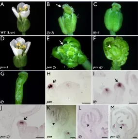
Fig. 1. pan lfydouble mutant flowers are indeterminate and fail to downregulate WUS. (A-G) Phenotypes of panand lfysingle and double mutant flowers. (A) Wild-type flower bearing four sepals, four petals, six stamens and two fused carpels in whorls one to four, respectively. (B)lfy-31flower with sepal-like organs in whorls one to three and fused sepalloid/carpelloid organs (arrow) in whorl four. (C) Flower of the well-characterized strong lfy-6allele displaying similar phenotypes to lfy-31. (D)pan-3flower bears extra sepals and petals but no carpel defects. (E)pan-3 lfy-31double mutant flower with sepal-like organs in whorls one to three and unfused
sepalloid/carpelloid organs (arrow) in whorl four. (F) The same pan lfyflower as in E, with several organs removed to expose the ectopic floral structures growing within (arrowhead). The approximate position of the removed fourth-whorl organs is indicated (dotted line). (G) The same lfy flower as in B, dissected to reveal a relatively normal gynoecium. (H-M) In situ localization of WUStranscript in stage 2 flowers (H-J), or in stage 7 or older flowers (K-M), of pan-3(H,K); lfy-6(I,L); or pan-3 lfy-31(J,M) plants. (H-J) Early WUSexpression (arrows) is similar in all three genotypes. (K-M) In older flowers, no WUSexpression is observed at the base of the gynoecium (arrowheads) of pan(K) and lfy(L) single mutants, but remains strong in pan lfyflowers (M) of a similar stage. Scale bars: 50μm.
Table 1. Identities and numbers of organs in lfyand panmutant flowers*
Mutant Sepals/sepal-like Petals Stamens Carpels/sepalloid-carpels Flowers with interior organs (%) n
pan-3 5.1±0.7 5.1±0.6 5.3±0.7 2 0 77
lfy-6 9.8±0.5 0 0 3.5±0.5 0 40
lfy-31 pan-3 11.2±1.9 0 0 6.9±1.1 89 38
*lfy-6and lfy-31 pan-3flowers were later-arising structures with floral and secondary inflorescence characteristics.
D
E
V
E
LO
P
M
E
N
[image:3.612.49.563.681.729.2]such redundancy, we generated a constitutively repressing form of PAN by fusing it to the SUPERMAN repressor domain motif (Hiratsu et al., 2003). Such constructions have been used to study bZIP factors in several model systems (Fukazawa et al., 2000; Rieping et al., 1994) and, in Arabidopsis, SUPERMAN repressor domain (RD) fusions of several transcription factors have been shown to phenocopy their corresponding loss-of-function mutants (Baudry et al., 2006; Hiratsu et al., 2003; Xu et al., 2006). The expression of such a PANfusion, either ubiquitously or from the endogenous promoter, yielded plants with severe growth defects, thereby masking any floral phenotypes (data not shown). In order to assay its effects on floral patterning, we thus used the flower-specific APETALA1 (AP1) promoter (Hempel et al., 1997). A 1.7 kb upstream fragment of the AP1locus drives expression throughout the early flower; in addition, and at variance with the normal AP1 expression pattern, it remains active in the entire flower, presumably due to the absence of additional regulatory elements (Fig. 2A; see Fig. S7 in the supplementary material; M. Yanofsky, personal communication).
In order to remove the effects of competition between PAN-RD and endogenous PAN, we introduced the pAP1(1.7-kb)::PAN-RD construct (hereafter referred to as PAN-RD) into pan-2mutant plants (Fig. 2B). The majority (82%) of these PAN-RD/+; pan/panprimary transformants bore flowers with increased numbers of petals and stamens (6.3±0.6 and 5.8±0.4 respectively, n=20), secondary
flowers in the axils of first-whorl organs (2.8±0.6 versus 0 in the wild type and pan), extra carpels in the fourth whorl (3.5±0.5) and severe indeterminacy (Fig. 2C,D). The other 18% (n=85) of transformants showed a slight enhancement of the pan organ number phenotypes without any determinacy defects (data not shown). To eliminate the possibility that the indeterminacy phenotypes were caused by the use of the AP1(1.7-kb) promoter fragment, we used an ethanol-inducible two component system (Deveaux et al., 2003; Maizel and Weigel, 2004) to drive PAN-RD expression under the endogenous PANpromoter. In the absence of induction, pan-2plants bearing this construct show no fourth-whorl phenotypes (see Fig. S8A in the supplementary material). However, after induction with ethanol, we observe flowers bearing unfused gynoecia and ectopic internal carpel structures (see Fig. S8B in the supplementary material). Thus, expression of a PAN-repressor domain fusion protein in the flower leads to the loss of floral determinacy, a phenotype observed at low frequency in panmutant plants grown in specific culture conditions (see above).
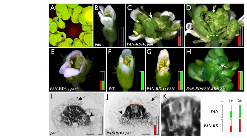
[image:4.612.52.474.56.292.2]A caveat to the use of dominant-negative alleles is that they may act as neomorphs, altering the expression of ectopic, rather than genuine, downstream targets of the protein under study. We decided to study this by varying the ratio of chimeric to wild-type protein. If PAN-RD behaves as a true dominant-negative allele, increasing doses of it should yield increasingly stronger phenotypes, whereas increasing doses of unmodified PANshould attenuate the phenotypes. We first Fig. 2. A dominant-negative PANchimera induces floral determinacy phenotypes. (A)pAP1(1.7-kb)::alcR--alcA::ER-GFPexpression in an inflorescence meristem. GFP signal (green) is observed in the central domes (arrowheads) of all visible flowers after induction with ethanol vapors. Autofluorescence from the shoot apical meristem is visible (red). (B-H) Floral phenotypes of PAN wild-type or mutant plants harboring one or more copies of PAN-RDunder the control of the AP1(1.7-kb) promoter. Red and green bars indicate the number of copies of PAN-RDor wild-type PAN, respectively. Open bar indicates no copies, half-filled indicates one copy and filled indicates two copies (see key, bottom right). (B)pan-2mutant flower. (C)panflower harboring one copy of PAN-RDdisplaying amplified pan-like phenotypes, as well as additional phenotypes such as extra carpels (arrow) and severe floral indeterminacy. (D) Side view of the flower in C with some organs removed to reveal ectopic floral structures (arrow) developing interior to the fourth whorl (demarcated by a dashed line). (E) A pan-like phenotype is observed in flowers arising from a cross of genotype in C to wild type. This flower harbors one copy of PAN-RDand is heterozygous at the PANlocus. (F) Wild-type flower. (G) Wild-type flower harboring one copy of PAN-RDdisplaying a pan-like phenotype. (H) Flower from progeny of the plant in G harboring two copies of PAN-RD presenting strong indeterminacy defects, similar to PAN-RD/+ panplants (C). (I-K) In situ localizations of AGtranscript in stage 5-6 flowers of pan-2 (I) or PAN-RD/+ pan-2(J,K) plants. AGlocalization appears unperturbed in panflowers (I), but in PAN-RD/+ pan-2flowers a central region within the AGdomain shows diminished signal intensity. Also marked are sepals (dashed arrow) that do not express AGand early stamen primordia
(arrowhead) that do. (K) Magnified view of the dashed box in J. Scale bars: 10μm.
D
E
V
E
LO
P
M
E
N
used pollen from the PAN-RD/+; pan/panflowers described above in a cross to wild type and examined the phenotypes of F1 plants selected for the presence of the transgene. We found that these flowers (PAN-RD/+; pan/+) had phenotypes similar to the panmutant (compare Fig. 2E with 2B). Thus the same PAN-RDinsertion that confers strong indeterminacy phenotypes on panflowers does not do so on pan/+ heterozygotes (instead yielding only the more sensitive organ number defect), showing that PAN-RDexpression does not ectopically induce floral indeterminacy. Next we examined primary transformants in wild-type plants, thus with two additional wild-type copies of PAN (Fig. 2F). Approximately 20% (n=60) of these plants (genotypically PAN-RD/+; PAN/PAN) phenocopied the panmutant (Fig. 2G; 4.3±0.5 sepals, 4.9±0.3 petals and 4.7±0.5 stamens; n=20), whereas the rest showed no discernable phenotypes. We then examined the progeny of these plants, to determine the phenotypes of plants harboring two copies of the transgene (see Materials and methods). These flowers (PAN-RD/PAN-RD; PAN/PAN) displayed strong phenotypes, including extra carpels and floral indeterminacy (Fig. 2H), and closely resembled PAN-RD/+; pan/panflowers (Fig. 2C,D). Taken together, these data show that the effects of the PAN-RD fusion protein are modified by wild-type PAN in a dosage sensitive manner. This suggests that PAN-RD and endogenous PAN compete for the same targets and that the PAN-RDphenotypes are due to the repression of genuine PAN targets.
To characterize the molecular basis of the PAN-RDphenotype, we performed in situ hybridizations to determine whether the PAN-RD transgene induced changes in AGexpression patterns. We observed that, as in the wild type, AGis expressed uniformly throughout the third and fourth whorls of stage 5-6 pan-2flowers (Fig. 2I). However, in similarly staged PAN-RD/+; pan/panflowers, AGexpression is patchy, with the central region of the floral meristem showing reduced expression (Fig. 2J,K). Since pAP1(1.7-kb) drives expression throughout the central dome of the flower during these stages, these results indicate that the PAN-RD chimera might exert an unequal influence on different cells within the AG-expressing region.
Thus our results show that expression of a PAN-RD fusion protein in the flower is sufficient to mimic panloss-of-function phenotypes and to produce floral indeterminacy. Since the indeterminacy phenotype occurs more stably in PAN-RDplants than in pansimple mutants, this role possibly requires other spatially or temporally restricted factors. Taken together with the phenotypes of the double mutant flowers described above, this suggests that PAN plays a role in the development of the fourth whorl, specifically in the proper regulation of the floral stem cell population, and that this role is achieved through the regulation of AG. Furthermore, as the effects of PAN-RDwere similar to the loss-of-function phenotypes of pan mutant alleles, the role of PAN in floral determinacy is likely to be that of an activator of a gene involved in the process.
PAN bindsAG regulatory sequences in vivo
We next sought to determine the precise mechanism by which PAN affects floral stem cell fate. As discussed above, AG is a key regulator of this process, by itself acting to repress WUSexpression. Because AGexpression is perturbed in PAN-RDflowers and because PAN is a predicted transcriptional activator, we reasoned that its role might be to positively regulate AGexpression. To test whether PAN directly associates with the AG promoter, we used chromatin immunoprecipitation (ChIP) assays, which examine the in vivo binding of transcription factors to DNA. We maximized the sensitivity of our assays by using a synchronized population of flowers at stages 5-7 (Wellmer et al., 2006) (see Materials and methods), as the stem cell organizing activity is known to be
terminated during this time. In addition, we used a characterized PAN-specific polyclonal antibody that, when used in immunohistochemical analyses, detects protein signals closely resembling PAN mRNA expression patterns, but showing no signal in mutant flowers (Chuang et al., 1999). We further pre-cleared this
antibody against tissue from pan-2 plants prior to
[image:5.612.313.560.57.215.2]immunoprecipitating intact protein-DNA complexes. We then performed quantitative real-time RT-PCR to assay for the enrichment of sequences within the 3 kb second intron of theAG locus with respect to input DNA, where important cis regulatory sequences are located (Fig. 3A) (Busch et al., 1999; Deyholos and Sieburth, 2000; Hong et al., 2003; Lohmann et al., 2001; Parcy et al., 1998; Sieburth and Meyerowitz, 1997). We observed that five amplicons out of a total of eight tested within this region are significantly enriched (Fig. 3B; amplicons B=9.2-fold±2.0, C=9.0±0.6, F=6.6±0.7, G=4.9±0.4 and H=40.5±9.2) when Fig. 3. PAN directly binds AGAMOUSregulatory sequences. (A) Structure of the 5.7 kb AGlocus. Exons and introns (top) are represented in bold and dashed lines, respectively. The second intron (asterisk) contains all known AGregulatory elements. The detailed view of the 3 kb intron shows the fragments used to determine enrichment in the chromatin immunoprecipitation assays (blue bars), the 3⬘ fragment used in co-bombardment experiments (red line), and evolutionarily conserved elements and known or predicted transcription factor binding motifs (Davies et al., 1999; Hong et al., 2003; Lohmann et al., 2001; Parcy et al., 1998). Black triangles, LFY/WUS binding sites; white triangle, predicted LFY binding site; stars, CArG boxes; circles, CCAAT boxes; diamond, AAGAAT motif; green squares, predicted core bZIP binding sites; vertical tick marks: 200 bp intervals. (B) Results of chromatin immunoprecipitation experiments from stage 5-7 flowers. Anti-PAN antiserum was used to isolate protein-DNA complexes and DNA enrichment levels were measured by quantitative real-time RT-PCR (see Materials and methods). Enrichment was calculated for antibody-bound DNA relative to total input DNA and was then normalized against an internal genomic control. Vertical bars show mean
enrichment levels from duplicate experiments for amplicons distributed along the intron (shown in 3A). The scale for amplicon ‘H’ is different and is shown in red. Results are shown only for amplicons with a coefficient of correlation (r2)>0.98. (C) Results of particle
co-bombardment experiments in onion epidermal cells. Vertical bars represent mean values from duplicate experiments for numbers of cells stained for GUS enzymatic activity. The pAGi-3⬘::GUSreporter, which recapitulates most facets of AGexpression in vivo, shows some background activity (left bar) in onion epidermal cells. When co-bombarded with p35S::PAN-VP16(right bar), the ‘numbers of cells’ stained for GUS activity is 4- to 5-fold greater. Error bars represent standard deviation from the mean.
D
E
V
E
LO
P
M
E
N
normalized relative to internal genomic controls. These data demonstrate that PAN, either alone or in a complex, binds AG regulatory sequences in vivo.
Because of the nature of ChIP assays, not every enriched fragment necessarily contains a PAN binding site. Four of the five ChIP-enriched amplicons (B, C, F and H) map to regions previously described as being very highly conserved (Hong et al., 2003). Of these, amplicon C contains binding sites for LFY and WUS (Lohmann et al., 2001; Parcy et al., 1998), whereas predicted bZIP core binding sites (Jakoby et al., 2002) are located within or close to two others (F and H). Hong et al. (Hong et al., 2003) have shown that an AG reporter containing a deletion within amplicon H loses expression at later floral stages, suggesting that it plays a role in the maintenance of AGexpression. In an accompanying article (Maier et al., 2009), the authors use one-hybrid assays to show that amplicon F contains PAN binding sites. Furthermore, they show that mutations in this bZIP motif disrupt binding in vitro and eliminate in vivo expression in the context of an AGreporter. Given the very high enrichment of amplicon H in our ChIP assays, we were interested in determining whether this region might also contain PAN binding sites. To this end, we made use of a published 3⬘AG reporter construct that reproduces the normal AGexpression pattern in vivo (Busch et al., 1999). In this reporter, an 800 bp 3⬘pAG fragment (Fig. 3A) drives expression of the uidA gene, which encodes the β-glucuronidase (GUS) enzyme (Busch et al., 1999). We performed particle co-bombardment experiments in onion epidermal cells with a putative constitutively activated form of PAN (p35S::PAN-VP16). Although the reporter alone presents basal reporter activity in onion cells (44.5±34.6 cells; Fig. 3C; see Fig. S9A,C in the supplementary material), perhaps due to the presence of minimal p35Ssequences, 4- to 5-fold greater numbers of cells (157±11.3; Fig. 3C; see Fig. S9B,D in the supplementary material) show GUS activity when co-bombarded with p35S::PAN-VP16. It is thus possible that PAN has two or more target sites within the AG promoter, to which it might bind with different affinities or different partners.
Flowers mutant for AG display stem cell overproliferation phenotypes and, in addition, also show homeotic transformations of stamens to petals (Bowman et al., 1989). Since PAN-RDdisrupts floral stem cell regulation without inducing the homeotic transformations associated with the loss of AGfunction, we asked whether PAN might function as a general regulator of AG, or only to modify its activity in the fourth whorl. To this end, we used the weakag-4allele, which produces flowers with reduced numbers of stamens and with fourth whorl organs replaced by another flower (Fig. 4A) (Sieburth et al., 1995). Mutations in several loci, including
HUA1and HUA2, REBELOTE(RBL) or ULTRAPETALA1(ULT1)
enhance the ag-4allele by fully or partially converting stamens to petals (Chen and Meyerowitz, 1999; Fletcher, 2001; Prunet et al., 2008). We reasoned that if PAN plays a general role in regulating AG expression, a pan allele might similarly enhance the weak ag phenotype. Conversely, if PAN participates primarily in the floral determinacy aspects of AG, the double mutant might not be significantly enhanced. In fact, we observe that pan-2 ag-4double mutant flowers (Fig. 4B) differ from ag-4single mutants only in that they present an additional pan-like phenotype: extra perianth organs (4.8±0.5 sepals and 4.8±0.5 petals compared with four each in ag-4; n=20 for both genotypes), as is the case for double mutants of pan and the strong ag-1allele (Running and Meyerowitz, 1996). The third-whorl stamens of pan-2 ag-4flowers appear morphologically normal, although they are reduced in number (5.5±0.5 compared with 5.8±0.4 in ag-4). This suggests either that the stamen identity
functions of the mutant protein encoded by the ag-4allele are robust and are not perturbed by the absence of PAN, or that PAN regulates AGexpression only in the fourth whorl.
Prunet et al. (Prunet et al., 2008) have recently proposed the existence of a distinct subdomain within the floral fourth whorl, in which a decrease in AG expression is sufficient to disrupt the specification of floral determinacy. Two observations indicate that the effects of PAN on the AGpromoter might be spatially restricted, perhaps to this subdomain. First, in PAN-RDflowers, there is a marked reduction in the accumulation of AGtranscript at the very center of the dome of the floral meristem. Second, unlike many other AGinteractors (Chen and Meyerowitz, 1999; Fletcher, 2001; Prunet et al., 2008), pan mutations have no effect on the third whorl phenotypes of a weak agallele. A restricted effect of PAN on AG expression might explain why mutations in PANattenuate the fourth whorl phenotype of supmutants (Running and Meyerowitz, 1996), which produce reduced or masculinized carpels (see Fig. S6G,H in the supplementary material) (Bowman et al., 1992). One model is that in the center of the fourth whorl of pan supdouble mutant flowers (see Fig. S6I in the supplementary material), even the mild reduction in AGexpression caused by the absence of PAN could lead to increased or prolonged WUSexpression, and thus to an increase in the size of this region. Since the stamen identity genes AP3and PIare excluded from the very center of supflowers (Bowman et al., 1992; Goto and Meyerowitz, 1994), the corresponding, but enlarged, region in pan sup flowers could now become specified into a functional carpel.
Like PAN, the broadly expressed APETALA2(AP2) gene, the absence of which causes only specific floral phenotypes (a change in sepal and petal identities), was recently shown, through the characterization of a semi-dominant allele, to play a role in the control of stem cells in the shoot (Würschum et al., 2006). Thus both PAN and AP2 have primary roles in specific aspects of floral patterning, but also have masked functions in stem cell regulation that are likely to require interactions with other domain- and/or stage-specific factors. Identifying these interactors through genetic screens or other methods could prove invaluable in gaining a full understanding of the complex regulatory mechanisms that control stem cell fate.
[image:6.612.346.513.60.153.2]As the main function of AG in floral determinacy appears to be to downregulate WUSexpression in a narrow temporal window, AG expression must be quickly upregulated in order to ensure the complete arrest of stem cell fate, and thereby ensure proper floral Fig. 4. panmutations do not enhance the third-whorl
phenotypes of a weak agallele. (A) Flower of the weak ag-4allele in a mixed background of wild-type accessions (L-er/Ws). Such flowers have normal outer organs but fourth-whorl carpels are replaced with a new flower (arrow). (B) A pan-2 ag-4flower in the same genetic background has the altered floral organ numbers of the panmutant and the new fourth-whorl flower phenotype of ag-4(arrow).
D
E
V
E
LO
P
M
E
N
patterning. We suggest that plants have evolved a complex, multiply redundant system to ensure the proper regulation of AG, and furthermore, that many of the factors involved in the regulation of floral stem cells probably also perform unrelated patterning functions.
We thank J. Lohmann for sharing unpublished results; Arnavaz Garda, Alexis Lacroix, Claudia Bardoux and Hervé Leyral for technical assistance; Ioan Negrutiu, Christophe Trehin, Nathanaël Prunet and Olivier Hamant for useful discussions and comments on the manuscript; Ioan Negrutiu for sharing unpublished results and seeds; and John Bowman and Marty Yanofsky for communicating unpublished results. This work was supported by a Caltech Division of Biology postdoctoral fellowship (to P.D.), a Marie Curie Incoming International Fellowship IIF-022002 (to P.D.), the EU Marie Curie SY-STEM Network (to J.T.) and a NSF Grant IOS-0544915 (to E.M.M.).
Supplementary material
Supplementary material available online at
http://dev.biologists.org/cgi/content/full/136/10/1605/DC1
References
Baudry, A., Caboche, M. and Lepiniec, L.(2006). TT8 controls its own expression in a feedback regulation involving TTG1 and homologous MYB and bHLH factors, allowing a strong and cell-specific accumulation of flavonoids in
Arabidopsis thaliana. Plant J.46, 768-779.
Bowman, J. L., Smyth, D. R. and Meyerowitz, E. M.(1989). Genes directing flower development in Arabidopsis. Plant Cell1, 37-52.
Bowman, J. L., Sakai, H., Jack, T., Weigel, D., Mayer, U. and Meyerowitz, E. M.(1992). SUPERMAN, a regulator of floral homeotic genes in Arabidopsis.
Development114, 599-615.
Busch, M. A., Bomblies, K. and Weigel, D.(1999). Activation of a floral homeotic gene in Arabidopsis. Science285, 585-587.
Chen, X. and Meyerowitz, E. M.(1999). HUA1and HUA2are two members of the floral homeotic AGAMOUSpathway. Mol. Cell 3, 349-360.
Chuang, C. F., Running, M. P., Williams, R. W. and Meyerowitz, E. M.(1999). The PERIANTHIAgene encodes a bZIP protein involved in the determination of floral organ number in Arabidopsis thaliana. Genes Dev.13, 334-344.
Davies, B., Motte, P., Keck, E., Saedler, H., Sommer, H. and Schwarz-Sommer, Z.(1999). PLENAand FARINELLI: redundancy and regulatory interactions between two AntirrhinumMADS-box factors controlling flower development. EMBO J.18, 4023-4034.
Deveaux, Y., Peaucelle, A., Roberts, G. R., Coen, E., Simon, R., Mizukami, Y., Traas, J., Murray, J. A., Doonan, J. H. and Laufs, P.(2003). The ethanol switch: a tool for tissue-specific gene induction during plant development. Plant J.36, 918-930.
Deyholos, M. K. and Sieburth, L. E.(2000). Separable whorl-specific expression and negative regulation by enhancer elements within the AGAMOUSsecond intron. Plant Cell12, 1799-1810.
Eshed, Y., Baum, S. F., Perea, J. V. and Bowman, J. L.(2001). Establishment of polarity in lateral organs of plants.Curr. Biol. 11, 1251-1260.
Fletcher, J. C.(2001). The ULTRAPETALAgene controls shoot and floral meristem size in Arabidopsis. Development128, 1323-1333.
Fukazawa, J., Sakai, T., Ishida, S., Yamaguchi, I., Kamiya, Y. and Takahashi, Y.(2000). REPRESSION OF SHOOT GROWTH, a bZIP transcriptional activator, regulates cell elongation by controlling the level of gibberellins. Plant Cell12, 901-915.
Gleave, A. P.(1992). A versatile binary vector system with a T-DNA organisational structure conducive to efficient integration of cloned DNA into the plant genome. Plant Mol. Biol. 20, 1203-1207.
Goto, K. and Meyerowitz, E. M.(1994). Function and regulation of the
Arabidopsisfloral homeotic gene PISTILLATA. Genes Dev.8, 1548-1560.
Hempel, F. D., Weigel, D., Mandel, M. A., Ditta, G., Zambryski, P. C., Feldman, L. J. and Yanofsky, M. F.(1997). Floral determination and expression of floral regulatory genes in Arabidopsis. Development124, 3845-3853.
Hiratsu, K., Matsui, K., Koyama, T. and Ohme-Takagi, M.(2003). Dominant repression of target genes by chimeric repressors that include the EAR motif, a repression domain, in Arabidopsis. Plant J.34, 733-739.
Hong, R. L., Hamaguchi, L., Busch, M. A. and Weigel, D.(2003). Regulatory elements of the floral homeotic gene AGAMOUSidentified by phylogenetic footprinting and shadowing. Plant Cell15, 1296-1309.
Ito, T., Takahashi, N., Shimura, Y. and Okada, K.(1997). A serine/threonine protein kinase gene isolated by an in vivo binding procedure using the Arabidopsis floral homeotic gene product, AGAMOUS. Plant Cell Physiol.38, 248-258.
Jakoby, M., Weisshaar, B., Droge-Laser, W., Vicente-Carbajosa, J., Tiedemann, J., Kroj, T. and Parcy, F.(2002). bZIP transcription factors in
Arabidopsis. Trends Plant Sci.7, 106-111.
Laux, T., Mayer, K. F., Berger, J. and Jurgens, G.(1996). The WUSCHEL gene is required for shoot and floral meristem integrity in Arabidopsis. Development 122, 87-96.
Lenhard, M., Bohnert, A., Jurgens, G. and Laux, T.(2001). Termination of stem cell maintenance in Arabidopsisfloral meristems by interactions between
WUSCHELand AGAMOUS. Cell105, 805-814.
Lohmann, J. U., Hong, R. L., Hobe, M., Busch, M. A., Parcy, F., Simon, R. and Weigel, D.(2001). A molecular link between stem cell regulation and floral patterning in Arabidopsis. Cell105, 793-803.
Maier, A. T., Stehling-Sun, S., Wollmann, H., Demar, M., Hong, R. L., Haubeiß, S., Weigel, D. and Lohmann, J. U.(2009). Dual roles of the bZIP transcription factor PERIANTHIA in the control of floral architecture and homeotic gene expression. Development136, 1613-1620.
Maizel, A. and Weigel, D.(2004). Temporally and spatially controlled induction of gene expression in Arabidopsis thaliana. Plant J.38, 164-171.
Mayer, K. F., Schoof, H., Haecker, A., Lenhard, M., Jurgens, G. and Laux, T.
(1998). Role of WUSCHELin regulating stem cell fate in the Arabidopsisshoot meristem. Cell95, 805-815.
Mizukami, Y. and Ma, H.(1995). Separation of AGfunction in floral meristem determinacy from that in reproductive organ identity by expressing antisense AG
RNA. Plant Mol. Biol. 28, 767-784.
Parcy, F., Nilsson, O., Busch, M. A., Lee, I. and Weigel, D.(1998). A genetic framework for floral patterning. Nature395, 561-566.
Prunet, N., Morel, P., Thierry, A. M., Eshed, Y., Bowman, J. L., Negrutiu, I. and Trehin, C.(2008). REBELOTE, SQUINT, and ULTRAPETALA1function redundantly in the temporal regulation of floral meristem termination in
Arabidopsis thaliana. Plant Cell20, 901-919.
Rieping, M., Fritz, M., Prat, S. and Gatz, C.(1994). A dominant negative mutant of PG13 suppresses transcription from a cauliflower mosaic virus 35S truncated promoter in transgenic tobacco plants. Plant Cell6, 1087-1098.
Running, M. P. and Meyerowitz, E. M.(1996). Mutations in the PERIANTHIA
gene of Arabidopsisspecifically alter floral organ number and initiation pattern.
Development122, 1261-1269.
Schultz, E. A. and Haughn, G. W.(1991). LEAFY, a homeotic gene that regulates inflorescence development in Arabidopsis. Plant Cell3, 771-781.
Sieburth, L. E. and Meyerowitz, E. M.(1997). Molecular dissection of the
AGAMOUScontrol region shows that ciselements for spatial regulation are located intragenically. Plant Cell9, 355-365.
Sieburth, L. E., Running, M. P. and Meyerowitz, E. M.(1995). Genetic separation of third and fourth whorl functions of AGAMOUS. Plant Cell7, 1249-1258.
Weigel, D. and Meyerowitz, E. M.(1993). Activation of Floral Homeotic Genes in Arabidopsis. Science261, 1723-1726.
Weigel, D., Alvarez, J., Smyth, D. R., Yanofsky, M. F. and Meyerowitz, E. M.
(1992). LEAFYcontrols floral meristem identity in Arabidopsis. Cell69, 843-859.
Wellmer, F., Alves-Ferreira, M., Dubois, A., Riechmann, J. L. and
Meyerowitz, E. M.(2006). Genome-wide analysis of gene expression during early Arabidopsisflower development. PLoS Genet.2, e117.
Würschum, T., Gross-Hardt, R. and Laux, T.(2006). APETALA2regulates the stem cell niche in the Arabidopsisshoot meristem. Plant Cell18, 295-307.
Xu, Y., Teo, L. L., Zhou, J., Kumar, P. P. and Yu, H.(2006). Floral organ identity genes in the orchid Dendrobium crumenatum. Plant J.46, 54-68.