PRIMER
Mitotic bookmarking in development and stem cells
Nicola Festuccia*, Inma Gonzalez*, Nick Owens and Pablo Navarro‡ABSTRACT
The changes imposed on the nucleus, chromatin and its regulators during mitosis lead to the dismantlement of most gene regulatory processes. However, an increasing number of transcriptional regulators are being identified as capable of binding their genomic targets during mitosis. These so-called‘mitotic bookmarking factors’ encompass transcription factors and chromatin modifiers that are believed to convey gene regulatory information from mother to daughter cells. In this Primer, we review mitotic bookmarking processes in development and stem cells and discuss the interest and potential importance of this concept with regard to epigenetic regulation and cell fate transitions involving cellular proliferation.
KEY WORDS: Mitotic bookmarking, Stem cells, Epigenetics, Mitotic inheritance, Transcription factors
Introduction
Developmental transitions and cell differentiation are driven by transcription factors, which establish cell type-specific transcription patterns (Spitz and Furlong, 2012). Both during embryogenesis, when millions of differentiated cells are generated from a single fertilised egg, and during adulthood, when the activation of stem cells amplifies the population of differentiating cells, the acquisition of cell identity occurs in actively dividing cells (Holtzer et al., 1972; Soufi and Dalton, 2016). As cells proliferate, they undergo replication and mitosis–two processes that profoundly influence the molecular events associated with gene regulation (Alabert and Groth, 2012; Chen et al., 2015; Steffen and Ringrose, 2014). During replication, different portions of the genome are sequentially and progressively targeted until full DNA duplication has occurred. In contrast, mitosis represents a short and sudden moment involving the most dramatic reorganisation of the nucleus and chromatin experienced during the life of a cell: structures such as the nuclear envelope are dismantled, and compact, rod-shaped mitotic chromosomes are formed (de Castro et al., 2016; Hirano, 2015). Notably, several mitotic mechanisms simultaneously converge on a common, global consequence: the inactivation of several transcription factors and a drastic downregulation of transcription (de Castro et al., 2016; Ma et al., 2015; Maeshima and Eltsov, 2008; Palozola et al., 2017; Wang and Higgins, 2013).
Understanding how these alterations impact developmental transitions is a key question at the centre of which lies the concept of the‘memory’of gene expression (Fig. 1). Although epigenetic mechanisms (see Box 1) have mainly been linked to the maintenance of gene silencing (Reik, 2007; Steffen and Ringrose, 2014), evidence for a memory of gene activity does exist: the
probability of transcribing a given gene after mitosis is significantly higher in cells derived from parent cells having experienced transcription of that gene compared with those that have not (Ferraro et al., 2016; Hormanseder et al., 2017; Zhao et al., 2011). It is possible that the organisation of transcription factors in self-reinforcing circuits encodes sufficient information to re-establish efficiently the appropriate transcription patterns in daughter cells (Fig. 1), at least to some extent, without invoking the need for a molecular memory driving gene reactivation (Egli et al., 2008; Ptashne, 2013). Nevertheless, increasing observations suggest that the mitotic behaviour of certain transcriptional regulators directly instructs gene reactivation after mitosis via a mechanism known as‘mitotic bookmarking’(Box 1; Fig. 1). This concept originally stems from the observation that metaphase cells are more sensitive to DNA denaturation (Darzynkiewicz et al., 1977a,b) and display increased levels of single-strand DNA (Juan et al., 1996) than cells in interphase. These features were directly related to gene regulation: promoters active in interphase display DNA unwinding in mitosis (Gazit et al., 1982; Martínez-Balbás et al., 1995) and signs of transcription factor binding (Michelotti et al., 1997). These early studies, which may be considered as the birth of the field of mitotic bookmarking, suggested that the propagation of an open chromatin structure associated with transcription factor binding may facilitate the prompt reactivation of transcription in interphase (Fig. 1). This led to the concept of mitotic bookmarking factors, defined as gene regulators that bind specific regulatory elements during mitosis to convey regulatory information to daughter cells (Box 1; Fig. 1). Over the years, a number of general and ubiquitous regulators of transcription have been proposed to act as bookmarking factors (Table 1). More recently, sequence-specific developmental regulators have also been identified as potentially binding mitotic chromatin. This has opened up a new avenue for understanding how transcription factors not only establish but also maintain cell type-specific transcription profiles across cell division.
The purpose of this Primer article is to provide a broad introduction to mitotic bookmarking processes by focussing on three key aspects: first, how it relates to the impact of mitosis on transcriptional and chromatin regulation; second, how transcriptional regulators interact with mitotic chromosomes and how this can be translated into function; and third, how cell identity might be regulated by bookmarking factors. This leads us to describe the implications of this phenomenon in light of our understanding of epigenetic gene regulation, lineage stability and phenotypic flexibility during development. Finally, we describe major technical difficulties in the analysis of mitotic bookmarking and discuss their implications for current and future studies.
Mitotic bookmarking in the face of the constraints imposed by mitosis
To bind its targets during mitosis, a transcription factor must overcome a large number of mitosis-specific regulations that generally lead to the suppression of binding (Fig. 2). Although an Epigenetics of Stem Cells, Department of Developmental and Stem Cell Biology,
Institut Pasteur, CNRS UMR3738, 25 rue du Docteur Roux, 75015 Paris, France. *These authors contributed equally to this work
‡Author for correspondence ( [email protected])
P.N., 0000-0002-2700-6598
DEVEL
O
exhaustive description of mitotic cells is beyond the scope of this article – excellent reviews describing this in detail are already available (de Castro et al., 2016; Ma et al., 2015; Wang and Higgins, 2013)–the aspects discussed below underscore the magnitude of changes entailed by mitosis and how these might influence the nature and potential importance of mitotic bookmarking.
Mitotic-specific phosphorylation of transcription factors
Mitosis coincides with a drastic downregulation of transcription (Palozola et al., 2017; Prescott and Bender, 1962), largely mediated
by the targeted phosphorylation and inactivation of several general transcription factors (Gottesfeld and Forbes, 1997). Sequence-specific transcription factors, and certain chromatin remodellers, can also be subjected to mitotic hyper-phosphorylation, triggering their degradation or inactivation. This is exemplified by the degradation of the myogenic factor Myf5 (Lindon et al., 1998), the stereotypical phosphorylation of the linker domain of C2H2 zinc-finger proteins (Dovat et al., 2002; Rizkallah et al., 2011), the inactivation of major developmental regulators such as Oct4 (also known as Pou5f1) and Sox2 (Qi et al., 2016; Shin et al., 2016), or that of some chromatin remodellers such as Brg1 (Smarca4) (Sif et al., 1998). Whether this phosphorylation-mediated inactivation is regulated during development or in distinct cell types remains unknown. For instance, the general transcription factor TBP, which retains the capacity to bind mitotic chromatin and represents an excellent candidate to bookmark active promoters (Chen et al., 2002; Christova and Oelgeschläger, 2002; Xing et al., 2008), has not been systematically found associated with mitotic chromosomes (Blobel et al., 2009; Komura et al., 2007; Segil et al., 1996; Varier et al., 2010). This suggests that mitotic bookmarking might be a regulated property. In contrast, the ectopic expression of other mitotic bookmarking factors in cells in which they are not endogenously expressed is often accompanied by mitotic chromosome association (Deluz et al., 2016; Liu et al., 2017), indicating that it is an inherent characteristic of certain proteins.
Mitotic condensation, chromatin accessibility and mitotic bookmarking factors
One should not expect every gene regulator that is not inactivated to be able to interact with mitotic chromatin. Several reasons may cause their lack of binding (Fig. 2), from the breakdown of the nuclear envelope, which increases the volume within which gene regulators can diffuse and, consequently, reduces their local concentration, to the severe chromatin reorganisation induced by
Daughter cell
Stability
Flexibility
A
B
C Interphase
Mitosis Mother cell
Time 0 Time 1 Time 2
1 2
1 2
Unaffected regulator Inactivated regulator Degraded regulator
1 2
1 2 2
1 2
Time 3
A
B
C
[image:2.612.90.525.59.249.2]High-affinity site Low-affinity site Active marks Repressive marks Key
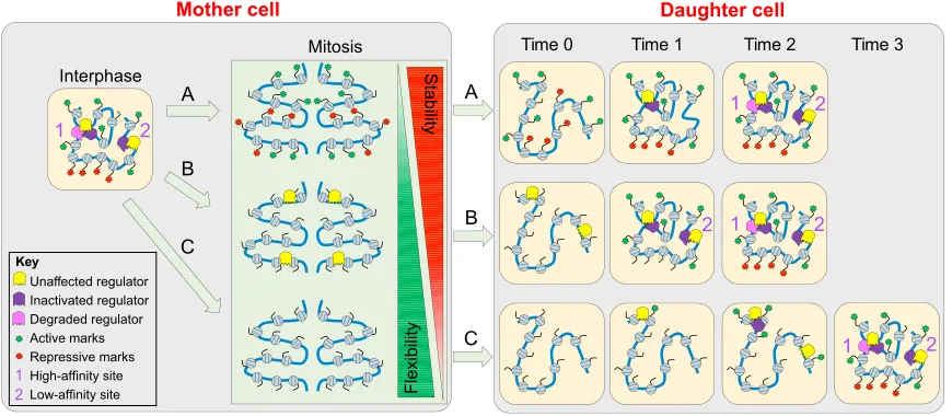
Fig. 1. Conveying gene regulatory architectures from mother to daughter cells.The regulatory architecture of the chromatin in interphase cells (shown in yellow boxes) depends on where and how various gene regulators (coloured Pacman-like icons) interact with high- and low-affinity sites in chromatin, which adopts a specific 3D organisation and can be modified with active (green) and repressive (red) marks. During mitosis (shown in the green box), three non-mutually exclusive scenarios are considered: (A) chromatin marks can be maintained; (B) the binding of particular regulators (i.e. mitotic bookmarking factors) can be maintained; or (C) all previous signs of gene regulation can be lost. A generalised outcome of each scenario, highlighting the different speeds and order of reactivation following each mitotic scenario, is illustrated. In scenarios A and B, gene regulators are able to find their targets more efficiently than in scenario C, which relies on the intrinsic self-organisation capacity of gene regulatory networks. Scenarios A and B thus provide a mitotic memory that canalises gene reactivation and, hence, stability. In scenarios B and C, however, the mere extinction of selected regulators is sufficient to avoid both their function and the memory of their function: they thus provide more flexibility than scenario A. Scenario B, which represents mitotic bookmarking, therefore displays the regulatory advantages of both scenarios A and C.
Box 1. Epigenetic (book)marks and mitotic bookmarking factors
Strictly speaking, the term‘epigenetic mark’should be restricted to those chromatin features that: (1) influence gene expression; (2) are maintained through replication and mitosis; and (3) are independent from the mechanisms that were used for their establishment. Thus, the concept of an epigenetic mark includes that of persistence through mitosis. However, because the term‘epigenetic mark’is increasingly being used in an informal manner, it is now common to encounter the additional qualification of certain DNA or chromatin marks as‘mitotic bookmarks’, to differentiate them from those that are not necessarily epigenetic in nature. Conversely, mitotic bookmarking factors should not be considered as epigenetic marks even if they contribute to the transmission of a‘memory’of gene regulation across cell generations. Indeed, it is now established that the nature of mitotic binding by transcription factors is highly dynamic. Moreover, mitotic bookmarking factors that autonomously bind chromatin through sequence-specific interactions represent both the initial trigger and the inherited information itself. Therefore, mitotic bookmarking should not be rigorously considered as an epigenetic mechanism either. In this Primer, we restrict the term ‘mitotic bookmarking factor’ to transcription and chromatin regulators that bind at regulatory elements during mitosis to convey gene regulatory information to daughter cells.
DEVEL
O
the formation of mitotic chromosomes. This reorganisation is largely mediated by alterations to the three-dimensional topology of the chromatin and the acquisition of mitotic-specific chromatin loops of ∼100 kb (Dekker, 2014; Naumova et al., 2013). These loops are laterally and longitudinally compacted 2- to 4-fold compared with interphase chromatin (Lleres et al., 2009; Vagnarelli,̀ 2012). Moreover, nucleosomes are phosphorylated and hypo-acetylated (Wang and Higgins, 2013), and shift position to occupy transcription start sites and compete with the transcriptional machinery (Kelly et al., 2010). Thus, during mitosis, the chromatin adopts a state that does not favour transcription factor binding, enhancer-promoter communications, and transcriptional activity. Despite all these features, however, virtually all active gene
regulatory regions in interphase are maintained in an accessible state during mitosis (Blythe and Wieschaus, 2016; Hsiung et al., 2015; Teves et al., 2016; Xu et al., 2017). This last observation has three major potential implications. First, it indicates that the nucleosomes that occupy gene transcription start sites in mitosis (Kelly et al., 2010) are nevertheless accessible, and, most likely, fragile (Xi et al., 2011). Second, it suggests that mitotic bookmarking by transcription factors is potentially more widespread than previously anticipated and directly responsible for maintaining gene regulatory regions in a permissive state. Supporting this view, two mitotic bookmarking factors, TBP and Hsf2, have been shown to locally recruit PP2a (Xing et al., 2008, 2005), a phosphatase that inactivates condensins, key players in the formation of condensed
Global decoration
Specific binding
Factor Live PCR NGS References
Transcri
pti
o
n factors
FoxA1 Caravaca et al., 2013
Esrrb Festuccia et al., 2016a
Klf4 Liu et al., 2017
Gata1 Kadauke et al., 2012
Rbpj Lake et al., 2014
Runx2 Ali et al., 2010; Pockwinse et al., 2011; Young et al., 2007
Myc Yang et al., 2013
FoxI1 Yang et al., 2014
TLE1 Ali et al., 2010
Pou5f1 Deluz et al., 2016; Liu et al., 2017; Teves et al., 2016
Sox2 Deluz et al., 2016; Liu et al., 2017; Teves et al., 2016
HSF2 Xing et al., 2005
HNF1b Lerner et al., 2016; Verdeguer et al., 2010 Utf1 Kooistra et al., 2009
Gbx2 Deluz et al., 2016 Klf5 Deluz et al., 2016 Rex1 Deluz et al., 2016 Tbx3 Deluz et al., 2016 Tcf3 Deluz et al., 2016
HMGB1 Caravaca et al., 2013; Pallier et al., 2003 HMGB2 Caravaca et al., 2013; Pallier et al., 2003 HMGN1 Pallier et al., 2003
Gata4 Caravaca et al., 2013 C/EBP-a Caravaca et al., 2013
RNAP
II
m
a
chi
n
ery
TBP Chen et al., 2002; Christova and Oelgeschlager, 2002; Segil et al., 1996; Xing et al., 2008
NC2 Christova and Oelgeschlager, 2002
TAF5 Christova and Oelgeschlager, 2002
TFIIB Christova and Oelgeschlager, 2002
RNAP
I an
d
II
I
ma
c
h
in
e
ry
TAF12 Segil et al., 1996
TFIIIB Fairley et al., 2003
TFIIIC2 Fairley et al., 2003
TFIII110 Fairley et al., 2003
UBF1 Chen et al., 2005; Young et al., 2007 Rpa43 Chen et al., 2005
Chrom
a
ti
n reg
u
la
tors
CTCF Burke et al., 2005; Yang et al., 2013
P300 Wong et al., 2014; Zaidi et al., 2003
Mll Blobel et al., 2009
Brd4 Dey et al., 2003; Dey et al., 2009; Zhao et al., 2011
Psc Buchenau et al., 1998; Follmer et al., 2012
Pc Follmer et al., 2012
Ash2l Blobel et al., 2009
RbBP5 Blobel et al., 2009
Menin Blobel et al., 2009 Dnmt1 Easwaran et al., 2004
ISWI Yokoyama et al., 2013 MeCP2 Brero et al., 2005
Brd2 Dey et al., 2003
Bmi1 Voncken et al., 2005; Voncken et al., 1999 dRing Follmer et al., 2012
CHD4 Yokoyama et al., 2013 Uhrf1 Uemura et al., 2000
[image:3.612.112.501.56.525.2]IF
Table 1. Known gene regulators potentially behaving as mitotic bookmarking factors.In this table we report general and sequence-specific transcription factors, together with other chromatin regulators, that might be acting as mitotic bookmarking factors. Only factors derived from microscopy [either using fixed samples, generally with methanol (immunofluorescence, IF), or through live imaging using fusion proteins] and/or chromatin immunoprecipitation studies [analysed by either PCR or next-generation sequencing (NGS) ChIP-seq], as indicated in the table, are reported. Note that for TBP, the evidence presented was not ChIP-seq but ChIP-on-chip. The factors highlighted in red represent particularly solid candidates. The cells highlighted in yellow denote conflicting results in the literature. Rex1 (Zfp42); NC2 (Dr1); UBF1 (UBTF); dRing (Sce).
DEVEL
O
mitotic chromosomes (Hirano, 2015). Third, condensins have been shown to be enriched at regulatory regions, both in interphase and in mitosis (Dowen et al., 2013; Sutani et al., 2015), and could directly impact the organisation of nucleosomal arrays and transcription factor binding, either by passive steric hindrance or by inducing positive writhe on the DNA (Hirano, 2015). Positive supercoiling can inhibit transcription factor binding and has been associated with the partial disassembly of octamers (Levchenko et al., 2005). Therefore, in addition to contributing to the condensation of chromatin fibres, condensins might paradoxically favour fragile nucleosomes and contribute to local chromatin accessibility at regulatory regions whilst inhibiting transcription factor binding. Moreover, it is known that chromosome condensation levels oscillate during different phases of mitosis and vary between different regions of the chromatids (Lleres et al., 2009), and that̀ several mitotic factors such as condensin I and topoisomerase II display dynamic exchange with the cytosol (Christensen et al., 2002; Gerlich et al., 2006; Tavormina et al., 2002). Therefore, nucleosomal arrays in mitotic chromosomes are not necessarily compacted and embedded within somewhat static chromatin fibres. Rather, they are intrinsically dynamic (Chen et al., 2005), and may allow mitotic bookmarking factors to bind to and influence key local properties of the chromatin.
Mitotic-specific histone modifications
One of the most recognisable hallmarks of mitosis is histone phosphorylation (Sawicka and Seiser, 2012). Histone H3
phosphorylation, which systematically takes place on the residue neighbouring a lysine, blocks the binding of specific readers to methylated H3K4, H3K9 and H3K27, as shown for TFIID, HP1 and Eed, respectively (Fischle et al., 2005; Hirota et al., 2005; Lau and Cheung, 2011; Varier et al., 2010). Therefore, during mitosis, even the most canonical epigenetic regulation of constitutive (HP1) and facultative (Eed) heterochromatin may be at least partially destabilised. Consequently, the mechanism by which histones are dephosphorylated in daughter cells to allow the recruitment of heterochromatin regulators represents an important aspect for maintaining gene regulatory states after division (de Castro et al., 2017). Moreover, mitotic H3T3 and H3S10 phosphorylation leads to histone H4K16 deacetylation (Vaquero et al., 2006; Wilkins et al., 2014) and, more generally, histone acetylation is known to be reduced in the context of mitotic chromatin (Kruhlak et al., 2001; McManus and Hendzel, 2006). Nonetheless, a large subset of regulatory regions maintain high levels of H3K27 acetylation in mitosis (Hsiung et al., 2016; Liu et al., 2017) and the histone acetyl-transferase p300 has been shown to retain binding to mitotic chromatin (Wong et al., 2014; Zaidi et al., 2003). Similarly, other chromatin remodelling factors have been suggested to associate with mitotic chromatin (Black et al., 2016). Further studies are needed to tease apart the function of such chromatin regulators during mitosis and shed light on their possible role in the maintenance of DNA accessibility and nucleosome remodelling.
Histone phosphorylation (eviction)
T
T
T
Condensin and other activities (inhibition)
T
Nucleosome reorganisation (eviction)
T
Phosphorylation (inactivation or degradation)
Accessible
Accessible Nuclear
breakdown (diffusion)
T
Accessible
Accessible Bookmarking
? ?
Slow reactivation
Mitosis Interphase
[image:4.612.144.466.60.345.2]Fast reactivation
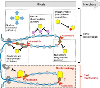
Fig. 2. Mitotic bookmarking in the face of mitosis.A large number of changes can alter the binding capacity of gene regulators (yellow Pacman-like icons) during mitosis. Nuclear envelope breakdown, for example, leads to a drop in the effective concentration of regulators and increases their free diffusion. Some gene regulators are inactivated or degraded, often via phosphorylation (red star). Chromatin can also be globally phosphorylated (pink stars on histone tails), leading to the eviction of specific chromatin readers. Condensins (grey half rings), together with other mitotic activities (e.g. topoisomerases, not depicted), bind at previously active regulatory regions (depicted in red within a blue DNA molecule) and inhibit the binding of gene regulators. The organisation of the nucleosome array is also modified, with some nucleosomes (green) occupying regulatory regions. Despite all of this, many regulatory regions remain globally accessible (red arrowheads) and some factors, known as mitotic bookmarking factors, are able to bind their targets during mitosis (bottom box), leading to fast reactivation dynamics in the following interphase.
DEVEL
O
The binding and modes of action of mitotic bookmarking factors
Most discoveries related to mitotic bookmarking start with the observation that the regulatory protein under study seems to coat the mitotic chromosomes. Although microscopy techniques have enabled the rapid identification of transcription regulators with bookmarking potential, the mitotic binding of only a handful of sequence-specific transcription factors has been characterised comprehensively (Table 1). These factors include Gata1 in erythroblasts (Kadauke et al., 2012), FoxA1 in hepatocytes (Caravaca et al., 2013), Myc in embryo-derivedDrosophilacells (Yang et al., 2013), Rbpj in embryonic carcinoma cells (Lake et al., 2014), and several pluripotency factors –Esrrb, Oct4, Sox2 and Klf4–in embryonic stem cells (Festuccia et al., 2016a; Liu et al., 2017). As we discuss below, studies of these and other factors have provided key insights into the modes by which mitotic bookmarking factors might function.
Site-specific recruitment of mitotic bookmarking factors
In all of the cases highlighted above, it has been shown that only a subset of their targets remain bound in mitosis: these mitotic bookmarking factors display a selectivity of binding ranging from 10% of bookmarked targets (Gata1, FoxA1 and Esrrb) to 40-70% (Myc, Rbpj, Klf4, Sox2 and Oct4). Moreover, binding during mitosis appears lower compared with that during interphase for most of these factors (Gata1, FoxA1, Esrrb, Rbpj and Myc). Why some factors are more efficient than others remains unclear, although it is likely that technical differences and limitations might underlie these discrepancies, which, in some cases, are remarkable. For instance, whereas one study showed that Sox2 does not exhibit site-specific bookmarking in mouse embryonic stem cells (ESCs), except for a few dozen loci (Deluz et al., 2016), another showed Sox2 mitotic binding at nearly half of its thousands of interphase targets (Liu et al., 2017). Setting these discrepancies aside (which are discussed further below), an important question is how and why only a subset of the interphase targets of a given transcription factor are occupied in mitosis–a question that is particularly intriguing in light of the retained accessibility of regulatory regions during division. Except for Myc, which preferentially binds to non-canonical E-boxes in mitosis (Yang et al., 2013), identical DNA motifs can be identified within bookmarked and non-bookmarked regions. Moreover, a higher proportion of the regions occupied by Esrrb or FoxA1 in mitosis possess a consensus binding sequence compared with interphase (Caravaca et al., 2013; Festuccia et al., 2016a), and overall these sites tend to be of better quality (Festuccia et al., 2016a). This is also reflected in the higher average occupancy that bookmarked regions show in interphase, compared with the regions that lose binding during mitosis. Hence, establishing robust and specific DNA interactions could play a determinant role in mitotic bookmarking. In agreement, the ectopic introduction of consensus Esrrb-binding sites in the genome is sufficient to establish regions bookmarked by Esrrb (Festuccia et al., 2016a). Thus, sequence-specific interactions with DNA seem to be sufficient for mitotic binding, and the overall quality of each binding site, in conjunction with the local chromatin environment and the interaction with mitotic-specific activities, might explain selective retention. Bookmarking by Oct4, Klf4 and Sox2, however, does not show a clear preference for regions possessing more or better binding sites (Liu et al., 2017). Nevertheless, the regions bookmarked by these three factors are particularly enriched for Esrrb-binding sites, whereas those sites that lose their binding are not (Liu et al., 2017). Hence, some bookmarking factors, such as
Esrrb, might play a preponderant role in re-structuring the genome-wide distribution of several other transcription factors during mitosis.
Chromosomal decoration by mitotic bookmarking factors
Whether the macroscopic coating of mitotic chromatids observable by microscopy is the result of the sum of the site-specific interactions described above, and what its functional relevance might be, remains controversial (Fig. 3). The DNA-binding domain of several transcription factors is required for their global association with mitotic chromatin, as shown by microscopy for pluripotency factors (Deluz et al., 2016; Festuccia et al., 2016a; Teves et al., 2016) and other regulators such as HNF1b (Lerner et al., 2016). In contrast, compromising the ability of FoxA1 to interact with its consensus binding sequence does not completely eliminate its coating of the chromatids (Caravaca et al., 2013). Notably, FoxA1 exhibits an atypical mode of interaction with the nucleosomes through a domain structurally related to linker histones (Clark et al., 1993). Similarly, Rbpj1 can bind to core nucleosomes
in vitroand the decoration of mitotic chromosomes is not completely
lost upon mutating its DNA-binding domain (Lake et al., 2014). Hence, both sequence-specific and non-specific interactions can contribute to mitotic binding (Fig. 3). Unconventional mechanisms might also mediate mitotic localisation, as shown by the ability of isolated nuclear localisation signals to drive mitotic chromosomal coating (Deluz et al., 2016; Teves et al., 2016), hinting at a possible involvement of importins in shuttling transcription factors to the chromatids. Also, because DNA-binding domains tend to display clusters of positively charged amino acids (Stawiski et al., 2003), mitotic bookmarking factors could engage with the chromosomes via non-specific electrostatic interactions with the negatively charged hyper-phosphorylated chromatin. More generally, the presence of gene regulators has been reported at the chromosome periphery (Booth et al., 2016; Ohta et al., 2010), a domain in which layers of proteins and RNAs accumulate during cell division. Overall, several mechanisms are available to explain the chromosomal retention of transcriptional regulators without the involvement of site-specific recruitment (Fig. 3).
From mitotic bookmarking to gene reactivation
Mitotic bookmarking factors are generally perceived to accelerate gene reactivation after mitosis (Fig. 2), during early G1, even though this has been clearly demonstrated for only a handful of factors. A canonical example is provided by Brd4 (Dey et al., 2003): live imaging of transcription using an artificial target engineered with large tandem arrays, has demonstrated that its mitotic presence around this target leads to rapid decondensation and resumption of post-mitotic transcription (Zhao et al., 2011). Moreover, Brd4-deficient mouse embryonic fibroblasts exhibit delayed gene expression in early G1 (Yang et al., 2008). Similarly, mitotic MLL1 (KMT2A) recruitment at active promoters in several cell lines has been correlated with accelerated transcriptional reactivation upon re-entry into interphase (Blobel et al., 2009). Finally, sequence-specific transcription factors have also been linked to post-mitotic gene reactivation: the genes bookmarked by Gata1 reactivate transcription faster at the mitosis-G1 (M-G1) transition in the presence of Gata1 (Kadauke et al., 2012), and the genes activated by Esrrb in early G1 are located proximal to bookmarked regions (Festuccia et al., 2016a). The mechanisms by which mitotic bookmarking factors achieve gene reactivation in the following interphase are, in contrast, poorly understood. A simple hypothesis would be that, by remaining bound to their targets during
DEVEL
O
mitosis, bookmarking factors accelerate the reassembly of regulatory complexes at promoters and enhancers, thereby fostering rapid transcriptional responses. However, just like in interphase, the binding of transcription factors in mitosis is dynamic, with residence times in the order of seconds (Chen et al., 2005). In mitosis, the kinetics of transcription factor exchange on and off chromatin are not greatly altered (Caravaca et al., 2013; Deluz et al., 2016; Festuccia et al., 2016a; Pallier et al., 2003; Pockwinse et al., 2011; Teves et al., 2016), and in most cases, faster dynamics have been observed. TBP constitutes a notable exception, most likely as a consequence of the impaired turnover of pre-initiation complexes in the near absence of transcription (Chen et al., 2002). An important implication of these observations is that the function of mitotic bookmarking by sequence-specific transcription factors cannot be simply achieved by carry-over of immobile factors that stably occupy regulatory elements throughout cell division. Together with the evidence that mitotic chromatin is dynamic and maintains similar accessibility profiles as interphase chromatin, these observations call for exploring alternative hypotheses, at least for sequence-specific transcription factors. A possibility is that the dynamic interaction of bookmarking factors with specific regulatory elements contributes to the maintenance of global accessibility or other chromatin features. In this sense, the activity of transcription factors in mitosis would not be conceptually dissimilar to that in interphase. Finally, the global coating of chromosomes could also be functional, either by serving a structural role or by simply increasing the local concentration of transcriptional regulators in the proximity of DNA, thereby reducing the search time for binding sites upon global decondensation. Nonetheless, qualifying such proteins as mitotic bookmarking factors would be misleading (Box 2).
The regulatory nature of mitotic bookmarking factors Mitotic bookmarking factors are generally thought to accelerate gene reactivation in interphase and, as discussed below, they generally control a functionally relevant subset of target genes that define the molecular identity of the cell. Moreover, additional functions have been proposed for mitotically bound transcription factors, be it in the preparation of future cell fate choices or in the control of other properties of the chromatin. In this section, we use multiple examples to illustrate these points.
Mitotic bookmarking, post-mitotic gene reactivation and the maintenance of cell identity
During mitosis, both Gata1, a key factor driving haematopoiesis, and FoxA1, which is required for liver differentiation, bind in the vicinity of genes highly expressed in the corresponding lineage (Caravaca et al., 2013; Kadauke et al., 2012). Similarly, Esrrb, Oct4, Klf4 and Sox2, four key transcription factors that sustain pluripotency and promote mouse ESC self-renewal, have been shown to bind in mitosis to their most active targets in interphase, including to clusters of enhancers located in the vicinity of cell identity genes (Festuccia et al., 2016a; Liu et al., 2017). Therefore, regions bookmarked by sequence-specific transcription factors tend to be among the most active enhancers and promoters in interphase, and are particularly associated with tissue-specific genes that define the transcriptional identity of the corresponding cell type (Fig. 4, green panel). Moreover, the analysis of Esrrb-responsive genes during the cell cycle in mouse ESCs has shown that genes located close to bookmarked regions are over-represented among the genes activated by Esrrb in early G1 (Festuccia et al., 2016a). As the cell cycle progresses, the genes up- and downregulated by Esrrb increase in number and lose their statistical enrichment for genes
Electrostatic interactions Periphery
Chromatin Scaffold
ATGCCA
Site-specific interactions Non-specific interactions
Decorated mitotic chromosome
+
-
- - -
+ + +
- -
+
-
ATGCCA
[image:6.612.106.510.56.305.2]Recruitment to chromosome periphery
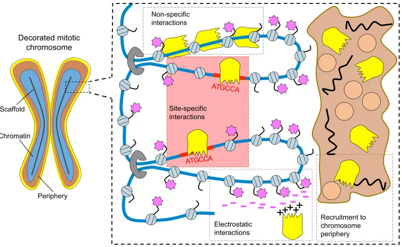
Fig. 3. Different modalities of recruitment to mitotic chromosomes.Mitotic chromosomes (left) are composed of an axial scaffold (black line), the chromatin (blue) and a peripheral compartment (brown). When visualised by microscopy, some gene regulators globally decorate mitotic chromosomes (yellow layer). This signal may be derived from different modalities of chromosomal retention, as shown in detail on the right. Three main possibilities may underlie the establishment of non-specific interactions between gene regulators (yellow Pacman-like icons) and chromosomes: via non-specific DNA/nucleosome interactions; via electrostatic interactions with phosphate groups ( pink stars) added to H3 tails; via recruitment to the chromosome periphery, which is a compartment rich in proteins (orange circles) and RNAs (black lines). In addition, gene regulators might be specifically recruited to selected regions of the chromatin, either through base-specific interactions with DNA motifs (e.g. transcription factors), or through interactions with specific histone or DNA modifications (e.g. chromatin readers). Only this last site-specific modality may represent a genuine mitotic bookmark, as discussed in the text.
DEVEL
O
located in proximity of bookmarked regions. As bookmarked regions are enriched for cognate binding sequences for Esrrb, this indicates that mitotic bookmarking leads to the activation of direct transcriptional targets in early G1, with further consequences being a reflection of indirect regulations mediated by the dynamics of the pluripotency network. Therefore, it is likely that mitotically bookmarked regions represent a core regulatory node that can re-initiate the progressive deployment of the transcriptional circuits sustaining cell identity (Figs 1 and 4). Remarkably, the genes exhibiting proximal mitotic Esrrb binding in mouse ESCs are expressed in pluripotent compartments of the embryo, whereas regions losing Esrrb in mitosis are associated with trophectodermal genes (Festuccia et al., 2016a), an extra-embryonic lineage in which Esrrb functions later during development. This indicates that mitotic bookmarking may enable a transient memory of gene activity as cell identities are progressively established (Fig. 4). However, despite recent efforts, a definitive demonstration of the functional importance of mitotic bookmarking in the control of cell identity is still lacking. For instance, it has been reported that the depletion of Oct4 (Liu et al., 2017) and Sox2 (Deluz et al., 2016) at the M-G1 transition leads to a minor decrease in self-renewal. Moreover, upon ectopic expression of Oct4 or Sox2 mutant proteins that are degraded at the M-G1 transition, the efficiency of reprogramming somatic cells back to pluripotency is either partially affected, such as in the case of Oct4 (Liu et al., 2017), or identical to the control, as shown for Sox2 (Deluz et al., 2016). This variable outcome is unexpected given that Sox2 and Oct4 tend to act synergistically as heterodimers (Rizzino, 2009). Moreover, transgenic expression of Sox2 is known to bias differentiation towards neuroectodermal lineages, a property that is slightly altered when an M-G1 defective Sox2 is ectopically expressed (Deluz et al., 2016). That Oct4 and
Sox2 might behave as mitotic bookmarking factors potentially represents a major discovery, because these factors are the only ones that are mandatory for self-renewal to occur. Unfortunately, no simple agreement can be reached among the currently available studies (Deluz et al., 2016, 2017; Liu et al., 2017; Teves et al., 2016), and an understanding of how the mitotic behaviour of these factors is connected to phenotypical changes remains rather elusive (Deluz et al., 2016, 2017; Liu et al., 2017). Additionally, two independent reports have shown that both Oct4 and Sox2 are phosphorylated by aurora kinases during mitosis and that this inhibits their DNA-binding activity (Qi et al., 2016; Shin et al., 2016). Current findings regarding the bookmarking activities of Oct4 and Sox2 therefore require careful evaluation. In conclusion, the impact of mitotic bookmarking by sequence-specific transcription factors on the preservation of cell identity, although extremely appealing, needs further experimentation to be convincingly documented. The studies published over the last few years, in particular those in mouse ESCs, should pave the way to rigorous evaluation of this important issue.
Mitotic-specific functions of some transcription factors
Although the above observations point to a potential canonical bookmarking function of certain regulators, it is possible that such factors play more complex functions. Indeed, the mitotic binding profiles of Gata1 and FoxA1, but not those of the pluripotency factors analysed so far, reveal that a small subgroup of targets are specific to mitosis (Caravaca et al., 2013; Kadauke et al., 2012). At these regions, mitotic binding events could be associated with functions other than that of specifying the genes that must be faithfully reactivated in daughter cells. Several other functions for the mitotic binding of gene regulators have been proposed. For instance, in embryonic carcinoma cells, Rbpj targets in interphase are enriched for stem cell maintenance genes and, in mitosis, for genes that are expressed later during development, upon neuronal differentiation (Lake et al., 2014). Whether mitotic bookmarking by Rbpj, the main actor of Notch signalling and, therefore, of various differentiation and developmental pathways, contributes to the silencing of developmental genes before differentiation and/or primes their future activation, is not known. Furthermore, in mitotic human ESCs, the activating H3K4me3 mark increases significantly at a subset of repressed promoters that will be strongly activated only upon differentiation (Grandy et al., 2015). Mll complexes, which methylate H3K4, are specifically recruited at these regions during G2 and mitosis, but evicted in G1. More strikingly, a recent report demonstrated a surprising function of Polycomb group proteins, which are considered as major players in the epigenetic repression of developmental programmes (Steffen and Ringrose, 2014), in ubiquitin-dependent gene activation after mitosis (Arora et al., 2016). Therefore, gene regulators in mitotic cells could ensure not only the maintenance of gene expression patterns established during the previous interphase (mitotic bookmarking; Fig. 4, blue and green panels), but also prepare developmental transitions (Fig. 4, red panel). How such‘mitotic pre-empting factors’impact development and mitotic bookmarking requires further attention.
Mitotic control of large-scale chromatin regulation
Alternative functions have also been suggested for mitotic binding events, in particular inDrosophila. Targets of the Polycomb group protein Psc are profoundly reconfigured in mitosis compared with interphase: they frequently overlap with borders of topologically associated domains, where they might nucleate the re-binding of other Polycomb group proteins and help their spreading to re-Box 2. Proposed minimal evidence to qualify a regulator
as a mitotic bookmarking factor
Although formally demonstrating that a given regulator is a mitotic bookmarking factor remains extremely challenging, we propose the following fundamentals as the minimal evidence necessary to claim mitotic bookmarking activity. All the bookmarking factors so far identified show a global decoration of mitotic chromosomes. To establish this, live imaging of fluorescent fusion proteins, ideally expressed from endogenous loci, remains the gold standard in the field to avoid the artificial depletion commonly observed with paraformaldehyde-based immunofluorescence (as discussed in the main text). However, the chromosomal decoration during mitosis does not prove mitotic bookmarking function; site-specific binding at least at a subset of the regulatory elements that are active in interphase should be demonstrated. Conversely, it is not theoretically impossible that factors that are apparently excluded from mitotic chromosomes, including by live-imaging approaches, are engaged in specific interactions with selected targets. Therefore, ChIP-seq (or at least ChIP-PCR) should be systematically performed. Alternative techniques, such as motif footprinting using DNase-, MNase- or ATAC-seq, are themselves not solely sufficient to establish bookmarking, but may provide corroborating evidence provided that their inherent sequence biases are carefully considered. The impact of remnant interphase cells must also be experimentally assessed. Finally, minimal evidence of transmission of regulatory information needs to be substantiated, either by showing that: (1) bookmarked genes reactivate faster after mitosis in the presence of the factor whereas non-bookmarked regions do not; (2) the genes directly controlled by a bookmarking factor in early G1 are enriched in the vicinity of bookmarked regions, whereas those responding only in subsequent phases of the cell cycle are not; and (3) abolishing mitotic bookmarking leads to changes in gene expression and/or phenotypical consequences.
DEVEL
O
establish repressive chromatin domains after division (Follmer et al., 2012). In this regard, it is noteworthy that Polycomb group proteins exhibit profoundly different binding dynamics in mitosis compared with interphase, showing up to 300-fold longer residence times (Fonseca et al., 2012), which adds to the notion that epigenetic inheritance is relatively stable (Box 1). Moreover, these observations suggest that the functional 3D organisation of the chromatin preserves some landmarks during mitosis, regardless of its global disruption (Naumova et al., 2013). Accordingly, the binding of Myc to mitotic chromosomes inDrosophilaembryonic cell lines does not occur at promoters or at enhancers but, instead, at insulator sequences also bound by CTCF (Lake et al., 2014; Shen et al., 2015). Both CTCF and cohesin, two major players in chromatin organisation during interphase, have been shown to maintain site-specific binding in mitotic mammalian cells (Burke et al., 2005; Yan et al., 2013), further suggesting that the topological organisation of chromatin might also be mitotically marked to direct its refolding in daughter cells (Giorgetti et al., 2013). However, CTCF is a C2H2 zinc-finger protein that does not seem to escape the global mitotic inactivation of this family of transcription factors (Rizkallah et al., 2011; Sekiya et al., 2017). Regardless of this issue, the series of events that re-establish a functional chromatin template
after mitosis, and their correlation with both local mitotic chromatin states and mitotic bookmarking factors, represents one of the major challenges for the future.
The impact of mitotic bookmarking on epigenetic regulation and development
Perhaps the most conceptually challenging subject of research in this area is whether mitotic bookmarking confers a memory of gene regulation. If proven, then it would clearly provide a fresh conceptual framework for not only gene regulation and developmental biology, but also areas of biology in which cell proliferation and transcription factors play a predominant role. Also, should mitotic bookmarking constitute a mitotic memory of gene regulation, identifying its interplay with other mechanisms such as epigenetic regulation (Box 1) could be particularly enlightening. Below, we discuss several ways in which the study of mitotic bookmarking may reveal important insights.
Insights into gene expression reprogramming and cell fate
The impact of mitosis on gene regulation and cell fate has been directly assessed in reprogramming experiments using nuclear transfer: the cytoplasm of mitotic zygotes displays increased
Reprogramming of cell fate Active chromatin
(tissue-specific)
Silent chromatin Active chromatin
(global)
Accessible
E1
E2
E3
E4
E5 P2
P1
P3
E1 P1
P2 E2
E3
E4
P3 E5
Maintenance of cell fate
Interphase
Mitosis
䊉 Promoters are kept open
䊉 Promoters may be bookmarked by TBP
䊉 Many enhancers maintain accessibility
䊉 Key developmental enhancers are
bookmarked by transcription factors
䊉 Some enhancers maintain accessibility
䊉 Transient shift to euchromatin of some
silent regulatory regions
䊉 Bookmarking factors bind at some regulatory
[image:8.612.56.512.50.483.2]regions specifically in mitosis
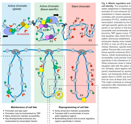
Fig. 4. Mitotic regulation and the control of cell identity.The properties of the regulatory states established in interphase at each promoter (P) and enhancer (E) that are maintained in mitosis depends on and correlates with several parameters. First, active promoters (P1/P2), whether they drive transcription (indicated by the black arrows) of cell type-specific genes (as shown in the green panel) or not (shown in the blue panel), remain accessible (red arrowheads) and are probably bound by TBP (green icons). This might ensure that daughter cells inherit the transcription pattern previously established. In contrast, enhancers display more variability: they may (E1/E2) or may not (E3) be accessible during mitosis. Moreover, specific bookmarking factors (yellow Pacman-like icon) primarily bind at tissue-specific enhancers (E2) over others (E1). Hence, mitotic bookmarking and enhancer accessibility could provide context-dependent specificity to the inheritance of gene regulation. Other enhancers close in mitosis (E3), leaving daughter cells with the option of re-opening or not. Similarly, silent regions (shown in the red panel) characterised by repressive marks (red stars), can transiently shift to active marks (green stars) in G2/M, and some regulators can bindde novoat these (E4) and other regions. This might contribute to changes in gene expression after interphase and pre-empt future cellular transitions.
DEVEL
O
competence to fully reprogramme adult somatic nuclei in interphase (Egli et al., 2007), and the efficiency of reprogramming mitotic somatic chromatin is dramatically enhanced (Halley-Stott et al., 2014). The molecular basis of the reprogramming advantage presented by mitotic samples is not fully understood but has been proposed to depend on histone ubiquitylation (Halley-Stott et al., 2014), which, as seen above, has been independently linked to an intriguing Polycomb-mediated post-mitotic reactivation (Arora et al., 2016). Moreover, and possibly promoted by the global mitotic accessibility of promoters, a large fraction of genes undergo a burst of transcription just after mitosis, which is stronger than at any other subsequent time in the cell cycle (Hsiung et al., 2016; Palozola et al., 2017) and leads to increased cell-to-cell variability (Hsiung et al., 2016). These observations support the notion that the remodelling of gene expression favoured by mitosis, albeit not mandatory, provides a platform from which changes in cell fate are then canalised in daughter cells (Chen et al., 2015). Perhaps not coincidentally, cell identity transitions are often initiated just after mitosis and the G1 phase tends to elongate during differentiation (Soufi and Dalton, 2016). Therefore, the gene regulatory events operating during mitosis, such as the maintenance of epigenetic marks, the preservation of chromatin accessibility, and the direct action of mitotic bookmarking factors, could play determinant roles during the developmental phases in which new cell identities are implemented (Fig. 4).
Insights into differentiation: balancing stability with flexibility Differentiation and the concurrent loss of cell potency are accompanied by increased levels of epigenetic repression acting on different groups of genes, depending on the lineage (Reik, 2007). These epigenetic mechanisms establish barriers to ensure lineage fidelity, as demonstrated in a wide range of reprogramming setups (Smith et al., 2016). The robustness of epigenetic mechanisms stems from the independence they acquire from the initial triggers that establish them (Berger et al., 2009; Henikoff and Greally, 2016). Although this undoubtedly provides stability to developmental cell fate choices, it consequently reduces the flexibility required to implement new regulatory architectures. Undifferentiated or early differentiating cells, such as early embryonic cells or activated adult stem cells and progenitors, face the opposing needs of rapidly adapting their transcriptome to change cell fate while concomitantly maintaining their transient cell states through division. For these cells, the mechanisms underlying their transcriptional memory must be stable enough to be propagated through one or more rounds of cell division, and yet sufficiently flexible and responsive to still allow for expedited changes in fate. Therefore, a conflict exists during development between stability and flexibility (Fig. 1), an issue that has been highlighted and discussed under the perspective of long- and short-term epigenetic memory (Reik, 2007). It is in this context that mitotic bookmarking may exhibit its developmental significance: it might convey substantial regulatory information from mother to daughter cells without compromising the required responsiveness to change cell fate. Indeed, repressing or inactivating a mitotic bookmarking factor not only abolishes its immediate function during interphase but also the memory of its effects. Moreover, different regulatory elements display distinct behaviours during mitosis. Promoters, for example, are generally accessible (Hsiung et al., 2015) and probably bookmarked by Tbp (Xing et al., 2008). Hence, basal activity and the availability of responsive promoters might not be particularly challenged in mitosis (Palozola et al., 2017), thereby ensuring the simple transmission of the set of active genes from mother to daughter cells (Fig. 4, blue and green panels).
This memory is developmentally reinforced by the activity of certain master transcription factors that bookmark subsets of enhancers, those associated with cell identity genes (Caravaca et al., 2013; Festuccia et al., 2016a; Kadauke et al., 2012; Liu et al., 2017), during mitosis (Fig. 4, green panel). However, not all enhancers are bookmarked by sequence-specific transcription factors or maintained in a fully accessible state, thereby opening a window of opportunity to generate new regulatory architectures in the following interphase (Fig. 4, red panel).
Insights into epigenetic regulation
It is generally unclear whether the active chromatin states observed in interphase are systematically epigenetic in nature (Box 1). Understanding how mitotic bookmarking factors exert their function might thus have important implications for this matter. Indeed, should mitotic bookmarking factors directly contribute to the maintenance of active chromatin states during mitosis, then a key defining property of epigenetic regulation, its independence from upstream regulators (Box 1), could be called into question. Rigorously speaking, if transcription factors are actively required for permissive chromatin states to be maintained during mitosis, then the various marks and open chromatin configurations observed in mitotic chromatin should not necessarily be considered epigenetic. Conversely, the regulation of chromatin also impinges upon transcription factor binding, mainly by restricting a subset of all the motifs present across the genome to be effectively occupied. This suggests that the reciprocal interactions between transcription factors and chromatin modifications that shape active regions in interphase remain partially active during mitosis. If the permanent action of transcription factors to maintain euchromatic properties during mitosis seems almost tautologically obvious (even though it has not been shown), the potential role of mitotic bookmarking factors in the control of heterochromatin is tantalising. Notwithstanding that some sequence-specific transcription factors have been shown to bind heterochromatic regions, including during mitosis (Raff et al., 1994), essentially nothing is known about the behaviour of mitotic bookmarking factors with respect to heterochromatin. Hence, studying their function could lead to a much more profound re-evaluation of how epigenetic memory is transferred between cellular generations. Recent studies indicating that sequence-specific transcription factors are required to propagate constitutive heterochromatin in yeast (Wang and Moazed, 2017) are suggestive of such a mechanism. Overall, should mitotic bookmarking be rigorously demonstrated as a mechanism of mitotic inheritance regulating developmental progression, then investigating how it complements and influences epigenetic regulation will impact our understanding of the mechanisms governing how cell identity is established, maintained and reprogrammed.
Studying mitotic bookmarking: experimental challenges and conceptual implications
Although the above discussion highlights exciting and potentially ground-breaking findings in the field, it should be pointed out that a number of technical issues and experimental limitations are inherent to the study of mitotic bookmarking. Below, we discuss those issues that we believe are essential to consider when studying this process, many of which have contributed to the accumulation of confounding results in the literature.
Fixation artefacts and mitotic behaviour of gene regulators
It is remarkable that the most common agent to fix cells for further analysis, ( para)formaldehyde, seems to artificially evict potential
DEVEL
O
bookmarking factors from mitotic chromosomes even when live-cell imaging demonstrates mitotic retention. This was observed and documented several years ago (Pallier et al., 2003), and was recently generalised (Teves et al., 2016). Because formaldehyde requires a minimal time of interaction between two partners to create a covalent bond efficiently (Schmiedeberg et al., 2009), and given that bookmarking factors are generally characterised by fast molecular dynamics, it is possible that formaldehyde fixation captures potential bookmarking factors when they are not bound to mitotic chromosomes (Teves et al., 2016). Nevertheless, several genomic localisation studies have now reported site-specific mitotic interactions using formaldehyde (Table 1). It may thus be concluded that the global coating of mitotic chromosomes is not due to the sum of site-specific interactions. Rather, formaldehyde might deplete the pool of factors that are engaged in non-specific, short-lived interactions with elements other than specific genomic sites, which would be primarily responsible for the global coating of the mitotic chromosomes (Fig. 3). However, given the difficulty of robustly identifying mitotic binding sites, and the variability in published results (Deluz et al., 2016; Liu et al., 2015), it remains possible that formaldehyde fixation is blurring our perception of mitotic bookmarking. To clarify this categorically, it is necessary to find alternative methods of fixation. Orthogonal approaches that are not based on crosslinking, such as transcription factor footprinting as measured by the activity of nucleases and transposases, could also be used as alternative evidence of local binding in mitosis (Teves et al., 2016), although it will be essential to consider the sequence biases of such protocols, which could either obscure a genuine footprint or imply the presence of a footprint when one is absent (Sung et al., 2016). Nonetheless, a major conclusion that can already be established based on these revelations is that the studies showing loss of binding during mitosis require critical re-evaluation
when they are solely based on immunostaining after
paraformaldehyde fixation. To what extent these considerations also impinge upon the study of cells in interphase (Schmiedeberg et al., 2009) also warrants increased attention.
Mitotic preparations and associated issues
An in-depth characterisation of mitotic binding requires access to relatively pure populations of mitotic cells. Synchronisation with nocodazole only enriches for mitosis, and thus needs to be coupled to cell sorting approaches after staining for mitotic markers (Kadauke et al., 2012) or, in the case of adherent cultures, mechanical harvesting of dividing cells by shake-off. However, nocodazole arrest has several major caveats. First, it blocks cells in prometaphase, restricting analyses to a specific stage of mitosis such that some conclusions might represent an oversimplification of a much more dynamic scenario (Chen et al., 2005). Second, nocodazole inhibition of microtubules could disrupt important and not yet understood mechanisms that might influence mitotic bookmarking, as shown for Brd4 (Nishiyama et al., 2012). Third, contamination from cells in other phases of the cell cycle is expected and this can be a significant confounding factor when assessing protein binding using conventional techniques such as chromatin immunoprecipitation (ChIP)-seq, which already poses quantitative issues (Meyer and Liu, 2014). As the signal in mitotic cells is almost always lower than that measured in interphase, the contamination from remnant interphase cells could be a major issue. Titrating pre-determined proportions of contaminant cells into chromatin prepared from cells depleted of the examined factor (Festuccia et al., 2016a; Liu et al., 2017) can, however, give insight into the magnitude of signal arising from contamination and into the
sensitivity of the ChIP. As a general rule, low efficiency ChIP that does not allow the detection of 5-10% of contaminant chromatin (Festuccia et al., 2016a; Nora et al., 2017), and mitotic preparations with more than 5% of contaminant interphase cells, should be treated with caution.
Addressing the function of mitotic bookmarking factors
A final major challenge is to fully disentangle the role of a given factor during mitosis from its function in interphase. Tagging the protein under study with domains derived from cell cycle-regulated proteins to drive mitotic degradation (Deluz et al., 2016; Kadauke et al., 2012; Liu et al., 2017), such as the degrons found in cyclins A and B, seems to be the gold standard in the field. However, results and interpretation must be carefully considered. First, the efficiency of such systems varies from factor to factor and, in some cases, could lead to hypomorphic levels of expression during the entire cell cycle. Second, this approach results in complete depletion towards the end of cell division, when the chromatin is decondensing, and extends during G1 (Festuccia et al., 2016a; Liu et al., 2017). Therefore, when this strategy is used to assess the effect of mitotic binding on the resumption of transcription after division, it is fundamental to consider that the tagged factors will be absent during G1. Hence, depending on the length of G1, the relationship between the measured effects and the activity of the factor during mitosis might be largely unrelated. Therefore, studying the biological consequences of a loss of mitotic bookmarking remains extremely complicated. Only once we understand the molecular basis of the mitotic behaviour of bookmarking factors, along with their molecular consequences, will we develop specific strategies to invalidate their mitotic function specifically.
Concluding remarks
Understanding how the cell cycle and developmental progression reciprocally influence each other has been a long-lasting theme in the field of gene regulation. Among the different hypotheses formulated in the past there is one, that of ‘quantal cell cycles’ (Holtzer et al., 1972), that seems worth reconsidering in light of mitotic bookmarking, albeit only partially. In this theory, it was proposed that, during differentiation, cells undergo ‘proliferative cycles’during which the identity of the mother cell is systematically reproduced in daughter cells. However, after a fixed number of divisions and after transiting a specific phase of the cell cycle, cells would undergo a ‘quantal cycle’, leading to their immediate progeny unfolding a new identity. Although this theory was dismissed (Grounds and McGeachie, 1987), the proposal that specific phases of the cell cycle represent a structural obstacle to gene regulation, and that this represents a challenge and an opportunity to control cell fate, is in fact a central theme in the field of mitotic bookmarking. Indeed, the dynamic interplay between local occupancy of transcription factors and the transcription machinery, on one side, and condensation and alterations in chromatin structure, on the other, makes the mitotic inheritance of regulatory states a potential means to control cell fate. Dissecting the reciprocal or hierarchical dependencies between mitotic bookmarking factors, epigenetic marks and mitosis-specific activities, and how these influence the restoration of a transcriptionally competent chromatin template in interphase, represents a stimulating area of research that could expand our general understanding of the molecular basis of cell identity. For this, comparative studies of several developmental stages and systems displaying differential requirements for epigenetic gene regulation, such as early mouse development (Festuccia et al.,
DEVEL
O
2016b), or of those displaying very different temporal scales with regard to cell cycle progression and lineage determination, such as the fast cell cycles of pre-gastrulation development in several species, might be necessary.
Acknowledgements
We acknowledge Shahragim Tajbakhsh, François Schweisguth and Alfonso Martinez-Arias for stimulating discussions.
Competing interests
The authors declare no competing or financial interests.
Funding
Research in the P.N. laboratory is supported by the Institut Pasteur, the Centre National de la Recherche Scientifique, the Agence Nationale de la Recherche (ANR)
Laboratoire d’Excellence Revive (Investissement d’Avenir; ANR-10-LABX-73), the
ANR DS0405-2016 program (MitMAT; ANR-16-CE12-0004), the Fondation ARC pour la Recherche sur le Cancer (PJA20161204705), and the Fondation
Schlumberger pour l’Education et la Recherche (cercle FSER-2016). N.F. was
funded by an European Molecular Biology Organization Long Term Fellowship
(ALTF 876-2013) and a Marie-Curie Actions Intra-European Fellowship (EFIMB–
626705). I.G. and N.O. are supported by Revive.
References
Alabert, C. and Groth, A. (2012). Chromatin replication and epigenome
maintenance.Nat. Rev. Mol. Cell Biol.13, 153-167.
Ali, S. A., Zaidi, S. K., Dobson, J. R., Shakoori, A. R., Lian, J. B., Stein, J. L., van Wijnen, A. J. and Stein, G. S.(2010). Transcriptional corepressor TLE1 functions
with Runx2 in epigenetic repression of ribosomal RNA genes.Proc. Natl. Acad.
Sci. USA107, 4165-4169.
Arora, M., Packard, C. Z., Banerjee, T. and Parvin, J. D.(2016). RING1A and BMI1
bookmark active genes via ubiquitination of chromatin-associated proteins.
Nucleic Acids Res.44, 2136-2144.
Berger, S. L., Kouzarides, T., Shiekhattar, R. and Shilatifard, A.(2009). An
operational definition of epigenetics.Genes Dev.23, 781-783.
Black, K. L., Petruk, S., Fenstermaker, T. K., Hodgson, J. W., Caplan, J. L.,
Brock, H. W. and Mazo, A.(2016). Chromatin proteins and RNA are associated
with DNA during all phases of mitosis.Cell Dis.2, 16038.
Blobel, G. A., Kadauke, S., Wang, E., Lau, A. W., Zuber, J., Chou, M. M. and
Vakoc, C. R.(2009). A reconfigured pattern of MLL occupancy within mitotic
chromatin promotes rapid transcriptional reactivation following mitotic exit.Mol.
Cell36, 970-983.
Blythe, S. A. and Wieschaus, E. F.(2016). Establishment and maintenance of
heritable chromatin structure during early Drosophila embryogenesis.eLife5,
e20148.
Booth, D. G., Beckett, A. J., Molina, O., Samejima, I., Masumoto, H., Kouprina,
N., Larionov, V., Prior, I. A. and Earnshaw, W. C.(2016). 3D-CLEM reveals
that a major portion of mitotic chromosomes is not chromatin. Mol. Cell 64,
790-802.
Brero, A., Easwaran, H. P., Nowak, D., Grunewald, I., Cremer, T., Leonhardt, H.
and Cardoso, M. C. (2005). Methyl CpG-binding proteins induce
large-scale chromatin reorganization during terminal differentiation.J. Cell Biol.169,
733-743.
Buchenau, P., Hodgson, J., Strutt, H. and Arndt-Jovin, D. J.(1998). The
distribution of polycomb-group proteins during cell division and development
in Drosophila embryos: impact on models for silencing.J. Cell Biol.141, 469-481.
Burke, L. J., Zhang, R., Bartkuhn, M., Tiwari, V. K., Tavoosidana, G., Kurukuti, S., Weth, C., Leers, J., Galjart, N., Ohlsson, R. et al.(2005). CTCF binding and higher order chromatin structure of the H19 locus are maintained in mitotic
chromatin.EMBO J.24, 3291-3300.
Caravaca, J. M., Donahue, G., Becker, J. S., He, X., Vinson, C. and Zaret, K. S.
(2013). Bookmarking by specific and nonspecific binding of FoxA1 pioneer factor
to mitotic chromosomes.Genes Dev.27, 251-260.
Chen, X., Hartman, A. and Guo, S.(2015). Choosing cell fate through a dynamic
cell cycle.Curr. Stem Cell Rep.1, 129-138.
Chen, D., Hinkley, C. S., Henry, R. W. and Huang, S.(2002). TBP dynamics in
living human cells: constitutive association of TBP with mitotic chromosomes.Mol.
Biol. Cell13, 276-284.
Chen, D., Dundr, M., Wang, C., Leung, A., Lamond, A., Misteli, T. and Huang, S.
(2005). Condensed mitotic chromatin is accessible to transcription factors and
chromatin structural proteins.J. Cell Biol.168, 41-54.
Christensen, M. O., Larsen, M. K., Barthelmes, H. U., Hock, R., Andersen, C. L., Kjeldsen, E., Knudsen, B. R., Westergaard, O., Boege, F. and Mielke, C.
(2002). Dynamics of human DNA topoisomerases IIalpha and IIbeta in living cells.
J. Cell Biol.157, 31-44.
Christova, R. and Oelgeschläger, T.(2002). Association of human TFIID-promoter
complexes with silenced mitotic chromatin in vivo.Nat. Cell Biol.4, 79-82.
Clark, K. L., Halay, E. D., Lai, E. and Burley, S. K.(1993). Co-crystal structure of
the HNF-3/fork head DNA-recognition motif resembles histone H5.Nature364,
412-420.
Darzynkiewicz, Z., Traganos, F., Sharpless, T. and Melamed, M. R.(1977a).
Different sensitivity of DNA in situ in interphase and metaphase chromatin to heat
denaturation.J. Cell Biol.73, 128-138.
Darzynkiewicz, Z., Traganos, F., Sharpless, T. and Melamed, M. R.(1977b).
Interphase and metaphase chromatin. Different stainability of DNA with acridine
orange after treatment at low pH.Exp. Cell Res.110, 201-214.
de Castro, I. J., Gokhan, E. and Vagnarelli, P.(2016). Resetting a functional G1
nucleus after mitosis.Chromosoma125, 607-619.
de Castro, I. J., Budzak, J., Di Giacinto, M. L., Ligammari, L., Gokhan, E., Spanos, C., Moralli, D., Richardson, C., de Las Heras, J. I., Salatino, S. et al.
(2017). Repo-Man/PP1 regulates heterochromatin formation in interphase.Nat.
Commun.8, 14048.
Dekker, J.(2014). Two ways to fold the genome during the cell cycle: insights
obtained with chromosome conformation capture.Epigenetics Chromatin7, 25.
Deluz, C., Friman, E. T., Strebinger, D., Benke, A., Raccaud, M., Callegari, A., Leleu, M., Manley, S. and Suter, D. M.(2016). A role for mitotic bookmarking of
SOX2 in pluripotency and differentiation.Genes Dev.30, 2538-2550.
Deluz, C., Strebinger, D., Friman, E. T. and Suter, D. M.(2017). The elusive role of
mitotic bookmarking in transcriptional regulation: insights from Sox2.Cell Cycle
16, 601-606.
Dey, A., Chitsaz, F., Abbasi, A., Misteli, T. and Ozato, K.(2003). The double
bromodomain protein Brd4 binds to acetylated chromatin during interphase and
mitosis.Proc. Natl. Acad. Sci. USA100, 8758-8763.
Dey, A., Nishiyama, A., Karpova, T., McNally, J. and Ozato, K.(2009). Brd4 marks
select genes on mitotic chromatin and directs postmitotic transcription.Mol. Biol.
Cell20, 4899-4909.
Dovat, S., Ronni, T., Russell, D., Ferrini, R., Cobb, B. S. and Smale, S. T.(2002). A common mechanism for mitotic inactivation of C2H2 zinc finger DNA-binding
domains.Genes Dev.16, 2985-2990.
Dowen, J. M., Bilodeau, S., Orlando, D. A., Hubner, M. R., Abraham, B. J.,
Spector, D. L. and Young, R. A.(2013). Multiple structural maintenance of
chromosome complexes at transcriptional regulatory elements.Stem Cell Rep.1,
371-378.
Easwaran, H. P., Schermelleh, L., Leonhardt, H. and Cardoso, M. C.(2004).
Replication-independent chromatin loading of Dnmt1 during G2 and M phases.
EMBO Rep.5, 1181-1186.
Egli, D., Rosains, J., Birkhoff, G. and Eggan, K. (2007). Developmental
reprogramming after chromosome transfer into mitotic mouse zygotes.Nature
447, 679-685.
Egli, D., Birkhoff, G. and Eggan, K. (2008). Mediators of reprogramming:
transcription factors and transitions through mitosis.Nature Rev. Mol. Cell Biol.
9, 505-516.
Fairley, J. A., Scott, P. H. and White, R. J.(2003). TFIIIB is phosphorylated,
disrupted and selectively released from tRNA promoters during mitosis in vivo.
EMBO J.22, 5841-5850.
Ferraro, T., Esposito, E., Mancini, L., Ng, S., Lucas, T., Coppey, M., Dostatni, N.,
Walczak, A. M., Levine, M. and Lagha, M.(2016). Transcriptional memory in the
drosophila embryo.Curr. Biol.26, 212-218.
Festuccia, N., Dubois, A., Vandormael-Pournin, S., Gallego Tejeda, E., Mouren, A., Bessonnard, S., Mueller, F., Proux, C., Cohen-Tannoudji, M. and Navarro,
P., (2016a). Mitotic binding of Esrrb marks key regulatory regions of the
pluripotency network.Nat. Cell Biol.18, 1139-1148.
Festuccia, N., Gonzalez, I. and Navarro, P.(2016b). The epigenetic paradox of
pluripotent ES cells.J. Mol. Biol.429, 1476-1506.
Fischle, W., Tseng, B. S., Dormann, H. L., Ueberheide, B. M., Garcia, B. A., Shabanowitz, J., Hunt, D. F., Funabiki, H. and Allis, C. D.(2005). Regulation of
HP1-chromatin binding by histone H3 methylation and phosphorylation.Nature
438, 1116-1122.
Follmer, N. E., Wani, A. H. and Francis, N. J.(2012). A polycomb group protein is
retained at specific sites on chromatin in mitosis.PLoS Genet.8, e1003135.
Fonseca, J. P., Steffen, P. A., Muller, S., Lu, J., Sawicka, A., Seiser, C. and
Ringrose, L.(2012). In vivo Polycomb kinetics and mitotic chromatin binding
distinguish stem cells from differentiated cells.Genes Dev.26, 857-871.
Gazit, B., Cedar, H., Lerer, I. and Voss, R.(1982). Active genes are sensitive to
deoxyribonuclease I during metaphase.Science217, 648-650.
Gerlich, D., Hirota, T., Koch, B., Peters, J.-M. and Ellenberg, J. (2006).
Condensin I stabilizes chromosomes mechanically through a dynamic
interaction in live cells.Curr. Biol.16, 333-344.
Giorgetti, L., Servant, N. and Heard, E.(2013). Changes in the organization of the
genome during the mammalian cell cycle.Genome Biol.14, 142.
Gottesfeld, J. M. and Forbes, D. J.(1997). Mitotic repression of the transcriptional
machinery.Trends Biochem. Sci.22, 197-202.
Grandy, R. A., Whitfield, T. W., Wu, H., Fitzgerald, M. P., VanOudenhove, J. J., Zaidi, S. K., Montecino, M. A., Lian, J. B., van Wijnen, A. J., Stein, J. L. et al.
(2015). Genome-wide studies reveal that H3K4me3 modification in bivalent genes