ISSN Online: 2150-4105 ISSN Print: 2150-4091
Heterogeneous Microstructure and Distribution
of Trace Elements in Coral Stylophora pistillata
Nursed in the Phosphate Loading Berth Site in
the Gulf of Aqaba
Ali Al-Sawalmih
1,2*, Jafar Megdadi
11Marine Science Station, The University of Jordan & Yarmouk University, Aqaba, Jordan
2Department of Coastal Environment, Faculty of Marine Sciences, The University of Jordan, Aqaba, Jordan
Abstract
Cultured Stylophora pistillata in Phosphate Loading Berth (PLB) sites in the Jorda-nian Gulf of Aqaba was investigated using Back-Scatter Scanning Electron (BSE) mi-croscopy and Energy-Disperse X-ray (EDX) to determine trace elements and calcium concentrations’ distribution within the micrometer scale. Ca, C, and O in addition to six trace elements (Mg, Sr, S, Cl, Ni, and Na) were mapped within the sample cross section. Samples from the PLB were as expected poorly mineralized as previously reported and showed mineralization heterogeneity in the micrometer length scale in the BSE image. In four selected positions within the cross sections, Ca concentration found to range between 0.44 - 1.80 wt% in low-mineralized regions and between 3.99 - 4.66 wt% in mineralized regions. The average calcium concentrations were in accor-dance to previous study; about 10% of the Ca existed in the same coral species from other coastal sites in the Gulf of Aqaba. This could be attributed to the role of phos-phate in inhibition of calcification and enhancement of photosynthesis. Amounts of trace elements in lower Ca concentration positions within the cross section were rel-atively very low except for chlorine, whereas positions with higher Ca contained amounts of Na and Sr. This study reports remarkable heterogeneity in mineral dis-tribution within the microstructure of the coral Stylophora pistillata under phos-phate pollution stress.
Keywords
Phosphate, Coral, Gulf of Aqaba, Calcification, Stylophora pistillata, EDX, SEM How to cite this paper: Al-Sawalmih, A.
and Megdadi, J. (2016) Heterogeneous Mi-crostructure and Distribution of Trace Ele-ments in Coral Stylophora pistillata Nursed in the Phosphate Loading Berth Site in the Gulf of Aqaba. Natural Science, 8, 541-552.
http://dx.doi.org/10.4236/ns.2016.812053
Received: May 3, 2016 Accepted: December 2, 2016 Published: December 5, 2016
Copyright © 2016 by authors and Scientific Research Publishing Inc. This work is licensed under the Creative Commons Attribution International License (CC BY 4.0).
http://creativecommons.org/licenses/by/4.0/
1. Introduction
Terrestrial runoff into seawater leads to increasing loads of nutrients, sediments and pollutants discharged from the land like using and handling of fertilizers that contain phosphate [1] [2], which is considered to be a growing concern for most of the coun-tries endowed with coral reefs [3] [4]. This is referred to high sensitivity of coral reef ecosystems to changes in the surrounding physicochemical environment [5].
Seawater contains trace elements in the form of K+, Na+, Mg2+, Ca2+, Cl− and 2 4 SO−
which have significant impacts on the formation and elemental composition of marine skeletons like corals [6]. Trace elements are secreted within the coral skeleton through either direct replacement of calcium (or carbonate) in coral aragonite structure for di-valent ions [7] [8] [9], while non-didi-valent ions are trapped by the inclusion of detritus materials into skeletal pore spaces, uptake of organic materials, incorporation of metals into coral skeletons, or coral feeding [7] [8] [10] [11] [12]. The incorporation of trace elements in coral skeletal aragonite crystals provides chronological proxies for physio-chemical changes in the surrounding environment [12] [13] [14].
Conventional loading systems of phosphates produced as a constituent of fertilizer result in very high levels of dust pollution in the surrounding coastal area, diffused and transported along the sea by winds [15]. Phosphate is an extremely dry and dusty ma-terial and its solubility in seawater is relatively high (20 - 56 micrograms per liter, de-pending on particle size) [16] [17]. In Aqaba seawater, phosphate dust was found to be soluble over the annual water temperature range and some algae may be able to solu-bilize particulate phosphate [18] [19].
Phosphate pollution has also been implicated as a contributor to the decline of reef ecosystems in the Gulf of Aqaba [20] [21] [22] [23]. Phosphate was reported to cause 36% inhibition of calcification in Stylophora pistillata at 2 ppm concentrations in sea-water, and stop it completely (99%) at 100 ppm [24]. It was found that phosphate is re-sponsible for increasing the death rate of Stylophora pistillata coral [25] while other study reported tolerance of coral species Stylophora pistillata to phosphate [26]. Al- Sawalmih [27] in his studies on branches Stylophora pistillata that are nursed in the Phosphate Loading Berth (PLB) site, found very low calcium composition in the arago-nitic skeleton, about 10% of it in the control samples. He also found that these micro-structures of these branches suffered from alteration due to dominance of organic fibers in the skeleton.
The Stylophora pistillata is among the important common coral species in the Gulf of Aqaba as well as in the Red Sea and known to be the fastest of the scleractinian cor-als. It is also very suitable for research because it can also be sampled without causing major damage to the colony [28] [29].
2. Materials and Methods
Small fragments of the model branching coral Stylophora pistillata were cultured in a mid-water floating nursery along the Jordanian coast of Gulf of Aqaba [30]. The frag-ments developed into small colonies to be used as bio-indicator for marine pollution in the selected sites.
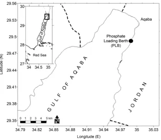
Coral samples were collected from the nurseries in the Phosphate Loading Berth (PLB) site (Figure 1), which lies about 3 km south of Aqaba city and is the single port for loading and exporting phosphate ore in Jordan.
Coral samples were cut perpendicular to the skeletal growth using low speed di-amond saw (BUEHLER Isomat, Germany) and cross section surfaces were further po-lished using ethyleneglycol lubricant to provide a flat surface that was coated with Au for the SEM imaging.
[image:3.595.210.540.390.676.2]Field-Emission Scanning Electron Microscope (JEOL JSM-7500F) with an Oxford Instruments detector was used for performing the EDX analysis to measure the ele-mental content in the coral samples and their corresponding emission spectra, as well as acquiring Back-Scatter Electron (VSE) microscopy images for the cross section of the sample. EDX analysis usually involves the generation of an X-ray spectrum from the entire scan area of the SEM, which shows the counts of X-rays received and processed by the detector and the energy level of those counts. The EDX instrument software as-sociates the energy level of the X-rays with the elements found in the scan area.
3. Results and Discussion
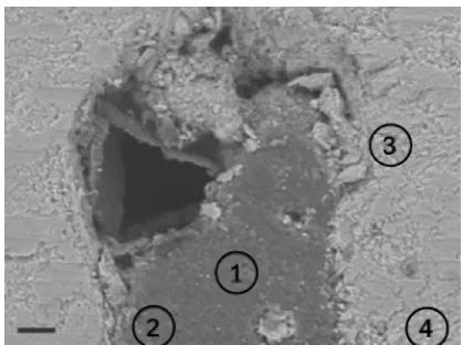
Figure 2 shows a Back-Scattering Electron (BSE) image for a polished surface of the coral sample. BSE can be used for rapid discrimination of phases in multiphase samples with high contrast of electron density, like organic and mineral phases, based on less- and more Ca concentrations.
Back-Scattered Scanning Electron Microscopy (BS-SEM) images of coral samples collected from the Phosphate Loading Berth (PLB) in the Jordanian Gulf of Aqaba (Figure 2) showed also that coral fragments from the phosphate pollution site is hete-rogeneous in the micrometer scale comprised of less-mineralized parts with very low calcium concentrations and mineralized parts with relatively higher calcium concentra-tions.
In Figure 2, the dark regions resemble the less-Ca phases and the bright ones indi-cates for the mineralized areas. This heterogeneous structure comes in accordance to the last study by Al-Sawalmih [27] on Stylophora pistillata coral samples from the phosphate pollution site, reporting dominance of organic/low-mineralized microstruc-ture in SEM images and elemental analysis. This can be attributed to the role of phos-phate in enhancement of coral zooxanthellae photosynthesis that is responsible for synthesis of organic matrix in the skeleton [31].
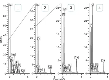
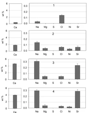
Four positions in the cross-section in the BSE image (Figure 2) were selected for elemental analysis using Energy-Dispersive X-rays (EDX); (1) and (2) in the less- mineralized “dark” region and (3) and (4) in the more mineralized “bright” region. The recorded EDX spectra from the positions 1-4 (Figure 3) were used by the instrument software to determine the weight percentage (wt%) of Oxygen (O), Carbon (C) and Calcium (Ca) in addition to the trace elements: N, Mg, S, Cl, Ni and Sr. The full quan-titative elemental analysis for the positions 1-4 are presented in Table 1.
[image:4.595.268.477.507.663.2]In all locations, Ca concentrations were very low as expected and recorded in pre-vious study on such coral in the same site [27] which was only 2.56 wt%, which is around 12% of the average in the control sample (21.95 wt%) but with a focus area
Figure 3. EDX spectra of the four selected positions for elemental analysis.
Table 1. Concentrations in wt% of C, O, Ca, and trace elements in the EDX scanned positions 1-4 in the coral cross section BSE image (Figure 2).
1 2 3 4
C 74.72 ± 0.11 57.86 ± 0.12 41.85 ± 0.14 37.93 ± 0.14 O 24.67 ± 0.11 40.01 ± 0.12 52.85 ± 0.14 57.40 ± 0.14
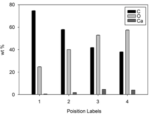
Ca 0.44 ± 0.01 1.80 ± 0.02 4.66 ± 0.03 3.99 ± 0.03
Na 0.03 ± 0.01 0.14 ± 0.02 0.31 ± 0.02 0.30 ± 0.02
Mg --- 0.04 ± 0.01 0.05 ± 0.01 0.04 ± 0.02
S --- --- --- ---
Cl 0.14 ± 0.01 0.06 ± 0.01 0.05 ± 0.01 0.03 ± 0.01
Ni --- 0.03 ± 0.01 --- 0.02 ± 0.01
Sr --- 0.06 ± 0.02 0.24 ± 0.03 0.29 ± 0.03
averages over the range of 100 m. In our study, the focus area for the EDX analysis was in the range of 1 m in order to map the heterogeneity in trace element distribution within the skeleton microstructure.
Figure 4. Bar chart representation of the concentrations in wt% of C, O, and Ca in the EDX scanned positions 1-4 in the coral cross section BSE image (Figure 2).
elements needs to be further investigated based on expanded experimental work and li-terature survey. In the less-mineral region, position (2) has quite higher Ca than (1) because the last is located close to the less- and higher mineralized interface (Figure 2), which may give an indicator to the gradient behavior of Ca concentration through this boundary.
The amount of trace elements (N, Mg, S, Cl, Ni, and Sr) in the four scanned positions in the coral sample cross section (Figure 2) are shown in Figure 5. The EDX elemental analysis shows that the coral samples contain considerable amounts of trace elements (see Figure 1) which is strongly related to the chemistry of surrounding seawater dur-ing skeleton formation [10] [32] [33].
Except for Chlorine (Cl), trace elements in the position 1 of the lowest calcium con-tent were very low (<0.1 wt%) so their amounts can’t be considered due to possible un-certainty in their values. Trace elements concentrations can be reliably considered only in regions of high Ca concentrations.
The relatively high Cl concentration in position (1) which is 3 times more than it in other positions cannot be explained. Chlorine (Cl) exist in coral skeletons (but not in the aragonitic structure), as potassium chloride or as sodium chloride phases and is considered to be toxic for corals [34]. The main source of excess of Cl in seawater is sewage treatment that during floods which may be mixed with seawater, in addition to biofouling treatment in industrial effluents [35].
Figure 5. bar chart representation of the concentrations in wt% of Ca, and trace elements in the EDX scanned positions 1-4 in the coral cross section BSE image (Figure 2).
0.015. However, the method used for this study is not optimal for calculating trace- element/Ca (mmole/mole) ratios.
Although Na is not a divalent cation (monovalent ion), i.e. it does not satisfy the electrostatic valence requirements of the Ca position in the aragonite lattice, but being the second most abundant ion in seawater [37], in addition to the similarity of its ionic radius, Na transfers rapidly from sea water into the crystallizing skeletal material [38]. The charge imbalances resulting from the incorporation of Na+ into aragonite lattices is compensated by lattice defects and distortions such as vacancy sites and interstitial ions that occur at higher density at the center of calcification [36] [39].
seawater temperature at the time of coral growth, but anthropogenic pollution in sea-water may effect on the temperature record [41].
Mg is generally found in much lower concentrations in aragonitic skeletons due to its smaller ionic radius than Ca [42], although being the third most abundant ion in sea-water, behind sodium (Na) and chloride (Cl) [6].
Due to its inhibition of calcification, high levels of phosphate are inversely associated with decreasing concentrations of strontium in the corals [43]. Nevertheless, morpho-logical alteration of the coral skeleton due increased levels of phosphate suggests also that strontium may provide an excellent indicator of environmental stress related to anthropogenic-induced elevated nutrient levels [41].
Sulphur (S) was not detected in our sample. It is the forth abundant ion in seawater (as sulfate 2
4
SO−) following close behind Mg [37]. It exists in coral skeletons as
occu-pying specific sites in the crystal lattice of marine aragonites [44] [45].
Nickel (Ni) concentrations in the four scanned positions were almost not detected. It is weakly incorporated into the aragonite skeleton relative to calcium concentrations due to structural incompatibility [46]. Although Ni is vital for the function of many or-ganisms, concentrations in some areas from both anthropogenic release and natural sources might increase its concentrations to toxic levels to living organisms [47] [48] [49].
The low calcium in corals that are stressed by phosphate dust pollution, is due to the enrichment of phosphate as nutrient in seawater which cause inhibition of calcification in corals and coralline algae as well [25] [50]. It was reported that phosphate in seawa-ter decrease calcification in the corals Pocillopora damicornis [51], through increasing eutrophication which effects on survival and stability of reef communities by disrupting normal carbon cycle functions [52] [53] [54]. However, the mechanisms by which dis-solved inorganic nutrients like phosphates can disturb corals are subject to controver-sial debate [55].
The environmental impacts of the deposited phosphate dust on marine ecosystem include not only siltation of the coral reef and depression of coral growth (Hawkins et al., 1991) but also increasing suspended solids and water turbidity, reduction of water clarity and light penetration [56]. Phosphate dust also has physical effects in reducing the light intensity and increasing sedimentation, both of which have been shown to have deleterious effects on reefs [57]. Reduced light, excess phosphate and sedimenta-tion were found to be responsible for reduced calcificasedimenta-tion and increased mortality of corals [25].
4. Conclusion
was found to be higher in the lowest-calcium concentrations than other regions. Fur-ther elemental analysis and microstructure studies could be conducted on oFur-ther coral species and several skeletal organisms that live in the Phosphate Terminal coastal site in order to find out their response to phosphate pollution. Moreover, trace element/Ca ra-tios should be considered in future similar studies using suitable investigation tech-niques for complete analysis.
Acknowledgements
The authors are indebted to Prof. Fuad Al-Horani for providing the coral samples used for this study and to Prof. Peter Fratzl for giving the chance to use the SEM laboratory at the Max-Planck Institute for Colloids and Interfaces (MPIKG). Many thanks to Su-sann Weichold and Heike Runge of the SEM lab Germany for their support and help during the measurements, also we are thankful to the divers Ali Al-Njadat and Omar Al-Momany for collecting the coral samples used in this study.
References
[1] Smith, S.V., et al. (2003) Humans, Hydrology, and the Distribution of Inorganic Nutrient Loading to the Ocean. Bioscience, 53, 235-245.
https://doi.org/10.1641/0006-3568(2003)053[0235:HHATDO]2.0.CO;2
[2] Tilman, D., et al. (2001) Forecasting Agriculturally Driven Global Environmental Change. Science, 292, 281-284. https://doi.org/10.1126/science.1057544
[3] Bryant, D.G., et al. (1998) Reefs at Risk: A Map-Based Indicator of Threats to the World’s Coral Reefs. World Resources Institute, Washington DC.
[4] Spalding, M., Ravilious, C. and Green, E.P. (2001) World Atlas of Coral Reefs. University of California Press, Berkeley.
[5] Johannes, R.E. (1972) Coral Reefs and Pollution. In: Ruivo M., Ed., Marine Pollution and Sea Life, Fishing News (Books), London, 364-374.
[6] Martin, D.F. (1972) Marine Chemistry. 2nd Edition, M. Dekker, New York.
[7] Correge, T. (2006) Sea Surface Temperature and Salinity Reconstruction from Coral Geo-chemical Tracers. Palaeogeography Palaeoclimatology Palaeoecology, 232, 408-428.
https://doi.org/10.1016/j.palaeo.2005.10.014
[8] Flor, T.H. and Moore, W.S. (1977) Radium/Calcium and Uranium/Calcium Determina-tions for Western Atlantic Reef Corals. 3rd International Coral Reef Symposium, 2, 555- 561.
[9] Weber, J.N. (1973) Incorporation of Strontium into Reef Coral Skeletal Carbonate. Geo-chimica et CosmoGeo-chimica Acta, 37, 2173-2190.
https://doi.org/10.1016/0016-7037(73)90015-X
[10] Hanna, R.G. and Muir, G.L. (1990) Red-Sea Corals as Biomonitors of Trace-Metal Pollu-tion. Environmental Monitoring and Assessment, 14, 211-222.
https://doi.org/10.1007/BF00677917
[11] Howard, L.S. and Brown, B.E. (1986) Metals in Tissues and Skeleton of Fungia-Fungites from Phuket, Thailand. Marine Pollution Bulletin, 17, 569-570.
https://doi.org/10.1016/0025-326X(86)90573-4
Thermometry in Coral Skeletons. Science, 204, 404-407.
https://doi.org/10.1126/science.204.4391.404
[13] Al-Rousan, S., Al-Shloul, R., Al-Horani, F. and Abu-Hilal, A. (2012) Heavy Metals Signa-ture of Human Activities Recorded in Coral Skeletons along the Jordanian Coast of the Gulf of Aqaba, Red Sea. Environmental Earth Sciences, 67, 2003-2013.
https://doi.org/10.1007/s12665-012-1640-0
[14] Stanley, S.M. and Hardie, L.A. (1998) Secular Oscillations in the Carbonate Mineralogy of Reef-Building and Sediment-Producing Organisms Driven by Tectonically Forced Shifts in Seawater Chemistry. Palaeogeography, Palaeoclimatology, Palaeoecology, 144, 3-19.
https://doi.org/10.1016/S0031-0182(98)00109-6
[15] Sandler, D. (1993) Protecting the Gulf of Aqaba: A Regional Environmental Challenge, En-vironmental Law Institute, Washington DC, 512 p.
[16] Bemert, G. and Ormond, R. (1981) Red Sea Coral Reefs. Kegan Paul International, London, 192 p.
[17] Freemantle, M.H., Hulings, N., Mulqi, M. and Watton, E.C. (1978) Calcium and Phosphate in Jordan Gulf of Aqaba. Marine Pollution Bulletin, 9, 79-80.
https://doi.org/10.1016/0025-326X(78)90454-X
[18] Grobler, D.C. and Davies, E. (1979) Availability of Sediment Phosphate to Algae. Water SA, 5, 114-122.
[19] McColl, R.H.S. (1975) Availability of Soil and Sediment Phosphorus to a Planktonic Alga. New Zealand Journal of Marine and Freshwater Research, 9, 169-182.
https://doi.org/10.1080/00288330.1975.9515557
[20] Fishelson, L. (1973) Ecology of Coral Reefs in Gulf of Aqaba (Red-Sea) Influenced by Pollu-tion. Oecologia, 12, 55-67. https://doi.org/10.1007/BF00345470
[21] Fishelson, L. (1977) Stability and Instability of Marine Ecosystems, Illustrated by Examples from Red-Sea. Helgolander Wissenschaftliche Meeresuntersuchungen, 30, 18-29.
https://doi.org/10.1007/BF02207822
[22] Loya, Y. (1975) Possible Effects of Water-Pollution on Community Structure of Red-Sea Corals. Marine Biology, 29, 177-185. https://doi.org/10.1007/BF00388987
[23] Al-Sawalmih, A., et al. (2016) Elemental Analysis of the Branched Coral Stylophora pistil-lata Nursed along the Jordanian Coast of Gulf of Aqaba. Marine Pollution Bulletin, In Preparation.
[24] Yamashiro, H. (1995) The Effects of Hebp, an Inhibitor of Mineral Deposition, upon Pho-tosynthesis and Calcification in the Scleractinian Coral, Stylophora pistillata. Journal of Experimental Marine Biology and Ecology, 191, 57-63.
https://doi.org/10.1016/0022-0981(95)00045-S
[25] Walker, D.I. and Ormond, R.F.G. (1982) Coral Death from Sewage and Phosphate Pollu-tion at Aqaba, Red-Sea. Marine Pollution Bulletin, 13, 21-25.
https://doi.org/10.1016/0025-326X(82)90492-1
[26] Loya, Y. (1976) Red-Sea Coral Stylophora pistillata Is a R-Strategist. Nature, 259, 478-480.
https://doi.org/10.1038/259478a0
[27] Al-Sawalmih, A. (2016) Calcium Composition and Microstructure of Coral Stylophora pis-tillata under Phosphate Pollution Stress in the Gulf of Aqaba. Natural Science, 8, 89-95.
https://doi.org/10.4236/ns.2016.83012
[28] Loya, Y. (1972) Community Structure and Species Diversity of Hermatypic Corals at Eilat, Red Sea. Marine Biology, 13, 100-123. https://doi.org/10.1007/BF00366561
Yonge, M. and Stoddart, D.R., Eds., Regional Variation of Indian Ocean Coral Reefs, Aca-demic Press, New York, 117-139.
[30] Al-Horani, F. (2013) Sustainable Resources of Corals for the Restoration of Damaged Coral Reefs in the Gulf of Aqaba, Red Sea. Life Science Journal, 10, 352-360.
[31] Jones, O.A. and Endean, R. (1973) Biology and Geology of Coral Reefs. Academic Press, New York.
[32] Esslemont, G. (2000) Heavy Metals in Seawater, Marine Sediments and Corals from the Townsville Section, Great Barrier Reef Marine Park, Queensland. Marine Chemistry, 71, 215-231. https://doi.org/10.1016/S0304-4203(00)00050-5
[33] Ramos, A.A., Inoue, Y. and Ohde, S. (2004) Metal Contents in Porites Corals: Anthropo-genic Input of River Run-Off into a Coral Reef from an Urbanized Area, Okinawa. Marine Pollution Bulletin, 48, 281-294. https://doi.org/10.1016/j.marpolbul.2003.08.003
[34] Johannes, R.E. (1975) Chapter 2: Pollution and Degradation of Coral Reef Communities. In: Wood, E.J.F. and Johannes, R.E., Eds., Elsevier Oceanography Series, Elsevier, Amster-dam, 13-51. https://doi.org/10.1016/s0422-9894(08)71107-3
[35] Wood, E.J.F. and Johannes, R.E. (1975) Tropical Marine pollution. Elsevier Oceanography Series 12, Elsevier, Amsterdam, 192 p.
[36] Mitsuguchi, T., Uchida, T. and Matsumoto, E. (2010) Na/Ca Variability in Coral Skeletons. Geochemical Journal, 44, 261-273. https://doi.org/10.2343/geochemj.1.0067
[37] Turekian, K.K. (1968) Oceans. Foundations of Earth Science Series, Prentice-Hall, Engle-wood, 120 p.
[38] Harriss, R.C. and Almy, J.C.C. (1964) A Preliminary Investigation into the Incorporation and Distribution of Minor Elements in the Skeletal Material of Scleractinian Corals. Bulle-tin of Marine Science, 14, 418-423.
[39] White, A.F. (1977) Sodium and Potassium Coprecipitation in Aragonite. Geochimica et Cosmochimica Acta, 41, 613-625. https://doi.org/10.1016/0016-7037(77)90301-5
[40] Wray, J.L. and Daniels, F. (1957) Precipitation of Calcite and Aragonite. Journal of the American Chemical Society, 79, 2031-2034. https://doi.org/10.1021/ja01566a001
[41] Rasmussen, C.E. (1988) The Use of Strontium as an Indicator of Anthropogenically Altered Environmental Parameters. 6th International Coral Reef Symposium Executive Committee, Townsville, 8-12 August 1988, 325-339.
[42] Chave, K.E. (1954) Aspects of the Biogeochemistry of Magnesium 1. Calcareous Marine Organisms. Journal of Geology, 62, 266-283. https://doi.org/10.1086/626162
[43] Senesi, N., Polemio, M. and Lorusso, L. (1983) Evaluation of Barium, Rubidium and Stron-tium Contents in Commercial Fertilizers. Fertilizer Research, 4, 135-144.
https://doi.org/10.1007/BF01053250
[44] Barmatthews, M., Wasserburg, G.J. and Chen, J.H. (1993) Diagenesis of Fossil Coral Skele-tons: Correlation between Trace-Elements, Textures, and U-234/U-238. Geochimica et Cosmochimica Acta, 57, 257-276. https://doi.org/10.1016/0016-7037(93)90429-Z
[45] Vielzeuf, D., et al. (2013) Distribution of Sulphur and Magnesium in the Red Coral. Chem-ical Geology, 355, 13-27. https://doi.org/10.1016/j.chemgeo.2013.07.008
[46] Rasmussen, C.E., Cuff, C. and Hopley, D. (1992) Evidence of Anthropogenic Disturbances Retained in the Skeleton of Massive Corals from Australia’s Great Barrier Reef. 7th Interna-tional Coral Reef Symposium, Guam, 22-27 June 1992.
https://doi.org/10.1016/j.toxlet.2004.02.018
[48] Haber, L.T., et al. (2000) Hazard Identification and Dose Response of Inhaled Nickel- Soluble Salts. Regulatory Toxicology and Pharmacology, 31, 210-230.
https://doi.org/10.1006/rtph.2000.1377
[49] Scott-Fordsmand, J.J. (1997) Toxicity of Nickel to Soil Organisms in Denmark. In: Ware, G.W., Nigg, H.N. and Bevenue, A., Eds., Reviews of Environmental Contamination and Toxicology: Continuation of Residue Reviews, Springer, New York, 1-34.
https://doi.org/10.1007/978-1-4612-2264-4_1
[50] Simkiss, K. (1964) Phosphates as Crystal Poisons of Calcification. Biological Reviews of the Cambridge Philosophical Society, 39, 487-504.
https://doi.org/10.1111/j.1469-185X.1964.tb01166.x
[51] Lamberts, A.E. (1974) Measurement of Alizarin Deposited by Coral. In: Cameron, A.M. and Cambell, B.M., Eds., The Second International Coral, The Great Barrier Reef Waters, Department of Zoology, University of Hawaii, Honolulu, 241-244.
[52] Sawall, Y., Teichberg, M.C., Seemann, J., Litaay, M., Jompa, J. and Richter, C. (2011) Nutri-tional Status and Metabolism of the Coral Stylophora subseriata along a Eutrophication Gradient in Spermonde Archipelago (Indonesia). Coral Reefs, 30, 841-853.
https://doi.org/10.1007/s00338-011-0764-0
[53] Godinot, C., Ferrier-Pagès, C., Montagna, P. and Grover, R. (2011) Tissue and Skeletal Changes in the Scleractinian Coral Stylophora pistillata Esper 1797 under Phosphate Enrichment. Journal of Experimental Marine Biology and Ecology, 409, 200-207.
https://doi.org/10.1016/j.jembe.2011.08.022
[54] Dunn, J.G., Sammarco, P.W. and LaFleur Jr., G. (2012) Effects of Phosphate on Growth and Skeletal Density in the Scleractinian Coral Acropora muricata: A Controlled Experimental Approach. Journal of Experimental Marine Biology and Ecology, 411, 34-44.
https://doi.org/10.1016/j.jembe.2011.10.013
[55] D’Angelo, C. and Wiedenmann, J. (2014) Impacts of Nutrient Enrichment on Coral Reefs: New Perspectives and Implications for Coastal Management and Reef Survival. Current Opinion in Environmental Sustainability, 7, 82-93.
https://doi.org/10.1016/j.cosust.2013.11.029
[56] Hashwa, F. (1980) The Phosphate Pollution in the Gulf of Aqaba. Khartoum.
Submit or recommend next manuscript to SCIRP and we will provide best service for you:
Accepting pre-submission inquiries through Email, Facebook, LinkedIn, Twitter, etc. A wide selection of journals (inclusive of 9 subjects, more than 200 journals)
Providing 24-hour high-quality service User-friendly online submission system Fair and swift peer-review system
Efficient typesetting and proofreading procedure
Display of the result of downloads and visits, as well as the number of cited articles Maximum dissemination of your research work
Submit your manuscript at: http://papersubmission.scirp.org/