blockade of talin-dependent integrin
aa
IIb
bb
3
(platelet GPIIb–IIIa) activation
Brian G. Petrich, … , Sanford J. Shattil, Mark H. Ginsberg
J Clin Invest.
2007;
117(8)
:2250-2259.
https://doi.org/10.1172/JCI31024
.
In vitro studies indicate that binding of talin to the
b
3integrin cytoplasmic domain (tail)
results in integrin
a
IIbb
3(GPIIb–IIIa) activation. Here we tested the importance of talin
binding for integrin activation in vivo and its biological significance by generating mice
harboring point mutations in the
b
3tail. We introduced a
b
3(Y747A) substitution that disrupts
the binding of talin, filamin, and other cytoplasmic proteins and a
b
3(L746A) substitution that
selectively disrupts interactions only with talin. Platelets from animals homozygous for each
mutation showed impaired agonist-induced fibrinogen binding and platelet aggregation,
providing proof that inside-out signals that activate
a
IIbb
3require binding of talin to the
b
3tail.
b
3(L746A) mice were resistant to both pulmonary thromboembolism and to ferric
chloride–induced thrombosis of the carotid artery. Pathological bleeding, measured by the
presence of fecal blood and development of anemia, occurred in 53% of
b
3(Y747A) and
virtually all
b
3-null animals examined. Remarkably, less than 5% of
b
3(L746A) animals
exhibited this form of bleeding. These results establish that
a
IIbb
3activation in vivo is
dependent on the interaction of talin with the
b
3integrin cytoplasmic domain. Furthermore,
they suggest that modulation of
b
3integrin–talin interactions may provide an attractive target
for antithrombotics and result in a reduced risk of pathological bleeding.
Research Article
Hematology
Find the latest version:
Research article
2250 The Journal of Clinical Investigation http://www.jci.org Volume 117 Number 8 August 2007
The antithrombotic potential of selective
blockade of talin-dependent integrin
α
IIb
β
3
(platelet GPIIb–IIIa) activation
Brian G. Petrich, Per Fogelstrand, Anthony W. Partridge, Nima Yousefi, Ararat J. Ablooglu, Sanford J. Shattil, and Mark H. Ginsberg
Department of Medicine, UCSD School of Medicine, La Jolla, California, USA.
In vitro studies indicate that binding of talin to the
β
3integrin cytoplasmic domain (tail) results in integrin
α
IIbβ
3(GPIIb–IIIa) activation. Here we tested the importance of talin binding for integrin activation in vivo
and its biological significance by generating mice harboring point mutations in the
β
3tail. We introduced
a
β
3(Y747A) substitution that disrupts the binding of talin, filamin, and other cytoplasmic proteins and a
β
3(L746A) substitution that selectively disrupts interactions only with talin. Platelets from animals
homozy-gous for each mutation showed impaired agonist-induced fibrinogen binding and platelet aggregation,
pro-viding proof that inside-out signals that activate
α
IIbβ
3require binding of talin to the
β
3tail.
β
3(L746A) mice
were resistant to both pulmonary thromboembolism and to ferric chloride–induced thrombosis of the carotid
artery. Pathological bleeding, measured by the presence of fecal blood and development of anemia, occurred
in 53% of
β
3(Y747A) and virtually all
β
3-null animals examined. Remarkably, less than 5% of
β
3(L746A)
ani-mals exhibited this form of bleeding. These results establish that
α
IIbβ
3activation in vivo is dependent on the
interaction of talin with the
β
3integrin cytoplasmic domain. Furthermore, they suggest that modulation of
β
3integrin–talin interactions may provide an attractive target for antithrombotics and result in a reduced risk
of pathological bleeding.
Introduction
Primary hemostasis following vascular injury is dependent on the aggregation of platelets and the formation of a platelet plug. In arterial disease, however, platelet aggregation can lead to an occlusive platelet thrombus, resulting in myocardial infarction or stroke. Platelet aggregation requires the binding of soluble mul-tivalent adhesive ligands, such as fibrinogen, fibronectin, or von Willebrand factor to platelet GPIIb–IIIa (integrin αIIbβ3). Indeed,
blockade of ligand binding to αIIbβ3 by agents such as abciximab,
eptifibatide, or tirofiban can completely inhibit platelet aggrega-tion. Thus, these agents are effective in the prevention and treat-ment of arterial thrombosis in the acute settings of percutaneous coronary intervention (1). In spite of the compelling mechanistic rationale for such pharmacological blockade of αIIbβ3, the chronic
administration of oral αIIbβ3 antagonists has not proved beneficial
in preventing recurrent thrombotic events (2). One plausible expla-nation for this failure is the relatively narrow therapeutic window for these agents because pathological bleeding associated with complete loss of αIIbβ3 function necessitates maintenance of less
than maximal blockade of the integrin (1). Thus, the role of αIIbβ3
in arterial thrombosis and the clinical success of acute blockade of this integrin indicate that it is a useful therapeutic target; how-ever, the failure of oral αIIbβ3 antagonists suggests that a better
understanding of how this receptor functions in thrombosis and in prevention of pathological bleeding in vivo could lead to advan-tageous new approaches to therapeutic platelet inhibition.
Regulation of the affinity of αIIbβ3 for adhesive ligands is central
to the control of platelet aggregation (3). αIIbβ3 is expressed in a
low-affinity form in resting platelets, and intracellular signals initiated by agonists, such as ADP or thromboxane A2, acting via distinct
receptors, result in increased affinity of αIIbβ3, often referred to as
“activation” (4). Agents that block signaling through ADP recep-tors (e.g., clopidogrel) or through the generation of thromboxane A2
(e.g., aspirin) are moderately beneficial in the chronic prevention of arterial thrombosis (5), suggesting that more effective blockade of the activation of αIIbβ3 might be a useful antithrombotic strategy.
Activation is an intrinsic property of the integrin αIIbβ3 and can
be due to changes in the conformation of the extracellular domain and/or to cooperative binding to multivalent ligands resulting from integrin clustering (6). Electron microscopic and immuno-chemical analyses suggest that long-range changes in the tertiary and quaternary structures of integrins also contribute to affinity regulation; however, the full range of these conformational chang-es remains to be determined (7–9). Recent in vitroanalyses, using model systems, indicate that the binding of talin to the cytoplasmic domain of β3 integrin and other integrin cytoplasmic domains is a
final common step in integrin activation (10–12), and they have defined critical structural features of the interaction (13). Taking into consideration insights from these structural studies, we have generated mice with mutations in the mouse β3 integrin tail that
selectively disrupt the binding of talin alone or the binding of talin and several other cytoplasmic proteins to β3 in platelets. We find
that binding of talin to the β3 integrin tail is critical for
agonist-induced αIIbβ3 activation in vivo. More importantly, we
demon-strate that selective disruption the of β3 integrin–talin interaction
protects mice from thrombosis without causing the anemia and gastrointestinal (GI) bleeding associated with the complete loss of
β3 integrin function in β3-null mice. These data suggest that
dis-rupting the β3-talin interaction may offer antithrombotic benefit
by widening the therapeutic window of αIIbβ3 antagonism.
Nonstandard abbreviations used: GI, gastrointestinal.
Conflict of interest: The authors have declared that no conflict of interest exists.
Results
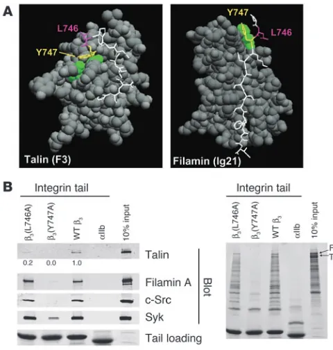
Integrin β3 cytoplasmic domain mutants that disrupt interactions with
platelet intracellular proteins. Previous in vitro studies with human proteins pointed to β3(Tyr747) and β3(Leu746) as potential
tar-gets for disrupting the intracellular interactions of the β3
cytoplas-mic domain with talin (13, 14). The crystal structure of β3
resi-dues Trp739 to Tyr747 in complex with talin F3 shows that both
β3(Tyr747) and β3(Leu746) make extensive contacts with talin.
In contrast, a homology model of β3 (Pro745 to Ile757) docked
into the structure of filamin A immunoglobulin-like domain 21 predicted hydrophobic contacts between β3(Tyr747) and a loop
between 2 β strands in filamin A. However, in contrast to what occurs in the integrin-talin interaction, β3(Leu746) makes no
contacts with filamin A (Figure 1A). These structures explain why
β3(Y747A) blocks the binding of both human talin and filamin,
whereas β3(L746A) blocks only talin binding (12). To test the role
of β3(Tyr747) and β3(Leu746) on the binding of proteins from
murine platelet lysates, we used recombinant WT and mutant
β3 integrin cytoplasmic domain proteins in affinity
chromatogra-phy experiments. WT β3 bound mouse talin and filamin. In
addi-tion, the β3 cytoplasmic domain bound the tyrosine kinases Syk
and c-Src (Figure 1B, left panel). β3(Y747A) disrupted interactions
with each of these mouse proteins. In sharp contrast, although
β3(L746A) bound much less talin than WT β3, it bound other
pro-teins to the same extent as WT β3. Inspection of protein-stained
gels underscored the specificity of the effect of the β3(L746A)
mutation on protein binding; there was no substantial reduction in any of the β3-binding proteins other than talin (Figure 1B, right
panel). These results indicate that both β3(Y747A) and β3(L746A)
mutations disrupt β3-talin interactions in mouse platelets.
How-ever, β3(Y747A) blocks binding of many other proteins, probably
because it disrupts the β turn formed by N744PLY (15) in addition
to disrupting direct contacts with the protein ligands. Thus, the effects of these point mutations on the binding of murine proteins to the β3 cytoplasmic domain enable us to evaluate the effects on
murine platelet function of selective blockade of talin binding ver-sus a more general blockade of protein interactions.
Generation of mice bearing β3(Y747A) and β3(L746A) mutations. To test the effects of β3(Y747A) and β3(L746A) mutations on
platelet function, we generated mice harboring either of these mutations using a gene-targeting approach similar to that previ-ously described for the construction of the β3(Y747,759F) integrin
subunit (16). Mouse 129/SvJ ES cells were electroporated with a targeting vector containing 6 kb of β3 genomic sequence, a loxP
-flanked neomycin (Neo) cassette inserted between exons 14 and 15, and either Y747A or L746A mutation in exon 15 (Figure 2A). ES clones were screened by PCR and confirmed by Southern blot-ting using cDNA probes 5′ and 3′ to the targeted sequence (Figure 2B) and with a Neo probe to confirm a single integration site of the targeting vector (data not shown). For each mutation, 2 inde-pendently derived clones of ES cells were injected into C57BL/6 blastocysts. Each ES cell clone generated chimeric mice that exhib-ited germline transmission of the mutation as determined by PCR (data not shown). Heterozygous animals were crossed with EIIa-Cre mice to obtain offspring in which the Neo cassette was excised. Heterozygous animals were crossed to obtain animals homozygous for the mutations or WT littermates as determined by PCR on genomic DNA isolated from ear biopsy samples (Figure 2C). The presence of the mutation in β3 integrin mRNA was confirmed by
sequencing reverse transcription–polymerase chain reaction prod-ucts generated using RNA isolated from spleens of homozygous mutant animals. Mice were backcrossed to a C57BL/6 strain for at least 3 generations, and WT sex-matched littermates were used as controls for homozygous mutant animals in all experiments. Simi-lar to β3-null animals (17), these mutant mice exhibited no gross
developmental abnormalities and had normal platelet and white blood cell counts (Table 1). Thus, the mice are suitable for exami-nation of blocking interactions of the β3 cytoplasmic domain on
platelet function in vivo and in vitro.
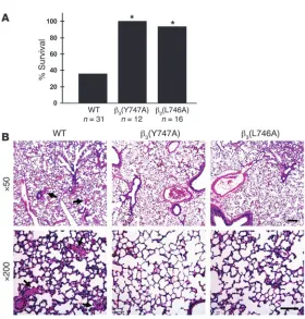
Protection from thrombosis in β3(Y747A) and β3(L746A) mice. Since
αIIbβ3 is required for platelet aggregation, we examined the effect of
the β3(Y747A) and β3(L746A) mutations on the formation of
occlu-sive platelet thrombi in a pulmonary thromboembolism model in which platelet activation was induced by tail vein infusion of col-lagen and epinephrine. Upon intravenous injection of a mixture of these platelet agonists, 64% of WT littermates (20 of 31 mice) died within 5 minutes (Figure 3A). Histological examination of lung tissue from these animals revealed platelet thrombi throughout the pulmonary vasculature (Figure 3B). In sharp contrast, 100% of
β3(Y747A) (12 of 12 mice) and 95% of β3(L746A) (15 of 16 mice)
[image:3.585.43.283.83.335.2]ani-mals survived the infusion of collagen and epinephrine (P < 0.005, Figure 1
Structure-based mutagenesis of the β3 integrin cytoplasmic domain.
(A) Space-filled models of talin F3 domain (left) or filamin A immuno-globulin–like domain 21 in complex with short internal fragments of the β3 integrin cytoplasmic domain (shown as sticks, with Tyr747 and
Leu746 in yellow and purple, respectively). (B) Affinity chromatography using recombinant WT and mutant β3 cytoplasmic domain proteins and
research article
2252 The Journal of Clinical Investigation http://www.jci.org Volume 117 Number 8 August 2007
WT versus either mutant; Figure 3A). The lungs of these mutant mice were largely devoid of platelet thrombi (Figure 3B).
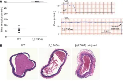
To examine the effects of the β3(L746A) mutations in a model
of arterial thrombosis initiated by vascular injury, we measured the time course of thrombus formation in response to ferric chlo-ride–induced injury of the common carotid artery. In this model, the carotid arteries of 6 of 6
WT mice completely occluded in an average of 6.4 ± 1.9 min-utes following injury (Figure 4A). In contrast, arteries of 0 of 5 β3(L746A) mice occluded
during the 30 minutes follow-ing injury despite similar lev-els of ferric chloride–induced endothelial damage (Figure 4, A and B). Thus, β3(L746A) mice
are protected from thrombosis in models involving impact to large arteries or the microcir-culation.
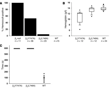
Reduced GI bleeding in β3(L746A) mice. Mice entirely
lacking integrin β3 exhibit
GI bleeding associated with reduced survival (17). Indeed, in our hands 100% (11 of 11) of β3-null animals had
detect-able blood in their stool (Figure 5A). In contrast, fewer β3(Y747A) animals (19 of
36) were positive for fecal blood, indicating that β3(Y747A) impairs vascular integrity
to a lesser extent than complete lack of β3.
In sharp contrast to the results in null and
β3(Y747A) mice, less than 5% of β3(L746A)
animals (1 of 23) exhibited fecal blood. The protection from GI bleeding in the
β3(L746A) mice was confirmed by their lack
of anemia relative to the β3(Y747A) animals
(Figure 5B and Table 1). Indeed, β3(L746A)
mice showed hematocrits and hemoglobin levels similar to those of WT animals. The pathological bleeding observed in β3(Y747A)
mice was associated with reduced prenatal and perinatal survival. Significantly fewer homozygous β3(Y747A)
mutant animals obtained from heterozygote by heterozygote breed-ings survived to weaning age (144 homozygotes versus 192 WT;
P < 0.00001). β3(L746A) animals, however, were observed at
expect-ed Mendelian ratios (106 homozygotes versus 108 WT; P = 0.97). Together, these results indicate that β3(Y747A) animals display a
Table 1
Hemograms for WT and β3 mutant mice
β3(L746A) β3(Y747A)
WT Homozygous WT Homozygous Mean SEM Mean SEM Mean SEM Mean SEM
wbc (×109/l) 4.2 0.4 3.2 0.3 4.2 0.9 2.6 0.3
rbc (×109/l) 9.0 0.1 8.2A 0.2 9.1 0.2 6.8A 0.4
Hemoglobin (g/l) 13.9 0.1 13.3 0.3 13.8 0.3 10.8A 0.6
Hematocrit (%) 49.1 0.7 46.7 1.1 48.4 0.6 39.8A 2.2
MCV (fl) 54.4 0.6 56.8 1.3 53.5 0.8 58.5A 0.9
MCH (pg) 15.4 0.1 16.2A 0.2 15.2 0.1 16.0A 0.3
MCHC (g/dl) 28.4 0.3 28.6 0.5 28.5 0.5 27.4 0.5 Platelets (×109/l) 618 57 616 57 633 69 689 51
Neutrophils (%) 41.5 3.8 39.8 5.7 32.5 4.0 36.7 5.0 Lymphocytes (%) 47.9 4.4 50.5 6.4 53.1 4.5 53.4 5.4 Monocytes (%) 6.8 1.1 6.5 1.0 9.5 1.0 7.2 0.9 Eosinophils (%) 3.4 0.7 3.3 0.8 5.1 1.1 2.8 0.9 AP < 0.05 versus WT. MCV, mean corpuscular volume; MCH, mean corpuscular hemoglobin; MCHC, MCH
[image:4.585.44.366.81.412.2]concentration.
Figure 2
Generation of β3(Y747A) and β3(L746A)
mutant mice. (A) Targeting vector containing 6 kb of the 3′ end of the mouse β3 integrin gene
(exons 14 and 15). Tyr747- and Leu7460-to-alanine mutations were inserted into exon 15. DTA, diphtheria toxin A. (B) Southern blot analysis of R1 ES cell genomic DNA trans-fected with the β3(Y747A) targeting vector
digested with BamHI (left) or EcoRI (right) using a 5′ probe or 3′ probe, respectively. (C) PCR genotyping of genomic DNA isolated from β3(Y747A) and β3(L746A) mouse ear
bleeding diathesis similar to that reported for β3-null animals. On
the other hand, β3(L746A) mice were much less predisposed to these
pathologies or to reduced survival.
The foregoing results indicated that the β3(L746A) mice were
protected from thrombosis yet were spared the frequent occurrence of GI bleeding and reduced survival observed in β3(Y747A) and
β3-null mice. To examine the ability of these animals to achieve
hemostasis following injury, we measured the time required to arrest bleeding following tail resection. Both β3(Y747A) and
β3(L746A) animals manifested impaired hemostasis as evidenced
by failure to arrest bleeding within 10 minutes compared with an average of 90 seconds in WT animals (Figure 5C). Thus, while spontaneous GI bleeding is markedly reduced in β3(L746A)
ani-mals, both β3(Y747A) and β3(L746A) animals showed impaired
hemostasis in a tail bleeding assay.
Disrupted inside-out signaling in platelets from β3 mutant mice. To
explore the cellular basis for the biological effects of the β3(Y747A)
and β3(L746A) mutations in mice, we evaluated agonist-induced
activation of αIIbβ3 by measuring fibrinogen binding to platelets.
As expected, WT β3 mice showed a large increase in the amount
of specifically bound fibrinogen in response to ADP/epinephrine, PMA, or a PAR4 thrombin receptor agonist peptide (Figure 6A).
β3(Y747A) and β3(L746A) platelets showed profoundly impaired
responses. In contrast, β3(Y747A) and β3(L746A) platelets bound
fibrinogen somewhat better than WT β3 platelets in the presence
of 0.5 mM MnCl2, an exogenous activator of the integrin
(Fig-ure 6B, left panel). This increased binding was accounted for by a modest increase in β3 integrin surface expression in β3(Y747A)
and β3(L746A) compared with control platelets (Figure 6B, right
panel). Neither platelets preincubated with the β3-blocking
anti-body 1B5 nor β3-null platelets bound fibrinogen in response to
the agonists, indicating that the fibrinogen binding was
depen-dent on the β3 integrin (data not shown). Consistent
with the fibrinogen-binding defects observed, agonist-induced platelet aggregation was impaired in both mutants (Figure 6C). It is noteworthy that similar functional impairments were observed with selective disruption of talin binding to β3(L746A) and a near
complete blockade of cytoplasmic protein binding to β3(Y747A). This provides additional evidence for
the critical and selective role of talin in this process. Thus, these mutant αIIbβ3 integrins are defective in
their capacity to respond with increased affinity to intracellular signals rather than in their innate capac-ity to bind fibrinogen.
Consistent with preservation of αIIbβ3
ligand–bind-ing function, static adhesion of platelets to immobi-lized fibrinogen was similar in WT, β3(Y747A), and
β3(L746A) platelets (Figure 6D). Platelet adhesion was
mark-edly reduced by 1B5, an αIIbβ3-blocking antibody, or in β3-null
platelets, indicating that adhesion is dependent on β3 integrins.
These results show that β3 integrins expressed on β3(Y747A) and
β3(L746A) platelets maintain ligand-binding function and
con-firm a previous report that αIIbβ3 activation is not required for
static adhesion of platelets to fibrinogen (18).
Intact outside-in signaling in platelets from β3(L746A) mice. Once
integrins have bound ligand, they can generate biochemical responses, such as activation of Src family kinases and tyrosine phosphorylation of focal adhesion kinase phosphoprotein of 125 kD (pp125FAK), that result in cell spreading. To examine the ability
of β3(L746A) to mediate this form of signaling, termed outside-in
signaling, we measured tyrosine phosphorylation of pp125FAK in
response to fibrinogen binding to platelets. When washed plate-lets were resuspended in buffer containing 250 μg/ml fibrinogen and stimulated by addition of 100 μM ADP/100 μM epinephrine, WT platelets, but not platelets from β3(L746A) animals, showed
an increase in pp125FAK phosphorylation (Figure 7). However,
in the presence of 0.5 mM MnCl2 to activate αIIbβ3 extrinsically,
β3(L746A) platelets exhibited fibrinogen-dependent pp125FAK
phosphorylation similar to that of WT platelets. This indicates that αIIbβ3(L746A) is able to mediate outside-in signaling when
ligated by fibrinogen. Thus, the reduced pp125FAK
phosphory-lation in β3(L746A) platelets shown in Figure 7 is ascribable to
their failure to increase fibrinogen binding in response to ADP/ epinephrine. β3(L746A) platelets showed reduced spreading on
fibrinogen in response to ADP or PMA (Figure 8). In the presence of MnCl2, however, spreading was similar in WT and β3(L746A)
platelets. Together, these data indicate that blocking talin binding to β3 inhibits αIIbβ3 activation but maintains outside-in signaling
[image:5.585.42.322.73.366.2]and platelet spreading if the αIIbβ3 is activated exogenously.
Figure 3
β3(Y747A) and β3(L746A) mice are protected from
micro-vascular thrombosis. (A) Mice were injected intravenously with 800 μg/kg collagen and 60 μg/kg epinephrine and monitored for 30 minutes. *P < 0.005. (B) Representative sections of H&E-stained lungs from a WT mice that died during the assay and β3(Y747A) and β3(L746A) mice that
survived and were sacrificed immediately following the assay. WT sections show extensive microthrombi through-out the lungs, while β3 mutant lungs were clear. Scale bars:
research article
2254 The Journal of Clinical Investigation http://www.jci.org Volume 117 Number 8 August 2007
Discussion
Here we have tested the importance of talin binding to integrin β3
in the activation of platelet αIIbβ3 and in hemostasis and
throm-bosis. Guided by insights derived from structural studies of the interactions between the β3 integrin cytoplasmic domain and
cyto-plasmic proteins, we have generated 2 mouse strains bearing β3
mutations that disrupt talin binding. One strain bears β3(Y747A),
a mutation that disrupts multiple protein interactions with the β3
integrin cytoplasmic domain; the second bears β3(L746A), a
muta-tion that selectively disrupts β3 integrin–talin interactions. Both
strains of mutant mice were protected from pulmonary thrombo-sis following intravenous injection of collagen and epinephrine. In addition, β3(L746A) animals showed protection from ferric
chloride–induced thrombosis of the carotid artery. Thus, using 2 distinct models of thrombosis, we have established that inhibition of the talin-β3 interaction confers an antithrombotic effect. In
con-trast, β3(L746A) mice were relatively resistant to the pathological
bleeding, as assessed by GI blood loss and anemia, that occurred in 53% of β3(Y747A) and virtually all β3-null mice (Figure 5 and ref.
17). Platelets from both β3(Y747A) and β3(L746A) mice showed
marked reduction in agonist-induced fibrinogen binding to αIIbβ3,
providing what we believe to be the first in vivo evidence for the talin dependence of integrin activation in mammals. However, platelet adhesion to immobilized fibrinogen under static condi-tions was unaffected by either β3 cytoplasmic domain mutation,
demonstrating that the αIIbβ3 in β3(Y747A) and β3(L746A)
plate-lets maintains αIIbβ3 ligand binding function. While both mutants
showed disrupted activation of αIIbβ3, platelets from the β3(L746A)
mice were able to spread on fibrinogen and exhibit tyrosine phos-phorylation of pp125FAK when α
IIbβ3 was activated exogenously by
MnCl2. Thus, β3(L746A) mice have a selective defect in the
inside-out signaling required for activation of αIIbβ3, yet their platelets
maintain the capacity to generate αIIbβ3-mediated outside-in
sig-nals required for platelet spreading. Together, these results estab-lish the importance of the talin–β3 integrin interaction in αIIbβ3
activation in platelets and show that blocking this interaction can have a potent antithrombotic effect without the kind of pathologi-cal bleeding associated with complete lack of αIIbβ3 function.
Previous studies using a variety of in vitroapproaches showed that talin binding to the β3 integrin cytoplasmic domain induces
αIIbβ3 activation (10, 11) and is a final common step in the process
(12). Platelets bearing β3(L746A), which leads to specific loss of
talin binding, bound significantly less fibrinogen in response to platelet agonists than WT platelets, indicating a marked defect in inside-out signaling (Figure 6). The low levels of fibrinogen binding observed with high agonist doses in β3(L746A) and
β3(Y747A) mutant platelets may have been due to either
incom-plete inhibition of talin binding to the integrin or to the existence of a talin-independent αIIbβ3 activation mechanism. Truncation
of the β3 cytoplasmic domain [β3(724X); ref. 19] or a point
muta-tion [β3(S752P)] (20) is associated with defective hemostasis and
reduced agonist-mediated αIIbβ3 activation in humans; however,
the β3(724X) mutation is expected to block binding of multiple
proteins (21), and the binding defect in β3(S752P) remains to be
defined (22). Furthermore, in contrast to β3(L746A), the 2 human
[image:6.585.92.492.84.347.2]mutations impair outside-in signaling (23, 24). Our results are Figure 4
β3(L746A) mice are protected from arterial thrombosis. (A) Thrombosis was induced in the carotid artery of mice by a 3-minute application of
5% ferric chloride to the surface of the vessel. The time to complete vessel occlusion was considered the time after injury to zero blood flow as measured with a Doppler flow probe. Representative Doppler flow tracings from a WT and a β3(L746A) mouse are shown. (B) H&E-stained
sec-tions of carotid arteries of WT and β3(L746A) mice obtained 30 minutes after ferric chloride injury. A section of β3(L746A) carotid artery distal to
complemented by the subtle hemostatic and platelet function defects observed in mice bearing β3(Y747,759F) mutations. Those
mice display a defect in secondary platelet aggregation ascribed to defective outside-in signaling and manifest rebleeding from tail wounds after initial hemostasis (16). The β3(Y747,759F)
muta-tion does not block talin binding, and platelets from those ani-mals manifested normal agonist-induced activation of integrin
αIIbβ3 and primary hemostasis. In contrast, as noted above, the
β3(L746A) and β3(Y474A) mice showed defective αIIbβ3 activation,
impaired primary aggregation, and prolonged bleeding times. Because of these differences in functional effects, it would be of great interest to examine the impact of the β3(Y747,759F)
muta-tions in comparison to the dramatic effect of the β3(L746A)
muta-tion on platelet-dependent thrombosis.
The importance of talin binding for integrin activation in vivo is likely to apply to other integrins. The critical structural features for talin binding are conserved in multiple mamma-lian (β1, β2, β3, β5, β6, β7) integrins and in invertebrate integrins
(13). Previous in vitro studies indicate that talin participates in the activation of β1 and β2 integrins (12, 25, 26). Mice
bear-ing β1(Y783,795A) manifest defective β1 integrin activation
in keratinocytes and platelets (27, 28). β1(Y783A) is known to
disrupt talin binding to β1 (14); hence, the defective activation
of β1(Y783,795A) integrins is consistent with a general role for
talin in activation of β1 integrins in vivo. Nevertheless, as shown
in Figure 1, a tyrosine-to-alanine substitution at this position within the β3 integrin (or a homologous mutation in β1
integ-rin; ref. 14) disrupts many protein interactions with the β inte-grin cytoplasmic domain. Thus, the results from the β3(L746A)
mutant mouse reported here, harboring a mutation that selec-tively disrupts β3 integrin–talin interactions, unambiguously
demonstrate that talin binding to the integrin β cytoplasmic domain is critical for in vivo integrin activation.
Blockade of talin binding to αIIbβ3 inhibits thrombosis with less
pathological bleeding than complete lack of αIIbβ3 function. Loss
of αIIbβ3 in humans, due to mutation in either αIIb or β3 integrin
genes (Glanzmann thrombasthenia) or as the result of pharma-cological blockade of ligand binding to αIIbβ3, is associated with
an increased risk of pathological bleeding (1, 29). Gene-targeted mice lacking αIIbβ3 are protected from thrombosis (30) but
mani-fest a greater than 95% incidence of GI bleeding and are severely anemic (ref. 17 and Figure 5). β3(L746A) animals displayed
negli-gible GI bleeding or anemia (Figure 5). The ability of β3(L746A)
platelets to adhere to immobilized fibrinogen and to generate outside-in signals may account for the lack of pathological bleed-ing. The increased GI bleeding in the β3(Y747A) mice is likely a
reflection of the lack of selectivity of β3(Y747A) for inhibition of
binding of cytoplasmic proteins, implying that β3(Y747A) can
dis-rupt many signaling events in addition to integrin activation (31, 32). Whereas β3(L746A) animals showed little sign of
pathologi-cal bleeding, they, like β3(Y747A) and β3-null animals, displayed
impaired hemostasis in response to tail resection. In β3(L746A)
mice, agonist-induced fibrinogen binding, a key step in aggrega-tion, was blocked, and tail resection resulted in bleeding from medium-sized veins and arteries. Thus, αIIbβ3-mediated platelet
aggregation is essential for arrest of bleeding from larger vessels, but the αIIbβ3-mediated adhesion and outside-in signaling may be
sufficient for hemostasis in the microcirculation.
The results of this study suggest that blockade of the
talin-αIIbβ3 interaction may offer a therapeutic target in pathological
thrombosis. Because ligand binding to integrin αIIbβ3 is
absolute-ly required for platelet aggregation, αIIbβ3 inhibitors are an
effec-tive monotherapy for preventing acute thrombosis in the setting of percutaneous coronary intervention (33). In sharp contrast, chronic blockade of αIIbβ3 by orally administered antagonists is
[image:7.585.44.407.78.363.2]ineffective in thrombosis protection, possibly because of the need Figure 5
Bleeding diathesis in β3 mutant mice.
(A) The presence of fecal blood was detected using a guaiac-based hemoc-cult test. Fecal specimens obtained from the indicated genotypes of mice at 6–12 weeks of age were scored as positive or negative in a blinded man-ner. (B) Box plot showing hemoglobin concentration measured in peripheral blood. Filled circles represent outliers and were included in statistical analy-sis. *P < 0.05 compared with WT. (C) Tail bleeding times. β3(Y747A)
and β3(L746A) homozygotes showed
research article
2256 The Journal of Clinical Investigation http://www.jci.org Volume 117 Number 8 August 2007
to limit dosage to avoid pathological bleeding (5). Effective oral antithrombotics, such as aspirin or clopidogrel, partially block
αIIbβ3 activation at the level of an agonist receptor or generation
of an endogenous platelet agonist. Here we show that disrupting the β3 integrin–talin interaction blocks αIIbβ3 activation and has
a dramatic antithrombotic effect and that blocking this interac-tion leaves the ligand-binding funcinterac-tion of platelet αIIbβ3 intact.
Similarly, inhibiting αIIbβ3 activation by agents such as aspirin and
clopidogrel also spares the ligand-binding function of αIIbβ3. This
preservation of ligand-binding function in combination with pres-ervation of outside-in signaling probably accounts for the mark-edly diminished pathological bleeding in the β3(L746A) mice
rela-tive to those with complete lack of β3 function. Thus, these studies
suggest the principle that the reduction in pathological bleeding associated with blocking the αIIbβ3-talin interaction creates a wider
therapeutic window than that achieved by blocking extracellular ligand binding to αIIbβ3. Furthermore, by preventing a final
com-mon event leading to integrin activation, disrupting this interac-tion might prove more effective than currently available oral anti-platelet agents that block a single agonist pathway. Small molecule inhibitors of integrin cytoplasmic domain protein-protein
interac-tions are feasible (34), and the critical structural features of the talin-integrin interaction have been defined (13), suggesting that the principle established here may pave the way to development of a new class of antithrombotic agents.
Methods
Generation of β3(Y747A) and β3(L746A) knock-in mice. A 6-kb fragment of the
β3 integrin gene encompassing exons 14 and 15 was amplified from R1 ES cell genomic DNA by PCR and inserted into pBluescript, and its identity was verified by sequencing. Mutations (coding for L746A or Y747A) in exon 15 were inserted by splice-overlap PCR (35) and confirmed by sequencing. A targeting vector was constructed by inserting a phosphoglycerate kinase (PGK) promoter–driven Neo cassette flanked by loxP sequences into a Hin-dIII site between exons 14 and 15 and a PGK–diphtheria toxin A cassette 5′
[image:8.585.74.517.81.376.2]to the β3 sequence (Figure 2A). The targeting sequence was isolated from pBluescript by NotI restriction digestion, agarose gel purified, and electro-porated into 129/SvJ ES cells at both the UCSD Transgenic Core Facility and the Fannie E. Rippel Transgenic Facility at the Massachusetts Institute of Technology (MIT). G418-resistant colonies were screened by PCR using primer A (5′-CACTTTGAGGTTTGAGGGTC-3′) and primer B (5′ -GCT-GATCCTCTAGAGTCGAC-3′) as depicted in Figure 2A. Positive clones
Figure 6
β3(Y747A) and β3(L746A) platelet interactions with fibrinogen. (A) FITC-labeled fibrinogen binding was measured by flow cytometry. Fibrinogen
binding was reduced in β3 mutant platelets in response to platelet agonists. Specific binding was defined as that which was inhibitable by 2 mM
EDTA. Mean fluorescence intensity (MFI) for each agonist treatment was normalized to the MFI obtained with 0.5 mM MnCl2 treatment. n = 7,
6, and 4 for WT, β3(Y747A), and β3(L746A), respectively. Epi, epinephrine. (B) Platelets from both WT mice and β3 mutants showed increased
fibrinogen binding in the presence of 0.5 mM MnCl2 (left). Surface β3 integrin expression of platelets obtained from β3 mutant animals (right).
Results are representative of 4 independent experiments and at least 4 mice per genotype. (C) Aggregation of platelets obtained from WT or
β3 mutant mice was measured in response to the addition of 100 μM ADP or 0.5 mM PAR4 peptide. Results are representative of at least 2
were further analyzed by Southern blotting of BamHI-digested genomic DNA using a 600-bp 5′ cDNA probe upstream of the targeted sequence (Figure 2A). This resulted in a 3.2-kb band from the WT allele and a 5.4-kb band from the targeted allele. When genomic DNA was digested with EcoRI and hybridized with a 600-bp 3′ cDNA probe complementary to a sequence downstream of the targeted sequence, we expected a 7-kb band from the WT allele and a 9.2-kb band from the targeted allele (Figure 2). Blots were hybridized with a cDNA containing the Neo resistance gene to detect random insertional events of the targeting vector. Positive clones (6 for each mutation), containing a single insertion, were karyotyped, and at least 2 clones for each mutant were injected into C57BL/6 host blas-tocysts. Chimeric male mice were bred to C57BL/6 females, and agouti offspring from these crosses were genotyped by PCR using genomic DNA extracted from ear biopsies and primer C (5′
-CAGTCCTCTACCTTA-CAGTG-3′) and primer D (5′-CTCTGCCCTCAGTTTCCTTA-3′) as depict-ed in Figure 2A. Two independently derivdepict-ed heterozygous animals for each mutation [β3(Y747A), 1 from MIT and 1 from UCSD; and β3(Y746A), from UCSD], were crossed with EIIa-Cre mice (The Jackson Laboratory) to delete the Neo cassette in germ cells (36). Deletion of Neo and presence of the targeted allele in offspring from β3 mutant and EIIa-Cre crosses were evaluated by PCR of genomic DNA using primers C and D (Figure 2A). All experiments were performed with independently derived lines for each mutation that were backcrossed to the C57BL/6 strain for at least 3 genera-tions. Sex-matched WT littermates were used as control animals. Mice were housed in the UCSD animal facility, and experiments were approved by the UCSD Institutional Animal Care and Use Committee.
[image:9.585.46.267.82.279.2]Molecular modeling. Modeling was performed using DeepView — Swiss-Pdb viewer (http://www.expasy.org/spdbv/; ref. 37). β3 integrin cytoplasmic
Figure 8
Agonist-stimulated platelet spread-ing. Platelets were allowed to spread on fibrinogen-coated coverslips (100
μg/ml) in the presence of MnCl2 (0.5
mM), ADP (100 μM), PMA (200 nM), or PAR4 peptide (1 mM), as indicated, for 45 minutes, fixed, and stained with rhodamine-phalloidin. The results were quantified from 2 independent experiments. *P < 0.05. Error bars represent SD. Original magnification, ×1,260.
Figure 7
pp125FAK phosphorylation. Platelets were incubated in suspension
with 250 μg/ml soluble fibrinogen with or without 0.5 mM MnCl2 and
ADP/epinephrine (100 μM each), as indicated, for 20 minutes at room temperature. Platelets were lysed, and pp125FAK phosphorylation was
measured by immunoblotting using an antibody against pp125FAK
(pTyr397). Blots were stripped and reprobed with an antibody to pp125FAK. Results are shown relative to maximum pp125FAK
[image:9.585.188.537.472.741.2]research article
2258 The Journal of Clinical Investigation http://www.jci.org Volume 117 Number 8 August 2007
domain interactions with filamin A were modeled based on the structure of β7 integrin cytoplasmic domain and filamin A (38). This approach is jus-tified by nuclear magnetic resonance spectroscopy and mutational studies that indicate that these integrin cytoplasmic domains bind to filamin A in a similar manner. Sequence threading was guided using the NPXY motif sequence, which is identical in the β3 and β7 integrins. The result-ing sequence alignment was submitted to Swiss-Model (http://swissmodel. expasy.org). The resulting model had no major errors as determined by the WhatCheck report on the expasy website.
Affinity chromatography with recombinant integrin cytoplasmic domains. Plate-lets were obtained from WT C57BL/6 mice as described below and lysed as previously described (11). Affinity chromatography was performed using recombinant integrin cytoplasmic domains bound to His-Bind Resin (Novagen) as previously described (11, 14). Samples were separated on a 4%–20% SDS-polyacrylamide gel (Novex; Invitrogen) and transferred for Western blotting with the following antibodies: talin 8d4 (Sigma-Aldrich), anti–filamin A (a kind gift from John Hartwig, Harvard Medical School, Boston, Massachusetts, USA), and c-Src and Syk (Santa Cruz Biotechnol-ogy Inc.). Signal was detected and results quantified using an Odyssey imaging system (LI-COR Biosciences).
Thrombosis assays. Pulmonary thromboembolism experiments were per-formed as described previously (39). Briefly, mice under isoflurane anes-thesia were administered 200 μl of saline containing 0.8 mg/kg collagen (Chrono-log Corp.) and 60 μg/kg epinephrine via tail vein injection. Ani-mals were monitored for death as determined by cessation of breathing for up to 30 minutes after injection. Lungs were removed, fixed overnight with 10% neutral buffered formalin, and embedded in paraffin. Sections were stained with H&E, and images were captured with a Leica DM LS micro-scope and Spot color digital camera (National Diagnostics).
Ferric chloride–induced thrombosis was induced as previously described (40) by applying a 1.2 × 1.2–mm piece of filter paper soaked in 5% ferric chloride to each side of the common carotid artery of a mouse under iso-flurane anesthesia. After 3 minutes, the filter paper was removed and the vessel was washed twice with saline. Flow through the carotid artery was monitored 2 minutes before injury and at least 30 minutes after removal of the ferric chloride using a 0.5 PSB Doppler flow probe and T402 flowmeter (Transonic Systems Inc.). Following the assay, the artery was ligated with sutures to stop the flow of blood, and the artery was removed, fixed in 3.7% formaldehyde, embedded in paraffin, sectioned, and stained with H&E.
Hemostasis assays. The presence of fecal blood was assessed with a guaiac-based hemoccult detection assay (Helena Laboratories) on freshly obtained stool samples. Tail bleeding assays were performed by resecting 1 mm of the tail followed by immersion in 37°C isotonic saline (17). All experiments were terminated at 10 minutes by cauterizing the tail.
Blood counts. Peripheral blood was collected from the retro-orbital plexus and transferred to tubes containing EDTA. Cell counts were performed using an MS9 automated cell counter (Melet Schloesing Laboratories) with veterinary parameters and reagents. Differential counts were performed manually on Wright-Giemsa–stained smears. Box-and-whisker plots of hemoglobin concentration were generated in which the box represents an interquartile range (IQR), a horizontal line represents the median, and ver-tical lines represent 1.5 × IQR. Outliers are represented by filled circles and were included in statistical analysis.
Platelet isolation and functional assays. Washed platelets were obtained from fresh anticoagulated blood and resuspended at 3 × 10⁸/ml in a platelet
incu-bation buffer (41). Soluble fibrinogen binding was performed by incubating platelets for 20 minutes with 150 μg/ml FITC-labeled fibrinogen followed by fixation with 1% formaldehyde for 10 minutes at room temperature and analyzed on a FACScan (BD). Surface expression of β3 integrin was measured by flow cytometry using an FITC-conjugated anti-mouse CD61 antibody (BD Biosciences). Platelet aggregation was performed as described previously (17) using platelet-rich plasma (PRP) at a platelet concentration of 3.5 × 10⁸ platelets/ml obtained from blood drawn into 0.1-volume 0.13 M sodium citrate. Aggregation of 300 μl of PRP was measured at 37°C while stirring using a 2-channel aggregometer (Chrono-log Corp.). Platelet adhe-sion assays were performed by incubating washed platelets for 1 hour at room temperature in fibrinogen-coated microtiter wells as previously described (42), and platelets were quantified by acid phosphatase assay (41). Platelet spreading was analyzed by confocal microscopy after cells were plat-ed on coverslips coatplat-ed with 100 μg/ml fibrinogen for 45 minutes at room temperature. Platelets were fixed with 4% formaldehyde/PBS for 10 minutes at room temperature and labeled with rhodamine-phalloidin. Images were captured with a Leica SP2 confocal microscope, and cell area was quanti-fied using ImageJ software (version 1.36b; http://rsb.info.nih.gov/ij/). To analyze FAK phosphorylation, platelets in suspension were incubated with 250 μg/ml fibrinogen with or without 0.5 mM MnCl2 and ADP/epineph-rine (100 μM each) for 20 minutes at room temperature. Platelets were then lysed by addition of one-half volume 3× lysis buffer (3% NP-40, 450 mM NaCl, 150 mM Tris-HCl, pH 7.4, 3 mM NaVO4, 1.5 mM NaF, 3 mM PMSF, 15 mM EDTA, and complete protease inhibitor; Roche) and clarified by cen-trifugation at 13,000 g for 10 minutes at 4°C. Forty micrograms of protein was separated on 4%–20% Tris-glycine gels (Novex; Invitrogen) and trans-ferred to nitrocellulose membranes for probing with anti-FAK(pTyr397) (Biosource) and FAK antibodies (Santa Cruz Biotechnology Inc.). Signal was detected and results quantified by infrared fluorescence spectrometry using an Odyssey imaging system (LI-COR Biosciences).
Statistics. Statistical significance was determined by Student’s t test for all experiments but the pulmonary thromboembolism study, which was ana-lyzed using Fisher’s exact test. A P value of less than 0.05 was considered statistically significant.
Acknowledgments
We gratefully acknowledge Aurora Burds Connor and Ella Kotheri for excellent assistance in generating gene-targeted animals. We thank Zaverio Ruggeri for valuable discussions concerning the fer-ric chloride thrombosis model. This work was supported by grants from the NIH (HL57900 and HL078784) and from the Cell Migra-tion Consortium, NIH (U54 GM064346). B.G. Petrich is a post-doctoral fellow of the American Heart Association. A.W. Partridge held a postdoctoral fellowship from the Tobacco-Related Disease Research Program. P. Fogelstrand has a postdoctoral fellowship from the Swedish Research Council.
Received for publication November 21, 2006, and accepted in revised form April 24, 2007.
Address correspondence to: Mark H. Ginsberg, Department of Medicine, University of California, San Diego, 9500 Gilman Drive, mail stop 0726, La Jolla, California 92093-0726, USA. Phone: (858) 822-6432; Fax: (858) 822-6458; E-mail: [email protected].
1. Quinn, M.J., Byzova, T.V., Qin, J., Topol, E.J., and Plow, E.F. 2003. Integrin alphaIIbbeta3 and its antag-onism. Arterioscler. Thromb. Vasc. Biol.23:945–952. 2. Chew, D.P., Bhatt, D.L., Sapp, S., and Topol, E.J.
2001. Increased mortality with oral platelet
gly-coprotein IIb/IIIa antagonists: a meta-analysis of phase III multicenter randomized trials. Circulation.
103:201–206.
3. Shattil, S.J., and Newman, P.J. 2004. Integrins: dynamic scaffolds for adhesion and signaling in
platelets. Blood.104:1606–1615.
4. Ginsberg, M.H., Partridge, A., and Shattil, S.J. 2005. Integrin regulation. Curr. Opin. Cell. Biol.
17:509–516.
therapeutic advances in antiplatelet therapy. Nat. Rev. Drug Discov.2:15–28.
6. Liddington, R.C., and Ginsberg, M.H. 2002. Integ-rin activation takes shape. J. Cell Biol.158:833–839. 7. Adair, B.D., et al. 2005. Three-dimensional EM struc-ture of the ectodomain of integrin αVβ3 in a com-plex with fibronectin. J. Cell Biol.168:1109–1118. 8. Campbell, I.D., and Ginsberg, M.H. 2004. The
talin-tail interaction places integrin activation on FERM ground. Trends Biochem. Sci.29:429–435. 9. Xiao, T., Takagi, J., Coller, B.S., Wang, J.H., and
Springer, T.A. 2004. Structural basis for allostery in integrins and binding to fibrinogen-mimetic therapeutics. Nature.432:59–67.
10. Calderwood, D.A., et al. 2002. The phosphotyrosine binding-like domain of talin activates integrins.
J. Biol. Chem.277:21749–21758.
11. Calderwood, D.A., et al. 1999. The Talin head domain binds to integrin beta subunit cytoplasmic tails and regulates integrin activation. J. Biol. Chem.
274:28071–28074.
12. Tadokoro, S., et al. 2003. Talin binding to integrin beta tails: a final common step in integrin activa-tion. Science.302:103–106.
13. Garcia-Alvarez, B., et al. 2003. Structural determi-nants of integrin recognition by talin. Mol. Cell.
11:49–58.
14. Pfaff, M., Liu, S., Erle, D.J., and Ginsberg, M.H. 1998. Integrin beta cytoplasmic domains differ-entially bind to cytoskeletal proteins. J. Biol. Chem.
273:6104–6109.
15. Ulmer, T.S., Yaspan, B., Ginsberg, M.H., and Camp-bell, I.D. 2001. NMR analysis of structure and dynam-ics of the cytosolic tails of integrin alpha IIb beta 3 in aqueous solution. Biochemistry.40:7498–7508. 16. Law, D.A., et al. 1999. Integrin cytoplasmic tyrosine
motif is required for outside-in alphaIIbbeta3 sig-nalling and platelet function. Nature.401:808–811. 17. Hodivala-Dilke, K.M., et al. 1999. β3-integrin-defi-cient mice are a model for Glanzmann thrombas-thenia showing placental defects and reduced sur-vival. J. Clin. Invest.103:229–238.
18. Savage, B., Shattil, S.J., and Ruggeri, Z.M. 1992. Modulation of platelet function through adhesion receptors. A dual role for glycoprotein IIb-IIIa (inte-grin alpha IIb beta 3) mediated by fibrinogen and glycoprotein Ib-von Willebrand factor. J. Biol. Chem.
267:11300–11306.
19. Wang, R., Shattil, S.J., Ambruso, D.R., and Newman, P.J. 1997. Truncation of the cytoplasmic domain of β3 in a variant form of Glanzmann thrombasthe-nia abrogates signaling through the integrin αIIbβ3 complex. J. Clin. Invest.100:2393–2403.
20. Chen, Y.P., et al. 1992. Ser-752→Pro mutation in the cytoplasmic domain of integrin beta 3 subunit and defective activation of platelet integrin alpha IIb beta 3 (glycoprotein IIb-IIIa) in a variant of Glanzmann thrombasthenia. Proc. Natl. Acad. Sci. U. S. A.89:10169–10173.
21. Liu, S., Calderwood, D.A., and Ginsberg, M.H. 2000. Integrin cytoplasmic domain-binding pro-teins. J. Cell Sci.113:3563–3571.
22. Ma, Y.Q., et al. 2006. Regulation of integrin alphaI-Ibbeta3 activation by distinct regions of its cyto-plasmic tails. Biochemistry.45:6656–6662. 23. Chen, Y.P., O’Toole, T.E., Ylanne, J., Rosa, J.P., and
Ginsberg, M.H. 1994. A point mutation in the integrin beta 3 cytoplasmic domain (S752→P) impairs bidirectional signaling through alpha IIb beta 3 (platelet glycoprotein IIb-IIIa). Blood.
84:1857–1865.
24. Ylanne, J., et al. 1993. Distinct functions of integ-rin alpha and beta subunit cytoplasmic domains in cell spreading and formation of focal adhesions.
J. Cell Biol.122:223–233.
25. Kim, M., Carman, C.V., and Springer, T.A. 2003. Bidirectional transmembrane signaling by cyto-plasmic domain separation in integrins. Science.
301:1720–1725.
26. Kuo, J.C., Wang, W.J., Yao, C.C., Wu, P.R., and Chen, R.H. 2006. The tumor suppressor DAPK inhibits cell motility by blocking the integrin-mediated polarity pathway. J. Cell Biol.172:619–631. 27. Chen, H., et al. 2006. In vivo beta1 integrin function
requires phosphorylation-independent regulation by cytoplasmic tyrosines. Genes Dev.20:927–932. 28. Czuchra, A., Meyer, H., Legate, K.R., Brakebusch,
C., and Fassler, R. 2006. Genetic analysis of beta1 integrin “activation motifs” in mice. J. Cell Biol.
174:889–899.
29. George, J.N., Caen, J.P., and Nurden, A.T. 1990. Glanzmann’s thrombasthenia: the spectrum of clinical disease. Blood.75:1383–1395.
30. Smyth, S.S., Reis, E.D., Vaananen, H., Zhang, W., and Coller, B.S. 2001. Variable protection of beta 3-integrin--deficient mice from thrombosis initiated
by different mechanisms. Blood.98:1055–1062. 31. Tahiliani, P.D., Singh, L., Auer, K.L., and LaFlamme,
S.E. 1997. The role of conserved amino acid motifs within the integrin beta3 cytoplasmic domain in triggering focal adhesion kinase phosphorylation.
J. Biol. Chem.272:7892–7898.
32. Woodside, D.G., et al. 2001. Activation of Syk protein tyrosine kinase through interaction with integrin beta cytoplasmic domains. Curr. Biol.
11:1799–1804.
33. Coller, B.S. 2001. Anti-GPIIb/IIIa drugs: current strategies and future directions. Thromb. Haemost.
86:427–443.
34. Ambroise, Y., Yaspan, B., Ginsberg, M.H., and Boger, D.L. 2002. Inhibitors of cell migration that inhibit intracellular paxillin/alpha4 binding: a well-documented use of positional scanning librar-ies. Chem. Biol.9:1219–1226.
35. Ho, S.N., Hunt, H.D., Horton, R.M., Pullen, J.K., and Pease, L.R. 1989. Site-directed mutagenesis by overlap extension using the polymerase chain reac-tion. Gene.77:51–59.
36. Lakso, M., et al. 1996. Efficient in vivo manipula-tion of mouse genomic sequences at the zygote stage. Proc. Natl. Acad. Sci. U. S. A.93:5860–5865. 37. Guex, N., and Peitsch, M.C. 1997. SWISS-MODEL
and the Swiss-PdbViewer: an environment for comparative protein modeling. Electrophoresis.
18:2714–2723.
38. Kiema, T., et al. 2006. The molecular basis of fila-min binding to integrins and competition with talin. Mol. Cell.21:337–347.
39. DiMinno, G., and Silver, M.J. 1983. Mouse anti-thrombotic assay: a simple method for the evalua-tion of antithrombotic agents in vivo. Potentiaevalua-tion of antithrombotic activity by ethyl alcohol. J. Phar-macol. Exp. Ther.225:57–60.
40. Konstantinides, S., Schafer, K., Thinnes, T., and Loskutoff, D.J. 2001. Plasminogen activator inhibi-tor-1 and its cofactor vitronectin stabilize arterial thrombi after vascular injury in mice. Circulation.
103:576–583.
41. Law, D.A., et al. 1999. Genetic and pharmacological analyses of Syk function in alphaIIbbeta3 signaling in platelets. Blood.93:2645–2652.
42. Arias-Salgado, E.G., et al. 2005. PTP-1B is an essen-tial positive regulator of platelet integrin signaling.