Protein aggregation in disease: a role for
folding intermediates forming specific
multimeric interactions
Arthur Horwich
J Clin Invest.
2002;
110(9)
:1221-1232.
https://doi.org/10.1172/JCI16781
.
For many decades, clinicians have been aware of the formation of insoluble protein
aggregates in particular diseases. For example, in sickle cell disease, the polymerization of
sickle hemoglobin into fibrils in the deoxygenating environment of the microvasculature
causes sickling of red cells and vaso-occlusion (1). In the most common form of
a
1-antitrypsin deficiency (see Perlmutter, this Perspective series, ref. 2), the presence of
a
1-antitrypsin inclusions in the endoplasmic reticulum (ER) of hepatocytes is associated with
deficient release of the enzyme into the circulation, placing both the inclusion-laden liver
and the antitrypsin-deficient lung at risk for damage (3). In Alzheimer disease (see Selkoe,
this series, ref. 4), the presence in the CNS of
b
-amyloid–containing plaques is associated
with neurodegeneration and dementia (5). Similarly, other neurodegenerative diseases
have recently been discovered to involve protein aggregation. For example, prion diseases
such as Creutzfeldt-Jacob disease and bovine spongiform encephalopathy are associated
with amyloid deposits of the PrP protein (6). Polyglutamine repeat diseases such as
Huntington disease are likewise associated with neuronal cytosolic and intranuclear
inclusions (7). These inclusions are composed of fibrils that stain similarly to amyloid (8).
Finally, in Parkinson disease, inclusions known as Lewy bodies, found in the cytoplasm of
cells of the basal ganglia, include amyloid-like aggregates of the protein
a
-synuclein (9, 10).
What do we know about the various processes […]
Perspective
Find the latest version:
For many decades, clinicians have been aware of the for-mation of insoluble protein aggregates in particular diseases. For example, in sickle cell disease, the poly-merization of sickle hemoglobin into fibrils in the deoxygenating environment of the microvasculature causes sickling of red cells and vaso-occlusion (1). In the most common form of α1-antitrypsin deficiency (see Perlmutter, this Perspective series, ref. 2), the pres-ence of α1-antitrypsin inclusions in the endoplasmic reticulum (ER) of hepatocytes is associated with defi-cient release of the enzyme into the circulation, placing both the inclusion-laden liver and the antitrypsin-defi-cient lung at risk for damage (3). In Alzheimer disease (see Selkoe, this series, ref. 4), the presence in the CNS of β-amyloid–containing plaques is associated with neurodegeneration and dementia (5). Similarly, other neurodegenerative diseases have recently been discov-ered to involve protein aggregation. For example, prion diseases such as Creutzfeldt-Jacob disease and bovine spongiform encephalopathy are associated with amy-loid deposits of the PrP protein (6). Polyglutamine repeat diseases such as Huntington disease are likewise associated with neuronal cytosolic and intranuclear inclusions (7). These inclusions are composed of fibrils that stain similarly to amyloid (8). Finally, in Parkinson disease, inclusions known as Lewy bodies, found in the cytoplasm of cells of the basal ganglia, include amyloid-like aggregates of the protein α-synuclein (9, 10).
What do we know about the various processes that produce these aggregates? Are there cellular mecha-nisms that can either prevent or respond to such processes? Can current understanding translate into prevention or therapy? The various articles in this Per-spective series address these questions and demon-strate the considerable progress that has been made toward explaining these events in molecular terms.
Resting on such understanding are new therapeutic strategies for some of these diseases, several of which are being tested in animal model systems.
Here, I offer an overview of the molecular mecha-nisms of both protein folding and misfolding, particu-larly in the formation of aggregates. This Perspective first revisits some of the seminal developments in pro-tein biochemistry that led to the idea that propro-tein aggregates contain specific, organized, polymeric struc-tures formed from partly structured folding interme-diates by alternative, off-pathway folding steps. The focus then turns to cellular responses in which aggre-gation is prevented or reversed by molecular chaper-ones, molecules dedicated to providing kinetic assis-tance to protein folding. The final section focuses on the structure and mechanism of formation of amyloid aggregates and comments on the prospects for treating the diseases with which their formation is associated.
Folding pathways of small and large proteins: studies in vitro and in bacteria
Two-state folding of small proteins. Mechanistic studies of both productive protein folding and misfolding/aggre-gation have been considerably advanced by the ability to observe these reactions in vitro with purified proteins. A purified native protein can be unfolded using denatu-rants such as 6 M guanidine-HCl or 8 M urea, then dilut-ed from denaturant into aqueous buffer and allowdilut-ed to fold or misfold. In the case of an enzyme, proper folding to native form can be detected by acquisition of enzy-matic activity; for other proteins, the acquisition of phys-ical properties characteristic of the native state must be monitored directly. The first such experiment was carried out by Christian Anfinsen and his coworkers in the late 1950s on the enzyme ribonuclease A (11). The ability of the reduced and unfolded protein to spontaneously fold into its native state established that the primary amino acid sequence of a protein contains all of the information necessary for proper folding into native form, a funda-mental principle for which Anfinsen received the Nobel Prize in Chemistry in 1972. The studies of Anfinsen and colleagues also implied that the native state of a protein lies at a free energy minimum and indicated a thermody-namic contribution to the folding process.
A large number of proteins of fewer than 150 amino acids can efficiently refold upon dilution from
denat-Protein aggregation in disease: a role for folding intermediates
forming specific multimeric interactions
Arthur Horwich
Howard Hughes Medical Institute and Department Of Genetics, Yale University School Of Medicine, New Haven, Connecticut, USA
J. Clin. Invest.110:1221–1232 (2002). doi:10.1172/JCI200216781.
PERSPECTIVE
Cellular responses to aggregated proteins associated with human disease | David H. Perlmutter, Series Editor
Address correspondence to:Arthur Horwich, Howard Hughes Medical Institute and Department of Genetics, Yale University School of Medicine, Boyer Center, 295 Congress Avenue, New Haven, Connecticut 06510, USA.
Phone: (203) 737-4431; Fax: (203) 737-1761; E-mail: [email protected].
Conflict of interest:No conflict of interest has been declared.
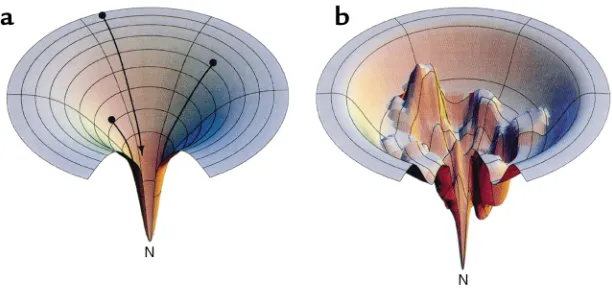
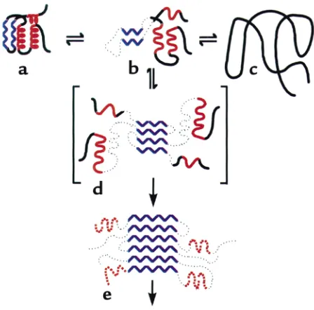
urant, many of them exhibiting what are called two-state kinetics, that is, traversing only between the unfolded and native states, usually on the time scale of a few seconds or less. A wealth of studies of such pro-teins has begun to define the modes by which propro-teins fold into their native forms. The process does not appear to be random, as the search time required would be impossibly long; rather, the unfolded protein under-goes specific kinetically preferred steps on the way to the native state, albeit that there may be multiple choic-es at any given point (see Figure 1a). The ability to examine the transition states for two-state folding reac-tions by mutation analyses, pioneered by Alan Fersht and his colleagues, allows determination of the ele-ments of structure that are present at the transition state and provides information on which structural fea-tures of the native protein have formed during folding up to a given point (12). Proteins appear to follow mul-tiple approaches to the transition state. For example, some proteins rapidly acquire secondary structure (which is present already at the transition state) before organizing tertiary structure; for others, a rapid col-lapse of hydrophobic regions to form a core can occur before or at the same time as secondary and tertiary structure formation. An interesting observation from the recent work of David Baker and coworkers is that proteins whose contiguous amino acid sequences remain in contact with each other via secondary or ter-tiary structure in the final three-dimensional native state fold faster than those whose local structures are formed from distantly separated sequences (in a paral-lel β-sheet, for instance), implying an entropic penalty for bringing together the distant segments of polypep-tide in the latter proteins (13).
[image:3.576.58.364.61.208.2]Partly folded intermediate states in the folding and aggregation of larger proteins. In contrast to the two-state proteins, refolding from denaturant of many proteins of more than 100–150 amino acids does not proceed effi-ciently. Indeed, in many such experi-ments, a precipitate of aggregated protein is recovered at the bottom of the tube. This finding implies that for many larger proteins, as well as for some smaller ones (see refs. 14, 15 for examples), there are kinetic barriers to reaching the native state. Thus, when left to fold on their own, these proteins become “kinetically trapped” in local energetic minima (Figure 1b). It is such intermediate, partly folded conformations that can associate to form aggregates. Some such intermediate states may be obligatory ones, whose confor-mation a folding protein must nec-essarily traverse in order to reach native form, whereas others may lie away from a productive pathway. Regardless, when such states are sig-nificantly populated, intermediate species are able to interact specifically with each other to form aggregates. Early evidence for this model of aggregate formation came from the classic experiment of Michel Goldberg and coworkers, who examined the behavior of the tetrameric enzyme tryptophanase, diluted from urea (16). Goldberg and colleagues observed that, after first unfolding tryptophanase with 8 M urea and then dilut-ing into buffer, both refolded native enzyme and inac-tive aggregate formed. With increasing concentrations of the protein, however, the yield of active enzyme was reduced, and the inactive enzyme sedimented in a sucrose gradient as a polydisperse population of mole-cules. Importantly, these workers showed that the pro-tein partitions in different ways between the native state and the inactive aggregate, depending on the con-centration of urea used at the renaturation step. In par-ticular, at 3 M urea, all of the protein is recovered in the aggregate, and no recovery ensues following removal of urea, suggesting that a folding intermediate is involved whose population is favored at this concentration of urea. Goldberg and his associates further showed that the self-association of this intermediate is specific, since when BSA or even a crude bacterial extract is denatured in the same tube with the tryptophanase, the enzyme’s refolding proceeds exactly as before. Hence, for any given concentration of tryptophanase, contacts between tryptophanase monomers, leading to either productive or nonproductive folding, are not affected by the presence of other denatured proteins. To account for both the concentration dependence and the specificity of the reaction, Goldberg and colleagues proposed that aggregation is driven by the same stere-ospecific interactions that are required for folding of
Figure 1
the polypeptide chain to the native form, but that these interactions occur between two separate chains (Figure 2); that is, intermolecular contacts become substituted for intramolecular ones.
Soon after, Rainer Jaenicke and his coworkers per-formed a study of refolding and aggregation of porcine tetrameric lactate dehydrogenase that revealed a simi-lar protein concentration–dependent behavior of aggregation (17). Using far ultraviolet circular dichro-ism spectroscopy, these authors showed that the aggre-gates resemble the native protein with regard to their
α-helical and β-strand content, providing direct sup-port for the idea that monomeric intermediate states responsible for aggregation exhibit significant native-like secondary structure. Unnative-like the rapid, first-order reaction for productive folding, the kinetics observed for aggregate formation was consistent with a compe-tition between two types of events, a unimolecular, con-centration-independent intrachain interaction, and a multimolecular, interchain one whose rate increases rapidly with the protein concentration.
Secondary structures and hydrophobic interactions that drive protein aggregation. More recent studies of the in vitro refolding of two monomeric proteins, phosphoglycer-ate kinase (PGK) and bovine growth hormone (bGH), provide information on the structures mediating aggregating interactions. In the case of PGK, the kinet-ics of aggregate formation is biphasic, with a first phase exhibiting first-order kinetics, corresponding to fold-ing of an intermediate, and a second phase that is con-centration-dependent. This second stage can be sup-pressed by chilling to 4°C, consistent with involvement of hydrophobic interactions in producing contact between the monomer intermediates (18). The aggre-gated species lacks substantial α-helical structure, nor-mally present at the surface of native PGK, and appar-ently exposes hydrophobic core β-strands. Presumably, some stereospecific combination of hydrophobic and
β-strand interactions between monomers constitutes the interchain interactions of aggregating PGK.
In experiments with bGH, as in the other studies, the intermediate species that accumulates shows consider-able native-like secondary structure (in this case all
α-helical), but it lacks the native tertiary structure. The unfolded bGH partitions between the aggregated and the native states (19). Remarkably, addition of a specif-ic peptide fragment (amino acids 96–133) of bGH can block aggregation. This peptide has been proposed to form an amphiphilic α-helix that associates through its hydrophobic aspect with a homologous exposed hydrophobic aspect of the same α-helix in the inter-mediate form of bGH, thus preventing intermolecular interaction and consequent aggregation.
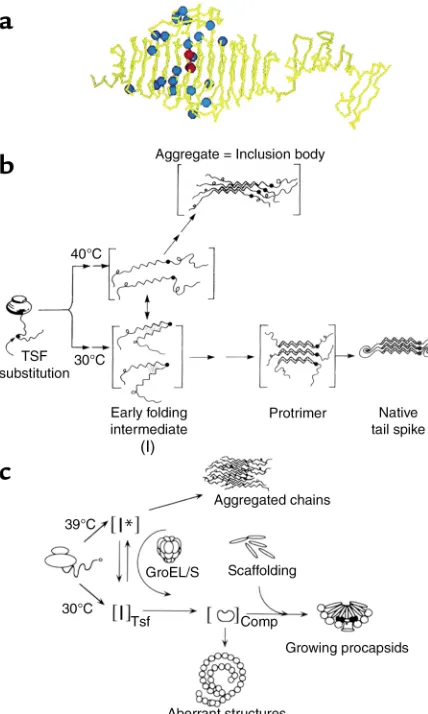
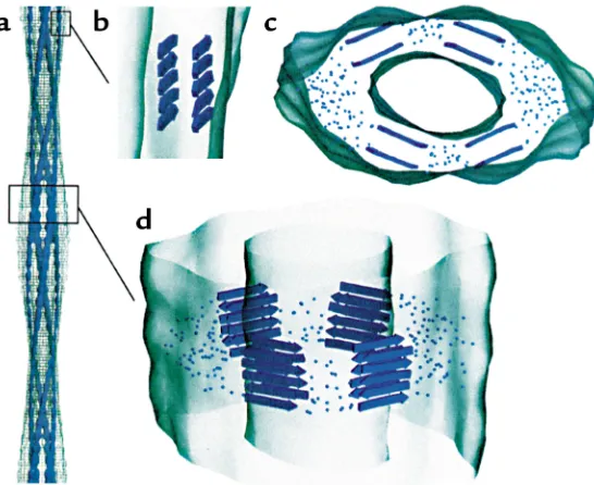
Inclusion body formation in vivo. Elegant studies of Jonathan King and coworkers, examining two Salmo-nellaphage proteins, show that the kinetic partitioning mechanism observed in vitro operates in vivo as well. These authors have studied the P22 tailspike protein, a homotrimeric assembly of 666 amino acid subunits that comprises a coiled array of parallel β-strands tak-ing the shape of a fish (ref. 20; subunit shown in Figure
3a), as well as a 48-kDa coat protein from the same bac-teriophage. King and colleagues have used tempera-ture-sensitive mutations in the tailspike protein to dis-sect the pathway of folding and assembly in intact
Salmonella(21) (Figure 3b). At high temperature, the newly synthesized tailspike subunits are initially found in a soluble fraction, but they then aggregate, forming refractile, dense, particulate structures known as inclu-sion bodies, which sediment at low speeds after cell breakage. By contrast, at lower (permissive) tempera-ture, the newly made tailspike subunits remain soluble, assembling into the native homotrimer. When cells incubated at high temperature are downshifted within a few minutes of synthesis, the tailspike subunits that were initially soluble can be recovered in the homotrimeric form, the result of redirecting the parti-tioning of an apparent thermolabile folding interme-diate toward the native state. However, if downshift is carried out at later times, subunits cannot be recovered into the soluble fraction or into a native trimer; aggre-gation and inclusion body formation supervene and become irreversible (Figure 3b).
[image:4.576.308.540.500.653.2]King and coworkers suggested that their tempera-ture-sensitive folding mutants, which map broadly across the middle 200 amino acids of the tailspike, in the midsection of the “fish” (Figure 3a, blue spheres), promote the conformational change of a thermolabile folding intermediate (designated I in Figure 3b) to yield aggregates. This intermediate, which exists in equilib-rium with the other conformational species even in the wild-type setting, forms a species that can self-associate (22). The crystal structure of the tailspike tends to sup-port this model (20), as the mutations map to the β-coil domain of the native structure (Figure 3a), mostly to solvent-exposed positions or to residues at or near the tight β-turns that enable formation of the β-coil. As such, these mutations would not be expected to affect the native structure, but they would be critical during
Figure 2
folding for producing the proper coiling and register-ing of the β-coil (Figure 3b).
Remarkably, suppression mutagenesis identifies two specific second-site mutations that can block the aggre-gation phenotype of the temperature-sensitive tailspike mutants (23). These global suppressor mutations, map-ping into the same central region of the tailspike (Fig-ure 3a, red spheres), are only active when present in the same subunit as the original mutation and do not affect the stability of the native (double-mutant) protein. They therefore appear to favor partitioning of the monomer-ic tailspike folding intermediate to native trimermonomer-ic form (Figure 3b), either by stabilizing the on-pathway inter-mediate forms or by destabilizing off-pathway confor-mations that lie in the direction of aggregation.
Temperature-sensitive folding mutations in the P22 coat protein behave similarly to the tailspike with respect to temperature-dependent partitioning into aggregates in vivo (24) (Figure 3c). King and colleagues have observed the folding and aggregation pathways of both tailspike and coat using nondenaturing gel analysis, an in vitro procedure that allows species from monomer up to higher-order multimers of either protein to be observed directly. Very interestingly, the two proteins, when placed together under conditions favoring aggre-gation, produce two distinct ladders of multimers in the
native gel, one corresponding to that observed with tail-spike aggregating alone and the other corresponding to that observed when coat protein aggregates alone (25). This experiment provides the clearest evidence that pro-teins aggregate independently of one another and strongly suggests that specific contacts, formed between folding intermediates, produce aggregates that are ordered arrays of a single protein.
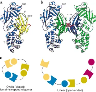
[image:5.576.77.291.60.417.2]Domain swapping between monomers. Surprising find-ings from David Eisenberg’s group, comparing the crystal structure of diphtheria toxin as a native monomer and as an acid-induced dimer, also have important implications for aggregation processes in general (26). These authors observed in the dimeric form that one of the three domains of the toxin monomer, the receptor-binding domain, had become rearranged in position such that its noncovalent con-tacts with the two other domains had been broken; it had rotated 180° and translated as much as 65 Å to form normal noncovalent contacts with the two domains of the other chain of the dimer (Figure 4, a and b). Such domain swapping has now been observed in crystals of several dozen other proteins. In all cases, as with Goldberg and colleagues’ early model of aggre-gation, the behavior is one of forming an intersubunit interface that is identical to an intrasubunit interface. In some cases, exchange observed in the crystals involves three or four subunits. The potential relevance of such higher-order exchange to processes of protein aggregation seems inescapable, as Eisenberg and coworkers consider the possibility of higher-order domain-swapped oligomers produced as closed or as linear assemblies (27) (Figure 4c). Indeed, the aggrega-tion of ZZ-type α1-antitrypsin (see Lomas and Mahade-va, this Perspective series, ref. 28) appears to be just the latter type of arrangement, in that a reactive center loop segment, which normally inserts as a β-strand into a gap in the center of a sheet within the same molecule,
Figure 3
inserts instead into the same position of another mol-ecule, a process termed loop-sheet polymerization (29). More detailed structural studies of additional aggre-gates will be necessary to evaluate whether domain swapping operates more generally or whether aggre-gates can be formed by intersubunit contacts that have no equivalent in the native state.
Protein folding in the cell: chaperones and protein biosynthesis
Binding of folding intermediates by chaperones. Work of the 1980s and early 1990s established that a class of spe-cialized heat-inducible proteins, molecular chaperones, can provide kinetic assistance to protein folding in a variety of contexts, in general preventing protein aggre-gation and promoting proper folding by binding to hydrophobic surfaces specifically exposed in non-native protein conformations (30, 31). Such behavior has been especially well demonstrated for the double-ring assem-blies known as chaperonins. For example, a host of pro-teins imported into yeast mitochondria in vivo fail to reach their refolded native state and become insoluble in a mutant deficient in the mitochondrial chaperonin Hsp60 (32). In vitro, likewise, subunits of such proteins as Rubisco, rhodanese, and malate dehydrogenase undergo wholesale aggregation following dilution from denaturant, failing to recover enzymatic activity. But the presence of the bacterial chaperonin GroEL, which sto-ichiometrically binds proteins in non-native states (33–35), allows these enzymes to be recovered quantita-tively in native form via a refolding reaction requiring
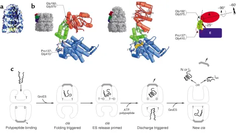
the cochaperonin GroES and ATP. With structural analysis of GroEL, it became clear that its open rings provide a hydrophobic binding surface (Figure 5a) that recognizes exposed hydrophobic surfaces specific to non-native polypeptides, surfaces that become buried in the interior of native proteins (36). These same hydrophobic surfaces in non-native proteins contribute, as outlined above, to the process of multimolecular aggregation, albeit that particular secondary-structural elements appear also to be involved. Nevertheless, the cavity surface of GroEL successfully competes for these hydrophobic sites — apparently without regard to their structural context, since both all–α-helical proteins and
β-sheet–rich ones can be bound (reviewed in ref. 37). Binding through hydrophobic contacts prevents such surfaces from promoting aggregation.
Not only can GroEL prevent aggregation via the step of binding, but it can also actively promote productive folding, triggered upon binding of ATP and GroES to the same ring to which the substrate polypeptide binds (31, 38) (Figure 5, a and b). Such binding is associated with rigid body movements of the peptide-binding api-cal domains of the bound GroEL ring, elevating and twisting their hydrophobic surfaces away from the cen-tral cavity and replacing the lining of the cavity with hydrophilic surfaces. These movements result in rapid release of the substrate protein from the cavity walls into a now encapsulated chamber, where it has no access to other monomers with which it could aggregate. The walls of the cavity, now hydrophilic in character, may encourage productive folding, insofar as they
energeti-Figure 4
Domain swapping between protein monomers in crystals. (aand b) Original demonstration of domain swapping of diphtheria toxin in a protein crystal (26). The receptor-binding domain of a monomer (shown in yellow in a) can become rotated and translated to form contacts with the other two domains of the toxin, as in b, where the blue receptor-binding domain of the subunit at the left has become associated with the other two domains of the green subunit at the right, and vice versa. This demonstrates the substitu-tion of interchain contacts for intrachain ones as proposed by Goldberg (see Figure 2). R, recep-tor binding domain. Reproduced with permis-sion from Proceedings of the National Academy of Sci-ences of the United States of America (26). (c) Models for mechanics of higher-order domain swapping. Reproduced with permission from
[image:6.576.230.537.428.723.2]cally favor conformations in which the substrate’s hydrophobic surfaces are buried and its hydrophilic sur-faces are exposed, as occurs in native proteins.
This folding-active ternary complex has a finite lifetime (12 seconds at 23°C) before the hydrolysis of ATP in the proximal ring and the subsequent binding of ATP to the opposite ring cause it to dissociate, releasing both GroES and the polypeptide substrate (31) (Figure 5c). If the pro-tein has reached native form during this time, it proceeds to carry out its function; in the case of oligomeric pro-teins, assembly with other subunits occurs in the bulk solvent. If the protein has failed to reach native form, or one committed to completing folding to the native state, it becomes rebound by the chaperonin or by another chaperone in the same cellular compartment, and it can attempt once again to fold correctly. Thus, chaperonins play into the kinetic partitioning schemes outlined earli-er for aggregation processes, shunting those folding intermediates that they recognize away from multimole-cular aggregation and toward the native state. An
excel-lent case of such partitioning is found in the P22 system, where aggregation of folding-defective mutant coat pro-teins has been observed in vivo to be suppressed by over-expression of GroEL and GroES (39) (Figure 3c).
Other cellular chaperones that reverse or prevent aggrega-tion. Once aggregation supervenes, chaperonins such as GroEL are unable to reverse the process, probably because they can only bind free monomeric subunits. In contrast, another ring-shaped cylinder, Hsp104, appears able to mediate ATP-dependent disaggregation of “soft” aggregates of up to 600 kDa; the action of additional chaperones of the Hsp70 family allows the components of such aggregates, once released, to refold and attain the native conformation (40). In vivo in intact yeast, such action is accomplished in the cytosol, where the heat-inducible Hsp 104 protein can dissolve morphologic aggregates produced during heat shock following temperature downshift (41).
[image:7.576.65.534.58.323.2]In addition to the active role played by chaperonins in productive folding, a number of other chaperones
Figure 5
bias kinetic partitioning in vivo away from aggregation. These include Hsp70s and their cooperating DnaJ-related partners, Hsp40s, which recognize and bind exposed hydrophobic side chains of non-native pro-teins in the context of an extended conformation (31). Similarly, small heat shock proteins (sHsp’s) of about 20 kDa prevent aggregation by forming oligomeric assemblies that bind incipiently denaturing proteins at their surfaces during heat shock or other cellular stress. In the case of Hsp70 proteins, ATP binding drives release of bound protein, allowing it a chance to fold correctly. In contrast, a return to normal cellular con-ditions is associated with release from sHsp’s.
A role of Hsp70 proteins in conformational quality control is highlighted by the ER version, BiP, which is involved with translocation of importing polypeptides and with their folding and assembly. Notably, BiP remains bound to many misfolded, incompletely mod-ified, or incompletely assembled species in this cellular compartment, preventing exit and progression down the secretory pathway. In many cases, misfolded pro-teins are retro-translocated from the ER to the cytosol where they are degraded by the proteasome (see Kauf-man, this Perspective series, ref. 42). In other cases, the oxidizing nature of the ER compartment contributes to the observation of disulfide–cross-linked aggregates within the compartment; correspondingly, a critical role in protein folding and unfolding has been recog-nized not only for classical chaperones like BiP, but also for oxidoreductases and for a lectin/glucosyltransferase system (see, for instance, refs. 43–45).
Amyloid formation in human disease
The term “amyloid” was coined in 1853 by Virchow to connote waxy, eosinophilic tissue deposits that he thought were composed of carbohydrate. Within 10 years, however, Friedrich and Kekule established that the deposits were composed of protein. In 1959, with examination by EM, it became clear that there were fib-rillar components to such deposits (46). Isolated fibrils were observed to be rigid and nonbranching and to stain characteristically with Congo red dye. EM further revealed that the fibrils were composed of several later-ally associated protein filaments, the latter measuring about 75 Å in diameter (47). In 1968 and 1969, two groups (48, 49) employed x-ray fiber diffraction to show that the spacing of strands within sheets of human amy-loid was consistent with a β-pleated sheet arrangement in which the sheets form a cross-βpattern whose spac-ing is dictated by the projectspac-ing side chains.
With improving methods to purify amyloid fibrils, Glenner and coworkers, in 1970, were the first to iden-tify a constituent protein of an amyloid, an Ig light chain in amyloid from patients with primary systemic amyloidosis (50). Glenner’s review of the field in 1980 was prescient. He described a “unifying definition of the amyloid fibril as having, in all cases, a β-pleated sheet structure,” and took the view that “amyloid accumula-tion is not the consequence of a single disease process . . . but a variety of different disease processes . . . result-ing in the deposition of twisted β-pleated sheet fibrils
formed from various proteins . . .” (51). At that point it was already known that amyloid plaques were charac-teristic of Alzheimer disease and of the spongiform encephalopathies, such as kuru and Creutzfeldt-Jacob disease, and that they were present in the cardiac system of many elderly patients. Today, we know that the deposits in these conditions contain Aβ-peptides, prion protein, and transthyretin (TTR), respectively. Joining this list are such other proteins as islet amyloid protein in the pancreas of patients with type 2 diabetes, lysozyme in the liver of patients with systemic amyloi-dosis, and cystatin C in patients with hereditary cerebral amyloid angiopathy (52).
Remarkably, the various proteins present in amyloid in these clinical disorders share no primary amino acid sequence, nor even tertiary structural features in their native forms, yet all can assume the same basic cross-β -sheet topology in amyloid fibrils. Recent results in vitro suggest that any protein can be converted to an amy-loid state if exposed to the proper partial-denaturing condition. Even the all–α-helical protein apomyoglo-bin has been incorporated into amyloid fibrils when exposed to pH 9 and 65°C (53). Thus, the amyloid state may be accessible to any protein as a very stable alter-native conformational state, a state that, despite involv-ing only noncovalent contacts between subunits, does not readily dissociate to monomer, even with exposure to powerful denaturants. Indeed, despite earlier assumptions to the contrary, proteins in amyloids may occupy a state that is even more thermodynamically stable than the native state.
Surely, this nonfunctional dead-end state is one to be avoided in vivo, and, as Dobson and others have com-mented, evolution has likely selected against residues or sequences within extant proteins that favor conver-sion to an amyloid conformation (54). Thus, with pro-tein sequences as they have evolved, very high kinetic barriers keep most of our proteins out of this alterna-tive energetic minimum. Why are amyloid structures so stable, why are apparently all proteins accessible to these states, and how are amyloid states accessed?
Amyloid structure. While we still lack a high-resolution structure for any protein in its amyloid form, recent fiber diffraction and EM studies have provided further insight into the cross-β-sheet structures found in amy-loid fibrils. Blake and Serpell used synchrotron radia-tion in fiber diffracradia-tion of TTR amyloids to derive a model of the amyloid fibril consisting of a helical stair-case of β-sheets in which four β-sheets form the cross section of a protofilament. The individual units in the model comprise structurally rearranged TTR mono-mers or dimono-mers (55). A cryoEM reconstruction study by Helen Saibil and coworkers of an amyloid formed from an SH3 domain also concluded that the protein must undergo structural rearrangement relative to its native fold (56) (Figure 6). Here, the fibril was found to be composed of four protofilaments, smaller than those of the TTR amyloid and consisting of two stacked
Additional EM studies of a yeast prion protein that can form amyloids in vitro indicate that the entire protein species is not required to become incorporated into the
β-sheet organization of a fibril. In this case, an NH2 -ter-minal region of the protein becomes incorporated into the fibrils themselves, while the remaining COOH-ter-minal portion of the protein is “marginated” to the out-side of the fibril (57). This region is likely to retain a native structure, and there are recent examples of fusion pro-teins incorporated into amyloids in which a nonamy-loidogenic “passenger” enzyme segment retains enzy-matic activity after the protein is incorporated into fibrils. A point that seems to emerge from both the diffrac-tion and the EM studies is that the primary contacts that glue an amyloid protofilament together along its length are the hydrogen bonds between β-strands. Since these are main-chain contacts, there may be min-imal side-chain involvement and therefore little or no sequence specificity to such behavior. Thus, it may be that any segment of polypeptide that can be folded or refolded into β-strand can ultimately participate in such a stable structure.
Production of amyloidogenic folding intermediates by desta-bilization of the native state. Evidence has emerged over the past several years to indicate that production of the amyloid state of a protein often involves destabilization of the already-folded native protein. That is, disruption of the globular structure, where a sequence of side chains has dictated a native fold, can dispose to refold-ing into a structure where main-chain contacts, rather than side-chain interactions, predominate.
It is important at this point to be clear about the def-initions of stability and destabilization. These terms are often used in a general structural sense to refer to the effects of mutations or conditions, such as low pH or the presence of denaturants, that disrupt the native conformation of a protein. It is desirable, however, to distinguish whether such a structural disruption
reflects an effect on thermodynamic or kinetic stability. The former refers to the free energy difference between the native state and a monomeric intermediate on the path-way to amyloidogenesis and is reflected in the equilibrium between the native and amy-loidogenic intermediate states. Mutations generally decrease the thermodynamic sta-bility of a protein by increasing the free ener-gy of the native state, although decreasing the free energy of the intermediate state is also possible. Kinetic stability refers to the height of the free energy barrier between the native state and the transition state for producing the amy-loidogenic intermediate and is reflected in the rate con-stant for conversion from native to intermediate. Muta-tions affect this rate either by increasing the free energy of the native state or by decreasing that of the transi-tion state. (Note that the relative free energy of the intermediate does not contribute to this effect.)
In many biological systems, both thermodynamic and kinetic effects occur simultaneously with mutation or changing conditions. However, as mentioned above, the kinetic barriers between native and amyloidogenic intermediate states are probably very large in the nor-mal cell, effectively precluding the establishment of an equilibrium between these states on biological time scales. In vitro, however, such a kinetic barrier can be surmounted by partial denaturation, as occurs when the pH is reduced to 3–4.5 or in the presence of mod-erate concentrations of alcohols.
Amyloid formation from mutant proteins. As with the inclusion body–forming aggregates, it is the formation of a non-native monomer intermediate that appears to be critical to the aggregation step in amyloid formation. Here, however, instead of forming from an unfolded state, the intermediate is typically formed from the already-folded native protein. While intermediates of both inclusion body and amyloid reactions have native-like properties, amyloid formation appears to also require the reformation or reorganization of some ele-ments of secondary and tertiary structure that disposes to association of these intermediates into amyloid fib-rils. Both in vitro and in vivo, this process can be poten-tiated by mutations that structurally destabilize the native state, as demonstrated particularly dramatically for monomeric lysozyme and tetrameric TTR (but see also ref. 58 concerning Ig light chain).
[image:9.576.54.327.61.284.2]In the case of lysozyme, a 14-kDa secretory protein composed of an α-helical and a β-sheet domain, two
Figure 6
known human mutations predisposing to amyloid-forming disease have been studied in detail (59). Crystal structures of the two mutant proteins in their native forms show them to resemble the wild-type version, although subtle changes suggest that the domain inter-face might be destabilized. Indeed, the activity of the variant proteins in vitro is more sensitive to heat treat-ment, which leads to the formation of fibrils with a pre-dominantly βstructure. Interestingly, fibrils obtained directly from patients, who are typically heterozygous for these (dominant) mutations, contain only the mutant lysozyme, indicating that only the mutant protein pop-ulates an amyloidogenic intermediate. Most astonishing in this regard, when hydrogen-deuterium exchange is carried out at physiologic temperature, the mutant pro-teins prove to be fully exchangeable on the time scale of minutes, whereas the wild-type protein contains 55 nonexchangeable protons. Thus, under conditions where the wild-type native protein is stable, the mutant proteins are very unstable, apparently populating aggre-gation-prone, partly folded, amyloidogenic states (Fig-ure 7b). Based on the earlier classic hen lysozyme folding studies of Dobson’s group (59), where the α-domain became structured in intermediate states while the
β-domain was still unstructured, it has been suggested that fibrillogenesis occurs through the β-domain of the unstable monomer (Figure 7, b and d).
In the case of TTR, destabilization of the tetramer’s quaternary structure leads to amyloid formation in vivo, either in the setting of dominant inherited muta-tions in familial amyloid polyneuropathy or, sporadi-cally, in senile cardiac amyloidosis; in vitro acid treat-ment (pH 3.5–4.0) has similar effects. The rate-limiting step appears to be tetramer dissociation, which is fol-lowed by a structural rearrangement of the product monomer to produce an amyloidogenic species that assembles into fibrils (summarized in ref. 60). Circular dichroism studies at pH 3.6 show that the amyloido-genic conformation retains a secondary-structure con-tent resembling that of the native protein (61). Consis-tent with a model requiring tetramer dissociation, a homotetramer of the most pathogenic TTR mutant, L55P, has been found to be much less stable than wild-type to acid treatment in vitro, dissociating and form-ing fibrils under mildly acidic conditions that do not affect wild-type TTR (62). Another prevalent amy-loidogenic mutant, V30M, is also sensitive to modest drops in pH but can be stabilized by the presence of a second mutation, T119M, either within the same sub-unit (62) or in trans, as one or more separate subunits in the same tetramer (63). This latter configuration cor-responds to the milder clinical condition observed in patients who are compound heterozygotes for V30M and T119M. Consistent with this, T119M tetramers are far more stable in vitro against acid disruption than are wild-type tetramers. Finally, a recent study in which a monomeric variant of TTR was engineered by mutat-ing residues in the dimerization interfaces (64) showed that the protein, which is stable and nonamyloidogenic at pH 7, undergoes a rapid conformational rearrange-ment and generates fibrils at mildly acidic pH. Here,
with tetramer dissociation no longer a requirement, presumably the only limiting step to fibril formation is tertiary structural rearrangement.
Efforts to better characterize the TTR amyloidogenic intermediate include tryptophan fluorescence studies suggesting that edge strands of one of the β-sheets become conformationally altered, and binding studies with the hydrophobic-binding dye ANS (8-anilino-naph-thalene-sulfonic acid), which suggest that this interme-diate does not expose significant hydrophobic surface (60). A deuterium-hydrogen–exchange nuclear magnet-ic resonance study has shown that the interstrand hydro-gen bonds of one of the sheets of TTR are disrupted at pH 4.5, indicating that sheet destabilization contributes to the amyloidogenic conformation (65).
Consistent with a role for kinetic control of TTR amyloid formation, the mutation V122I has recently been shown to increase the rate of TTR tetramer dis-sociation under certain conditions (66). A case for kinetic control has also been made for the prion pro-tein, where many of the inherited mutations disposing to disease have been found to have no effect on ther-modynamic stability of PrP, as measured by urea denaturation curves following circular dichroism spec-tra (67, 68). Similar analyses of mouse PrP show that the amyloid form is thermodynamically more stable than the native α-helical form but is separated from it by a high kinetic barrier (69).
[image:10.576.310.534.435.656.2]Thermodynamic control of polymerization by amyloido-genic intermediates. Amyloidogenic intermediates must polymerize to form fibrils. As with many other poly-merization reactions (microtubule growth, flagellum
Figure 7
formation, and viral coat assembly, for instance), nucle-ation may be a crucial step in this process for at least some amyloid species (70). An oligomeric assembly of a nucleus of a critical size can form from amyloido-genic monomers by a series of association reactions that have, overall, an unfavorable equilibrium. Hence, nucleus formation is under thermodynamic control. Once formed, however, the nucleus can rapidly poly-merize into an amyloid fibril.
This behavior predicts that there will be a critical con-centration of monomers below which amyloid cannot form; that, once the threshold concentration is reached, there will be a lag time before the association reactions permit production of a nucleus; and that addition of a preformed nucleus during the lag can result in immediate production of amyloid fibrils. While these predictions have been met in vitro for PrP, for Aβ-peptide, and for several other amyloid precur-sors, this mechanism remains to be established in vivo. For example, only prion diseases show the phenome-non of seeding, where inoculation of amyloid material produces disease. Nevertheless, nucleation-polymer-ization behavior might account, at least in part, for the very long development times for amyloid disease, even in the setting of mutations that destabilize the native state of an amyloidogenic protein.
At a further level of complexity, two recent studies indi-cate that nonfibrillar, in one case amorphous, aggregates of amyloidogenic proteins, formed early on a fibrilloge-nesis pathway, may be toxic to cells or brain function (71, 72). By contrast, mature fibrils do not exhibit such toxi-city. Whether these amorphous structures are incipient fibrils or are structurally disordered states from which ordered aggregates are formed remains unknown.
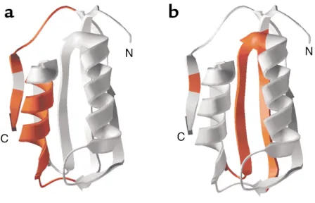
Domain structures that influence amyloidogenesis. Recent studies in vitro of the two-state folding protein acylphosphatase suggest that different regions of this protein are responsible for favoring pathways of folding to native form versus those leading to amyloid (73) (Fig-ure 8). In particular, mutations in two regions of the protein, amino acids 16–31, mapping into α-helix 1, and amino acids 87–98, mapping into a terminal β-strand,
interfere with amyloid formation promoted by denatu-ration in trifluoroethanol (Figure 8a). These regions exhibit significant hydrophobicity and β-sheet–forming propensity, despite the fact that amino acids 16–31 form an α-helix in the native state. By contrast, a dis-tinct region, exhibiting structure even in the transition state, has been found to be critical for productive fold-ing in aqueous solvent (Figure 8b). Thus, properties of different regions of the same chain can dictate which intermediates form and thereby influence the protein’s final fate, albeit that under physiologic conditions, evo-lution seems to have chosen that de novo folding origi-nating from the ribosome should favor the pathway to the native state.
Prospects for clinical control of amyloid diseases. The fore-going considerations have important implications for the formation of amyloids in the clinical setting. First, thankfully, it appears that only a limited number of pro-teins have access to an amyloid state in vivo. Almost all of those that can form amyloid are secreted proteins like lysozyme, TTR, and PrP. At this point, it is unclear whether these proteins undergo conformational con-version in the intracellular environment, perhaps in the mildly acidic compartment of the endosome, or whether conversion occurs outside the cell. Regardless, it is clear that chaperone systems that can normally forestall aggregation events fail to prevent these pro-teins from undergoing the transition to form amyloids. It may be simply a compartmentation problem: If amy-loid formation occurs in the endosomes or outside of the cell, chaperones are absent and there is no safeguard system available. Alternatively, if the intermediates formed are sufficiently structured, failing to expose sig-nificant hydrophobicity, as appears to be the case with TTR, the chaperone system might not recognize these species. Perhaps this is the case for other amyloids, formed in compartments that are well-populated by molecular chaperones. For example, polyglutamine repeat expansion disorders involve proteins that are cytosolic or nuclear (74). While chaperones as well as proteasome components have been found associated with polyglutamine aggregates, their role in aggregation remains unclear. Likewise, α-synuclein aggregation in Parkinson disease occurs in the cytosol, and occasional aggregates observed in postmortem tissue stain posi-tively for Hsp70 or Hsp40 chaperones. Finally, recent studies have suggested that aggregates themselves may inhibit the normal proteasome system that helps clear misfolded or aggregation-prone proteins (75).
Among the various amyloidogenic proteins, PrP is unique with regard to its infectious behavior. The notion that the prion form, PrPSc, can produce conformational conversion of the wild-type PrPc protein predicts a behavior that is not generally observed for the other amy-loidogenic species (76). Whether this relates somehow to the pathway of clinical infection or to the conforma-tional states of the protein remains to be addressed.
[image:11.576.63.287.60.204.2]If structural destabilization of the native state is the major contributor to amyloidogenesis in these various clinical conditions, then, as proposed by Kelly and col-leagues, stabilizing the native form of a protein by
Figure 8
binding a ligand may offer one of the most direct means of preventing disease (77). In the case of TTR, for example, providing a compound that mimics the normal ligand, thyroxine, and stabilizes the tetramer against dissociation is an approach currently being tested (78). By contrast, disease produced by Aβ -pep-tides will not be easily addressable in this manner, as these peptides are proteolytic products that have no favored conformational state other than the amy-loidogenic ones (5). Obviously, stabilizing intact APP itself against βor γproteolysis, or developing inhibitors of the proteases, may help prevent such amyloid for-mation. For prion disease a number of compounds are being tested, including quinacrine and chlorpro-mazine, that block PrPSc production and may also enhance its clearance (79). In sum, the particular con-formational pathway taken by each of the clinically rel-evant amyloidogenic proteins provides, in each case where a pathway becomes understood, a specific avenue for development of compounds that block par-titioning of that protein to amyloid.
Acknowledgments
I thank Wayne Fenton for discussion and critical read-ing of the manuscript, and Howard Hughes Medical Institute for its generous support.
1. Weatherall, D.J., Clegg, J.B., Higgs, D.R., and Wood, W.G. 2001. The hemoglobinopathies. In Metabolic and molecular bases of inherited disease. C. Scriver, A. Beaudet, D. Valle, and W. Sly, editors. McGraw-Hill. New York, New York, USA. 4571–4636.
2. Perlmutter, D.H. 2002. Liver injury in α1-antitrypsin deficiency: role of autophagy and mitochondrial injury in response to aggregated protein in the endoplasmic reticulum. J. Clin. Invest. In press.
3. Cox, D.W. 2001. α1-Antitrypsin deficiency. In Metabolic and molecular bases
of inherited disease. C. Scriver, A. Beaudet, D. Valle, and W. Sly, editors. McGraw-Hill. New York, New York, USA. 5559–5584.
4. Selkoe, D.J. 2002. Deciphering the genesis and fate of amyloid-βprotein yields novel therapies for Alzheimer disease. J. Clin. Invest. In press. 5. Selkoe, D. 1997. Cellular and molecular biology of the beta-amyloid
pre-cursor protein and Alzheimer’s disease. In The molecular and genetic basis of neurological disease. R.N. Rosenberg, S.B. Prusiner, S. DiMauro, and R.L. Barchi, editors. Butterworth-Heinemann. Boston, Massachusetts, USA. 601–611.
6. DeArmond, S.J., and Prusiner, S.B. 1997. Molecular neuropathology of prion diseases. In The molecular and genetic basis of neurological disease. R.N. Rosenberg, S.B. Prusiner, S. DiMauro, and R.L. Barchi, editors. Butter-worth-Heinemann. Boston, Massachusetts, USA. 145–163.
7. DiFiglia, M., et al. 1997. Aggregation of huntingtin in neuronal intranu-clear inclusions and dystrophic neurites in brain. Science.277:1990–1993. 8. Scherzinger, E., et al. 1997. Huntintin-encoded polyglutamine expan-sions form amyloid-like protein aggregates in vitro and in vivo. Cell.
90:549–558.
9. Conway, K.A., Harper, J.D., and Lansbury, P.T. 2000. Fibrils formed in vitro from α-synuclein and two mutant forms linked to Parkinson’s dis-ease are typical amyloid. Biochemistry.39:2552–2563.
10. Serpell, L.C., Berriman, J., Jakes, R., Goedert, M., and Crowther, R.A. 2000. Fiber diffraction of synthetic α-synuclein filaments shows amy-loid-like cross-β conformation. Proc. Natl. Acad. Sci. USA.97:4897–4902. 11. Anfinsen, C.B. 1973. Principles that govern the folding of protein chains.
Science.181:223–230.
12. Fersht, A.R. 1995. Characterizing transition states in protein folding: an essential step in the puzzle. Curr. Opin. Struct. Biol.5:79–84.
13. Baker, D. 2000. A surprising simplicity to protein folding. Nature.
405:39–42.
14. Kim, P.S., and Baldwin, R.L. 1990. Intermediates in the folding reactions of small proteins. Annu. Rev. Biochem.59:631–660.
15. Silow, M., Tan, Y.-J., Fersht, A.R., and Oliveberg, M. 1999. Formation of short-lived protein aggregates directly from the coil in two-state folding.
Biochemistry.38:13006–13012.
16. London, J., Skrzynia, C., and Goldberg, M.E. 1974. Renaturation of
Escherichia colitryptophanase after exposure to 8 M urea. Eur. J. Biochem.
47:409–415.
17. Zettlmeissl, G., Rudolph, R., and Jaenicke, R. 1979. Reconstitution of lac-tic dehydrogenase. Noncovalent aggregation vs. reactivation. Biochem-istry.18:5567–5571.
18. Mitraki, A., Betton, J.-M., Desmadril, M., and Yon, J.M. 1987. Quasi-irre-versibility in the unfolding-refolding transition of phosphoglycerate kinase induced by guanidine hydrochloride. Eur. J. Biochem.163:29–34. 19. Brems, D.N. 1988. Solubility of different folding conformers of bovine
growth hormone. Biochemistry.27:4541–4546.
20. Steinbacher, S., et al. 1994. Crystal structure of P22 tailspike protein: interdigitated subunits in a thermostable trimer. Science.265:383–386. 21. Haase-Pettingell, C.A., and King, J. 1988. Formation of aggregates from a thermolabile in vivofolding intermediate in P22 tailspike maturation.
J. Biol. Chem.263:4977–4983.
22. King, J., Haase-Pettingell, C., Robinson, A.S., Speed, M., and Mitraki, A. 1996. Thermolabile folding intermediates: inclusion body precursors and chaperonin substrates. FASEB J. 10:57–66.
23. Mitraki, A., Fane, B., Haase-Pettingell, C., Sturtevant, J., and King, J. 1991. Global suppression of protein folding defects and inclusion body for-mation. Science.253:54–58.
24. Gordon, C.L., and King, J. 1993. Temperature-sensitive mutations in the phage P22 coat protein which interfere with polypeptide chain folding.
J. Biol. Chem.268:9358–9368.
25. Speed, M.A., Wang, D.I.C., and King, J. 1996. Specific aggregation of par-tially folded polypeptide chains: the molecular basis of inclusion body composition. Nat. Biotechnol. 14:1283–1287.
26. Bennett, M.J., Choe, S., and Eisenberg, D. 1994. Domain swapping: entangling alliances between proteins. Proc. Natl. Acad. Sci. USA.
91:3127–3131.
27. Bennett, M.J., Schlunegger, M.P., and Eisenberg, D. 1995. 3D domain swapping: a mechanism for oligomer assembly. Protein Sci.4:2455–2468. 28. Lomas, D., and Mahadeva, R. 2002. α1-Antitrypsin polymerization and the serpinopath: pathobiology and prospects for therapy. J. Clin. Invest.
In press.
29. Lomas, D.A., Evans, D.L., Finch, J.T., and Carrell, R.W. 1992. The mech-anism of Z α1-antitrypsin accumulation in the liver. Nature.357:605–607. 30. Pelham, H.R. 1986. Speculations on the functions of the major heat
shock and glucose-regulated proteins. Cell.46:959–961.
31. Bukau, B., and Horwich, A.L. 1998. The Hsp70 and Hsp60 chaperone machines. Cell.92:351–366.
32. Cheng, M.Y., et al. 1989. Mitochondrial heat-shock protein Hsp60 is essential for assembly of proteins imported into yeast mitochondria.
Nature.337:620–625.
33. Goloubinoff, P., Christeller, J.T., Gatenby, A.A., and Lorimer, G.H. 1989. Reconstitution of active dimeric ribulose bisphosphate carboxylase from an unfolded state depends on two chaperonin proteins and MgATP.
Nature.342:884–889.
34. Martin, J., et al. 1991. Chaperonin-mediated protein folding at the sur-face of groEL through a “molten globule”-like intermediate. Nature.
352:36–42.
35. Ranson, N.A., Dunster, N.J., Burston, S.G., and Clarke, A.R. 1995. Chap-eronins can catalyze the reversal of early aggregations steps when a pro-tein misfolds. J. Mol. Biol.250:581–586.
36. Braig, K., et al. 1994. The crystal structure of the bacterial chaperonin GroEL at 2.8 Å. Nature.371:578–586.
37. Grantcharova, V., Alm, E.J., Baker, D., and Horwich, A.L. 2001. Mecha-nisms of protein folding. Curr. Opin. Struct. Biol.11:70–82.
38. Xu, Z., Horwich, A.L., and Sigler, P.B. 1997. The crystal structure of the asymmetric GroEL-GroES-(ADP)7 chaperonin complex. Nature.
388:741–750.
39. Gordon, C.L., Sather, S.K., Casjens, S., and King, J. 1994. Selective in vivo
rescue by GroEL-ES of thermolabile folding intermediate to phage P22 structural proteins. J. Biol. Chem.269:27941–27951.
40. Glover, J.R., and Lindquist, S. 1998. Hsp104, Hsp70, and Hsp40: a novel chaperone system that rescues previously aggregated proteins. Cell.
94:73–82.
41. Parsell, D.A., Kowal, A.S., Singer, M.A., and Lindquist, S. 1994. Protein disaggregation mediated by heat-shock protein Hsp104. Nature.
372:475–478.
42. Kaufman, R. 2002. Orchestrating the unfolded protein response in health and disease. J. Clin. Invest. In press.
43. Tu, B.P., Ho-Schleyer, S.C., Travers, K.J., and Weissman, J.S. 2000. Bio-chemical basis of oxidative protein folding in the endoplasmic reticulum.
Science.290:1571–1574.
44. Tsai, B., Rodighiero, C., Lencer, W.I., and Rapoport, T.A. 2001. Protein disulfide isomerase acts as a redox-dependent chaperone to unfold cholera toxin. Cell.104:937–948.
45. Helenius, A., and Aebi, M. 2001. Intracellular functions of N-linked gly-cans. Science.291:2364–2369.
micro-scopic analysis of the amyloid fibril. J. Cell Biol. 33:679–708.
48. Eanes, E.D., and Glenner, G.G. 1968. X-ray diffraction studies on amyloid filaments. J. Histochem. Cytochem.16:673–677.
49. Bonar, L., Cohen, A.S., and Skinner, M.M. 1969. Characterization of the amyloid fibril as a cross-βprotein. Proc. Soc. Exp. Biol. Med.131:1373–1375. 50. Glenner, G.G., Harbaugh, J., Ohms, J.I., Harada, M., and Cuatrecasas, P. 1970. An amyloid protein: the amino-terminal variable fragment of an immunoglobulin light chain. Biochem. Biophys. Res. Commun.41:1287–1289. 51. Glenner, G.G. 1980. Amyloid deposits and amyloidosis: the β-fibrilloses.
N. Engl. J. Med.302:1283–1292.
52. Kelly, J.W. 1996. Alternative conformations of amyloidogenic proteins govern their behaviour. Curr. Opin. Struct. Biol.6:11–17.
53. Fändrich, M., Fletcher, M.A., and Dobson, C.M. 2001. Amyloid fibrils from muscle myloglobin. Nature. 410:165–166.
54. Dobson, C.M. 1999. Protein misfolding, evolution and disease. Trends Biochem. Sci.24:329–332.
55. Blake, C., and Serpell, L. 1996. Synchrotron X-ray studies suggest that the core of the transthyretin amyloid fibril is a continuous β-sheet helix. Struc-ture.4:989–998.
56. Jimenez, J.L., et al. 1999. Cryo-electron microscopy structure of an SH3 amyloid fibril and model of the molecular packing. EMBO J.18:815–821. 57. Glover, J.R., et al. 1997. Self-seeded fibers formed by Sup35, the protein determinant of [PSI+], a heritable prion-like factor of S.cerevisiae. Cell.
89:811–819.
58. Hurle, M.R., Helms, L.R., Li, L., Chan, W., and Wetzel, R. 1994. A role for destabilizing amino acid replacements in light-chain amyloidosis. Proc. Natl. Acad. Sci. USA.91:5446–5450.
59. Booth, D.R., et al. 1997. Instability, unfolding and aggregation of human lysozyme variants underlying amyloid fibrillogenesis. Nature.385:787–793. 60. Lai, Z., Colon, W., and Kelly, J.W. 1996. The acid-mediated denaturation pathway of transthyretin yields a conformational intermediate that can self-assemble into amyloid. Biochemistry.35:6470–6482.
61. Colon, W., and Kelly, J.W. 1992. Partial denaturation of transthyretin is sufficient for amyloid fibril formation in vitro. Biochemistry.
31:8654–8660.
62. McCutchen, S.L., Lai, Z., Miroy, G.J., Kelly, J.W., and Colon, W. 1995. Com-parison of lethal and nonlethal transthyretin variants and their relation-ship to amyloid disease. Biochemistry.34:13527–13536.
63. Hammarström, P., Schneider, F., and Kelly, J.W. 2001. Trans-suppression of misfolding in an amyloid disease. Science.293:2459–2462.
64. Jiang, X., et al. 2001. An engineered transthyretin monomer that is nonamyl-oidogenic, unless it is partially denatured. Biochemistry.40:11442–11452.
65. Liu, K., Cho, H.S., Lashuel, H.A., Kelly, J.W., and Wemmer, D.E. 2000. A glimpse of a possible amyloidogenic intermediate of transthyretin. Nat. Struct. Biol. 7:754–757.
66. Jiang, X., Buxbaum, J.N., and Kelly, J.W. 2001. The V122I car-diomyophathy variant of transthyretin increases the velocity of rate-lim-iting tetramer dissociation, resulting in accelerated amyloidosis. Proc. Natl. Acad. Sci. USA.98:14943–14948.
67. Swietnicki, W., Petersen, R.B., Gambetti, P., and Surewicz, W.K. 1998. Familial mutations and the thermodynamic stability of the recombinant human prion protein. J. Biol. Chem.273:31048–31052.
68. Liemann, S., and Glockshuber, R. 1999. Influence of amino acid substi-tutions related to inherited human prion diseases on the thermodynam-ic stability of the cellular prion protein. Biochemistry. 38:3258–3267. 69. Baskakov, I.V., Legname, G., Prusiner, S.B., and Cohen, F.E. 2001.
Fold-ing of prion protein to its native alpha-helical conformation is under kinetic control. J. Biol. Chem.276:19687–19690.
70. Harper, J.D., and Lansbury, P.T. 1997. Models of amyloid seeding in Alzheimer’s disease and scrapie. Annu. Rev. Biochem.66:385–407. 71. Bucciantini, M., Giannoni, E., Chiti, F., Baroni, F., and Formigli, L. 2002.
Inherent toxicity of aggregates implies a common mechanism for protein misfolding diseases. Nature. 416:507–511.
72. Walsh, D.M., et al. 2002. Naturally secreted oligomers of amyloid β pro-tein potently inhibit hippocampal long-term potentiation in vivo. Nature.
416:535–539.
73. Chiti, F., et al. 2002. Kinetic partitioning of protein folding and aggrega-tion. Nat. Struct. Biol. 9:137–143.
74. Perutz, M.F., Johnson, T., Suzuki, M., and Finch, J.T. 1994. Glutamine repeats as polar zippers: their possible role in inherited neurodegenera-tive diseases. Proc. Natl. Acad. Sci. USA.91:5355–5358.
75. Bence, N.F., Sampat, R.M., and Kopito, R.R. 2001. Impairment of the ubiq-uitin-proteasome system by protein aggregation. Science.292:1552–1555. 76. Prusiner, S.B. 2001. Shattuck lecture: neurodegenerative diseases and
pri-ons. N. Engl. J. Med.244:1516–1526.
77. Miroy, G.J., et al. 1996. Inhibiting transthyretin amyloid fibril formation via protein stabilization. Proc. Natl. Acad. Sci. USA.93:15051–15056. 78. Klabunde, T., et al. 2000. Rational design of potent human transthyretin
amyloid disease inhibitors. Nat. Struct. Biol.7:312–321.
79. Korth, C., May, B.C.H., Cohen, F.E., and Prusiner, S.B. 2001. Acridine and phenothiazine derivatives as pharmacotherapeutics for prion disease.
Proc. Natl. Acad. Sci. USA.98:9836–9841.
80. Dill, K.A., and Chan, H.S. 1997. From Levinthal to pathways to funnels.