Efficient Pattern Matching Algorithm for
Classified Brain Image
R. Harini
Ph.D. Research Scholar Dept. of Computer Science
Periyar University Salem
C. Chandrasekar,
PhD. Reader & Associate ProfessorDept. of Computer Science Periyar University
Salem
ABSTRACT
The primary notion relying in image processing is image segmentation and classification. The intention behind the processing is to originate the image into regions. Variation formulations that effect in valuable algorithms comprise the essential attributes of its region and boundaries. Works have been carried out both in continuous and discrete formulations, though discrete version of image segmentation does not approximate continuous formulation. An existing work presented unsupervised graph cut method for image processing which leads to segmentation inaccuracy and less flexibility. To enhance the process, our first work describes the process of formation of kernel for the medical images by performing the deviation of mapped image data within the scope of each region. But the segmentation of image is not so effective based on the regions present in the given medical image. To overcome the issue, we implement a Bayesian classifier as our second work to classify the image effectively. The segmented image classification is done based on its classes and processes using Bayesian classifiers. With the classified image, it is necessary to identify the objects present in the image. For that, in this work, we exploit the use of pattern matching algorithm to identify the feature space of the objects in the classified image. An experimental evaluation is carried out to estimate the performance of the proposed efficient pattern matching algorithm for classified brain image system [EPMACB] in terms of estimation of object position, efficiency and compared the results with an existing multi-region classifier method.
General Terms
The journal is based upon Pattern Matching Algorithms for Medical Image Processing
Keywords
Image segmentation, Classification, Pattern matching, similarity measure
1. INTRODUCTION
Numerous applications in image dealing out and computer vision involve identifying a distinct model in an image, pattern toning. To be practical in carry out, pattern matching techniques must be habitual, common, rapid and vigorous. Pattern matching is classically achieved by examining the whole image, and assessing a distance measure among the model and a confined rectangular window. The method proposed in this paper is pertinent to some outline shape, yet a non-contiguous one. We utilize the concept of “window” to envelop all probable shapes.
Over the years, pattern-matching has been regularly utilized in diverse computer requests, for instance, in editors, recovery of information (from text, image, or sound), and probing nucleotide or amino acid series patterns in sequence databases. The present day pattern-matching algorithms counterpart the model precisely or roughly inside the text. An accurate pattern-matching is to discover all the incidences of a scrupulous model (x = x1, x2,…,xm) of m-characters in a
classified image (y = y1 y2 ... yn) of n-characters which are
constructed over a limited set of classified image indicated by α and the size of this set is identical to σ.
The shortest method is to evaluate the first m-characters of the image and the model in some predefined classify and, after a match or a mismatch, decrease the complete pattern by one character in the onward route of the image. This process is repetitive until the prototype is situated at the (nm+1) location of the image. This technique is normally recognized as a brute-force method. To assist this task, numerous algorithms have been planned, and these have their individual merits and demerits based on the prototype length, periodicity, and the kind of the image. Most of the eminent algorithms processed in two phases: i.e., the preprocessing stage and the seek phase. In the preprocessing stage, process the model and use this information in the seek phase to decrease the whole number of assessments and therefore decrease the general implementation time. The effectiveness of an algorithm largely depends on the seek stage.
Nearly all people who exercise computers appreciate and utilize pattern matching in several form. Search engines on the Web employ outline matching to establish information of attention. Patterns can be precise or fairly common, employing diverse wildcards that counterpart numerous endings, words, or strings. Many databases contain an analogous ability, in which a concept can be comprehensive in numerous directions. Most bioinformatics databases include analogous pattern-matching abilities. Flexible prototype matching is called parallel searching. Similarity is a proportion series match among diverse sequences. The assumption is that similarity recounts to functionality—if two series are comparable, they will have associated functionalities. Only a similarity match creates intellect for probing between parallel functions. The prospect of a match presented by similarity searches surrenders a more practical amount for contrasting sequences than a precise match. There are numerous ways to attain the pattern matching employed in similarity searching.
2. LITERATURE REVIEW
A basic crisis in computer revelation, image segmentation has been the focus of a huge amount of hypothetical and sensible studies. Numerous studies have relied on discrepancy formulations since they effect in the most efficient algorithms. Very quick techniques have been devised in a separate manner. The most competent were those which exercise graph cuts [1]. In this structure, images are analyzed as distinct functions over a positional array.
Parallel matching among images has a set to supply to the image processing society. In [2], proposed an easy image matching system supported on the location of the pixel values in the descriptions evaluated. When doing so we have restricted our work to images not containing a revolving discrepancy.
Pattern toning is a significant job of the prototype detection process in today's world for eradicating the structural and efficient activities in a given image. Even though the pattern matching algorithm [4] is normally utilized in computer science and information processing, it can be established in daily tasks. The approach in [3] used to evade redundant assessments in the DNA series. Owing to this, the number of assessments slowly reduces and assessment per character ratio of the planned algorithm decreases consequently contrasts to the some of the presented trendy methods.
The Devaki-Paul algorithm [5] for numerous pattern toning needs a pre-processing of the specified input text to organize a counter of the incidences of the 256 member ASCII character set. The piecewise constant data term representation [6], and its Gaussian simplification, have been intensively utilized in the circumstance of unsupervised graph cut methods because user interference is not necessary and, particularly, the data term can be printed in the type necessary by the graph cut algorithm [7]. Nevertheless, though practical, these representations are not usually appropriate. For example, medical images are best explained by the Gamma allocation and Wishart distribution [8]. Level set partitioning [9] is also being used for the pattern discovery process based on the patterns chosen. Region growing segmentation [10] is also being used with the allocation of segmented image parts by adapting the medical images. But the segmentation process does not provide better user intervention outcome.
The mainstream of study in medical image segmentation pertains to its use for MR images, mainly in brain imaging [11]. Instantly evaluation of different methods for segmenting MR images is also accessible [12]. To enhance the medical image segmentation process, on this work, we present a pattern matching algorithm to identify the feature space of the objects in the classified image. Our previous work elaborately described the classification of medical image with the corresponding classifiers
3. PROPOSED EFFICIENT PATTERN
MATCHING
ALGORITHM
FOR
CLASSIFIED BRAIN IMAGE
The proposed work is efficiently designed for identifying the feature space of the object in the image. The proposed efficient pattern matching algorithm for classified brain image system processed under three different phases. The first phase describes the process of formation of kernel for the medical images by performing the deviation of mapped image data
within the scope of each region from the piecewise constant model and based on the regularization term based on the function of indices value of the region. The second phase describes the process of implementation of a Bayesian classifier for classifying the image based on classes’ presents. Before classification, the given image is segmented using nearest neighbor classifiers which segments the image with high performance. The third phase describes the process of implementing the pattern matching algorithm based on similarity measures among the medical images. The architecture diagram of the proposed efficient pattern matching algorithm for classified brain image system is shown in fig 3.1.
The above figure describes the process of identifying the feature object space for a given medical image processing. At first, a medical image is given as input and identifies the deviation of mapped image data. After that, a kernel formation is used to identify the objective functional minimization and derived piece-wise constant value. Using graph-cut method, a given input medical image is segmented and processed. Then Bayesian classifier is used to classify the segmented image based on its classes it belongs to. After classification, the proposed pattern matching algorithm is used to identify the patterns present in the classified image. With the patterns, identify the similarity measures of the given pattern to identify the object feature space present in the given medical image.
3.1 NN classifier for image segmentation
Let an image be represented as M (m1, m2, m3…. mK ),
where m denotes individual pixel within multi-dimensional data and K denotes the number of pixels. The method works through an optimization using graph cut procedure to reduce the functional objective which is defined as follows:
2 1
1
||
y x||
s
x c xy K
y
a
m
v
A
,
1
c
---> (1)where
a
cxy represents the degree of membership ofdata elements my in the x th
xth segment, s is the number of segments to be partitioned, || my - vx || is a norm (distance measures are commonly applied)
representing the distance which states the similarity between measured multi-feature data and the segment centroid, and c is a real constant greater than 1 which controls the resulting partition.
In nearest-neighbor classifier, each pixel is categorized in the similar class as the training data with the closest intensity with majority vote of the closest training data. The nearest-neighbor classifier is measured a nonparametric classifier since it makes no fundamental assumption about the statistical structure of the data. It assumes that the pixel intensities are independent samples from a mixture of probability distributions, usually Gaussian. This mixture, called a finite mixture model, is given by the probability density function.
Kk
k j k k
j
F
y
q
F
1
)
,
(
)
,
,
(
---> (1)where qj is the intensity of pixel j, Fk is a component
probability density function parameterized by θk, and θ = [θ1,
θ2, ….. θk]. The variables Пk are mixing coefficients that weight
the contribution of each density function and П = [П1, П2,
……Пk]. Training data is collected by obtaining
representative samples from each component of the mixture model and then estimating each θk accordingly.
We evaluated the accuracy of segmentations via the percentage of misclassified pixels (PMP) defined, in the two-region segmentation case, as
100 * ) | |
| | | | 1 ( (%)
m m
s m s m
f b
f f b b MP
---> (3)
where bm and fm denote the background and
foreground of the ground truth (correct segmentation), bs and
fs denote the background and foreground of the segmented
image. Classification of new data is obtained by assigning each pixel to the class with the highest posterior probability. When the data truly follows a finite Gaussian mixture distribution, the ML classifier can perform well and is capable of providing a soft segmentation composed of the posterior probabilities.
3.2 Bayesian classifier for classification for segmented image
Image classification is defined here as a crisis of handing over images to diverse classes consistent with the classes they contain. The Bayesian classifier permits formation of high-level classes that cannot be represented by individual pixels or regions. Besides, learning of these classifiers need only a few training images. The input to the system is a collection of training images that have instance images for each class defined by the user. Represent these classes by w1, w2,….,ws.
Calculate the number of times for every probable region group (combinatorial created using all probable relationships among all probable prototype regions) is established in the set of training images for all class. A region group of interest is the one that is regularly established in a scrupulous class but hardly ever exists in other classes. For each region group, this can be measured using class separability which can be designed in terms of inside class and between class variances of the counts as
)
1
log(
22
W
B
--- 4Where
var{
|
}
1 2
i j
s
i
i
z
j
w
v
W
is the within classvariance
vi is the amount of training images for class wi ,
zj is the counts of the region group found in training image j,
}
,..,
2
,
1
|
var{
2
i
w j
j
i
s
z
B
Is the between-classvariance, and var {.} denotes the variance of a sample.
Choose the top t region groups with the prevalent class separability values. Let x1, x2,… xt be Bernoulli arbitrary
variables for these section groups, where xj = T if the province
group xj is established in an image and xj = F or else. Let p (xj
= T ) = ϴj. Then, the counts of xj is identified in images from
class wi has a allocation where vij is the number of teaching
images for wi that enclose xj . The Bayes estimate for ϴj
becomes
P (xj = T | wi) = vij+1 / vi+2 --- > 5
The Bayes estimation for an image fit in to class wi (i.e.
enclosing the object defined by class wi) is calculated as
s
i i i i
s v v w p
1
1 )
( --- > 6
For an unidentified image, investigate for each of the t region groups (determine whether xj = T or xj = F) and consign that
image to the finest identical class using the provisional independence assumption.
3.3 Pattern Matching algorithm to identify feature space
After classification of image using NN classifies, now it is necessary to extract the features in the image. Using image segmentation results, the following features of segmented objects are extracted.
1) Area: By determining the number of pixels in the segmented image parts consider I of the t-th frame, compute the area of the object ai (t).
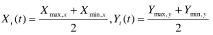
2) Width and height: Extract the positions of the pixel Pxmax,
Pxmin, Pymax, Pymin and determine the width wi (t) and hi (t) as
follows,
y y
i x x
i
t
X
X
h
t
Y
Y
w
(
)
max,
min,,
(
)
max,
min,3) Positions: Identify the positions of each object (Xi (t) and
2
)
(
,
2
)
(
max,x min,x i max,y min,yi
Y
Y
t
Y
X
X
t
X
4) Color: Using image data Pxmax, Pxmin, Pymax, Pymin, define the
color features of the object in the given medical image.
4
/
)]
(
)
(
)
(
)
(
[
)
(
xmax xmin ymax ymini
t
R
P
R
P
R
P
R
P
R
The proposed pattern matching algorithm is done with image classification and feature extraction. For this, utilize a minimum distance search between (t,i) and all objects in the preceding frame (t-1, j). The object (t, i) is identified with the object in the (t-1)-th frame which has the minimum distance from (t, i). By repeating the matching procedure all the time, it is easy to track all the objects in the segmented image parts. Consider an object i in the t-th frame. Determine the distance among the neighboring objects (t, i) and (t-1, j) as D (t,i;t-1,j). Consider an algorithm below described the pattern matching based on minimum distance search in the feature space.
Step 1: Extract the area, width, height, positions, and color data for segment i in the t-th frame
Step 2: Pattern matching is done using,
Step 3: Determine the calculation of distances,
j
j
t
i
t
D
(
,
;
1
,
),
Step 4: Search for the minimum distance,
)
,
1
;
,
(
min
)
,
1
;
,
(
t
i
t
k
jD
t
i
t
j
D
Step 5: Identify the distance measure for feature object space tracking
Step 6: Identify the Euclidean distance,
2
)
(
i
i i
E
x
y
D
Step 7: Estimate the positions of segment i in the next segmented parts
[image:4.595.55.273.70.106.2]Step 8: Repeat the matching procedures for all segments to identify the feature object space.
Fig 3.2 Process of pattern matching algorithm
With the above process, the pattern matching is done efficiently with the Euclidean distance measure for feature objects in the segmented image parts. To start the pattern matching, it is necessary to define the calculation of distance among the neighboring objects in the given image. Determine the minimum distance and identify the distance measure of each object in the given image. With the Euclidean distance, identify the positions of segment i in the next segmented parts. With the patterns, similarity matching is also being done effectively based on the identification of nearing objects.
4. EXPERIMENTAL EVALUATION
The performance of the proposed efficient pattern matching algorithm for classified brain image system is
implemented in Mathworks Matlab 7.0. The intention of the proposed efficient pattern matching algorithm for classified brain image system is to show how the feature space of the objects in the classified image are identified in which it belongs to using Bayesian classifier with no prior assumption regarding image model. The medical image (brain image) is taken as training set which is segmented into three regions. Segmented regions are based upon prior medical knowledge. At first, the input medical image is segmented using nearest neighbor classifiers. Then the Bayesian classifier is used for the classification of segmented image based on its appropriate class for enhancing the image processing scheme. After classification, implement pattern matching algorithm to identify the feature spaces of the object present based on its similarity measures. The purpose of this evaluation is to express the ability of the proposed efficient pattern matching algorithm for classified brain image system to adapt without human intervention to this class of images obtained with special techniques other than the common ones. These results with gray level images demonstrate that the proposed efficient pattern matching algorithm for classified brain image system] are robust and flexible with various types of images. The performance of the proposed efficient pattern matching algorithm for classified brain image system is measured in terms of
i) Estimation of object position ii) Average matching accuracy iii) Efficiency
5. RESULTS AND DISCUSSION
In this work, we have seen that how the feature space of the object is efficiently identified with the proposed efficient pattern matching algorithm based on its classes in which it belongs to using pixel values. The experimental results evaluated in terms of number of images used, number of segmented portions. The below table and graph describes the performance of the proposed efficient pattern matching algorithm for classified brain image system [EPMACB].
Table 5.1 Segmented image parts vs. Estimation of object position
Segmented image
parts
Estimation of object position (%)
Proposed EPMACB
IBCSI NN classifier
Existing
graph-cut method
5 54 50 48 32
10 62 54 52 35
15 70 60 56 40
20 76 64 62 42
The above table (table 5.1) describes the accurate estimation of object position in the image segmented parts. The estimation of object position accuracy by the proposed efficient pattern matching algorithm for classified brain image system is compared with our previous works and an existing multi-region graph-cut method.
Fig 5.1 Segmented image parts vs. Estimation of object position
0 20 40 60 80 100
0 5 10 15 20 25 30
Segmented image parts
E
s
ti
m
a
ti
on
of
ob
e
jc
t
po
s
it
ion
(
%
)
[image:5.595.56.271.177.277.2]Proposed EPMACB IBCSI NN classifier Existing graph cut method
[image:5.595.310.539.470.650.2]Fig 5.1 describes the estimation accuracy of object position in the image segmented parts. An identification of object at a particular position in a given image is a challenging task. To overcome the challenge, in this work, we proposed pattern matching algorithm to identify the object feature space on the segmented image by extracting the features at first. After that, based on the width and position of the object in the image, the minimum distance of the neighboring objects are identified and processed. With the distance measure, object in the image has been tracked efficiently in the proposed EPMACB. But in out previous work, it simply concentrated on the classification of image with the features and does not help to identify the feature space. Existing multi-region graph cut method used generic model for all issues and provide the outcome with less efficiency. Compared with an existing and other works, the proposed efficient pattern matching algorithm for classified brain image system provides an accurate estimation and the variance is 40-50% high in the proposed EPMACB.
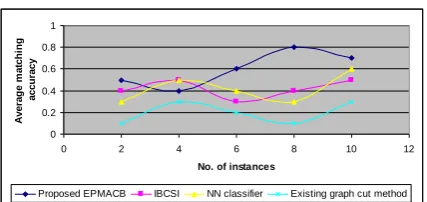
Table 5.2 No. of instances vs. average matching accuracy
No. of instances
Average matching accuracy
Proposed EPMACB
IBCSI NN classifier
Existing graph-cut
method
2 0.5 0.4 0.3 0.1
4 0.4 0.5 0.5 0.3
6 0.6 0.3 0.4 0.2
8 0.8 0.4 0.3 0.1
10 0.7 0.5 0.6 0.3
The above table (table 5.2) describes the average matching accuracy of the object positioned in the image segmented parts. The average matching accuracy by the proposed efficient pattern matching algorithm for classified brain image
system is compared with our previous works and an existing multi-region graph-cut method
.
Fig 5.2 No. of instances vs. average matching accuracy
0 0.2 0.4 0.6 0.8 1
0 2 4 6 8 10 12
No. of instances
A
v
e
ra
ge
m
a
tc
hi
ng
a
c
c
ura
c
y
Proposed EPMACB IBCSI NN classifier Existing graph cut method
Fig 5.2 describes the average matching accuracy of the object positioned in the image segmented parts. We illustrate the average matching accuracy values on different kinds based on the number of training instances. The proposed EPMACB method gets a large development on kinds with few training instances. This is significant since the allocation of objects in the genuine world is long-tailed. To enhance the process of matching algorithm, the pattern matching algorithm is used here. The pattern matching algorithm started its matching process based on the distance measure of the objects in the given image. Compared to the previous works simply concentrated on the classification of image with the features and does not help to identify the feature space. Existing multi-region graph cut method used generic model for all issues and provide the outcome with less efficiency. Compared with an existing and other works, the proposed efficient pattern matching algorithm for classified brain image system provides an accurate estimation and the variance is 50-60% high in the proposed EPMACB.
Table 5.3 Pixels in image vs. Efficiency
Pixels in image
Efficiency
Proposed EPMACB
IBCSI NN classifier
Existing graph-cut method
100 64 60 58 32
200 70 64 62 45
300 74 70 67 56
400 82 73 72 62
500 87 76 75 69
[image:5.595.48.268.508.699.2]0 20 40 60 80 100
0 100 200 300 400 500 600
Pixels in image
E
ff
ic
ie
nc
y
[image:6.595.69.264.72.167.2]Proposed EPMACB IBCSI NN classifier Existing graph cut method
Fig 5.3 Pixels in image vs. Efficiency
Fig 5.3 describes the efficiency of the process of pattern matching algorithm in the image segmented parts. The previous works simply concentrated on the classification of image with the features and does not help to identify the feature space. Existing multi-region graph cut method used generic model for all issues and provide the outcome with less efficiency. Compared with an existing and other works, the proposed efficient pattern matching algorithm for classified brain image system provides an efficient estimation and the variance is 60-70% high in the proposed EPMACB.
Finally, it is being observed that the proposed pattern matching algorithm efficiently identified the feature space of the objects in the classified image. The feature space of the image is easily identified with the patterns by extracting the features of the image.
6. CONCLUSION
In this paper we have investigated pattern matching algorithm to identify the feature spaces of the object present based on its similarity measures under medical images by achieving mapped image data deviation. Normally used geometric classifiers entail a group of training data to efficiently compute the ethereal and textural signatures for pixels and also cannot do classification supported on high-level user concepts because of the deficient of spatial information. The proposal of our work gains the advantages of pattern matching scheme under similarity measures. An effectiveness of the proposed pattern matching algorithm for segmented image classification method can be evaluated using quantitative comparative performances over a large number of medical images especially brain images. The performance evaluation is carried out by using quality metrics which show that compared to the existing multi-region graph-cut method; the proposed pattern matching algorithm brings different advantages with regard to accuracy attains 85% in classification process and flexibility attains 80% while analyzing the medical images.
7. REFERENCES
[1] M. Ben Salah, A. Mitiche et. Al., “Image partitioning with kernel mapping and graph cuts”, Proceedings of
2010 IEEE 17th International Conference on Image Processing September 26-29, 2010, Hong Kong
[2] Mikiyas Teshome1 et. Al., “A simple binary image similarity matching method based on exact pixel matching”, 2009 International Conference on Computer Engineering and ApplicationsIPCSIT vol.2 (2011) [3] Raju Bhukya et. Al., “Exact Multiple Pattern Matching
Algorithm using DNA Sequence and Pattern Pair, International Journal of Computer Applications (0975 – 8887)Volume 17– No.8, March 2011
[4] Yong ki Lee et. Al., “Tag-based object similarity computation using term space dimension reduction”, Proceeding SIGIR '09 Proceedings of the 32nd international ACM SIGIR conference on Research and development in information retrieval, Pages 790-791
[5] Ziad A.A Alqadi, et. Al., ‘Multiple Skip Multiple Pattern Matching algorithms’. IAENG International. Vol 34(2),2007.
[6] Devaki-Paul, “Novel Devaki-Paul Algorithm for Multiple Pattern Matching” International Journal of Computer Applications (0975 – 8887) Vol 13– No.3, January 2011.
[7] D. Cremers, M. Rousson, and R. Deriche, “A review of statistical approches to level set segmentation: integrating color, texture, motion and shape,” IJCV, 72(2), 2007.
[8] M. Ben Salah, A. Mitiche, and I. Ben Ayed, “A continuous labeling for multiphase graph cut image partitioning,” In Adv. In Visu. Comp., LNCS, G. Bebis et al. (Eds.), vol. 5358, pp. 268- 277, Springer-Verlag, 2008.
[9] I. Ben Ayed, A. Mitiche, and Z. Belhadj, “Multiregion Level-Set Partitioning of Synthetic Aperture Radar Images,” IEEE TPAMI, vol. 27, no. 5, pp. 793-800, 2005.
[10] G.LAVANYA,et. Al., “BREAST TUMOUR DETECTION AND CLASSIFICATION USING NAÏVE BAYES CLASSIFIER ALGORITHM”, International Journal of Emerging trends in Engineering and Development ISSN 2249-6149 Issue 2, Vol.3 (April-2012)
[11] Mohamed Ben Salah, Amar Mitiche, and Ismail Ben Ayed, ” Multiregion Image Segmentation by Parametric Kernel Graph Cuts”, IEEE Transactions On Image Processing, vol. 20, no. 2, Feb 2011.