Lucrări ştiinŃifice Zootehnie şi Biotehnologii, vol. 41 (1) (2008), Timişoara
DIFFERENTIATION OF EMBRYONIC STEM CELLS:
LESSONS FROM EMBRYONIC DEVELOPMENT
DIFERENTIEREA CELULELOR STEM EMBRIONARE:
MODEL PENTRU DEZVOLTAREA EMBRIONARA
PALL EMOKE *, LICHNER ZSUZSANNA **, BONTOVICS BABETT **, GOCZA ELEN **
*University of Agricultural Science and Veterinary Medicine, Cluj-Napoca, Romania **Agricultural Biotechnology Center, Gödöllı, Hungary
Embryonic stem (ES) cells, the undifferentiated cells of early embryos are established as permanent lines and are characterised by their self-renewal capacity and the ability to retain their developmental capacity in vivo and in vitro. The pluripotent properties of ES cells are the basis of gene targeting technologies used to create mutant mouse strains with inactivated genes by homologous recombination. There are several methods to induce the formation of EBs. One of them the formation by aggregating ES cells in hanging drops, using gravity as an aggregation force. This method presents the advantage of obtaining well-calibrated EBs almost identical in size. We used at our experiment the mouse ES cell line KA1/11/C3/C8 with a normal karyotype, at 14th passages. Immunohistochemical examination was aimed to identify tissue-restricted proteins for the two differentiated lineages: titin as a cell-specific antigen for cardiac and skeletal muscle, betaIII-tubulin for the neuronal differentiation, cytokeratin Endo-A (TROMA) for the presence of mesenchymal progenitor cells, Oct-4 for the presence of the undifferentiated ES cells. The beating cardiac muscle clumps showed more synchronous rhythm than those seen in EBs obtained from suspension culture method, where the beating cardiac muscle clumps appeared later, had a lower frequency and were uneven. The synaptic networks of neuronal cells were best developed in EBs from suspension, compared to those observed in EBs from hanging-drop method.
Key words: embryonic stem cells, in vitro differentiation, cardiac, neuronal
Introduction
pluripotent properties of ES cells are the basis of gene targeting technologies used to create mutant mouse strains with inactivated genes by homologous recombination (Wobus et al., 1984).
ES cells cultivated as embryo-like aggregates, called embryoid bodies (EBs), differentiate in vitro into cellular derivatives of all three primary germ layers of endodermal, ectodermal, and mesodermal origin. ES cell lines develop from an undifferentiated stage resembling cells of the early embryo into terminally differentiated stages of the cardiogenic, myogenic, neurogenic, hematopoietic, adipogenic, or chondrogenic lineage, as well as into epithelial, endothelial, and vascular smooth muscle (VSM) cells (Hesler et al., 1997).
Terminally differentiated ES cells also show pharmacological and physiological properties of specialised cells: in vitro differentiated cardiomyocytes have characteristics typical of atrial-, ventricular-, Purkinje-, and pacemaker-like cells, and neuronal cells are characterized by inhibitory and excitatory synapses. Neuronal, cardiac, and VSM cells express functional receptors typical for each cell type. Differentiation of ES cells in EBs provides a suitable model not only to understand the process of early embryonic development but also to identify molecules involved in the regulation in the differentiation processes (Wobus et al.,
2005).
There are several methods to induce the formation of EBs. One of them the formation by aggregating ES cells in hanging drops, using gravity as an aggregation force. This method presents the advantage of obtaining well-calibrated EBs almost identical in size. When grown as EBs, ES cells spontaneously differentiate into many cell types, including cardiomyocytes. ES cells may differentiate into endothelial progenitors, positive for early markers, such as CD31, also known as platelet-endothelial cell adhesion molecule (PECAM)-1 and vascular endothelial growth factor receptor (VEGFR)-2. The addition of angiogenic factors in the medium, such as endothelium-specific VEGF (or VEGF-A or VEGF-165) efficiently promotes the differentiation of ECs and their organisation into vascular structures, which may contain haematopoietic cells (Strübing et al., 1995).
Materials and Methods
In vitro cultivation of mouse ES cell lines
(GIBCO), 1000 units/ml of leukaemia inhibitory factor (ESGRO) and 20% fetal calf serum (FCS) (HyClone).
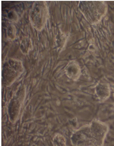
Fig. 1.: ES cell colonies growing on fibroblast layer
In vitro differentiation of ES cells into cardiac and neuronal lineages Two days before the differentiation, ES cells were passaged into gelatin (0.1%)(SIGMA) coated Petri dishes (Greiner) in the ES culture medium.
On the appointed day 0 of differentiation, we passage the cells into 10 cm bacteriological dishes, containing 5x10⁶ ES cells in 10 ml differentiation medium (This is the Day0 of differentiation). As differentiation medium the IMDM (Gibco) medium supplemented with 0.6m/m% penicillin, 1m/m% streptomycin and 20v/v% FCS was employed. MTG (monothyoglycerol) 3 µl/ml was always freshly added to the differentiation medium. For hanging drop production 2x10⁴ cells/ml containing cell-suspension was prepared in IMDM differentiation medium (Fig.2).
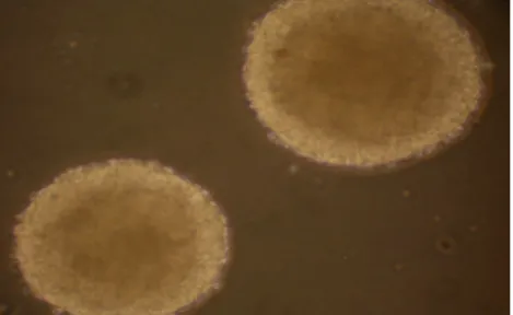
From this suspension, three Petri dish covers with 70 hanging-drops (a drop of 20 µl, contained 400 mouse ES cells) and one suspension Petri dish (3 ml cell suspension) were set in culture for two days (Day 2). Cell aggregates were obtained after 5–6 hrs, while EBs were morphologically completed after 2 days, by each procedure (Fig.3). The second step was to plate the EBs on a gelatin-coated surface for 17 days (Day2+7). Best EBs from the hanging-drops and from the cell suspension were separately harvested and pooled EBs from each variant were further grown on 24-well tissue culture dishes. Prior cultivation of EBs, a rounded shaped histologic cover slip was introduced within each well in order to later perform immuno-staining. All culture dishes so prepared, were gelatin-coated before addition of EBs.
Fig. 2.: Hanging drop differentiation method.
Fig. 3.: EBs at Day2+2 Hanging drop differentiation method was used.
Immunohistocehemical analysis of differentiated ES cells
Immunohistochemical examination was aimed to identify tissue-restricted proteins for the two differentiated lineages: titin as a cell-specific antigen for cardiac and skeletal muscle, (Wobus et al., 2005) betaIIItubulin for the neuronal differentiation (Bouhon et al., 2005), cytokeratin Endo-A (TROMA) for the presence of mesenchymal progenitor cells, Oct-4 for the presence of the undifferentiated ES cells.
and placed on Vecta-Shield Mounting Medium on chemically clean microscopic slides and examined with a fluorescence microscope.
Results and Discussions
Cardiomyocytes differentiation
From the 7th day of plating (Day2+7) we could observed contractions in a synchronous rhythm (Table 1)(Fig.4). Best contracting cells, showing titin expression (Fig.5), were observed within the EBs grown in IMDM medium derived form the hanging-drop variant, compared to EBs from suspension culture. The EBs from the suspension variant were smaller and the pulsing was retarded and uneven compared to the hanging-drop variant. In the RA treated plates the overall contractions of EBs were present, but at a lower frequency than in the IMDM medium, while contractions within EBs from suspension culture were only rare noticed.
Table 1.: Proportion of pulsing EBs in the four culture variants
Examinatio n day
Pulsing EBs (%)
IMDM hanging-drops
IMDM Suspension
culture
IMDM + RA
hanging-drops
IMDM + RA Suspension
culture
Day2+7 47.82 25 0 0
Day2+8 78.26 50 47.82 0
Day2+9 91.3 62.5 65.21 0
Day2+10 91.3 62.5 65.21 0
Day2+11 95.65 83.33 91.3 17.39
Day2+12 95.64 83.33 91.3 17.39
Day2+13 95.65 87.33 91.3 17.39
Day2+14 95.65 87.5 91.3 17.39
Day2+15 86.95 76.19 91.3 17.39
Neuronal differentiation was clearly seen under the RA treatment and
Fig. 4.: Percentage of pulsing clumbs in four different culture conditions.
Conclusions
Hanging-drop method provides a practical way of generating single EBs from a defined number of cells. EBs from the suspension culture method were smaller and unequal in size, compared to the hanging-drop procedure. Cardiomyocytes begun to contract spontaneously seven days after plating of EBs (Day2+7). They were localised between an epithelial and a basal mesenchimal layer. The hanging-drop method showed better results on the field of cardiac differentiation. The beating cardiac muscle clumps showed more synchronous rhythm than those seen in EBs obtained from suspension culture method, where the beating cardiac muscle clumps appeared later, had a lower frequency and were uneven.
Immunocytochemical detection of specific cellular antigens proved the specificity of EBs commitment towards cardiomyocytes and neuronal lineages.
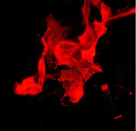
Fig. 5.:
Titin expression was found in beating clumps of attached EBs on
gelatin-coated surface.
Fig. 6.:
BetaII-tubulin expression in attached EB on gelatin coated surface.
Fig. 7.: Cytokeratin expression in attached EB on gelatin coated surface.
This work was supported by RO-43/2007 Bilateral project, 1A/060/2004 NKFP and 405/2006 ETT governmental projects.
Bibliography
1. Wobus, A.M., Kenneth R.B. (2005): Prospects for developmental biology
and cell terapy. Physiol Rev. Bethseda 85:635-678;
2. Nagy, A., Rossant, J., Nagy, R., Abramow-Newerly, W. and Roder, J.
(1993): Derivation of completely cell culture-derived mice from
3. Bouhon, A.I., Kato, H., Sidharthen, C., Allen N.D. (2005): Neural
differentiation of mouse embryonic cells in chemically defined medium.
Brain Research Bulletin 68:62-75;
4. Heschler, J., Fleischmann, B.K., Lentin, S.,. Maltsen V.A., Rotwedel, J., Wobus, A.M., Addicks, K. (1997): Embryonic stem cells: a model to
study structural and functional properties in cardiomyogenesis, Cardiovascular Reserch, 36:149-162;
5. Strübing, C, Ahnert-Hilger, G., Shan, J., Wiedenmann, B., Hescheler, J., Wobus, A.M. (1995): Differenciation of pluripotent embryonic stem
cells into the neuronal lineage in vitro gives rise to mature inhibitory and excitatory neurons. Mechanisms of Development 53:275 – 287;
6. Wobus, A.M., Holzahausen, H., Jakel, P., Schoneich, I. (1984):


![The People's Republic of China and the European Community. Information [External Relations] 106/75](data:image/gif;base64,R0lGODlhAQABAIAAAP///wAAACH5BAEAAAAALAAAAAABAAEAAAICRAEAOw==)