The Angiogenic Effect of Endothelial Specific Deletion of BMPR2 in the Developing Lung
By Hong Min Lee
Approved: Victoria Bautch, PhD, Thesis Advisor
John Bruno, PhD, Reader
Abstract
Angiogenesis, the growth of new blood vessels from pre-existing vessels, is a critical process for proper embryonic development. BMPs (bone morphogenetic proteins) are key angiogenic
players that are highly expressed during lung development, but the role of these proteins in the context of lung angiogenesis is not well characterized. A previous study showed that over-expressing Matrix Gla protein, an upstream BMP inhibitor, decreased lung vasculature. Importantly, our lab has also shown that overexpression of bmp2b, a BMP ligand, induces ectopic vessel sprouting in developing zebrafish. Additionally, endothelial-specific deletion of BMP receptor II (BMPR2) leads to decreased vascular sprouting in the postnatal mouse retina. Taken together, this evidence suggests that modulations in BMP signaling lead to perturbations in vessel development and sprouting. Therefore, I predict that deletion of BMPR2 in the
Introduction
The establishment of a vascular network is a crucial process in the developing embryo. The earliest vascular development occurs de novo via differentiation of angioblasts into
endothelial cells that then fuse to form larger vessel networks in a process called vasculogenesis (Baldwin et al.,1996). Distinct from vasculogenesis, angiogenesis, the process by which new blood vessels form from pre-existing vessels by sprouting of endothelial cells from a parent vessel, is an especially vital process by which oxygen and nutrient delivery to organs is maintained throughout development. These processes occur simultaneously and collaborate during lung vascular development. Angiogenesis occurs centrally in the lung forming the axial and lateral arteries and veins while vasculogenesis occurs distally in the periphery, forming isolated blood lakes that fuse with the central branches (de Mello et al., 1997). In later stages of gestation, these vessels remodel into a network of more uniform vessels.
endofin, a negative regulator of the BMP pathway, in osteoblasts increased vascular density in long bones (Zhang et al., 2009). Further studies in our lab, in which the BMP type II receptor, BMPR2, was deleted in endothelial cells of the post-natal mouse retina, resulted in decreased sprouting at the vascular front and decreased branching in the vessel plexus behind the vascular front (Chong, unpublished). This evidence suggests that BMP signaling is pro-angiogenic, such that increased BMP signaling leads to increased branching whereas decreased BMP signaling reduces branching and sprouting angiogenesis.
In lungs, BMPs are closely associated with branching morphogenesis of the lung epithelium. BMPs are expressed in intricate and stereotyped patterns in the developing lung (Danesh et al., 2009). As the primitive lung buds begin to develop off the primordial gut tube around embryonic day (E) 9.5, distinct BMP4 patterning is detected (Weaver et al.,
Development, 1999). This expression of BMP4 is dynamic throughout the stages of lung
development from E10.5 to E17.5 and tends to localize in the endoderm at the tips of the
growing buds starting from E11.5 (Bullusci et al., 1996). Despite the many studies of how BMP expression and signaling regulates lung budding and branching morphogenesis, its function as a regulator of lung vascular development has been less studied. Upstream regulators of the BMP pathway are implicated in vascular development of the lung, although the effects of direct manipulations to the BMP signaling pathway have not been analyzed in this organ. The deletion of matrix gla protein, an upstream negative regulator of BMP, leads to increased branching in lung vasculature (Yao et al., 2011). This suggests that BMP signaling is pro-angiogenic in the lung.
marker during different embryonic stages (E10.5-E12.5) of lung development. We showed, through this initial characterization, a trend in which branching increases steadily over this period. By genetically deleting BMPR2 in the endothelium using the Cre-Lox system, we are discovering the effects of decreased BMP signaling on the developing vasculature of the lung. Preliminary results in E11.5 lungs suggest decreased branching as a result of BMPR2 deletion in endothelial cells. We are currently solidifying and expanding the characterization of E11.5 lungs with BMPR2 deletion.
Materials and Methods
Animals. Wild-type embryos were generated from matings between Flk1-GFP and CD-1 albino mice. This produced embryos containing an endothelial cell-specific Flk1 promoter driving expression of green fluorescent protein (GFP) for facilitated visualization of vessels. These embryos were collected on gestational stages E10.5, E11.5, E12.0, and E12.5, counting the morning of the vaginal plug as E0.5. We dissected the embryos in PBS and fixed in 4% paraformaldehyde (PFA) for 1 hour. The embryos were then washed three times with PBS in five-minute intervals. Embryos that were GFP-positive were separated from GFP-negative. The lungs were dissected from embryos in PBS and kept in PBS at 40C until sectioned or mounted for analysis.
Mutant embryos were generated by mating a Flk1-GFP; Cdh5-CreERT2; Bmpr2flox/flox male to a
Bmprflox/flox; Rosa-tdTomato females in order to create a line of embryos with Flk1-GFP;
Cdh5-CreER; Bmpr2flox/flox; Rosa-tdTomato. Cdh5-CreER is a transgenic line which expresses the
injected intraperitoneally. The administration of tamoxifen activates the Cre-recombinase and excises the loxP flanked exons four through five of the Bmpr2 locus (Beppu et al., 2005). To determine Cre-excision efficiency, we included a Rosa-tdTomato reporter in the same line of embryos. The Rosa-tdTomato reporter contains a lox-STOP-lox cassete with a downstream
tdTomato gene inserted in the Rosa26 endogenous locus (Madison et al., 2010). Therefore, when
tamoxifen is administered, the STOP codon is also excised and the downstream tdTomato reporter protein, a red fluorescent protein, is expressed. The outcome is an embryo in which BMPR2 deleted in endothelial cells (BMPRiECKO) expresses ROSA-tdTomato to aid in determining excision efficiency. We estimated and confirmed BMPR2 deletion in endothelial cells via the co-localization of ROSA-tdTomato reporter (red-fluorescent protein) and the FLK:GFP expressed under the vessel-specific, Flk1 promoter. The embryos were dissected and a small piece of the limb or tail was collected for genotyping. The embryos were fixed in 4% PFA individually in tubes for 1 hour. Then, they were washed three times with PBS in five-minute intervals.
Genotyping. The Viagen Direct-PCR lysing system was used to lyse the samples collected from the embryos. After lysing, PCR reactions were performed using 2X PCR Master Mix (Thermo Scientific). The following primers were used to detect BMPR2 flox: 5’ TTA TTG TAA GTA CAC TGTT TGC TGT C 3’, 5’GGC AGA CTC TGA CTT TGA CGC TAG 3’. A flox band is 315 bp and a WT band is 260 bp. To detect Cdh5CreER mutants we used primers 5’GAC CAG CTT CGT TCA CTC A3’, 5’TAG CGC CGT AAA TCA AT 3’. The Cdh5 Cre ER mutants
Vibratome Sectioning. The lungs were embedded in 4% low melting agarose and sectioned into 250-micrometer slices using a Leica VT1200S Vibratome. The surface sections were kept and mounted with Vectashield to reduce variability between sections.
Whole Mount. The lungs were embedded in 1% low melt agarose on a Matek dish. The Matek dish was filled with 4% low melt agarose to stabilize the area surrounding the lung. The sample was covered with PBS to prevent drying in the dish before imaging.
Imaging. An FV-1200 Olympus confocal microscope with 40x silicon-oil immersion objective was used to image vibratome sections. Imaging was done both in the XY plane and in the Z plane. The step size used for Z-plane imaging ranged from 3-5 microns and the number of Z planes imaged ranged from 28-36. The multi-area acquisition tool was used to image large regions of the lung and the stitch tool was used to stitch these regions together to obtain one image of the complete lung. An Olympus two-photon microscope with 25x gel immersion objective was used to image the whole-mounted lungs. The Z-step size used was 1.0 um and 150-200 planes were imaged.
Analysis. The images of the lungs were compressed along the Z plane at max intensity using ImageJ. Vessels were traced in Adobe Photoshop. In cases of discrepancies between vessel connections, the uncompressed Z-stacks were consulted. If the vessels structures could not clearly be deciphered as was the case near the tips of the lung buds due to low GFP expression, then these regions of vessels were not traced. These tracings were re-opened on ImageJ and the “skeletonize” and “analyze skeletonize” tools were used to obtain branching data. Imaris
software is currently be used to create 3-D surfaces and reconstructions of the vasculature of the lung. We plan to use the “filament tracing” tool in Imaris to analyze branching using the 3-D
Statistics. Both one-way ANOVA and student-t tests were used to determine statistical
significance in sectioned wild-type lungs using Prism. Unpaired student t-tests were performed on every combination of two-stages of sectioned wild-type lungs. Unpaired student t-tests were performed on whole-mount sectioned lungs to determine statistical significance between E10.5 and E11.5. Unpaired student t-tests were also performed to determine statistical significance between sectioned and whole-mount E11.5 WT and mutant lungs. In all cases, significance was set at p≤ 0.05
Results
E
E10.5 E11.5 E12.0 E12.5
Fl
k-G
FP
Figure 1. Branching increases in sectioned wild-type lungs as developmental stage increases. (A-D) representative images of vibratome sections from stages E10.5, E11.5, E12.0, and E12.5, respectively. Yellow outline indicates individual lung buds in the image. Scale bars denote 100 micrometers. (E) Graph with quantified branches/mm by embryonic stage. Asterisks denote significance p≤0.05 as determined by an unpaired student t-test.
Figure 2. Branching increases in whole-mounted wild-type lungs as developmental stage increases. (A,B) representative images of E10.5 and E11.5 whole-mounted lungs, respectively. Scale bars denote 100 micrometers. (C) branching in whole mounted E10.5 and E11.5 lungs. Left bud (L), right caudal bud (RCd), right medial bud (RMd), right cranial bud (RCr). Right medial buds at E11.5 were not quantified.
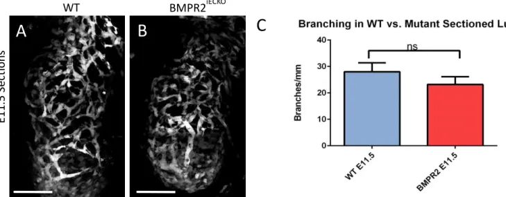
Figure 3. Branching decreases when BMPR2 is excised in endothelial cells in sectioned lungs. (A) representative image of sectioned wild-type lung (B) representative image of sectioned BMPR2iECKO lung. Scale bars denote 100 micrometers. (C) branching in sectioned WT vs BMPR2 iECKOE11.5 lungs.
Fl k-G FP E11.5 E10.5
A
B
C
L RCd L RCd RMd RCr
C
WT BMPR2iECKOFigure 4. Branching decreases when BMPR2 is excised in endothelial cells in whole mount lungs. (A) representative image of whole mount lung (B) representative image of whole mount BMPR2iECKO lung. Scale bars denote 100 micrometers. (C) branching in whole mount WT vs BMPR2 iECKOE11.5 lungs.
Branching increases in sectioned wild-type lungs as developmental stage increases. To characterize wild-type angiogenesis throughout the embryonic phase of lung development, we utilized Flk1-GFP mice to visualize vessels. Flk1-GFP mice have GFP inserted into the Flk1
locus such that green fluorescent protein is expressed via the Flk1 promotor, which is selective for endothelial cells of developing blood vessels (Ema et al., Blood, 2006). We quantified
branching from individual lung buds in sectioned lungs by tracing vessels at stages E10.5, E11.5, E12.0, and E12.5 (Figure 1A-D). The number of branch points was normalized to the total length of the vessels. The number of branches/mm for embryonic stages E10.5, E11.5, E12.0, and E12.5 were 25.9±2.9, 26.0±3.3, 33.0±3.7, and 37.8±0.5 branches/mm, respectively (Figure 1E). There was no statistical significance between the groups using one-way ANOVA, however, as
expected, the trend of the data demonstrates that as the stage of the lungs increase, there is an WT BMPR2iECKO
E1
1
.5
W
h
o
le
M
o
u
n
t
increase in branching. Furthermore, using a student t-test, we found statistical significance in branching between E10.5 and E12.5 with a p value of 0.01 and between E11.5 and E12.5 with a p-value of 0.03 (Figure 1E). Overall, this data shows that branching of vessels increases during lung development and our method for branching calculations are reliable and reproducible.
Branching increases in whole mount wild-type lungs as developmental stage increases. To determine if this trend of increased branching could also be detected in whole mount wild-type lungs, we embedded E10.5 and E11.5 lungs as whole mounts in agarose and utilized a two-photon microscope to capture vessels at a greater depth (Figure 2A, B). We traced vessels and analyzed branching in the same manner as the sections were quantified, by normalizing the number of branch points to the total length of vessels in millimeters. We found that E10.5 and E11.5 lungs had 30.4±2.3 branches/mm and 35.5±0.8 branches/mm, respectively (Figure 2C). Again, we saw that branching increased during lung development, although there was no
statistical significance between these two stages as determined by a student t-test (Figure 2C). In both E10.5 and E11.5 stages, the whole mount had more branching than the sections, suggesting that this method captures more branching than sectioning and using confocal microscopy.
Endothelial cell genetic deletion of BMPR2 decreases branching in embryonic lung sections.
Once we quantified branching in wild type lungs and established reliable methods for
generate an embryo in which Bmpr2 in endothelial cells could be excised by a tamoxifen-dependent Cre-ER recombinase and also expressed the vessel-specific marker FLK1:GFP (see Methods). We induced Cre-ER recombinase with tamoxifen, such that our embryos had
genetically deleted Bmpr2 in endothelial cells starting at E9.5. We dissected the lungs at E11.5. Using the co-localization of Rosa-tdTomato reporter (see Methods) and FLK:GFP, we estimated that Cre-mediated excision ranged from 30-60% of endothelial cells in the imaged mutant lungs. Despite incomplete excision of Bmpr2, we saw reduced vessel branching in lungs of embryos that also had the Cre-driver transgene. The mutant lungs at E11.5 had 23.2±3.0 branches/mm, which was on average approximately 5 branches less than the wild-type E11.5 lungs, which had 28.0±3.4 branches/mm. This difference is currently not statistically significant, however, the lack
of significance can be attributed to the low cre-excision efficiency and our low sample size. Through future experiments, we are planning to expand our sample size and increase excision of BMPR2 in endothelial cells in our mutant lungs.
Endothelial cell genetic deletion of BMPR2 decreases branching in embryonic lung whole-mount.
thus, Cre-excision efficiency and our low sample size is also a caveat for this experiment. However, overall trends show that there is decreased branching in BMPR2iECKO lungs when
compared to wild-type embryonic mouse lungs.
Discussion
Although BMPs have been implicated in vessel sprouting and branching, the role of BMPs in early vessel patterning during lung development has not been well characterized. We hypothesized that decreased BMP signaling would lead to decreased angiogenesis in the developing lung. In this study we analyzed lungs throughout the embryonic phase of
development, E9.5 to E12.5, for evidence of blood vessel branching. We demonstrate that branching of vessels in wild-type lungs increases from stages E10.5 to E12.5. Preliminary experiments with BMPR2iECKO at E11.5 stages show that, compared to the wild-type, there is a
decrease in branching, suggesting the BMP signaling is involved in regulating angiogenesis in the lung. To date, this data is not statistically significant, however, the trend of the data supports our hypothesis and corroborates with previous studies that suggest that BMP is a pro-angiogenic cue.
To characterize angiogenesis throughout the embryonic phase of lung development, we quantified branching of vessels. Although we saw some evidence of sprouting, the abundance of sprouting was significantly less than other classical in vivo angiogenesis models, such as the postnatal mouse retinal model. These were not the only differences we noted between lung vessels and these other sprouting models. When we first started imaging lung vessels, we
uniform and characterized largely by sprouting angiogenesis. In contrast, our lung vessels looked irregular and variable in size with small holes or divots. Previous studies of the microvasculature in post-natal rat lungs corroborate this characterization of lung vessels and demonstrated that the primary mechanism of angiogenesis in the lung takes the form of intussusceptive, non-sprouting, vessel proliferation (Caduff et al., 1986). This process of intussesceptive angiogenesis in which perforations or holes in the vessels creates new vessels is not as well-understood as sprouting angiogenesis (Risau et al., 1997). Given the lack of sprouting angiogenesis and similar observations of “tiny holes” in the vessels of our embryonic lungs, this suggests that
intussesceptive angiogenesis is also the primary mechanism of lung vessel development during mouse embryonic development.
The process of branching may be driven by BMP-induced manipulations on endothelial cell migration and/or proliferation in lung vessels. Previous studies have implicated BMPs in endothelial cell migration and proliferation in vivo. BMP-4,-6,-9 for example, induced proliferation of mouse embryonic-stem-cell-derived endothelial cells when exogenously added in culture (Suzuki et al., 2010). BMPs also have demonstrated effects on migration. When BMP-4 was added to these cells, there was also an increase in random migration over a 24 hour period (Suzuki et al., 2008). This provides one explanation by which BMP signaling may be involved in regulating angiogenesis during development.
effects on branching in the lung may suggest that BMPs have undiscovered effects on transluminal tissue pillar formation, a hallmark of intussusceptive angiogenesis that affects vessel branching in lungs (Djonov et al., 2000). One experiment by which we can probe the migration and proliferative capabilities of endothelial cells in the embryonic lung is to embed ex-vivo wild-type and BMPR2iECKO lungs in a matrix and quantify migration and sprouting of labeled endothelial cells. This will help determine whether BMP also plays a role in migration and proliferation of lung endothelial cells during development.
We also consistently noted that in E10.5 and in some E11.5 lungs, the tips of the lung buds had low GFP expression. At first, we suspected that GFP expression was being quenched during the process of imaging. However, this GFP expression remained consistent even when protocols were amended so that imaging was performed sooner after dissection. This observation suggests that vasculogenesis is still actively occurring at distal ends and angiogenesis is the not the sole process by which the embryonic lung vasculature develops (Parera, 2005). We suspect that the low, irregular GFP expression is indicative of angioblasts that are actively differentiating into endothelial cells. As of yet, we have not performed BMPR2 deletion experiments on E10.5, where this pattern of GFP expression has been most prevalent. However, BMPs also have demonstrated effects on endothelial cell differentiation. In a previous study human embryonic stem cells (hSECs) cultured with high-dosage treatment of exogenous BMP lead to an increase in hSECs committed to the endothelial cell lineage. This same study also noted a higher rate in endothelial cell differentiation (Goldman et al., 2009). This suggests that a decrease in BMP signaling would also decrease vasculogenesis at the periphery in the early stages where
Importantly, studies on the effect of endothelial cell BMPR2 deletion on branching has relevancy for disease. Pulmonary arterial hypertension (PAH) is a disease characterized by defective remodeling of peripheral blood vessels in the lung. Heterozygous germline mutations in BMPR2 are highly correlated with pulmonary arterial hypertension (Cai et al., 2012). It would be interesting to see if PAH lungs have perturbed or dampened branching as has been suggested from our BMPR2 deletion experiments. In any case, elucidating the role of BMP signaling during lung development will aid in unraveling the complexities of this pathway as well as provide therapeutic benefits for diseases in which vessels are abnormal due to BMP signaling.
References
Baldwin, H.S. (1996) Early embryonic vascular development. Cardiovasc Res 31 E34-E45. Beppu, H., Lei, H., Bloch, K.D., Li, E. (2005) Generation of a Floxed Allele of the Mouse Bmp Type Ii Receptor Gene. Genesis 41:133-137
Bellusci, S., Henderson, R., Winnier, G., Oikawa, T., Hogan, B.L. (1996) Evidence from normal expression and targeted misexpression of Bmp4 plays a role in mouse embryonic lung
morphogenesis. Dev 122:1693-1702.
Burri, P.H., Hlushchuk, R., Djonov, V. (2004) Intussusceptive angiogenesis: Its emergence, its characteristics, and its significance. Dev Dyn 231(3):474-488.
Caduff, J.H. (1986) Scanning electron microscopic study of the developing microvasculature in the postnatal rat lung. Anat Rec 216:154-164
Cai, J., Pardali, E., Sanches-Duffhues, G., tenn Dijke, P. (2012) BMP signaling in vascular disease. FEBS Lett 586:1224-1232.
Danesh, S.M., Villasenor, A., Chong, D., Soukup, C., Cleaver, O. (2009) BMP and BMP receptor expression during murine organogenesis. Gene Expr Patterns 9(5): 255-265. de Mello, D.E., Sawyer, D., Galvin, N., Reid, L.M. (1997) Early fetal development of lung vasculature. Am J Respir Cell Mol Biol 16(5):568-81.
Ema M., Takahashi, S., Rossant, J. Deletion of the selection cassette, but not cis-acting elements, in targeted Flk1-LacZ allele reveals Flk1 expression in multipotent mesodermal progenitors.
Blood 107(1):111-117.
Goldman O., Feraud, O., Boyer-Di Ponio, J., Driancourt, C., Clay, D. Le Bousse-Kerdiles, M.C. Bennaceur-Griscelli, A., Uzan, G. (2009) A boost of BMP4 accelerates the commitment of human embryonic stem cells to the endothelial lineage. Stem Cells 27(8):1750-1759.
Kushner, E.J., Bautch, V.L. (2013) Building blood vessels in development and disease. Curr
Opin Hematol 20(3):231-236.
Madisen, L., Zwingman, T. A., Sunkin, S. M., Oh, S. W., Zariwala, H. A., Gu, H., Ng, L. L., Palmiter, R.D, Hawrylycz, M. J., Jones, A. R., Lein, E. S., and Zeng, H.(2010) A Robust and High-Throughput Cre Reporting and Characterization System for the Whole Mouse Brain, Nat
Neurosci, 13: 133-40.
Parera M.C., van Dooren, M., van Kempen, M., de Krijger, R., Grosveld, F., Tibboel, D., and Rottier, R. (2005) Distal angiogenesis: a new concept for lung vascular morphogenesis. Am J Phys 288:141-149.
Sörensen, I., Adams, R.H., and Gossler, A. (2009) Dll1-Mediated Notch Activation Regulates Endothelial Identity in Mouse Fetal Arteries, Blood 113: 5680-88.
Suzuki Y., Montagne, K., Nishihara, A., Watabe, T., Miyazano, K. (2008) BMPs promote proliferation and migration of endothelial cells via stimulation of VEGF-A/VGFR2 and angiopoietin-1/Tie2 signaling. J Biochem 143(2):199-206.
Suzuki Y., Ohga, N., Morishita, Y., Hida, K., Miyazono, K., Watabe, T. (2010) BMP-9 induces proliferation of multiple types of endothelial cells in vitro and in vivo. J of Cell Sci 123: 1684-92.
Weaver M., Yingling J.M., Dunn, N.R., Bellusci S., Hogan B.L. (1999) Bmp signaling regulates proximal-distal differentiation of endoderm in mouse lung development. Development 126: 4005-4015.
Wiley D.M., Kim J.D., Hao, J., Hong C.C., Bautch, V.L., Jin S.W. (2011) Distinct signaling pathways regulate sprouting angiogenesis from the dorsal aorta and the axial vein. Nat Cell Bio
13: 686-692.
Yao Y., Jumabay, M., Wang, A., Bostrom, K. (2011) Matrix Gla protein deficiency causes arteriovenous malformations in mice. J Clin Invest 121(8):2993-3004.