Serum Ferritin and Iron/TIBC of Pregnant Women
Attending Nnamdi Azikiwe University Teaching
Hospital, Nnewi, Anambra State, Nigeria: A
Longitudinal Study
Oluchi Aloy-Amadi
1, Augustine U. Akujobi
2, Johnkennedy Nnodim
1, Joy Ndudim-Dike
1,
Amaka Edward
1, Michael Anokwute
31Department of Medical Laboratory Science, Imo State University, Owerri, Imo State, Nigeria, 2Department
of Optometry, Imo State University, Owerri, Imo State, Nigeria, 3Department of Haematology and Blood
Transfusion, Nnamdi Azikiwe University Teaching Hospital, Nnewi, Anambra State, Nigeria
ABSTRACT
Background: During pregnancy, serum ferritin level is decreased because of the increased need of iron which triggers ferritin mobilization from its stores. Iron is needed during pregnancy to expand the red blood cell mass and for fetal and placental growth. Aim: The study was aimed at determining the levels of serum ferritin and serum iron/TIBC of pregnant women at different trimesters and comparing them with that of non-pregnant women. Methods: One hundred and sixty apparently healthy pregnant women attending antenatal clinic at Nnamdi Azikiwe University Teaching Hospital, Nnewi at booking in their first trimesters constituted the study population. Similarly, 160 age-matched non-pregnant women were used as the control groups. Five milliliters of venous blood were collected from each subject by means of a hypodermic needle and syringe. One milliliter of blood was aliquoted into potassium CDTA container and used for malaria parasite screening. The remaining 4 ml was placed into gel tubes and used for screening for HIV I and II, Hepatitis B surface Antigen (HBSAg), Hepatitis C virus (HCV), Veneral Disease Research Laboratory (VDRL), and for serum ferritin and Iron. The same procedures were conducted on same pregnant women at the second and third trimesters. At the second trimester, only 156 pregnant women were followed up, while 140 women completed the study at the last trimester. Results: Serum ferritin (ng/ml) and serum iron (µg/dl) were significantly decreased from the first trimester (28.83 ± 19.39) and (74.18 ± 23.92), second (23.76 ± 18.74) and (67.77 ± 17.22), and to the third (20.45 ± 18.42) and (61.23 ± 17.35) (F=10.2, P < 0.001) and (F = 21.9, P < 0.001), respectively. On the other hand, TIBC increased significantly from the first trimester (349.50 ± 52.69), second (364.67 ± 54.53), and to the third (374.40 ± 55.40) (F = 10.9, P < 0.001), respectively. When compared to non-pregnant controls, the serum ferritin and iron in the first (28.83 ± 19.39) and (74.18 ± 23.92), second (23.76 ± 18.74) and (67.77 ± 17.22), and third (20.45 ± 18.42) and (61.23 ± 17.35) trimester were significantly decreased compared to the non-pregnant controls (39.67 ± 54.70) and (80.78 ± 38.19) (F = 14.2, P < 0.001) and (F = 33.7, P < 0.001), respectively. The TIBC in the first (349.50 ± 52.69), second (364.67 ± 54.53), and third (374.40 ± 55.40) trimesters showed a significant increase compared to the controls (338.37 ± 57.68) (F = 17.1, P < 0.001), respectively. Conclusion: This study showed that levels of serum ferritin and serum iron/TIBC in pregnant women are altered in pregnancy. Therefore, there is need to monitor pregnancies at risk to prevent adverse outcomes.
Key words: Longitudinal, pregnancy, serum ferritin, serum iron/TIBC
Address for correspondence:
Oluchi Aloy-Amadi, Department of Medical Laboratory Science, Imo State University, Owerri, Imo State, Nigeria. Tel.: 08037424672.
INTRODUCTION
F
erritin is a protein that performs an iron storage function in mammals. It is found mainly in the liver, spleen, and bone marrow and to a lesser extent throughout the tissues. The concentrations of serum ferritin present a close correlation with total reserves of iron in the body. This protein can, therefore, be used as a reliable estimator of iron reserves in the organism. During gestation, levels of serum ferritin fall by 50%. This is a consequence of the normal heme-iron dilution process during pregnancy, and also of the extraction of iron by the fetus.[1] The utility offerrotherapy during gestation is still a matter of controversy. Some studies have found beneficial effects for the mother including lower rate of anemia Karimi et al., 2000, and for the fetus where higher levels of ferritin have been found in newborns when the mothers received iron supplements during their pregnancy.[2] Serum ferritin is considered as a better parameter to detect latent iron deficiency especially before the change of red cell morphology and red cell indices. A high degree of correlation has been shown between serum ferritin and bone marrow iron stores. In the stage of latent iron deficiency (absence of storage iron), as assessed by marrow iron content, serum ferritin concentration is decreased, but the transferrin saturation, serum iron, and Hb levels may remain unchanged.[3]
During pregnancy, low serum ferritin concentrations in the presence of normal hemoglobin indicate deficient iron stores. Such females are prone to develop overt iron deficient anemia. In pregnancy, serum ferritin concentration is the maximum at 12–16 weeks of gestation, and then the levels start decreasing as the pregnancy advances. Low serum ferritin levels during second and third trimesters predict low hemoglobin levels in late pregnancy.[4] Serum ferritin levels showed significantly lowest values in second trimester, with slight increase again in third trimester. Increasing gravity had no significant effect on serum ferritin levels.[4] Iron stores that are elevated in pregnancy are associated with preterm delivery, pre-eclampsia, and gestation diabetes mellitus. Women with ferritin levels that are elevated for the third trimester of pregnancy (>41 ng/ml) have a gravity increased risk of preterm and very preterm delivery that has been attributed to intrauterine infection. Another plausible mechanism for high ferritin levels is failure of the maternal plasma volume to expand.
Ferritin production is increased with infection and inflammation as part of the acute phase response. In the presence of infection, macrophages produce inflammatory cytokines that generate reactive oxygen species, releasing free iron ferritin.[5]
The body of an adult human contains about 4 g (0.005% body weight) of iron, mostly in hemoglobin, and myoglobin.
These two proteins play essential roles in vertebrate metabolism, respectively oxygen transport by blood and oxygen storage in muscles. To maintain the necessary levels, human iron metabolism requires a minimum of iron in the diet.[6]
Iron requirements are greater in pregnancy than in the non-pregnant state. Although iron requirements are reduced in the first trimester because of the absence of menstruation, they steadily increase thereafter. The total requirement of a 55 kg woman is approximately 1000 mg. Translated into daily needs, the requirement is approximately 0.8 mg Fe in the first trimester, and > 6 mg in the third trimester. Absorptive behavior changes accordingly; a reduction in iron absorption in the first trimester is followed by a progressive rise in absorption throughout the remainder of pregnancy. The amounts that can be absorbed from even an optimal diet, however, are less than the iron requirements in later pregnancy and stores of ≥300 mg if she is to meet her requirements fully.[7]
Iron deficiency continues to be the leading single nutritional deficiency in the world, despite considerable efforts to decrease its prevalence.[8]
Women in developing countries are always in a state of precarious iron balance during their reproductive years. Their iron stores are not well developed because of poor nutritional intake, recurrent infections, menstrual good loss, and repeated pregnancies.[9] During the first two trimesters of pregnancy, iron deficiency anemia increases the risk for preterm labor, low birth weight babies, and infant mortality and predicts iron deficiency in infants after 4 months of age.[10] It is estimated that anemia accounts for 3.7% and 12.8% of maternal deaths during pregnancy and childbirth in Africa and Asia, respectively.[11]
This study was aimed at determining the levels of serum ferritin and serum iron/TIBC of pregnant women attending Nnamdi Azikiwe University Teaching Hospital, Nnewi (NAUTH), Anambra State, Nigeria.
MATERIALS AND METHODS
Study areaThe research study was conducted at NAUTH, Nnewi, from January to December, 2016.
Subjects
The study constituted of 160 apparently healthy pregnant women who presented for booking at the antenatal clinic of NAUTH in their first trimesters. Their ages were between 20 and 40 years. They were enrolled after ethical approval from NAUTH and written informed consent from the pregnant women was obtained. Similarly, 160 age-matched
non-pregnant women consisting of NAUTH staff and health science students were used as the control groups.
METHODOLOGY
This research was a cohort study and was carried out at the antenatal clinic of NAUTH, Nnewi, from January to December, 2016. All pregnant women who presented for booking for antenatal care in their first trimester were recruited for the study and were enrolled after providing their informed consent. Questionnaires were administered to them to obtain their medical and obstetrics history. Ultrasound scan was used to confirm the pregnancy and its duration.
At the first trimester visit and at subsequent trimesters, the blood pressure was measured. The weight and height were also measured and used to calculate the body mass index (BMI). Five millimeters of venous blood were collected from each subject by means of a hypodermic needle and syringe. One milliliter of blood was aliquoted into potassium EDTA container and used for screening for malaria parasite. The remaining 4 ml was put into gel tubes and used for screening for HIV I and II, Hepatitis B Surface Antigen (HBsAg), Hepatitis (Virus (HCV), Veneral Disease Research Laboratory (VDRL), and for serum ferritin and iron.
After the screening exercise, pregnant women who were not eligible were excluded. One hundred and sixty apparently healthy pregnant women were enrolled in the study as the test group, while an equivalent number of age-matched non-pregnant women served as controls. Pregnancy test was to confirm they were not pregnant. The same procedures were conducted on same pregnant women at the second and third trimesters. 156 pregnant women were followed up at the second trimester, while only 140 completed the study. Results from pregnant women were compared across the trimesters and comparisms were also made with the non-pregnant controls.
Ethical approval
The ethical approval was obtained from the ethics review Committee of Nnamdi Azikiwe University Teaching Hospital (NAUTH), Nnewi, and written informed consent was obtained from the pregnant women before sample collection.
Statistical analysis
Statistical analysis was performed using computer software Statistical Package for the Social Sciences (SPSS). One-way analysis of variance was used to compare between pregnant women at all trimesters and non-pregnant women (Independent ANOVA), and for comparisons across the trimesters (the repeated measure ANOVA). Statistical
significance was calculated using post hoc test to analyze the results of the experimental data. Differences were considered significant at P < 0.05.
RESULTS
Table 1 shows mean levels of serum Ferritin and serum iron/ TIBC of pregnant women at different trimesters.
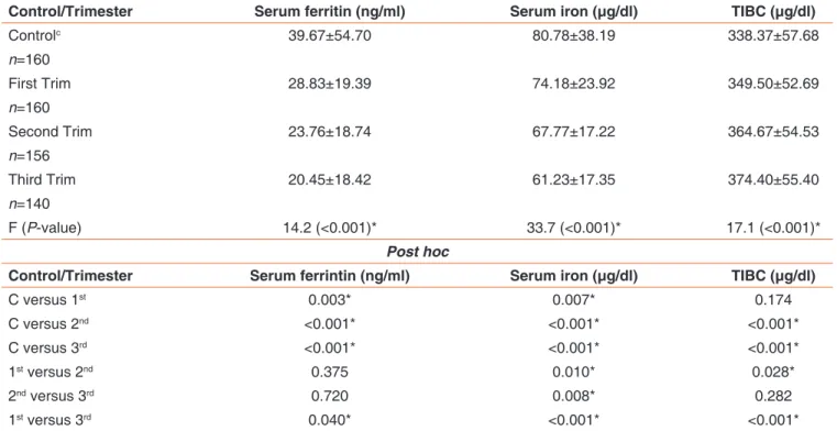
Table 2 shows mean values of serum ferritin, serum iron, and TIBC in pregnant women compare to non-pregnant women. The mean level of serum ferritin in the control subjects (39.67 ± 54.70) was significantly increased statistically when compared to the first (28.83 ± 19.39), second (23.76 ± 18.74), and third (20.45 ± 18.42) trimesters (F = 14.2, P < 0.001). Similarly, the serum, iron mean levels in the controls (80.78 ± 23.19) were significantly increased compared to the first trimesters (74.18 ± 23.92), second (67.77 ± 17.22), and third (61.23 ± 17.35) trimesters (F = 33.7, P < 0.001). However, the TIBC mean level in the controls (338.37 ± 57.69) was significantly increased statistically compared to the first (349.50 ± 52.69), second (364.67 ± 54.53), and third (374.40 ± 55.40) (F = 17.1, P < 0.001).
Post hoc analysis
Serum ferritin in the first (28.83 ± 19.39), second (23.76 ± 18.74), and third (20.45 ± 18.42) trimesters showed a statistically significant decrease when compared to the control subjects (39.67 ± 54.70) (P = 0.003 and <0.001). A non-statistically significant decrease was seen when the second trimester (23.76 ± 18.74) was compared to the first (28.83 ± 19.39) (P = 0.375), and the third trimester (20.45 ± 18.42) (P = 0.720), but a statistically significant decrease was observed when the third trimester (20.45 ± 18.42) was compared to the first (28.83 ± 19.39) (P = 0.040). Serum iron showed a statistically significant decrease when the first (74.18 ± 23.92), second (67.77 ± 17.22), and third (61.23 ± 17.35) trimesters were compared to the control subjects (80.78 ± 38.19) (P = 0.07 and <0.001). Similarly, a statistically significant reduction was observed when the second trimester (67.77 ±17.22) was compared to the first (74.18 ± 23. 92) (P = 0.010), and the third (61.23 ± 17.35) (P = 0.008), and when the third trimester (61.23± 17.35) was compared to the first (74.18 ± 23.92) (P < 0.001). However, TIBC in first trimester (349.50 ± 52.69) showed a statistically insignificant increase when compared to the controls (338.37 ± 57.68) (P = 0.174), but a statistically significant increase in the second (364.67 ± 54.53) and third (374.40 ± 55.40), compared to the controls (338.37 ± 57.68). A statistically significant increase was also observed when the second (364.67 ± 54.53) and third (374.40 ± 55.40) were compared to the first (349.50 ± 52.69) (P = 0.028 and <0.001). On the
other hand, a non-statistically significant increase was seen when the third trimester (374.40 ± 55.40) was compared to the second (364.67 ± 54.43) (P = 0.282).
DISCUSSION
This study has shown that serum ferritin decreased significantly in pregnancy as compared to the non-pregnant women.
The decrease may be because during pregnancy there is increased need of iron which triggers ferritin mobilization
from its stores. This is in line with the work done by Namama,[12] who showed that levels of serum ferritin were decreased in pregnancy compared to that in non-pregnancy,[13] also showed that non-pregnant women had more iron stores, therefore had less need for iron than their pregnant counterparts. The higher iron need in pregnancy triggered its mobilization from its stores. During pregnancy, there is an immense stress on iron metabolism and it frequently induces iron deficiency which is characterized, by a reduced ferritin level.[4] Furthermore, some of the pregnant women started pregnancy with low iron stores, hence the reason for low ferritin in pregnancy compared to the controls.
Table 1: Mean values of serum ferritin and serum iron/tibc of pregnant woman at different trimesters (mean±SD)
Trimester Serum ferritin (ng/ml) Serum iron (µg/dl) TIBC (µg/dl)
First trim N=160 28.83±19.39 74.18±23.92 349.50±52.69 Second trim N=156 23.76±18.74 67.77±17.22 364.67±54.53 Third trim N=140 20.45±18.42 61.23±17.35 374.40±55.40 F (P-value) 10.2 (<0.001)* 21.9 (<0.001)* 10.9 (<0.001)* Post hoc 1st versus 2nd 0.019* 0.003* 0.014* 2nd versus 3rd 0.179 0.002* 0.166 1st versus 3rd <0.001* <0.001* <0.001* Significant at P<0.05
Table 2: Mean values of serum ferritin, serum iron, and TIBC in pregnant women compare to non-pregnant women (mean±SD)
Control/Trimester Serum ferritin (ng/ml) Serum iron (µg/dl) TIBC (µg/dl)
Controlc 39.67±54.70 80.78±38.19 338.37±57.68 n=160 First Trim 28.83±19.39 74.18±23.92 349.50±52.69 n=160 Second Trim 23.76±18.74 67.77±17.22 364.67±54.53 n=156 Third Trim 20.45±18.42 61.23±17.35 374.40±55.40 n=140 F (P-value) 14.2 (<0.001)* 33.7 (<0.001)* 17.1 (<0.001)* Post hoc
Control/Trimester Serum ferrintin (ng/ml) Serum iron (µg/dl) TIBC (µg/dl)
C versus 1st 0.003* 0.007* 0.174 C versus 2nd <0.001* <0.001* <0.001* C versus 3rd <0.001* <0.001* <0.001* 1st versus 2nd 0.375 0.010* 0.028* 2nd versus 3rd 0.720 0.008* 0.282 1st versus 3rd 0.040* <0.001* <0.001*
Ferritin is an acute phase reactant protein and is sometimes found elevated independent of the iron status during illness and inflammation. According to Bain et al.,[14] serum ferritin decreases in early pregnancy and usually remains low throughout pregnancy, even when supplementary iron is given. Pregnancy is commonly associated with urinary tract infections and some occult infections. In such individuals, high serum ferritin levels are likely to be seen despite iron deficiency.[15]
Ferritin progressively decreased from the first to the third trimester. This could be due to increased demand for fetal growth and development as pregnancy progressed. This disagrees with the study done by Naghmi et al. and Namama[4,12] who showed that ferritin decreased from first to second trimester with a slight rise in the third trimester. It is in line with Okwara et al.,[13] who stated that serum ferritin declined progressively from first to the third trimester. The immense stress on iron metabolism during pregnancy frequently induces iron deficiency, hence the reduction in ferritin level. This also implies a progressive mineral transfer from the mother to the fetus. Serum ferritin also showed significantly lowest values in the third trimesters.
The demand for iron is variable during the three trimesters and the practice of iron supplementation is also not uniform. Furthermore, the decrease in serum ferritin level may be associated to plasma volume expansion and the higher need of iron in pregnancy caused its mobilization from its stores; therefore, serum ferritin levels can be variable during different trimesters of pregnancy.
In this study, serum iron was decreased significantly at all stages of pregnancy compared to the controls. The decrease might be because iron is needed during pregnancy to expand the red blood cell mass and for fetal and placental growth. This is in line with the work of Bothwell[7] who stated that iron was more reduced in pregnancy than in non-pregnant women because iron requirements are significantly greater in pregnancy than in non-pregnant state, despite the temporary respite from iron losses incurred during menstruation. Similarly,[16] Chaudari et al. also showed the same pattern. During pregnancy, hemodynamic changes lead to expansion of blood plasma volume up to 50% and increase in red cell mass up to 20% which iron required greater than that in the non-pregnant state. Iron is needed for expansion of the red blood cell mass and for transfer placental in structures.[17] Furthermore, iron was reduced in pregnancy may be because some of the pregnant women started pregnancy with low or because they consumed diets of low iron bioavailability. Serum iron was decreased from the first to the third trimester. There is unequal distribution of iron requirement during pregnancy, as iron is needed for fetal and placement
development (Bothwell, 2000). There is a significant increase in the amount of iron required to increase the red cell mass, expand the plasma volume and allow for the growth of the fetal-placental unit.[18]
Total iron binding capacity (TIBC) was insignificantly increased in the first trimester, but increased significantly in the second and third trimesters compared to non-pregnant women. When compared across the trimesters, the values increased from the first to the third trimester. This agrees with the work done by Amah-Tariah et al. and Chaudari et al. Okwara et al.[13,16,19] TIBC is known to be increased in pregnancy and during iron overload.[20]
CONCLUSION
The findings have shown that, by longitudinal analysis, serum ferritin and serum iron were decreased with increased in gestational age, while TIBC was increased as pregnancy progressed. Furthermore, serum ferritin and serum iron were decreased in each trimester compared to the non-pregnant controls, whereas TIBC was increased in all the trimesters compared to the controls. Therefore, there is need to monitor pregnancies at risk to prevent adverse outcomes.
REFERENCES
1. Alper BS, Kimber R, Reddy KA. Using ferritin levels in pregnancy. J Fam Pract 2000;49:829-32.
2. Casanova BF, Samuel MD, Macones GA. Development of a clinical prediction rule for iron deficiency anemia in pregnancy. Am J Obstet Gynecol 2005;193:460-6.
3. Hyder SM, Person L, Chowdhury M, Lonnerdal BO, Ekstorm EC. Anemia and iron deficiency during pregnancy in rural Bangladesh. Public Health Nutr 2004;7:1065-70. 4. Naghmi A, Khalid H, Shaleen M. Comparison of serum ferritin
levels in the trimesters of pregnancy and their correlation with increasing gravity. Int J Pathol 2007;5:26-30.
5. Scholl TO. High third trimester ferritin concentration: Associations with very pre delivery, infection, maternal nutritional status. J Obstet Gynecol 1998;92:161-5.
6. Micronutrient Information Centre. “Iron” Micronutrient Information Centre. Corvallis, Oregun: Linus Pauling Institute, Oregun State University; 2016.
7. Bothwell TH. Iron requirements in pregnancy and strategies to meet them. Am J Clin Nutr 2000;72;265-71.
8. World Health Organization. Iron Deficiency Anemia: Assessment, Prevention, and Control. A Guide for Programme Managers. Geneva: World Health Organization; 2001. 9. Mukherji J. Iron deficiency anemia in pregnancy. Ratinal Drug
Bull 2002;12:2-5.
10. Brabin BJ, Hakiri M, Pellertier D. An analysis of anaemia and pregnancy related maternal mortality. J Nutr 2001;131:6045-145.
11. Khan KS, Wojdyla D, Say L, Gulmezoglu AM, Van Look PF. WHO analysis of causes of maternal death: A systemic review. Lancet 2006;367:1066-74.
12. Namama ST. Monitoring levels of iron, TIBC, Hb, Transferrin and ferritin during pregnancy in Sulaiman city, Iraq. Int J Med Health Res 2015;1:15-8.
13. Okwara JE, Nnabuo LG, Nwosu DC, Ahaneku JE, Anolue F, Okwara NA, et al. Iron status of some pregnant women in Orlu town, Eastern Nigeria. Niger J Med 2013;22:15-8.
14. Bain BJ, Bates I, Laffan MA, Levis SM. Basic Haematological Techniques: Dacie and Lewis Practical Haematology. 11th ed.
Amsterdam, Netherlands: Elsevier; 2012. p. 23-9, 59, 110-7. 15. Hon J, Suzanne P, Oliver BA. Maternal serum ferritin and fetal
growth. Obstet Gynaecol 2000;95:447-52.
16. Chaudari H, Dixit R, Jadeja JM. Serum level of iron and transferrin in normal and anaemicpregnant women. Int J Basic Appl Physiol 2013;2:123-6.
17. Short M. Iron deficiency anemia: Evaluation and management. Am Fam Phys 2013;87:98-104.
18. Yip R. In: Bowman B, Russel RM, editors. Present Knowledge in Nutrition. 8th ed. Washington, DC: ILS Press; 2001. p. 311-8.
19. Amah-Tariah FS, Ojeka SO, Dapper DV. Haematological values in pregnant women in Port Harcourt, Nigeria: Serum iron and transferrin, total and unsaturated iron-binding capacity and some red cell and platelet indices. Niger J Physiol Sci 2011;26:173-8.
20. Wessling-Resnick M. Iron. In: Ross AC, Caballero B, Cousin RJ, Tucker KL, Ziegler RG, editors. Modern Nutrition in Health and Disease. 11th ed. Baltimore, MD. Lippincott
Williams and Wilkins; 2014. p. 178-88.
How to cite this article: Aloy-Amadi O, Akujobi AU, Nnodim J, Ndudim-Dike J, Edward A, Anokwute M. Serum Ferritin and Iron/TIBC of Pregnant Women Attending NnamdiAzikiwe University Teaching Hospital, Nnewi, Anambra State, Nigeria: A Longitudinal Study. Clin Res Obstetrics Gynecol 2020;3(2):1-6.