ORGANIZATION AND FUNCTION OF MOLECULAR DOMAINS IN MYELINATED NEURONS
James Allen Green
A dissertation submitted to the faculty of the University of North Carolina at Chapel Hill in partial fulfillment of the requirements for the degree of Doctor of Philosophy in Cell and
Molecular Physiology
Chapel Hill 2013
Approved by:
iii ABSTRACT
JAMES ALLEN GREEN: Organization and Function of Molecular Domains in Myelinated Neurons
(Under the direction of Manzoor A. Bhat)
Myelinated neurons are organized into distinct molecular domains that are essential for electrical conduction. These domains include the axon initial segment, nodes of Ranvier, paranodes, juxtaparanodes, and internode. The paranode acts as a molecular fence,
maintaining separation of the sodium enriched nodes from the potassium channel-enriched juxtaparanodes via axo-glial septate junctions. In demyelinating diseases like multiple sclerosis and Guillain-Barré, the clustering and segregation of such domains and structures are disrupted, resulting in altered neuronal function.
iv
and transmission electron microscopic analyses of Whrn-/- mice revealed cellular swellings in cerebellar Purkinje axons filled with mitochondria and vesicles. Such Purkinje axon findings were similar to the Caspr, another paranodal protein, knockout mouse cerebellar phenotype. Additional higher magnification, electron micrograph images of the paranodal,
juxtaparanodal and internodal region in Whrn-/- mice reveal abnormal accumulation of mitochondria and vesicles. Taken together, our studies revealed Whirlin plays an important role in paranodal and juxtaparanodal organization, maintaining cytoskeletal organization, and preventing accumulation of mitochondria and vesicles. In addition to these core findings, results are included from a study characterizing fluorescent imaging of
v
DEDICATION
To my father, James Lee Green: For a man, a hero, a friend who I lost at 10 years-old but still think about every day. Your death to cancer never silenced you. It made your
message more important: to live every day to the fullest, to work hard in school, to love and care for my family, and to never fear creativity and risk in the kitchen. Until the end of my days, I will love and miss you.
To my mother, Margaret Gardipee: While your brain injury took away so much from you, it never took away your love and concern for your children’s well-being. You will always be the strong, proud, Native American woman who reminds me of the Gros Ventre language, traditions, and history. Your laugh will always be the most potent cure for my sorrow, and your tears will remain the most sinking weight on my soul. Somewhere inside those beautiful brown eyes is the mother who can do it all, but to the mother that is here and now, know this, “You are perfect and loved just the way you are.”
To my uncle and aunt, Dennis and Carol Goulet: Thank you for raising me and finding the resolve to pick up the pieces after your brother died. Thank you for all you have given: your dedication to my well-being and growth even when you were in pain, your surrounding me with good church friends, your insistence to work hard and finish every journey I start, and your unconditional love to me and our family.
vi
loved. We were never the beneficiaries of luck, money or fame in life, but we found that all we needed was each other as brothers and sisters through the highs and lows.
vii
ACKNOWLEDGEMENTS
The work presented in this dissertation would not have been possible without the support and guidance of my colleagues and friends at University of North Carolina-Chapel Hill as well as recent colleagues and friends at University of Texas Health Science Center at San Antonio.
I would like to thank my advisor, Manzoor Bhat, for always providing resources and advice needed to keep my projects moving forward. Together, we made an eventful transition from one university to another and kept the lab ship afloat. Thank you for all your trust and loyalty to my academic success over these years.
I would also like to acknowledge my committee members, Kathleen Caron, Richard Cheney, Jay Brenman, and Silva Markovic-Plese for their encouragement and input towards the completion of this work.
I also thank the members of the Bhat lab, both past and present, including Swati Banerjee, Kevin Blauth, Liz Buttermore, Yu-Chi Chen, Liz Fisher, Rosa Mino, Raehum Paik, Anil Pillai, and Courtney Thaxton for the immeasurable time and support they have given to me during lab meetings, practice presentations, technical assistance, and countless questions I have asked them.
viii
I would also like to thank the former and current administration and staff in the MD/PhD program office for their constant support and responsiveness to our changing situations. Nine years is a long academic commitment to any student, let alone trying to manage eighty or more of them is something of an unappreciated miracle, so my deepest thanks to Dr. “O” (Eugene Orringer), David Siderovski, Liz Garman, Alison Regan, and Carol Heiron.
I’d like to acknowledge, particularly during the recent transition to Texas and back, the hard work of the faculty and staff in the Cell and Molecular Physiology Department in helping organize the framework and path that allowed me to reach this point in my career. Thank you Carol Otey, Robert Sealock, Eleni Tzima, Michael Goy, Ben Philpot, John Rawls, Mark Zylka, Adriana Tavernise, Janice Warfford, and Vicki Morgan.
Finally, parts of this work were done in collaboration with the labs of Thomas Friedman and Bechara Kachar at the National Institute on Deafness and Other
ix PREFACE
Portions of this dissertation have been published or are in preparation to be published. Thank you to all the co-authors and those who contributed reagents and time to these
publications.
Chapter 2 results derive from a 2009 collaboration between the Manzoor Bhat (MB) and Glenn Matsushima (GM) labs. Specifically, Lorelai Taylor (LT) treated the mice with or without cuprizone. Perfusion, fixation, tissue slicing, immunostaining, imaging, figure creation, and writing were performed by James Green (JG).
Chapter 3 is a slightly revised version of Green, J., Yang, J., Grati, M., Kachar, B., and Bhat, M.A. (2013). Loss of Whirlin, a Cytoskeletal Scaffolding Protein, Disrupts Proper Compaction of the Paranodal Region in Myelinated Axons. Manuscript accepted with revisions. James Green (JG) performed genotyping and phenotype quantitation,
immunostaining and confocal imaging; Jun Yang (JY) provided Whrn exon 1 knockout mouse strain; M’hamed Grati (MG) and Bechara Kachar (BK) provided whirler mouse strain; JG and Manzoor Bhat (MB) participated in the design of the study, assembled figures, and drafting of the manuscript. All authors approved the final manuscript.
x
http://www.jneurosci.org/site/misc/about.xhtml#permission. M’hamed Grati (MG), Peter G.
xi
TABLE OF CONTENTS
LIST OF TABLES ………. xv
LIST OF FIGURES ………... xvi
LIST OF ABBREVIATIONS ………. xviii
Chapter 1. INTRODUCTION: ORGANIZATION AND FUNCTION OF MOLECULAR DOMAINS IN MYELINATED NEURONS ………...…….………. 1
1.1 The axon initial segment (AIS) ...………... 1
1.2 The node of Ranvier (node) ...……….………….. 11
1.3 The paranode ...………. 17
1.4 The juxtaparanode (JXP) ……….. 23
1.5 The internode ...………. 25
1.6 Myelination and evolutionary model systems ...………... 28
1.7 Neuropathies, myelinopathies and related domain pathology ..……… 31
2. CUPRIZONE AND CASPR-CRE MODEL MOUSE SYSTEMS ……… 38
2.1 Introduction ...………... 38
2.1.1 Mus musculus, an animal model for human biomedical research ……… 38
2.1.2 Mouse utilization in neuroscience and contributions to axonal domain organization research ……..………... 39
2.1.3 The cuprizone mouse model ……….. 40 2.1.4 Mouse recombinant technology, site-specific
xii
(Cre-LoxP) knockout mouse lines ………. 42
2.2 Materials and Methods ...……….. 43
2.2.1 Cuprizone Treatment in Animals .………….………. 43
2.2.2 Generation of Caspr-Cre mutant animals ...………49
2.2.3 Tissue preparation, antibodies and immunostaining ……….. 53
2.2.4 Image Analysis and Software ...………..……… 54
2.3 Results ...………..………. 54
2.3.1 Cuprizone-treated mice reveal significant losses of paranodal Caspr and glial Myelin Basic Protein in cerebral and cerebellar white matter tract ………. 54
2.3.2 Identification of three Caspr-Cre recombinant lines (A9, E10, dB1) ……… 63
2.4 Discussion ………. 68
2.4.1 The cuprizone mouse model establishes a reproducible demyelination/remyelination model for the study of myelinated axonal domain disorganization and reformation ...……….. 68
2.4.2 Identification and application of three Caspr-Cre recombinant lines ……… 71
3. WHIRLIN, A CYTOSKELETAL SCAFFOLDING PROTEIN, STABILIZES THE PARANODAL REGION AND AXONAL CYTOSKELETON IN MYELINATED AXONS ………. 74
3.1 Introduction ...………... 74
3.2 Materials and Methods ...……….. 76
3.2.1 Animals ...……….……….. 76
3.2.2 Genotyping ...……….………. 77
3.2.3 Generation of Whrn Antibody ...……….……… 77
xiii
3.2.5 Other Antibodies and Immunostaining Reagents ..…….……… 79
3.2.6 Immunostaining ...……….……… 79
3.2.7 Immunoblotting ...……….…………... 80
3.2.8 Electrophysiology ...……….……… 81
3.2.9 Image Analysis and Software ……….……… 81
3.2.10 Quantification of Phenotype and Statistics ....………….…………... 82
3.2.11 Transmission electron microscopy ...……….. 83
3.3 Results ……….. 84
3.3.1 Whrn is Expressed in Central and Peripheral Nervous System Tissues Throughout Development ….………. 84
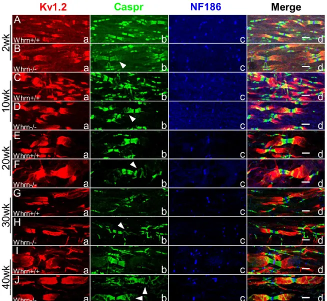
3.3.2 Whrn Knockout Mice Reveal a Quantifiable Paranodal Compaction Phenotype in Peripheral Myelinated Axons………... 87
3.3.3 Whrn Knockout Mouse Sciatic Nerve and Spinal Cord Myelinated Fibers Display Paranodal Compaction Abnormalities Throughout Development ………... 93
3.3.4 Whirlin knockout mice have cerebellar Purkinje cells with bead-like, axonal swellings ……….. 100
3.3.5 Ultrastructural Abnormalities in Whrn Knockout Sciatic Nerve, Spinal Cord Fibers, and Cerebellar Purkinje Axons ……… 101
3.4 Discussion ……….. 107
3.4.1 Whrn Alternative Splicing and Phenotypes ….……….…... 107
3.4.2 Whrn Localization in Myelinated Axons ……….……… 108
xiv
4. LOCALIZATION OF PDZD7 TO THE STEREOCILIA ANKLE-LINK ASSOCIATES THIS SCAFFOLDING PROTEIN WITH THE USHER
SYNDROME PROTEIN NETWORK ………. 112
4.1 Introduction ...………. 112
4.2 Materials and Methods ...……… 114
4.2.1 Mass spectrometry………. 114
4.2.2 Antibodies .………... 114
4.2.3 Inner ear tissue ..………... 115
4.2.4 Immunocytochemistry ………. 115
4.2.5 Complementary DNA expression vectors ……… 116
4.2.6 Culture and transfection of rat inner ear tissue ………. 116
4.2.7 Culture and transfection of LLC-PK1 and COS7 cells ..……..………... 117
4.2.8 Scanning electron microscopy ……….. 117
4.3 Results .………... 117
4.3.1 Detection of PDZD7 by mass spectrometry ………. 117
4.3.2 Localization of PDZD7 at the stereocilia base …..………….…….. 120
4.3.3 PDZD7 colocalization with stereocilia ankle-link proteins ………. 120
4.3.4 PDZD7 interactions with Usher proteins in COS7 and LLC-PK1 cells………... 126
4.4 Discussion ...………... 128
xv
LIST OF TABLES
Table 2.1 Mouse Cre lines established by NIH Neuroscience
xvi
LIST OF FIGURES
Figure 1.1 Molecular domains in myelinated axons ……….. 2 Figure 1.2 Molecular organization of myelinated axon domains ……….. 4 Figure 2.1 Corpus callosum white matter tracts reveal generally
decreased levels of Caspr and Myelin Basic Protein in animals treated for 3, 5 and 6 weeks with 0.2% cuprizone
compared to untreated animals ……….. 56 Figure 2.2 Cerebellar white matter tracts show generally decreased
levels of Caspr and Myelin Basic Protein signal in 3, 5 and 6 week cuprizone-treated animals compared to untreated
animals ………..……….. 60 Figure 2.3 Generation of Caspr-Cre recombinant mouse ……… 65 Figure 3.1 Whirlin (Whrn) is a PDZ-containing protein expressed
throughout the central and peripheral nervous system ………... 85 Figure 3.2 Loss of Whirlin in the peripheral nervous system results in
disrupted paranodal compaction ………. 89 Figure 3.3 Loss of Whirlin contributes to abnormal paranodal
compaction and cytoskeleton instability throughout sciatic
nerve age ………. 94 Figure 3.4 Loss of Whirlin contributes to abnormal paranodal
compaction throughout development in the spinal cord ………. 97 Figure 3.5 Whirlin alone and combination 4.1B/Whirlin knockout mice
have cerebellar Purkinje cells with bead-like, axonal swellings ……… 99 Figure 3.6 Ultrastructural examination of three month-old Whirlin
knockout sciatic nerves reveals organelle accumulation and
cytoskeletal disruption ……….. 102 Figure 3.7 Ultrastructural examination of Whirlin knockout central
nervous system tissues reveals organelle accumulation in
myelinated axons ……….. 105 Figure 4.1 Detection of PDZD7 in hair cells stereocilia ……… 118 Figure 4.2 PDZD7 colocalization with ankle-link proteins in maturing
xvii
Figure 4.3 PDZD7 colocalization with stereocilia ankle-link proteins
in mature rat hair cells ……….. 124 Figure 4.4 PDZD7 interactions with Usher proteins in COS7 and
xviii
LIST OF ABBREVIATIONS 4.1B 4.1 brain protein
AGSJs axo-glial septate junctions AIS axon initial segment Ank/ANK Ankyrin
BAC bacterial artificial chromosome Calb Calbindin
CAM cell adhesion molecule Caspr Contactin-associated protein Caspr2 Contactin-associated protein-2 CGT ceramide galactosyltransferase CMT Charcot-Marie Tooth disease CNS central nervous system Cont Contactin
H2O water
DRG dorsal root ganglion EBP50 ezrin binding protein-50 ECM extracellular matrix
EDTA Ethylene-diamine-tetra-acetic acid EM electron microscopy
xix IHC immunohistochemistry
MBP myelin basic protein
Kv#.# voltage-gated potassium ion channels
Nav#.# voltage-gated sodium ion channels
NF155 155kDa isoform of Neurofascin locus NF186 186kDa isoform of Neurofascin locus PCR polymerase chain reaction
PBS phosphate buffer saline
PDZ Postsynaptic density protein 95/Drosophila disc large tumor suppressor/Zonula occludens-1 protein
PNS peripheral nervous system PSD-93 postsynaptic density protein-93 PSD-95 postsynaptic density protein-95
RT-PCR reverse transcription polymerase chain reaction SDS sodium dodecyl sulfate
SEM standard error of the mean shRNA short hairpin RNA
Tag1 transiently expressed axonal surface glycoprotein-1 TEM transmission electron microscopy
Whrn Whirlin gene
CHAPTER 1
INTRODUCTION: ORGANIZATION AND FUNCTION OF MOLECULAR DOMAINS IN MYELINATED NEURONS
1.1 The axon initial segment (AIS)
The axon initial segment (AIS) is the most proximal section of the outgoing axon from the neuron cell body and just distal to the axon hillock (Hedstrom and Rasband 2006). Like all the molecular domains of the myelinated axon, this region has a broad collection of transmembrane proteins necessary for protein interactions and/or ion channels as well as cytoskeletal elements to anchor protein complexes to their recognized region. Functionally, the AIS is an electrical gatekeeper, the decision point as to whether or not to initiate an action potential. The AIS performs this function by summing all excitatory and inhibitory inputs until a critical firing threshold is reached and the action potential initiated.
Several ion channels cluster at the AIS and contribute to the final summation including sodium (Na+, Nav1.6, Nav1.2) channels, promoting membrane depolarization and
subsequent firing, as well as potassium (K+, KCNQ2/3, Kv1.1/1.2) channels, regulating
action potential frequency and modulating excitability (Pan, Kao et al. 2006, Johnston, Griffin et al. 2008, Kole, Ilschner et al. 2008). Sodium and potassium channels are
2
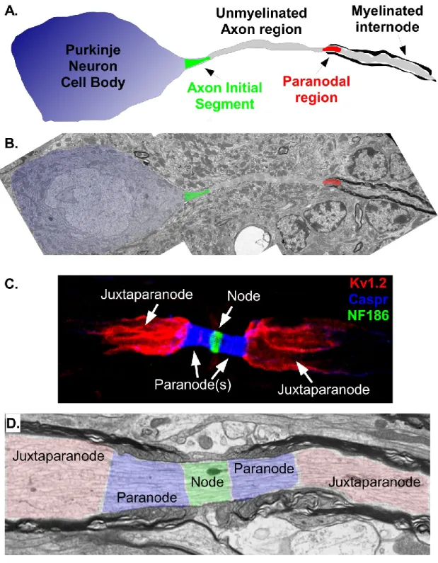
Figure 1.1. Molecular domains in myelinated axons
3
(grey), first paranode (red), and remaining myelin-wrapped juxtaparanode and internode (grey). B. Transmission electron micrograph through a 3 month-old mouse cerebellar Purkinje neuron with color matched overlay from schematic in A. Note this slice fails to show the unmyelinated segment of the axon (middle pseudo-colored grey segment) either above or below the imaged plane. AIS region is peusdo-colored in green and paranode in red. Note the distinctive electron dense layers of myelin wrapped around the axon past the first paranode. C. Immunostaining of a 4 month-old mouse sciatic nerve fiber immunostained against potassium channels (Kv1.2) to label the juxtaparanode (red), Contactin-associated
4
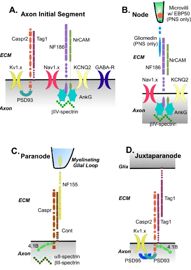
Figure 1.2. Molecular organization of myelinated axon domains
A-D. Labeled diagrams showing the known proteins responsible for formation and
5
at the axon initial segment (Punkinje neuron example), including ion channels (Kv1.x in dark
yellow, Nav1.x in pink, KCNQ2 in light yellow, GABA-R in dark blue), cell adhesion
molecules (Caspr2 in orange, Tag1 in violet, NF186 in light purple, NrCAM in green) and cytoskeletal scaffolding proteins (PSD93 in blue-green, IV-spectrin in green, AnkG in cyan). B. Molecular components at the node of Ranvier, including ion channels (Nav1.x in
pink, KCNQ2 in light yellow), cell adhesion molecules (Gliomedin in cyan, NF186 in light purple, NrCAM in green) and cytoskeletal scaffolding proteins (EBP50 in orange, IV-spectrin in green, AnkG in cyan). Note microvilli are unique structures found on myelinating Schwann cells in the peripheral nervous system and thus EBP50 and Gliomedin only found there and not in the central nervous system. C. Molecular components at the paranode, including cell adhesion molecules (Caspr in dark orange, Cont in brown, NF155 in yellow) and cytoskeletal scaffolding proteins (II-spectrin in pale green,II-spectrin in dark green, 4.1B in green). The protein complex of Caspr, Cont, and NF155 make up the axo-glial septate junctions critical to paranodal barrier function. D. Molecular components at the juxtaparanode, including ion channels (Kv1.x in dark yellow), cell adhesion molecules
6
intracellular interactions (Isom 2002, Yu and Catterall 2003). Also present at the AIS are numerous cell adhesion molecules (CAMs). Many of these CAMs belong to the L1
subfamily of immunoglobulin (Ig) which include neurofascin186 (NF186, 186kDa isoform of Neurofascin), contactin-associated protein-2 (Caspr2), neuron-related cell adhesion molecule (NrCAM), and transiently expressed axonal surface glycoprotein-1 (Tag1) (Dodd, Morton et al. 1988, Volkmer, Hassel et al. 1992, Davis, Lambert et al. 1996, Grumet 1997, Brummendorf, Kenwrick et al. 1998, Hedstrom, Xu et al. 2007). As mentioned previously, several adaptor and scaffolding proteins are responsible for anchoring these membrane-bound surface proteins to the AIS’s underlying actin/spectrin cytoskeleton (Zhou, Lambert et al. 1998, Boiko, Vakulenko et al. 2007). Such adaptors and scaffolding proteins include IV-spectrin (b4spec), ankyrinG (AnkG), and postsynaptic density protein 93 (PSD-93) (Jenkins and Bennett 2001, Kole, Ilschner et al. 2008).
AnkyrinG (AnkG) is the initial intrinsic ‘AIS master regulator’ (Bennett and Baines 2001). Historically, the ankyrin class of proteins is known for stabilizing groups of plasma membrane-bound proteins to specific cellular regions for interactions between cells and efficient signaling (Bennett and Baines 2001). Independent in vitro and in vivo studies have confirmed this decisive role for AnkG in AIS organization (Zhou, Lambert et al. 1998, Hedstrom, Xu et al. 2007, Hedstrom, Ogawa et al. 2008). Using shRNA in a targeted knockdown of AnkG, cultured hippocampal neurons failed to cluster any other
7
AnkG in mature hippocampal neurons where the mature AIS had already clustered proteins like sodium channels and cytoskeletal elements led to destabilization of such proteins (Hedstrom, Ogawa et al. 2008). Using an in vitro mouse model, it was shown that genetic ablation of AnkG in the cerebellum resulted in a lack of clustering of Nfasc and sodium channels at the Purkinje AIS (Zhou, Lambert et al. 1998). Once such effects regarding AnkG on the AIS were independently verified, reports shifted to the exact molecular mechanism as to how AnkG stabilizes this region. If AnkG is the initial AIS molecule, what cellular signals get AnkG to the AIS and recruit other protein partners? One series of in vivo experiments suggested that phosphorylated inhibitor of kappa B alpha (B) acts as a cofactor in trafficking AnkG to the AIS (Schultz, Konig et al. 2006, Sanchez-Ponce, Tapia et al. 2008, Rasband 2010), but lack the scientific support of in vitro results. Once at the AIS, AnkG, through an unphosphorylated tyrosine on a conserved motif, binds NrCAM and NF186 at the AIS (Davis, Lambert et al. 1996, Garver, Ren et al. 1997, Zhang and Bennett 1998, Lustig, Zanazzi et al. 2001). Without this AnkG interaction, protein accumulation and stabilization with Na+ channels fails to occur at the AIS in numerous studies (Zhou, Lambert et al. 1998, Hedstrom, Xu et al. 2007, Zonta, Desmazieres et al. 2011, Buttermore, Piochon et al. 2012). Taken together, these results strongly suggest AnkG represents the master organizer and regulator of the AIS.
8
fact the default fate for any neuronal projection. Axonal identity and specialization is the ultimate consequence of AIS formation and associated AnkG intrinsic regulation. This diffusion barrier role in the AIS was first discovered when some membrane proteins were found to diffuse more slowly through the AIS than other proteins (Winckler, Forscher et al. 1999). Moreover, disruption of the filamentous actin cytoskeleton nullified this differential mobility, thereby suggesting AIS barrier function required the interaction of the cytoskeleton with membrane proteins. Such AIS barrier activity restricts both cytoplasmic and membrane-bound proteins from simply diffusing into the axon. More recent studies have taken
advantage of fluorescent reporters to show that tagged, unsaturated phospholipids halt their diffusion across the AIS within the same developmental window of AIS protein clustering and maturation of associated protein complexes (Nakada, Ritchie et al. 2003). In support of this finding, another study found large-molecular weight dextrans would not diffuse into the axon once the AIS was formed in contrast to smaller-molecular weight dextrans used (Song, Wang et al. 2009). Likewise, large dextrans could diffuse into the axon once AIS barrier function was compromised with disruption of the filamentous actin cytoskeleton. In addition to molecular size, the AIS permits entry of specific molecules depending on their cargo or associated proteins. In this study, the authors found kinesin motor proteins carrying axonal component cargos were permitted entry while dendritic component cargos were blocked from entering the axon through the AIS (Song, Wang et al. 2009). Taken together, these studies reveal the AIS is critical for establishing and maintaining axonal identity in addition to initiating action potential propagation.
9
tuning frequency, size, and location which differ greatly between cellular subtypes, brain nuclei, and cortical regions (Konishi 2003, Kuba, Ishii et al. 2006, Lorincz and Nusser 2008, Fried, Lasker et al. 2009, Grubb, Shu et al. 2011). Depending on the requirement of the individual neuron within the circuit, a range of plasticity can be observed. While much of the AIS research has been relatively recent, direct evidence of AIS plasticity comes from two independent studies. The authors of one such study found an increase in AIS length of
auditory brainstem neurons following auditory deprivation of birds (Kuba, Oichi et al. 2010). In the second highly technical study, ion flux could be directly monitored through live
imaging of the associated AIS (Grubb and Burrone 2010, Grubb, Shu et al. 2011). The in vitro study found spatial shifting in AIS position distally along the axon as it was exposed to long-term increased excitatory activity (Grubb and Burrone 2010). The positional shift in the AIS required prolonged two-day stimulation as well as recruitment of voltage-gated calcium channel activity. Such results suggest AIS function responds to environmental and circuitry cues to control firing rate in a plastic fashion through alterations in cellular organization, movement of channels up or down the AIS region, and recruitment of non-standard ionic channels. However, it should be noted the extent of plasticity in older animals has not been demonstrated. So while studies in certain neurons of younger animals display remarkable AIS plasticity, reports also suggest molecules such as AnkG and NF186 in mice promote the long-term stabilization and rigidity of the AIS (Zonta, Desmazieres et al. 2011). Examining the transitional periods of AIS function, recent data shows maturation of the AIS depends on NF186 expression (Buttermore, Piochon et al. 2012). A transitional developmental switch from Nav1.2 to Nav1.6 within the AIS is critical to AIS function in adult myelinated neurons
10
(Nav1.6) does not enrich at the Purkinje AIS segment of NF186 deficient Purkinje neurons
compared to wild-type Purkinje neurons (Buttermore, Piochon et al. 2012), suggesting differing mechanisms between primary formation through AnkG and secondary maturation through NF186 of the AIS. Supporting this hypothesis, electrophysiological measurements of Purkinje cells lacking an intact AIS demonstrated an inducible action potential firing was possible, but the resulting waveform of that action potential was altered (Zonta, Desmazieres et al. 2011, Buttermore, Piochon et al. 2012). Notably, the loss of the AIS disrupted the spontaneous firing of the Purkinje cell (Zonta, Desmazieres et al. 2011, Buttermore, Piochon et al. 2012). Together, it is highly likely the cellular mechanisms responsible for neuronal AIS plasticity overlap with mechanisms for AIS maturation.
Finally, the AIS can be further subdivided using sodium channel isoform localization with Nav1.2 channels segregated to the proximal AIS and Nav1.6 channels restricted to the
distal AIS (Hu, Tian et al. 2009, Buttermore, Piochon et al. 2012). Interestingly, the distal AIS and associated Nav1.2 channels are activated at a lower electrophysiological threshold
relative to proximal AIS and its Nav1.6 channels (Hu, Tian et al. 2009). In order to explain
this finding, it was suggested by the authors that proximal AIS sodium channels are
responsible for back-propagation of the action potential while the distal AIS sodium channels and higher-voltage threshold regulate the axonal action potential initiation. Bifurcation of AIS organization and function vis-à-vis sodium channel isoforms highlights the precise relationship between molecular form and function in regulating axonal activity. Such
11 1.2 The node of Ranvier (node)
The nodes of Ranvier (nodes) are ~1m long, unmyelinated molecular domains enriched with voltage-gated sodium channels and necessary for saltatory action potential propagation (Waxman and Ritchie 1993, Salzer 2003, Hedstrom and Rasband 2006, Rasband 2006, Thaxton and Bhat 2009). Unlike one AIS per neuron, numerous nodes are distributed at regular intervals along the axonal projection. In conjunction with the insulating properties of myelin, the nodal configuration and associated sodium channel enrichment allows the action potential to leap or ‘saltare’ down the axon at a faster velocity than unmyelinated axons. Spacing and assembly of nodes is coordinated with cues from myelinating glia, allowing for decreased membrane capacitance and increased membrane resistance (Court, Sherman et al. 2004, Susuki and Rasband 2008, Thaxton, Pillai et al. 2011). For example, spacing between nodes and the diameter of axons in the avian brainstem are regulated so interaural time differences are detectable (Jeffress 1948, Rasband 2010, Seidl, Rubel et al. 2010). Of note, a significant number of protein complexes are conserved between the AIS and node including Nav1.2, Nav1.6, KCNQ2/3, IV-spectrin, AnkG, NrCAM, and NF186
(Volkmer, Hassel et al. 1992, Berghs, Aggujaro et al. 2000, Boiko, Rasband et al. 2001, Komada and Soriano 2002, Salzer 2003). AnkG interacts with CAMs, Nav1.6, and indirectly
to the underlying cytoskeleton through IV-spectrin at the node (Bennett and Lambert 1999). Like the AIS, a developmental switch in sodium channel isoform expression from Nav1.2
(immature nodes) to Nav1.6 (mature nodes) takes place (Boiko, Rasband et al. 2001, Kaplan,
12
In contrast to AIS intrinsic formation which depends on AnkG localization prior to CAM and sodium channel clustering, external signals aid the development of the node (Pedraza, Huang et al. 2001, Eshed, Feinberg et al. 2005). Notably, different mechanisms of node formation are thought to occur whether in the CNS versus PNS due to the different glial cells types that contribution to myelination. Schwann cells, the myelinating glia in the PNS, have microvilli protrusions that extend out toward the unmyelinated nodal gap. These microvilli in turn secrete proteins as part of the extracellular matrix (ECM) (Eshed, Feinberg et al. 2005, Eshed, Feinberg et al. 2007). The nodal ECM becomes a developmental partner in node formation with various glycoprotein constituents necessary for stabilizing Schwann cell microvilli at the node (Saito, Moore et al. 2003, Melendez-Vasquez, Carey et al. 2005). Several independent studies have identified an initial formative event for the node is the binding of gliomedin on the Schwann cell microvilli to the axonal NF186 (Lambert, Davis et al. 1997, Eshed, Feinberg et al. 2005, Schafer, Custer et al. 2006). This interaction between gliomedin and NF186 stabilizes and recruits subsequent nodal proteins, and when gliomedin expression was knocked-down, it resulted in a disruption to clustering of NF186 and sodium channels (Dzhashiashvili, Zhang et al. 2007) Surprisingly, ectopic gliomedin clusters were observed along the internodes of co-cultured Scwhann cell-myelinated dorsal root ganglion (DRG) neurons when only the extracellular domain of NF186 was added to the media (Eshed, Feinberg et al. 2005). Such findings suggest that the extracellular domain of NF186 was critical to node formation, and thus greater research effort has been spent on determining the involvement and role of NF186 in myelinated neurons.
13
immunoglobulin domains, three fibronectin type III repeats, and a mucin domain (Thaxton and Bhat 2009). The intracellular region of NF186 contains an FIGQY motif (Garver, Ren et al. 1997) which interacts and recruits AnkG to the nodal domain. Sodium channels are typically found bound to NF186 through the cytoskeletal protein AnkG, but the -subunit of sodium channels can also independently bind to NF186 (Malhotra, Kazen-Gillespie et al. 2000, Lemaillet, Walker et al. 2003, Brechet, Fache et al. 2008, Rasband 2008). In vitro knockdown studies of NF186 in Schwann cell-DRG co-cultures have confirmed that loss of NF186 disrupts sodium channel enrichment at the node in the PNS (Dzhashiashvili, Zhang et al. 2007). However, this knockdown of NF186 was unable to rescue nodal formation after adding a different NF186 construct deficient in the intracellular domain responsible for binding AnkG, further reinforcing the recruitment role of NF186 for AnkG and sodium channels (Dzhashiashvili, Zhang et al. 2007).By demonstrating the external and intracellular domains of NF186 were required for nodal protein clustering, it reinforces the hypothesis that at least some glial involvement is necessary for nodal organization in the PNS. These PNS studies, however, have limited insight into the formation and stabilization of CNS nodes.
The importance of other cell types in facilitating the organization of CNS nodes remains elusive. Some analogous structures can be proposed as with perinodal astrocytes in the CNS and the Schwann cell microvilli in the PNS. EM data reveals that perinodal
14
in mice however did not demonstrate significant deficits in nodal organization (Brakebusch, Seidenbecher et al. 2002) which could suggest CNS nodal organization may require
interaction of Brevican in conjunction with other yet unidentified molecules. Some of these proteins could come from the oligodendrocyte protein secretion prior to myelination, which has been linked to nodal protein clustering (Kaplan, Cho et al. 2001). Clearly, this lack of knowledge as to the signaling components necessary for CNS node formation suggests more research is needed. Once nodes are formed, numerous studies have demonstrated several neuronally-derived proteins for organization and stabilization of the node. For example, deficits in IV-spectrin showed vesicle-filled membrane protrusions in the nodal area of the axon (Yang, Lacas-Gervais et al. 2004). This suggests cytoskeletal organization at the node is linked to proper IV-spectrin function. Also, loss of IV-spectrin resulted in disrupted
axonal conduction and significant drops in the expression level of AnkG and sodium channels at the node, suggesting it is vital for stabilizing the nodal complex (Komada and Soriano 2002, Lacas-Gervais, Guo et al. 2004).
Some controversial and conflicting reports have been found concerning the role of Neurofascin in CNS nodal domain development. To begin, Neurofascin null mice have disruption of both CNS and PNS nodes, but die at post-natal day six before the nervous system starts most of its myelination (Sherman, Tait et al. 2005). Such mice lack both
dominant isoforms in the nervous system, the 186kDa (NF186) isoform and 155kDa (NF155) isoform. NF155 is produced by the myelinating glia and localized to the paranodal region (see section 1.4). NF186 is expressed by neurons, transported and localized to the AIS and node. Given the significant involvement of Neurofascin in several neuronal domains,
15
day six. To overcome this technical problem, re-expression of transgenic NF186 in
Neurofascin null mice revealed AnkG and Nav1.6 localized to nodes and was independent of
paranode formation (Zonta, Tait et al. 2008). Controversially, the same group also found re-expression of transgenic NF155 in Neurofascin null mice rescued CNS nodal components (Zonta, Tait et al. 2008). Such results suggested paranodal formation was linked in a dependent manner with nodal formation in the CNS. These results challenge the findings where nodal clustering and organization were unaffected in paranodal mutant mice like Caspr, Contactin, and NF155 null mice (Bhat, Rios et al. 2001, Boyle, Berglund et al. 2001, Pillai, Thaxton et al. 2009). Additionally, scanning electron microscopy (SEM) of freeze-fracture samples show early differentiation events still take place in the node prior to paranodal axo-glial septate junction (AGSJ) formation (Tao-Cheng and Rosenbluth 1983). Recent in vitro work in co-cultures of Schwann cell and DRGs have contributed further to the scientific debate. In such co-cultures from wild-type and Neurofascin null mice which lack in vivo environmental cues, sodium channel clustering at mature nodes appears
dependent on paranodes and independent of NF186 (Feinberg, Eshed-Eisenbach et al. 2010). Loss of paranodal AGSJs appear to result in destabilization of sodium channels, furthering the hypothesis of linked paranodal-dependent nodal stabilization (Ishibashi, Dupree et al. 2002, Poliak and Peles 2003, Rios, Rubin et al. 2003, Rosenbluth, Dupree et al. 2003). In order to definitely resolve these conflicting reports concerning Neurofascin and qualifying nodal-paranodal regional independence, in vivo studies need to be performed in which paranodes remain intact while node-organizing proteins are altered.
16
independence (Thaxton, Pillai et al. 2011). It was found in this study that loss of NF186 disrupted clustering of the node but left paranodal and juxtaparanodal regions unaffected. Additionally, it presented a unified story for NF186 nodal organization in both the PNS and CNS tissues, independent of paranodal influence on either. Just as the literature suggested the semi-permeable barrier function of the AIS, the node seemed to act as barrier to the
neighboring paranodal domains in normal mice. Paranodal myelin loops typically never occupy the ECM spare around the nodal axolemma. However, transmission electron microscopy of the neuronal knockout of Neurofascin revealed that paranodal loops had encroached from both sides and overlapped each other, thereby covering the nodal gap (Thaxton, Pillai et al. 2011). Also highlighted in the study was the differences in supposed mechanistic paths of AIS and node formation with relation to NF186 as nodal loss of NF186 resulted in failed clustering of AnkG, NrCAM, and either gliomedin or ezrin binding protein (EBP50) in the PNS (Thaxton, Pillai et al. 2011). Such findings correlate with previous results that demonstrate NF186 can localize to nodes independently of interaction or
stabilization by AnkG and that NF186’s arrival at the possible node occurs before any other known nodal molecules (Lambert, Davis et al. 1997, Lustig, Zanazzi et al. 2001, Koticha, Maurel et al. 2006, Dzhashiashvili, Zhang et al. 2007, Thaxton, Pillai et al. 2011). An independent study also revealed that the fibronectin type-III domain within NF186 was necessary for interaction with PNS-specific gliomedin and subsequent clustering at the node (Labasque, Devaux et al. 2011). Further reports suggested NF186 as the master organizer of the node by demonstrating NF186 can directly interact with the -subunit of sodium channels as a secondary non-AnkG mediated means of clustering them at nodes (Ratcliffe,
17
While significant evidence exists for NF186’s role in node formation, the subsequent steps of nodal organization remain elusive. Some of the mechanistic puzzle has begun to take shape with recent scientific work. One study used an in vitro co-culture system, but this time transected the axons in order to remove the soma and its contribution to nodal organization prior to induced myelination (Zhang, Bekku et al. 2012). Such a design allowed researchers to isolate whether components were diffusely localized in the axonal membrane already or had to be translocated from the neuronal soma prior to clustering at the node. The authors found CAMs were already expressed on the axolemma and only needed to be ensnared by induced Schwann cells and associated ligands for nodal accumulation. In contrast, nodal molecules such as sodium channels and cytoskeletal components required production and/or transport from the soma to the targeted node (Zhang, Bekku et al. 2012). Curiously, it was also found in the study that recycling of mature nodal components was only possible if the soma remained attached to the axon. Taken together, such data suggests the formative nodal molecule NF186 interacts with the myelinating glia and/or extracellular environment to become clustered, signal and recruit other nodal components regardless of axonal trafficking.
1.3 The paranode
18
19
Denisenko-Nehrbass, Oguievetskaia et al. 2003). Additionally, Caspr complexes with 4.1B, spectrin and actin to likely anchor the membrane-bound AGSJs to the axonal cytoskeleton (Garcia-Fresco, Sousa et al. 2006, Buttermore, Dupree et al. 2011). Unlike the nodal or JXP regions where ion channels cluster, the paranode has no such clustering of any channels. Instead, the paranodal region forms and subsequently accumulates proteins necessary to fulfill its primary role as molecular barrier between node and JXP.
Formation and stabilization of the paranode requires both neuronal and glial components; therefore the paranode is devoid of intrinsic or independent formation
20
complexes. One cytoskeletal protein of importance is 4.1B. Paranodal mutants show diffusion of 4.1B along the axon rather than enrichment at the paranode, suggesting paranodal interactions between 4.1B with Caspr (Gollan, Sabanay et al. 2002). Likewise, recent studies demonstrate that paranodal proteins Caspr and Cont traffic and localize to the paranode but lack stability in the absence of protein 4.1B (Buttermore, Dupree et al. 2011, Cifuentes-Diaz, Chareyre et al. 2011). Similarly, electron microscopy revealed that the loss of 4.1B triggers a destabilization of AGSJs, pulling away of AGSJs from paranodal loops and axolemma, and ultimately broken paranodes (Buttermore, Dupree et al. 2011). Such data correlate to earlier findings that the paranodal axonal cytoskeleton is maintained as a complex between Caspr and proteins 4.1B, II-spectrin, II-spectrin, and actin (Gollan, Sabanay et al. 2002, Denisenko-Nehrbass, Oguievetskaia et al. 2003, Garcia-Fresco, Sousa et al. 2006, Ogawa, Schafer et al. 2006), suggesting AGSJ formation is possible without
cytoskeletal interaction but that their long-term stabilization is diminished. Finally, the cytoskeletal influence from the glial side through NF155 remains a mystery.
A separate line of investigation suggests paranodal organization depends not only on paranodal proteins but also the glial membrane lipid composition of such cells (Simons and Trotter 2007). The composition of myelin is predominately lipids, making up approximately 70-85% of the dry weight, followed by proteins (Morell, Toews et al. 1994). Of the lipids involved in myelination, nearly one-third are gylcosphingolipids, galactosylceramide (GalC), and its sulfated derivative, sulfatide (Coetzee, Fujita et al. 1996). In particular, it was
21
Dupree, Girault et al. 1999, Dupree and Popko 1999, Marcus, Dupree et al. 2002, Marcus, Honigbaum et al. 2006). Loss of CGT resulted in AGSJ disruption and breakdown in segregation of nodal sodium channels from juxtaparanodal potassium channels (Dupree, Girault et al. 1999, Marcus, Dupree et al. 2002). After subcellular fractionation studies of glial NF155 (Schafer, Bansal et al. 2004), the evidence supporting the lipid raft hypothesis was strengthened by showing lipid rafts form in the paranodal loops to help organize glial paranodal components. While lipids make up a majority of the myelin sheath, protein
constituents are also critical to its formation and maintenance. Vital proteins for proper ASGJ formation include NF155, ceramide galactosyltransferase (CGT), proteolipid protein (PLP), myelin-basic protein (MBP), myelin-associated glycoprotein (MAG), 2’,3’-cyclic nucleotide 3’-phosphodiesterase and Nkx6-2 (Rosenbluth 1981, Trapp, Andrews et al. 1989, Coetzee, Fujita et al. 1996, Klugmann, Schwab et al. 1997, Honke, Hirahara et al. 2002, Lappe-Siefke, Goebbels et al. 2003, Southwood, He et al. 2004, Rasband, Tayler et al. 2005, Garcia-Fresco, Sousa et al. 2006). In summary, the paranode is a complex, interdependent structure whose formation and maintenance require glial and neuronal, protein and lipid interaction.
Returning to paranodal function, loss in the three critical AGSJ components NF155, Caspr, or Cont results in subsequent dissolution of these structures (Bhat, Rios et al. 2001, Boyle, Berglund et al. 2001, Pillai, Thaxton et al. 2009). Once ASGJs are lost, the distance between glial paranodal loop membrane and the axolemma widens. This loss of AGSJs have fundamental consequences on axonal conductance and survival as mice with ablation of Caspr have severe problems with conduction velocity, waveform and amplitude and
22
lamina in the PNS helps maintain the overall myelin structure (Poliak and Peles 2003). Behavioral outcomes likely relate to the loss in paranodal barrier function and diffusion of potassium channels away from the JXP, as seen in other paranodal mutants (Dupree, Girault et al. 1999, Bhat, Rios et al. 2001, Garcia-Fresco, Sousa et al. 2006, Pillai, Thaxton et al. 2009). Additional to its barrier role between nodal and juxtaparanodal regions, paranodal complexes act as an intracellular organizer of the axonal cytoskeleton. Electron microscopic examination of Caspr and NF155 paranodal mutants shows the presence of swellings and accumulations along the axon (Garcia-Fresco, Sousa et al. 2006, Pillai, Thaxton et al. 2009). Such swellings reveal tangles and knots of axonal cytoskeletal elements rather than normal parallel arrays. Also, organelle accumulation was a dominant feature within these
cytoskeletal tangles and a sign of disrupted axonal transport that eventually led to axon degeneration (Garcia-Fresco, Sousa et al. 2006). These results suggested the axonal
cytoskeleton was highly sensitive to disruption in paranodal organization and stability (Sousa and Bhat 2007). Measurements of phosphorylated neurofilaments in NF155 mutants showed increased levels in swellings, a secondary indication of cytoskeletal and domain
23
axolemma. Ultimately, the formation and stabilization of paranodal proteins, lipids, and scaffolding proteins at AGSJs are necessary for axonal stability and health.
1.4 The juxtaparanode (JXP)
The juxtaparanode is the axonal domain located between the paranode and internode. In normal myelinated neurons, this region’s molecular signature is an enrichment of
potassium channels, in particular delayed rectifier potassium channels Kv1.1 and Kv1.2
(Wang, Kunkel et al. 1993, Rhodes, Strassle et al. 1997). Myelin compaction is required for potassium channel enrichment and stabilization at JXP (Baba, Akita et al. 1999). A
24
(Ogawa, Horresh et al. 2008). The internal c-terminus of Caspr2 like Caspr contains a FERM binding domain that mediates its binding to protein 4.1B (Menegoz, Gaspar et al. 1997, Peles, Nativ et al. 1997, Poliak, Gollan et al. 1999, Gollan, Sabanay et al. 2002, Denisenko-Nehrbass, Oguievetskaia et al. 2003). To better characterize this interaction and determine its significance, GST-fusion proteins of Caspr2’s c-terminus were generated with subsequent mutations both in the PDZ-binding motif as well as in the 4.1-binding domain. The study found only Caspr2 mutations lacking its 4.1-binding domain failed to bind with membrane associated guanylate kinases, suggesting those interactions between Caspr2 and 4.1 may be of greater significance for JXP organization (Horresh, Bar et al. 2010). As expected, protein 4.1B is enriched at the paranode and JXP, and subsequent loss of 4.1B results in diffusion of potassium channels, Caspr2, and PSD95 from the JXP (Horresh, Bar et al. 2010, Buttermore, Dupree et al. 2011, Cifuentes-Diaz, Chareyre et al. 2011). Finally, a report shows 4.1B is required for clustering of JXP components but not necessary for interaction between Caspr2 and potassium channels (Buttermore, Dupree et al. 2011).
25
depolarization by sodium channels (Benatar 2000). This however is only one function. Potassium channels also can push the membrane potential back further into hyperpolarized state. Such a hyperpolarized state prevents back propagation of action potentials (Purves, Augustine et al. 2004). Importantly, the abnormal localization of potassium channels does not disrupt conduction velocity of action potentials but rather makes neurons more
hyperexcitable (Cifuentes-Diaz, Chareyre et al. 2011). After in vivo analysis of 4.1B null mice revealed potassium channels were mislocalized, spontaneous and evoked repetitive activity was also found in mutants, characteristic of hyperactivity, compared to control axons (Cifuentes-Diaz, Chareyre et al. 2011). Such findings agree with previous literature which demonstrates potassium channels modulate neuronal activity as well as display
hyperexcitability when disrupted as in the case of disorders such as epilepsy (Sutherland, Williams et al. 1999, Lopantsev, Tempel et al. 2003). In summary, studies characterizing the importance of the juxtaparanodal domain and potassium channel function underscore their necessity for neuronal activity and firing patterns. Disruptions to these domains can result in devastating physiological consequences.
1.5 The internode
The internode is the longest and largest myelinated domain flanked by distant
26
Bunge 1975, Maurel, Einheber et al. 2007). Spread along the internode of mature
myelinating glial cells are spiraling cytosplasmic channels known as Schmidt-Lanterman incisures (SLIs). At SLIs, links form between the adaxonal membrane (glial membrane that contacts the axon) and the abaxonal membrane (glial membrane that contacts the ECM) (Poliak and Peles 2003). The exact role for these SLIs has yet to be determined, but it has been proposed these channels transport materials across the myelin to provide maintenance of the myelin sheath (Ghabriel and Allt 1981, Arroyo, Xu et al. 2001). Significant cellular communication is required between axon and myelinating glia in order for these internodal structures to maintain proper compaction and organization.
In addition to interaction between axon and myelinating glia, mechanisms within both sense cellular signals from the axon for proper myelination. During development and
myelination of the axon, the axonal diameter must be estimated by the myelinating glial cell so that the proper number of myelin wraps and thickness is achieved (Smith, Blakemore et al. 1982). Investigators of this process have found a conserved ratio of axon diameter to myelin thickness known as the g ratio. Curiously, the g ratio is determined differently by Schwann cells and oligodendrocytes, suggesting the cellular context between the PNS and CNS matters. In the PNS but not CNS of mice with altered levels of phosphorylated
27
their binding partners are critical to axo-glial interactions at the internode and subsequent downstream effects on myelin compaction and thickness.
Here we discuss proteins localized to the internode and/or Schmidt-Lanterman incisures (SLIs) that could potentially influence their organization and stabilization. The cellular adhesion molecule myelin associated glycoprotein (MAG) is found along the
internode and SLIs (Trapp and Quarles 1984, Trapp, Quarles et al. 1984, Trapp 1990). Given the propensity of myelinated axonal domains to utilize proteins with immunoglobulin
domains (Ig superfamily) like Caspr and Caspr2, a similar protein like MAG could play a role in axo-glial interactions within the internode (Lai, Brow et al. 1987, Salzer, Holmes et al. 1987). Reports demonstrate that MAG mutant mice have typical overall myelination and organization, but small differences along the internodal space were observed between the axon and myelin (Li, Tropak et al. 1994, Montag, Giese et al. 1994) Another protein
28
Shingai, Ikeda et al. 2003). Necl proteins seem particularly abundant in their interactions as SLIs within the internode also reveal homophilic and heterophilic interactions (Maurel, Einheber et al. 2007). Expanding the protein players at the internodal region, a recent report found protein 4.1G is a novel component of internode organization (Ivanovic, Horresh et al. 2012). Expression of 4.1G was found in Schwann cells and colocalized with Necl4 (Horresh, Bar et al. 2010, Ivanovic, Horresh et al. 2012). However, in 4.1G null sciatic nerve fibers, Necl4 failed to properly cluster at the SLIs (Ivanovic, Horresh et al. 2012). Further
investigation revealed paranodal proteins appeared broken into segments near the paranodal and JXP components in the absence of 4.1G. Proteins, like Kv1.2 channels, were ectopically
accumulated by the broken paranodal segments and no longer properly distributed along the inner mesaxon. In summary, these studies demonstrate the conserved and unique molecules and mechanisms responsible for formation and stabilization of not only the internodal region but also the juxtaparanodal, paranodal, nodal and axon initial segment regions within the myelinated neuron.
1.6 Myelination and evolutionary model systems
29
to link to the actin-spectrin cytoskeleton through AnkG evolved in early chordates (Garrido, Giraud et al. 2003, Lemaillet, Walker et al. 2003, Hill, Nishino et al. 2008, Rasband 2008). Therefore, sodium channels clustered at the AIS likely before the node had evolved. Furthermore, potassium channels that localize to the AIS and node also contained this conserved AnkG motif (Pan, Kao et al. 2006, Rasband 2008). Since potassium channels are necessary for saltatory conduction, the potassium channel motif and myelination were likely concurrent in their evolutionary process (Hill, Nishino et al. 2008). Invertebrates lack nodes in their nervous system but do contain structures similar to vertebrate AIS. Consequentially, the invertebrate site of action potential initiation can occur at a variable distance from the soma as one neurite extension can split into both dendritic and axonal specializations (Meyrand, Weimann et al. 1992, Rolls, Satoh et al. 2007, Maniar, Kaplan et al. 2012). In C. elegans, axonal domains are analogous to the vertebrate AIS. For example, AnkG clusters with microtubule-stabilizing protein UNC-33 in vertebrates or protein collapsing response mediator protein-2 (CRMP-2) in C. elegans (Maniar, Kaplan et al. 2012). In vertebrates, the CRMP-2 protein takes on the additional role for axon specification (Maniar, Kaplan et al. 2012). Therefore, molecules and mechanisms responsible for sodium channel clustering (in the AIS) and axonal organization have evolutionary conservation.
While we’ve focused on the conserved motifs and regions in neurons like the AIS and node, protein complexes like the AGSJs were also conserved during evolution (Banerjee, Sousa et al. 2006). Septate junctions are regularly found in the epithelia and nervous system of invertebrates (Banerjee, Sousa et al. 2006) but only found at vertebrate AGSJs.
30
NeurexinIV/Cont/Neuregulin respectively (Faivre-Sarrailh, Banerjee et al. 2004, Banerjee, Sousa et al. 2006). Even molecular interactions are shared between these orthologs. For example, Neurexin IV, like Caspr, contains a 4.1-binding sequence in its c-terminus
(Baumgartner, Littleton et al. 1996, Bellen, Lu et al. 1998, Bhat 2003, Bhat 2004, Banerjee, Sousa et al. 2006). Likewise, the Drosophila 4.1 family ortholog Coracle colocalizes with Neurexin IV at septate junctions (Fehon, Dawson et al. 1994). Comparison of the
31
1.7 Neuropathies, myelinopathies and related domain pathology
Neuropathies are functional disturbances to neurons, often first identified in peripheral nerves and typically manifest by numbness, tingling, pain, and/or weakness. Neuropathies can be subdivided into acquired (diabetic, ischemic, sarcoid, toxic, etc.) or hereditary (sensory and autonomic, sensory radicular, progressive hypertrophic). Unlike most cells in the body with relatively short life spans and regenerative capacity, post-mitotic neurons must survive and adapt to insults for decades with their limited regenerative capacity. A great deal of mystery still remains between the human reports of such neuropathic symptoms and the cellular and molecular pathology that cause them. Myelinopathies are any disease which negatively impacts myelin including hypomyelination, dysmyelination, leukodystrophies, myelinolytic disease, spongiform
32
The AIS region has had significant investigation concerning injury and genetic disease, but in vitro culture data have not always agreed with in vivo genetic models. A recent report found AIS as a target of ischemic injury (Schafer, Jha et al. 2009). During stroke, the protease calpain degrades AnkG and IV-spectrin, critical cytoskeletal scaffolds in the AIS, resulting in loss of neuronal polarity. Given that this damage was independent from axonal degeneration, it suggests the AIS represents a vulnerable myelinated domain region during ischemic events. Dysfunction of the AIS has also been identified in diseased brain states. In one such study, pyramidal neurons in the hippocampus of Angelman
Syndrome mice were found to have elongated AIS regions with altered membrane properties (Kaphzan, Buffington et al. 2011). Due to increased sodium/potassium channel pumps, investigations found initial resting potential of neurons in the Angelman Syndrome mice was more hyperpolarized. Further investigation found action potential amplitude and maximal rise rate were greater in Angelman Syndrome mice. These results were due to increased Nav1.6 and AnkG within at the hippocampal AIS but not somatosensory cortex. Overall, these alterations resulted in decreased neuronal excitability (Kaphzan, Buffington et al. 2011). Proper neuronal function depends on the balance of ion channel expression at the AIS, and further insight into the mechanisms of AIS formation and maintenance will be the
cornerstone of therapy for AIS dysfunction and injury. But how does this apply to human disease? Research into schizophrenic patients has found a decrease in GABA release at the synapses between the cortical pyramidal AIS and chandelier neurons (Lewis, Hashimoto et al. 2005, Rasband 2010). This decrease in GABA transmission does not describe the
33
2009). As a reminder, a decrease in AnkG disrupts localization of other AIS components like Neurofascin. Likewise, Neurofascin disruption alters GABA receptor clustering as well as targeting of GABAergic synapses to the AIS (Ango, di Cristo et al. 2004, Burkarth, Kriebel et al. 2007, Cruz, Weaver et al. 2009, Buttermore, Piochon et al. 2012). Alterations in AIS stability likely play a role in disrupting neuronal activity homeostasis found in patients affected with schizophrenia. While imbalances in excitatory and inhibitory transmission within the brain are the hallmark of psychiatric disorders like autism and schizophrenia, significant literature has expanded such imbalances to other disorders such as epilepsy and seizures. Within the epilepsy field of research, one identified cause for epilepsy is sodium channel mutations. For example, the authors of one study reported 1-subunit NaV channel
mutations resulted in altered subcellular localization of these channels and hyper-excitable neurons (Meadows, Malhotra et al. 2002). In addition, another study using an animal model for epilepsy discovered that at the AIS AnkG and Nav1.6 levels were increased (Chen, Chen et al. 2009). Therefore, a common means of seizure activity seems to involve an increase in sodium channel activity at the AIS. With each study, the fact is reiterated that a balance of ion channel function is required within the axon, particularly the AIS, for proper neuronal function. In summary, the study of the AIS represents an axonal region rich in discovery for potential therapies related to ion channel localization and stabilization.
34
contributions to nodal organization are however not the only contributions to nodal health. Nodes are also destroyed through acquired disease states like multiple sclerosis. In MS, sodium channels clusters fail to maintain stability at nodes (Craner, Newcombe et al. 2004, Coman, Aigrot et al. 2006). This disruption is thought to contribute to axonal degeneration pathology and loss of neuronal function in MS patients (Craner, Newcombe et al. 2004). Given the difficulty in obtaining and analyzing human MS tissue, the exact mechanisms responsible for disruption of nodal sodium channels remain a mystery. One possible mechanism is auto-immunity to nodal proteins. Reports have shown auto-antibodies have been discovered to Neurofascin protein in MS patients (Mathey, Derfuss et al. 2007). Further investigation in in vitro co-cultures of DRGs and Schwann cells that found addition of
antibodies that block the function of CAMs resulted in loss of voltage-gated sodium channels and AnkG accumulation at the nodes (Lustig, Zanazzi et al. 2001). One hypothesis is that in MS patients with auto-antibodies these antibodies prevent NF186 from functioning properly at the node. Functionally, these antibodies somehow result in destabilization of voltage-gated sodium channels from this domain. This immune-mediated mechanism of molecular
35
Nodal organization and maintenance represent an important area of research so human therapies can be expanded and elucidated for these devastating disorders.
Turning to the next axonal domain, our understanding the organization and maintenance of the paranode have significant implications in human disease and injury. There are already numerous complex interactions necessary between neurons and glial cells for proper paranodal maturation which suggests more potential mechanisms to interfere and cause pathology. Myelinopathies like Charcot-Marie Tooth and multiple sclerosis disease ultimately progress to axonal degeneration in affected neurons. Such disorders result from various causes. For example, genetic mutations in a Connexin isoform lead to CMT disease while autoimmune antibodies target tissue in MS. The final result in both cases was axonal domain disorganization (Lubetzki, Williams et al. 2005, Oguievetskaia, Cifuentes-Diaz et al. 2005, Berger, Niemann et al. 2006, Shy 2006, Nave, Sereda et al. 2007, Trapp and Nave 2008). Not only are nodes disrupted, but MS patients also present with disrupted paranodes. While human mutations in NF155, Caspr, or Cont have not been directly associated with disease predisposition and onset, they have been linked to significant disruption in disease pathology (Wolswijk and Balesar 2003, Coman, Aigrot et al. 2006, Howell, Palser et al. 2006, Mathey, Derfuss et al. 2007). These studies revealed loss of Caspr enrichment at the paranode and disrupted juxtaparanodal potassium channel localization (Wolswijk and Balesar 2003, Coman, Aigrot et al. 2006, Howell, Palser et al. 2006). Initially, it was identified that NF155 expression was reduced in the paranodes of MS patients and
36
Baron et al. 2007). As previously mentioned, antibodies against Neurofascin are found in MS patients and likely contribute to disruption of this protein at the node (NF186) and paranode (NF155) (Mathey, Derfuss et al. 2007). Curiously, increasing titers of auto-antibodies were found in patients with chronic progressive MS relative to those with relapsing MS. Such findings suggest that as the disease progresses in severity it continually targets these critical proteins, preventing the regeneration of the paranodes and nodes. Due to the targeted
degradation of both Neurofascins, the oligodendrocytes likely lack the ability to remyelinate affected lesions and reorganize axonal domains, thereby promoting the axonal degeneration observed in chromic MS patients. The fact that loss of Caspr expression precedes
demyelination in MS patients offers additional support for degeneration of paranodes in MS lesions (Wolswijk and Balesar 2003). Destabilization of these paranodal proteins and structures resulted in AGSJ disruption of barrier function as shown by the movement of potassium channels into the paranodal region (Howell, Palser et al. 2006). In addition, axonal swellings that result from disorganization of axonal domains are commonly found in
neurodegenerative disease like amyotrophic lateral sclerosis (ALS), CMT, Alzeheimer’s disease, Wallerian degeneration, and cerebrospinal ataxia (Collard, Cote et al. 1995, Brownlees, Ackerley et al. 2002, Stokin, Lillo et al. 2005). In the early stages of ALS, the AIS diameter of associated neurons was increased (Sasaki and Maruyama 1992), so this may be a conserved mechanism of a diseased axon. These authors suggest that such swellings found in disease and seen at paranodes of AGSJ mutant mice may represent a conserved response to axonal distress, demyelination, and an early sign of axonal degeneration. Finally, we mention a few reports concerning juxtaparanodal proteins and human neurological
37
broad learning and cognition disorders which suggest CAMs have significant roles in the human nervous system function (Alarcon, Abrahams et al. 2008, Bakkaloglu, O'Roak et al. 2008, Savvaki, Panagiotaropoulos et al. 2008, Vernes, Newbury et al. 2008). To date, genetic ablation in mice of axonal domain proteins has not been linked to a particular disease
manifestation, but it is clear from the literature that these domain proteins are critical in precluding the progression of demyelinating neuropathies. In order to generate a full
38 CHAPTER 2
CUPRIZONE AND CASPR-CRE MODEL MOUSE SYSTEMS
2.1 Introduction
2.1.1 Mus musculus, an animal model for human biomedical research
The laboratory mouse (Mus musculus) represents a powerful system for mammalian genetic and biomedical research. The shared anatomy, physiology, and genetics between mice and humans make the mouse an ideal model for complex human diseases and testing drug effectiveness. Among the advantages to using the mouse as a model organism, the most important is their 95% genetic homology to humans. The use of mice for biomedical research has grown exponentially in the past decade. Mice have become effective models for diseases and conditions such as atherosclerosis (Mukhopadhyay 2013), cancer (van der Weyden and Adams 2013), diabetes (Scroyen, Hemmeryckx et al. 2013), glaucoma (Wong and Brown 2012), hypertension (Ellison 2013), osteoporosis (Wasserman, Webster et al. 2013),
neurological (Figiel, Szlachcic et al. 2012) and neuromuscular disorders (Michaud, Arnoux et al. 2010), as well as many rare diseases.
39
manageable and efficient. Additionally, natural variation among inbred mouse strains
provides a means to study complex diseases involving the interaction of multiple genes, such as hypertension and atherosclerosis, allowing for identification of genetic risk factors in the human population. Spontaneous, naturally occurring mutations have often caused afflictions in mice that mimic similar human genetic diseases. Finally, our technical ability to quickly and reproducibly manipulate the mouse genome has provided an incredibly powerful tool to control gene expression and chronicle the resulting physiological outcomes. This
recombinant technical knowledge has steadily grown and been routinely utilized in understanding complex neurological processes such as axonal domain organization.
2.1.2 Mouse utilization in neuroscience and contributions to axonal domain organization research
At the anatomical, cellular, and molecular levels, study of the mouse brain has contributed greatly to our understanding of the human brain and neuroscience in general. By the numbers, the mouse brain contains 70 million neurons compared to human’s 86 billion neurons, although both animals retain 20% of those neurons within their cortexes
(Herculano-Houzel, Collins et al. 2007). Interestingly rodent brains scale hypermetrically in size with their numbers of neurons while primate brain size increases approximately
40
highlighting preserved cellular structures and functions between mice and humans. And in examining molecular similarities, the mouse has arisen as the dominant animal model for human research given its ease with genetic manipulation and iteration.
While other animal models such as C. elegans and Drosophila have yielded insights into molecular axon-glial interactions, the mouse model has been broadly utilized in
characterizing the organization of myelinated axonal domains. As described in detail in Chapter 1, the myelinated neuron can be subdivided into several segments: axon initial segment, node of Ranvier, paranode, juxtaparanode, and internode. Common to all
myelinated neurons, specific molecules, both membrane-bound and cytoskeletal, are enriched within these neuronal subdivisions (Fig. 1.2). Researchers have consistently utilized
recombinant mouse genomic techniques to control and manipulate the time, amount, and presence of these molecular domain constituents to define their relative effects on neuronal and behavioral function. Alternatively, researchers have used pharmacological methods to selectively target and perturb neuronal or glial populations within the mouse nervous system in order mimic human outcomes in such tissues. Overall, there are numerous practical and historical reasons why the mouse represents the best neuroscience animal model for studying the organization of myelinated neuronal domains.
2.1.3 The cuprizone mouse model
41
selective toxicity and death of mature oligodendroglia. Demyelination occurs in lesion sites with significant size to allow detection of relevant cellular, molecular, and biochemical properties. Additionally, remyelination can take place once cuprizone is removed from the diet of mice. However, demyelination and remyelination are not mutually exclusive in the cuprizone model, and remyelination can occur simultaneously with demyelination at certain periods of cuprizone treatment.
A significant cascade of biochemical and cellular events take place during cuprizone-induced demyelination and refractory cuprizone-removed remyelination. Myelin gene expression quickly declines to a minimum at 3 weeks and quickly reverses to a 5 week maximum. Reduction in myelinated fibers and mature oligodendrocytes follow a slow steady decline until their nadir at 5 weeks and gradual increase to original levels at 9 weeks. Mature oligodendroglia apoptosis peaks at 3 weeks; astrocytes, microglia and macrophage
accumulation peak between 4-5 weeks. Interestingly, oligodendroglia progenitor accumulation peaks at 4-5 weeks but does not fall to original baseline levels even after twelve weeks post-cuprizone treatment.