IMPACT OF HEPATIC TRANSPORTER MULTIPLICITY AND LOSS-OF-FUNCTION ON HEPATOCELLULAR DISTRIBUTION AND EXCRETION OF DRUGS
Nathan D. Pfeifer
A dissertation submitted to the faculty of the University of North Carolina at Chapel Hill in partial fulfillment of the requirements for the degree of Doctor of Philosophy
in the Eshelman School of Pharmacy
Chapel Hill 2013
iii ABSTRACT
Nathan D. Pfeifer: Impact of Hepatic Transporter Multiplicity and Loss-of-Function on Hepatocellular Distribution and Excretion of Drugs
(Under the direction of Kim L.R. Brouwer)
The objective of this research project was to develop preclinical and clinical tools to assess hepatocellular exposure and multiplicity of transport. A variety of model systems were employed, including membrane vesicles, sandwich-cultured hepatocytes (SCH), rat isolated perfused livers (IPLs), and in vivo human studies; a truly translational approach. This research focused primarily on three drugs to evaluate transporter function: 99mTechnetium-mebrofenin (MEB), ritonavir (RTV), and rosuvastatin
(RSV). A semi-physiologically-based pharmacokinetic (semi-PBPK) modeling and in vitro systems translational approach suggested a transporter-mediated drug-drug interaction (DDI) between MEB and RTV at both hepatic and extrahepatic sites of MEB distribution, observed using a novel clinical protocol. Rather than a specific multidrug resistance-associated protein (MRP) 2 probe, this clinical study
iv
v
ACKNOWLEDGEMENTS
I want to thank Dr. Kim Brouwer for guiding my personal and scientific development, and for the opportunities she has provided in the course of this research project and my future career in science. I also want to thank all the members of my dissertation advisory committee who have been instrumental in the progression of this project and my scientific development: Dr. Thakker, for taking over as my chair and the timely guidance at critical moments; Dr.Pollack, for serving as my early chair and all of the modeling advice; Dr. Hawke, for all of the support through my clinical experience; and Giulia, for sharing your first-hand knowledge of working with 99mTc-mebrofenin.
I could not have done this without the help of my colleagues, Brouwer lab members past and present, with whom I have shared countless days and nights in the lab: Brandon Swift for showing me the ropes, Jin Lee, Tracy Marion, Grace Yan, LaToya Griffin, Wei Yue, Kevin Watt and Jason Slizgi. My contemporary partners in crime, humor, science and PK modeling: Brian Ferslew, Kyunghee Yang, Kathleen Köck and Rhiannon Hardwick. The best undergraduate research assistant, with whom I had the opportunity to work and share my only Duke-UNC basketball game, Kevin Harris. All of our
undergraduate work-study students over the years who have graciously cleaned our glassware and helped with many experiments, notably: Travis Brown, Chastity Caulder, Rhabia Choudhary and Pearl Nguyen. PK/PD, drug development and T32 fellows from whom I have learned a great deal: Ahsan Rizwan, Noelia Nebot, Dora Dumitrescu, Christina Mayer, Mario Sampson, Curtis Johnston, and Danny
vi
interests and responsibility: Christina Won, Melanie Nicol, Christine Walko, Amanda Corbett, and my blast from the past, Julie Dumond - the only person in these walls who knows from whence I came and I never had to explain myself.
vii
TABLE OF CONTENTS
LIST OF TABLES ... viii
LIST OF FIGURES ... ix
CHAPTER 1. Introduction ... 11
CHAPTER 2. Effect of Ritonavir on 99mTechnetium-Mebrofenin Disposition in Humans:
A Semi-PBPK Modeling and In Vitro Approach to Predict Transporter-Mediated DDIs ... 64 CHAPTER 3. Determination of Intracellular Unbound Concentrations and Subcellular
Localization of Drugs in Rat Sandwich-Cultured Hepatocytes Compared to Liver Tissue ... 103 CHAPTER 4. Basolateral Efflux Transporters Contribute Significantly to the Excretion
of Rosuvastatin in Rat and Human Hepatocytes: Characterization of Basolateral vs.
Biliary Clearance Using a Novel Protocol in Sandwich-Cultured Hepatocytes ... 125 CHAPTER 5. Summary and Future Directions ... 157
viii
LIST OF TABLES
Table 1.1. Summary of hepatic efflux transporter expression changes in disease states. ... 40
Table 2.1. Pharmacokinetics of 99mTc-mebrofenin in control and ritonavir pre-treated subjects. ... 87
Table 2.2. Parameter estimates derived from the 99mTc-mebrofenin semi-PBPK model
based on the scheme depicted in Figure 2A ... 89
Table 2.3. Demographic information for clinical study participants. ... 94
Table 2.4. Physiologic parameters for semi-PBPK model ... 95
Table 2.5. Total accumulation (cells+bile, standard HBSS) and BEI of [3H]taurocholate and
[3H]rosuvastatin (1μM; ~100nCi/mL) in 24-well human SCH ... 96
Table 2.6. 99mTc-Mebrofenin in vitro intrinsic clearance values and predicted in vivo clearance
from data generated in human SCH ... 97
Table 3.1. Total tissue concentrations, unbound fraction and calculated tissue-to-medium
or -perfusate partition coefficients (Kp and Kpu,u). ... 115
Table 4.1. Summary of recovered parameter estimates based on the model scheme depicted in Figure 4.1B describing rosuvastatin disposition in rat wild-type (WT) and Mrp2-deficient (TR-) sandwich-cultured hepatocytes (SCH) in the absence (Control) or
presence of GF120918, and rosuvastatin disposition in human SCH. ... 143
Table 4.2. Summary of recovered parameter estimates based on the model scheme depicted in Figure 4.2 describing rosuvastatin disposition in wild-type (WT) and Mrp2-deficient
ix
LIST OF FIGURES
Figure 1.1. Localization of Human Transport Proteins Involved in Hepatocellular
Disposition of Drugs. ... 41
Figure 1.2. Fold change in exposure as a function of fe at various [I]/KI ratios as
described by eq. 1. ... 42
Figure 1.3. Scheme depicting potential experimental uptake/efflux protocols in the
sandwich-cultured hepatocyte (SCH) model. ... 43
Figure 1.4. Fluorescence intensity in bile networks observed following administration of
1 μM carboxydichlorofluorescein diacetate (CDFDA) ... 44
Figure 2.1. 99mTc-Mebrofenin (A) blood concentration versus time curves;
(B) liver scintigraphy versus time curves by treatment group;
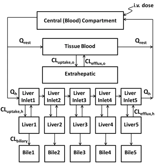
(C) liver-to-plasma ratio versus time curves; mean data by treatment group. ... 78 Figure 2.2. Semi-PBPK model scheme (A) representing 99mTc-mebrofenin disposition
in humans. Simulations based on the semi-PBPK model and observed blood, liver and bile curves for 99mTc-mebrofenin in subjects with quantitative scintigraphy data for
(B) subject 8 (control), and (C) subject 17 (2x300mg ritonavir). ... 80
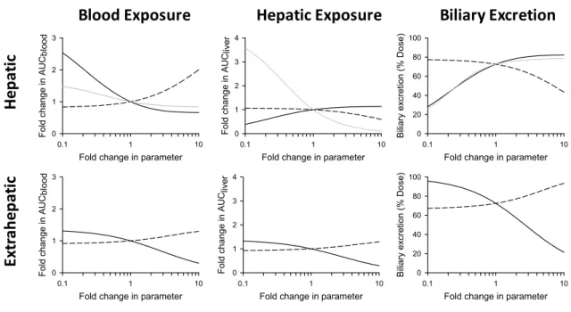
Figure 2.3. Sensitivity analysis of parameter estimates determined from the semi-PBPK model ... 82
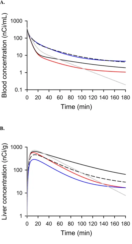
Figure 2.4. Simulations based on the 99mTc-mebrofenin semi-PBPK model scheme
(Figure 2) for 99mTc-mebrofenin (A) blood and (B) liver concentration-time data, and
(C) cumulative % dose excreted in bile ... 83
Figure 2.5. (A) 99mTc-Mebrofenin accumulation and biliary excretion index (BEI)
in cells+bile (closed bars) and cells (open bars) in the presence of increasing concentrations of extracellular ritonavir. 99mTc-Mebrofenin BEI values (above the bars) represent mean data.
(B) Ritonavir accumulation in hepatocytes (Ca2+-free HBSS) when co-administered
with 99mTc-mebrofenin, represented as intracellular total concentration (C
cell,total) ... 85
Figure 3.1. The tissue-to-plasma unbound concentration ratio (Kpu,u) applied to
hepatobiliary drug disposition. ... 116
Figure 3.2. Distribution and recovery of organelle-specific marker enzyme activity
following differential centrifugation of whole liver tissue (A) and SCH (B). ... 117
Figure 3.3. Subcellular distribution and recovery of ritonavir, rosuvastatin
and furamidine following differential centrifugation of whole liver tissue and SCH. ... 119
Figure 4.1. (A) Scheme depicting uptake/efflux protocol in sandwich-cultured hepatocytes (SCH). (B) Model schemes depicting the disposition of rosuvastatin in SCH studies. ... 145
Figure 4.2. (A) Scheme depicting the experimental protocol in isolated perfused livers (IPLs).
x
Figure 4.3. (A) Extracted ion current (XIC) chromatograph of rosuvastatin, rosuvastatin pentanoic acid, and d6-rosuvastatin in TR- Control liver tissue compared to blank liver
tissue (inset). (B) High resolution product ion spectra of rosuvastatin (top), d-rosuvastatin
(middle), and rosuvastatin pentanoic acid (bottom). ... 147
Figure 4.4. [3H]Rosuvastatin (RSV) mass versus time data in wild-type (WT) and
Mrp2-deficient (TR-) rat SCH in the absence (Control) or presence of GF120918
during the uptake and efflux phase………148 Figure 4.5. [3H]Rosuvastatin (RSV) mass versus time data in human SCH during the
uptake and efflux phase. ... 149
Figure 4.6. Rosuvastatin (RSV) biliary excretion (A. and B.) and outflow perfusate (C. and D.) rate versus time data in isolated perfused livers from wild-type
(WT: A. and C.) and Mrp2-deficient (TR-: B. and D.) rats in the absence
(open symbols; dashed line) or presence (closed symbols; solid line) of GF120918 ... 150
Figure 4.7. Recovery of rosuvastatin (solid bars) and rosuvastatin pentanoic acid (gray bars) in perfusate, liver and bile, as well as total recovery from wild-type (WT) and Mrp2-deficient
(TR-) isolated perfused livers in the absence or presence of GF120918. ... 151
Figure 4.8. ATP-dependent uptake of (A) 20 μM [3H]E
217G (MRP3) or (B) 2 μM [3H]DHEAS
(MRP4) in membrane vesicles from non-transfected (NT) and MRP3- or MRP4-overexpressing (control) HEK293 cells, respectively, and inhibition by 50 μM rosuvastatin (RSV) and MK571. (C) Time-dependent transport of [3H]RSV in membrane vesicles from NT (●) MRP3- (▲) and
MRP4-transfected (■) cells. (D) Concentration-dependent transport of RSV in membrane vesicles from MRP4-transfected HEK293 cells, and corresponding Vmax and Km values obtained from
11 CHAPTER 1
Introduction
Role of Hepatic Efflux Transporters in Regulating Systemic and Hepatocyte Exposure to Xenobiotics
Introduction
The liver is arguably the single most important organ for drug disposition, serving as the primary site of drug metabolism in the body, providing a “gatekeeper” function for systemic exposure following first-pass of portal blood, and being one of the major routes for final elimination of substances from the body. Compounds are presented to hepatocytes via sinusoidal blood, and gain access to the intracellular space by passive diffusion and/or active uptake mediated by transport proteins expressed on the
basolateral membrane. The propensity and relative contribution of these mechanisms depends largely on physicochemical properties of the compound. Once inside hepatocytes, the fate of compounds depends on susceptibility to the diverse strategies available for the cell to regulate concentrations of useful and potentially harmful chemicals constantly present in the cellular milieu. This includes subcellular sequestration and mechanisms of elimination, such as biotransformation (metabolism) and/or excretion. It has become clear that transport proteins are responsible for the hepatocellular disposition of highly polar or charged drugs and metabolites, which cannot exit hepatocytes at a sufficient rate without the aid of an active efflux mechanism.
This review provides a summary of transporter-mediated efflux of drugs and metabolites from
12
hepatocytes into sinusoidal blood and/or bile. The impact of altered transport function, resulting from drug-drug interactions (DDIs), genetic variation and disease states, on systemic and hepatic exposure is discussed. Related issues including multiplicity of transporters, interplay of metabolism and transport, and model systems used to elucidate the role of hepatic efflux on drug disposition are addressed and updated with recent contributions to this area of active research.
Canalicular Efflux
Canalicular efflux transporters include several well-known drug transporters such as ABCB1 (P-gp; MDR1), ABCC2 (MRP2), and ABCG2 (BCRP); in addition, ABCB11 (BSEP), ABCB4 (MDR3), ABCG5/G8, and SLC47A (MATE1) are responsible for bile acid, phosphatidylcholine, and sterol excretion(1-3). MRP2, BCRP, and P-gp are important determinants of biliary excretion of organic anions, organic cations, and Phase II xenobiotic products including glutathione, glucuronide, and sulfate conjugates.
MRP2 is responsible for the bile acid-independent portion of bile flow via secretion of reduced glutathione(1; 4). Additionally, MRP2 is responsible for biliary excretion of organic anions and sulfate-, glucuronide-, and glutathione-conjugates such as methotrexate, etoposide, and acetaminophen-glucuronide, as well as glucuronide and sulfate bile acid conjugates(4-6). Perhaps more importantly, MRP2 transports bilirubin conjugates into bile. Deleterious polymorphisms in the ABCC2 gene lead to the condition known as Dubin-Johnson Syndrome, which is characterized by hyperbilirubinemia despite normal secretion of bile acids via BSEP(5; 6). BCRP is a half transporter containing six transmembrane domains and one ATP-binding cassette in contrast to the common twelve transmembrane domains and two ATP-ATP-binding cassettes that comprise other ABC transporters(7; 8). Studies have shown that in order to be functional, BCRP most likely needs to homodimerize or oligomerize(7; 8). BCRP mediates the biliary excretion of sulfate and glucuronide conjugates and bile acids(4; 9). Additionally, BCRP appears to be responsible for the partial compensatory excretion of such conjugates in scenarios of diminished MRP2 function(6). P-gp transports a wide variety of substrates including immunosuppressants, antibiotics, steroids, anticonvulsants, and Ca2+
13
role in biliary excretion of drug conjugates, it is one of the major determinants of cation excretion into bile(6; 10). Unlike MRP2 and BCRP, P-gp is not considered as having a major role in the formation of bile flow(4).
Due to their wide substrate specificity, canalicular efflux transporters play a central role in limiting systemic exposure to xenobiotics and their conjugates. In the classic view, xenobiotics and conjugates are excreted into bile by canalicular transporters for delivery to the intestine and removal from the body via fecal excretion. However, some xenobiotics and conjugates that undergo biliary excretion evade fecal elimination, at least in part, by reabsorption in the intestine and re-delivery to the liver. This process, which may occur numerous times for a given compound, is known as enterohepatic recirculation (ER). ER can lead to a prolonged half-life of compound in the systemic circulation, and frequently is manifested by multiple peaks in the plasma concentration-time profile.(11; 12) ER often involves cleavage of phase II conjugates in the gut and reabsorption of parent drug, a process that can be facilitated by intestinal microflora.(12; 13) Drugs (or their metabolites) that are known to undergo ER to a significant extent include mycofenolic acid(14), glycyrrhizic acid(15), morphine(12), warfarin(12), amlodipine(16), indomethacin(17; 18), spironolactone(12), sulindac(12), sorafenib(19), valproic acid(20), and the irinotecan metabolite, SN-38(21).
14
(PFIC1), or ABCB4 (PFIC3). On the other hand, xenobiotic inhibition of BSEP can result in an acquired cholestatic condition, which may also lead to severe hepatocellular damage, toxicity, and liver failure, known as drug-induced liver injury (DILI), which will be discussed below(2; 23).
Basolateral/Sinusoidal Efflux
Basolateral efflux commonly is regarded as a compensatory route of elimination to protect hepatocytes in the setting of cholestasis or otherwise impaired biliary excretion, by mediating excretion of endo- and xenobiotics from hepatocytes into the sinusoidal blood. Transport proteins that have been shown to mediate basolateral efflux of drugs are limited primarily to MRP3 and MRP4. These
transporters serve also to facilitate systemic exposure and resulting efficacy, toxicity and/or non-hepatic (e.g., renal) elimination of metabolites that are formed in the liver. This is particularly true of phase II metabolites such as sulfate, glucuronide and glutathione conjugates(6). A classic example is
acetaminophen, which undergoes extensive conversion to glucuronide and sulfate conjugates that are recovered largely in urine, and are known to be substrates for MRP3 and/or MRP4(6). Another example is enalaprilat, the dicarboxylic acid and active metabolite of enalapril, which is formed primarily in the liver and relies on active transport for efflux into the systemic circulation(24). Additionally, increased systemic concentrations of the pharmacologically active phase II metabolite, ezetimibe-glucuronide, correlated with increased expression of Mrp3 and altered localization (internalization) of Mrp2 in a rodent model of NASH(25). This shift from biliary excretion to plasma by basolateral efflux transporters diverts the active ezetimibe-glucuronide metabolite away from the site of action in the intestine, and illustrates a mechanism of altered efficacy as a result of disrupted hepatobiliary efflux. Basolateral efflux transporters also may play a role in the disposition of parent drugs, such as fexofenadine. The loss of Abcc3 (but not Abcc4) in mice reduced outflow perfusate recovery in the isolated perfused liver system(26). Another example is methotrexate, which will be discussed under Interplay of Multiple Hepatic Efflux Systems (below).
15
glucuronides by Schinkel and colleagues(27). The interplay of Oatp-mediated hepatic uptake and efflux by Mrp2 and Mrp3 was investigated by observing alterations in the disposition of bilirubin glucuronides in single- and multiple-knockout mice(27-29). These studies elegantly challenged the prevailing perception that extensive biliary excretion of bilirubin glucuronides results from efficient vectorial transport from sinusoidal blood to bile on the level of an individual hepatocyte. The relative contribution of basolateral vs. canalicular excretion may be much greater than previously thought. Rather than efficient excretion from the cellular space into bile, it is possible that multiple rounds of less efficient cycling, or “hepatocyte hopping,” along the length of the liver sinusoid result in extensive biliary excretion. In this way, basolateral efflux was suggested to be a major contributor to the efficiency of the liver as a “flexible and robust” organ of detoxification, “potentially distributing the biliary excretion load over all hepatocytes within the liver lobule”(27).
A number of other efflux transporters are known to be expressed on the basolateral/sinusoidal membrane of hepatocytes, including ABCC5 (MRP5) and ABCC6 (MRP6). Although expression levels are generally low in healthy individuals, there is evidence of upregulation in disease states(30; 31), suggesting that these proteins may play an important compensatory role in the setting of impaired biliary excretion pathways, or otherwise compromised hepatocellular function. Although a number of drugs and endogenous compounds have been identified as substrates and/or inhibitors in isolated expression
systems(32), there are no known examples where these transport proteins play a significant role in the disposition of compounds in vivo. It may be that these proteins are responsible (in part or in whole) for the significant “residual” basolateral efflux of sulfate and glucuronide conjugates observed in Abcc3-/-,
Abcc4-/- and Abcc3-/-/Abcc4-/- animals(33).
In addition to the unidirectional, ATP-driven ABC family transporters, a number of facilitated diffusion mechanisms exist that can participate in uptake or efflux depending on the electrochemical gradient of substrates and co-substrates. One example is SLC51 (polyspecific organic solute carrier, OSTα/β), which is thought to be involved in bile acid and sterol reabsorption under normal
16
limited currently to bile acids, steroid conjugates and eicosanoids, but the ability to transport digoxin suggests that further characterization of potential drug substrates is needed. It has been suggested that the SLC isoforms (OATPs, OATs and OCTs) are capable of bidirectional transport under certain
conditions(38; 39), which would open the door to a large number of well-known substrate drugs, including the statins. However, the role of bi-directional transport by OATPs and other SLC family members is controversial and has not been shown definitively to play a role for drug molecules.
Functional Impairment Mechanisms
Drug Interactions Involving Hepatic Efflux Transport
Inhibition of hepatocellular efflux activity affects normal hepatic function, including excretion of endogenous substances such as glutathione, bile acids and bilirubin, and can alter the pharmacokinetic profile of drugs(40). Therefore, affinity of drug candidates for hepatic efflux transporters may represent a liability to serve as victims and/or perpetrators of drug interactions(40). For example,
clinically-significant differences in mycophenolic acid (MPA) pharmacokinetics are observed when this medication is co-administered with the calceiunurin inhibitors tacrolimus or cyclosporine A (CsA) for maintenance of immunosuppression following solid organ transplant(14; 41). MPA is metabolized primarily to the inactive phenolic glucuronide conjugate, MPAG, which is excreted into bile via MRP2, hydrolyzed back to MPA in the gut, and undergoes ER(14; 41; 42). Inhibition of MRP2-mediated biliary excretion and decreased ER explains some of the ~50% decrease in MPA trough concentrations in patients
17
decreased the biliary excretion of DPDPE (a metabolically stable opioid peptide) in isolated perfused livers from Abcc2-/Mrp2-deficient TR- rats(50). P-gp inhibition may contribute to other DDIs, such as
the reduced biliary excretion of colchicine, doxorubicin and digoxin by erythromycin or ketoconazole(44; 45). The role of Mrp2 and Bcrp as the primary mechanisms for biliary excretion of rosuvastatin was confirmed in rat livers from wild-type and TR- rats in the presence and absence of GF120918, which had
been suggested previously using in vitro systems and single knockout animals.(51; 52) A number of drugs have been shown to inhibit biliary excretion of conjugated bile salts mediated by BSEP, which can result in DILI, as demonstrated by several cases of drug withdrawals or black-box warnings (e.g., troglitazone, bosentan, erythromycin, nefazodone)(53-58). In addition to BSEP, MRP3 and MRP4 also are involved in the export of bile acids and drugs/metabolites. Recent studies suggest that inhibition of MRP4 is a risk factor for the development of DILI; considering the potential for drug candidates to inhibit MRP4, in addition to BSEP, could aid in predicting compounds with liability for DILI at an earlier stage in drug development(59).
For lists of compounds that inhibit hepatic efflux transporters, the reader is directed to a number of excellent reviews that have collated available data for individual ABC family transport proteins: MRP2(5; 32; 60), BCRP(7; 60-62), P-gp(60; 63), BSEP(64), MRP3(6; 32) and MRP4(6; 32). These reviews typically include information such as the model system that was used to generate inhibition data, the probe substrate, and a mathematical value corresponding to the inhibitory potential of the compound of interest, such as a percent inhibition at a given concentration, IC50 or Ki. These data tend to be
collected from reports of individual or small sets of inhibitors generated in different experimental
systems/conditions. As such, it can be difficult to compare the relative inhibitory potential of compounds from different sources. In addition to these reviews, several large-scale, public, web-based efforts are underway to curate the historic and growing body of experimental data reporting interactions between chemicals and transport proteins. These databases include “TP-Search” (www.tp-search.jp)(65), the Transporter Classification Database (TCDB; www.tcdb.org)(66), TransporterDB
18
(pharmacogenetics.ucsf.edu)(68). A large-scale QSAR modeling effort was reported recently by Tropsha and colleagues, which included automated curation of the aforementioned databases and literature sources, resulting in the largest known repository of transporter inhibition data(69).
Mechanisms of DDIs Involving Hepatic Efflux Transporters
The vast majority of data describing transporter DDIs suggest a competitive inhibition mechanism, resulting from the aforementioned wide and overlapping substrate specificity of transport proteins. However, drugs may interact with transport proteins by a variety of mechanisms and, therefore, cis-inhibition as well as mechanism-based and trans-inhibition, allosteric-induced modulation and induction of transport function may be important mechanisms of transporter-mediated DDIs(70). For example, trans-inhibition of BSEP has been demonstrated for estrogens, possibly contributing to cholestasis of pregnancy(71; 72). Time-dependent inhibition of hepatic uptake transporters has been shown with cyclosporine A(73; 74). Of the multiple binding sites proposed for P-gp, some are reported to interact in a positive cooperative manner, resulting in mutual stimulation of P-gp-mediated transport of several substrates, including rhodamine 123, anthracyclines, some flavonoids, and the p53 inhibitors QB102 and QB11(75). Similarly, in 2003, two groups independently reported potentiation of estradiol-17-β-D-glucuronide transport by MRP2, which was followed by additional reports from large-scale screening approaches(76). Another classic mechanism leading to increased transport activity is induction. Treatment of cultured primary rat hepatocytes with the chemical carcinogen 2-acetylaminofluorene (2-AAF), the antineoplastic drug cisplatin, and the protein-synthesis inhibitor cycloheximide led to a dose-dependent and time-dose-dependent increase in gene expression of Mrp2, with corresponding plasma
19
elimination(45). Although many of these atypical mechanisms have been observed only in in vitro systems, it is clear that competitive inhibition should not be regarded as the only mechanism of DDIs resulting in modulation of transporter function/activity.
Effect of Genetic Polymorphisms on Transporter Expression and Function
Several studies have investigated the impact of transporter polymorphisms on drug
pharmacokinetics; however, difficulty exists in demonstrating reproducible functional effects in vitro with successful clinical translation. A brief description of selected polymorphisms in specific efflux
transporters, and some of the controversies surrounding them, will be discussed. For a more detailed review of transporter pharmacogenetics, the reader is directed elsewhere.(5; 10; 79-81)
ABCC2/MRP2
ABCC2, next to ABCB1, is one of the most illustrious of the efflux transporters with regard to pharmacokinetics. Several polymorphisms in the ABCC2 gene locus have been associated with the hyperbilirubinemic disorder known as Dubin-Johnson Syndrome including the R412G, I1173F, R1392del, and M1393del protein variants(5). Of those associated with Dubin-Johnson Syndrome, the I1173F, R1392del, and M1392del variants lead to improper trafficking of MRP2 protein to the apical membrane of hepatocytes and loss of transport function(82; 83). Two independent groups identified the 2366C>T (S789F) and 4348G>A (A1450T) alleles as conferring decreased ATPase and transport activity, along with altered membrane localization(84; 85). In contrast, the 1249G>A (V417I) allele was shown to decrease affinity(85), or have little effect on transport function in vitro.(84) Additional in vitro
experiments discovered a reduction in transport activity for the K324A, K483A, R1210A, and R1257A MRP2 protein variants as well as diminished protein expression of the K578A variant(86). The 4544G>A allele has been associated with susceptibility to nonalcoholic fatty liver and cholestatic liver diseases, resulting in decreased ATPase activity and diminished transport of lopinavir, calcein, and
-1774Gdel/-20
1549G>A/-24C>T haplotype appears to reduce promoter activity(89). In contrast, the -24C>T allele alone and within the -24C>T/-1019A>G/-1549G>A haplotype leads to an increase in promoter activity(90). Additional ABCC2 haplotype studies investigating the -24C>T, 1249G>A, and 3972C>T alleles identified the -24C/1249A/3972C haplotype as conferring an increase in MRP2 protein levels, which corresponded to transport activity in vitro and altered bioavailability of talinolol in vivo(91). In contrast, the -24C/1249G/3972T, -24T/1249G/3972C, and -24T/1249G/3972T haplotypes resulted in diminished protein expression in vitro(91). Still others have shown a preference for transcription of the 3972C allele in comparison to 3972T, which did not correlate with hepatic or renal MRP2 expression(90).
ABCC3/MRP3
In 2004, Lang and colleagues determined that homo- and heterozygous carriers of the -211C>T variant exhibited decreased hepatic mRNA expression of ABCC3, and that this promoter variant affected binding of transcription factors(92). The investigators concluded that the -211C>T allele affected hepatic expression of the transporter, although this effect has been controversial(93). Additional studies using reporter gene activity experiments discovered that the length of the promoter, rather than the -211C>T variant affected gene expression(94). A later study discovered improper post-translational processing and localization of the MRP3 R1381S protein variant in vitro(95). Additionally, investigators identified the MRP3 S346F and S407N protein variants to confer decreased transport activity in Sf9 membrane
vesicles, the three polymorphisms were postulated to be possible risk factors for hepatotoxicity(95). In an effort to translate in vitro polymorphism studies to in vivo pharmacokinetics, Sasaki, et al., utilized a reporter gene assay to identify decreased transcriptional activity of the -1767A>G allele(96). However, the -1767A>G variant did not appear to affect human hepatic ABCC3 mRNA expression, nor was it associated with 4-methylumbelliferone-glucuronide pharmacokinetics(96).
ABCC4/MRP4
21
transfected cell system(97); however, the clinical significance of these variants has not been determined. Others identified the -1393T>C allele to be associated with improved event-free survival and decreased methotrexate plasma concentrations in pediatric acute lymphoblastic leukemia as opposed to an
association between the 934A>C allele with decreased event-free survival and increased toxicity(98). In contrast, a later study tested the results of Ansari, et al., in adult ALL patients and found no association between event-free survival and genotype(99). The impact of age of disease-onset and the discrepancies in these two findings have not been determined.
ABCB1/P-gp
The majority of the >100 ABCB1 polymorphisms have been studied in the context of gut expression of P-gp protein and altered bioavailability of drugs such as digoxin, talinolol, and
fexofenadine(10; 100). However, Elens, et al., investigated ABCB1 polymorphisms in ~150 liver donors and their subsequent effects on blood and hepatic tacrolimus concentrations(101). This study found that ABCB1 polymorphisms, specifically the 1199G>A and 2677G>T/A alleles, independently exerted a significant effect on tacrolimus liver, but not plasma concentrations, the primary endpoint used for therapeutic monitoring(101). Owen, et al., when investigating the 3435C>T and 2677T>G alleles determined that the large variation observed in human hepatic ABCB1 mRNA and P-gp protein
expression could not be attributed to genetic variation alone(102). In contrast, Song, et al., identified an increase in hepatic ABCB1 mRNA for homozygous carriers of the 3435C allele, and a decrease in those homozygous for the 2677T allele(103).
ABCG2/BCRP
22
BCRP protein in vitro(105). The Q141K variant also has been implicated in improper membrane localization of BCRP protein(106).
Despite major advances in the field of molecular biology and pharmacogenomics, the ability to translate in vitro polymorphic effects on transporter expression and function to the clinic remains a daunting task.
Expression of Hepatic Efflux Transporters in Disease States
Historically, investigations to determine the effect of liver disease on efflux transporter
expression have sought to provide insight to disease pathogenesis. However, alterations in the expression of efflux drug transporters can have significant effects on the elimination of xenobiotics and endogenous molecules. It has been proposed that upregulation of efflux transporters may serve as an adaptive
mechanism to limit cellular exposure to hepatotoxicants(107); however, systematic, comprehensive investigations of efflux transporter expression are severely limited for several disease states. The following will provide a brief overview of efflux transporter expression in human liver tissue from selected liver diseases. A summary of the effects discussed is provided in Table 1 below to aid in comparisons among diseases.
Primary Biliary Cirrhosis
Primary biliary cirrhosis (PBC) arises from inflammation and irritation of bile ducts leading to stasis of bile flow (cholestasis) which can severely damage hepatocytes and ultimately lead to cirrhosis. Much work has been done to understand the effects of cholestasis on transporter expression and function. ABCC2 mRNA remains largely unchanged in PBC compared to normal liver(107-109). While some have reported no change in ABCC4 mRNA(110), others have discovered an increase in ABCC4 mRNA in cholestasis(111). There is overall agreement that MRP3 and MRP4 protein are elevated in
23
Nonalcoholic Fatty Liver Disease & Nonalcoholic Steatohepatitis
Nonalcoholic fatty liver disease (NAFLD) and nonalcoholic steatohepatitis (NASH) represent a spectrum of liver damage that has emerged recently as a prevalent condition due to its association with the obesity epidemic. It is characterized by the accumulation of lipids within hepatocytes, termed simple steatosis. NAFLD progresses via inflammation, oxidative stress, and increased lipid accumulation to NASH, which also may incorporate fibrosis and eventual cirrhosis(31). ABCC1, ABCC3, ABCC4, ABCB1, and ABCG2 mRNA levels are increased with progression of NAFLD from simple steatosis to NASH, while ABCC2 mRNA levels remain unchanged(31). Protein levels of all efflux transporters appear to be increased with disease progression, although the localization of MRP2 most likely shifts to the subapical region, away from the plasma membrane(31).
Hepatitis C Virus
Hepatitis C virus (HCV) induces several response pathways in hepatocytes including oxidative and endoplasmic reticulum stress(112). Chronic HCV infection can lead to fibrosis and development of severe cirrhosis, propagating hepatocellular injury with potential for numerous alterations in gene expression patterns(113). In comparisons of HCV-positive tissue with HCV-negative, ABCC1, ABCC3, ABCC4, and ABCB1 mRNA are elevated in non-cirrhotic tissue(113; 114). ABCG2 and ABCC2 mRNA expression is diminished in HCC(113; 114) with ABCC2 mRNA decreasing as fibrosis severity
increases(112). Studies comparing mRNA expression in cirrhotic HCV tissue with HCV-negative, noncirrhotic tissue are in agreement with those comparing HCV-positive to noncirrhotic, HCV-negative tissue, suggesting that the changes in gene expression are driven primarily by infection status. ABCB1, ABCC1, ABCC4 mRNA is elevated, while ABCC2 and ABCG2 levels are decreased(114). Additionally, ABCC2 mRNA has been demonstrated to be reduced in HCV/HCC-positive tissues compared to
24 Hepatocellular Carcinoma
Hepatocellular carcinoma (HCC) is a common form of cancer that arises from cirrhosis due to chronic liver disease(116). Studies have shown an increase in mRNA levels of several efflux transporters including ABCC1, ABCC2, ABCC4, and ABCB1(116; 117). ABCG2 mRNA remains unchanged in comparison to healthy adjacent tissue(116). However, ABCC3 mRNA increased(116) or remain
unaltered(117). Information regarding protein levels of efflux transporters in HCC as compared to normal tissue remains limited.
Progressive Familial Intrahepatic Cholestasis.
Progressive familial intrahepatic cholestasis (PFIC) refers to a group of autosomal recessive disorders affecting biliary membrane proteins including ATP8B1, BSEP, and MDR3, referred to as PFIC1, 2, and 3, respectively. The resultant dysfunction in each generally culminates in disrupted formation of bile. Investigations assessing the expression of efflux transporters in PFIC are limited to small groups of patients. Keitel, et al., determined mRNA levels in PFIC 2 and 3 for a number of efflux transporters and discovered an increase in ABCC4 mRNA in both PFIC2 and 3, while protein was increased in PFIC3(118). ABCC1 and ABCB1 mRNA was increased in PFIC3 as well(118). ABCC2 mRNA was unaltered in PFIC2 and 3, but protein may be decreased in PFIC3(118). ABCC3 mRNA was unaltered in both PFIC2 and PFIC3(118). Unfortunately, due to low patient numbers (n=4-5) and thus low statistical power, generalizations on efflux transporter expression due to PFIC are tentative at best.
Though systematic studies including both mRNA and protein data for efflux transporters are minimal, a significant obstacle in the application of findings from such studies remains the ability to absolutely quantify transporter protein. Understanding the absolute expression level of one specific efflux transporter compared to another may provide better insight regarding which transporters may exert a more dominant function in various disease states and enable better prediction of drug response.
Trafficking of Hepatic Efflux Transporters
25
physiological demand(119). It has been postulated that these intracellular reservoirs of transporters serve as a means of increasing transporter expression in the membrane, and thus transport activity, without initiating the energetically expensive processes of transcription and translation(120). In contrast, other studies have demonstrated the endocytic retrieval of transporters from the membrane as a consequence of xenobiotic treatment or disease states. Whether changes involve transporter delivery to, or internalization from, the membrane, the net result is alteration of the steady-state levels of efflux transporters residing in the membrane, and thus modulation of their transport function. However, the majority of such studies have focused on canalicular efflux transporters including P-gp and MRP2, with less attention directed towards sinusoidal efflux transporters.
Much work has been done to determine the mechanisms governing P-gp trafficking from large intracellular pools to the canalicular membrane. Administration of cAMP or taurocholate to rats results in increased levels of hepatic P-gp in the membrane(120-122). Furthermore, in vivo pulse-chase
experiments revealed that newly-synthesized P-gp is targeted directly from the Golgi apparatus to the canalicular membrane rather than progressing through the transcytotic pathway via temporary insertion in the sinusoidal membrane(123). Additional studies in HepG2 cells revealed the involvement of protein kinase A (PKA)-RIIα, which can be activated by cAMP, in the trafficking of newly-synthesized P-gp to the membrane(124). Interestingly, the basal rate of P-gp delivery to the membrane in HepG2 cells differed from that of MRP2(124). Non-hepatic cell lines have provided further insight into the proteins responsible for regulating the insertion into and endocytic retrieval of ABCB1/P-gp from the membrane via perimembranous pools. Concomitant overexpression of green fluorescent-tagged P-gp in HeLa cells with constitutively-active forms of the small GTPases Rab5 or RalA resulted in an increase in
26
cells, Rab4-mediated retrieval of P-gp correlated with increased intracellular levels (reduced efflux) of an P-gp substrate(126).
Extensive investigations have focused on the dynamic regulation of MRP2 in the canalicular membrane. Several experimental cholestatic and oxidative stress-inducing conditions such as bile duct ligation or treatment with estradiol-17β-D-glucuronide, lipopolysaccharide, phalloidin, ethacrynic acid, or buthionine sulfoximine, are known to induce endocytic retrieval of Mrp2 from the canalicular membrane in rats(127-129). Additionally, disrupted canalicular localization of MRP2 has been demonstrated in primary biliary cirrhosis, drug-induced liver injury, poorly drained obstructive jaundice, autoimmune hepatitis, sclerosing cholangitis, and nonalcoholic steatohepatitis patients(31; 130; 131). The specific mechanisms governing the endocytic retrieval and exocytic insertion ofMRP2 in the canalicular
membrane are convoluted and involve multiple kinases. Ethacrynic acid-induced oxidative stress resulting in depletion of glutathione leads to rapid internalization of Mrp2 which can be rescued by subsequent glutathione treatment(127; 132). Estradiol-17β-D-glucuronide-induced cholestasis also causes internalization of Mrp2 resulting in diminished biliary excretion of Mrp2 substrates; however, this process is reversed by cAMP administration(133). The endocytic retrieval of Mrp2 from the membrane is largely a microtubule-independent process involving activation of Ca2+-dependent protein kinase C
(PKC), and de-phosphorylation of radixin, which is responsible for tethering actin filaments to membrane proteins(128; 134-137). De-phosphorylation of radixin destabilizes its interaction with Mrp2 leading to their dissociation and movement away from the membrane(134; 136). Once internalized,
phosphatidylinositol 3-kinase (PI3K) has been proposed to regulate Mrp2 retention within the cell via interactions with extracellular signal-regulated kinases 1 and 2(137; 138). Furthermore, sustained buthionine sulfoximine-induced oxidative stress has been shown to lead to not only internalization of Mrp2, but increased ubiquitination and subsequent degradation of the protein(129). Conversely,
27
also have been implicated in the exocytic reinsertion of Mrp2 in the membrane through signaling events involving p38 mitogen activated protein kinase(132; 134; 138; 139).
Glycosylation has been implicated as an additional factor affecting the membrane localization of efflux transporters. N-linked glycosylation of BCRP has been implicated as a possible mechanism affecting protein stabilization, proper routing of the transporter to the membrane, and functional
activity(140-142). Others have shown full glycosylation of MRP2 to be necessary for proper localization within the membrane and effective transport activity(143; 144). The interconnection of glycosylation pathways regulating transporter routing to the membrane and kinase pathways involved in the dynamic localization of efflux transporters remains to be determined.
Localization studies investigating sinusoidal efflux transporters are beginning to emerge; however, there is a substantial gap between the breadth of knowledge of canalicular efflux transporter trafficking and that of sinusoidal efflux transporters. Recent studies have identified Na+/H+ exchanger
regulatory factor 1 (NHERF1) and nexin 27 as binding partners of MRP4 necessary for internalization of the transporter(145; 146). Further studies are necessary to determine the pathways involved and clinically relevant states of MRP4 internalization. Nonetheless, whether reduced membrane expression of efflux transporters is the result of improper post-translational processing and trafficking to the membrane or endocytic retrieval and intracellular sequestration, the net result is diminished extrusion of substrates out of the hepatocyte.
Interplay Between Multiple Hepatic Efflux Systems
28
involvement of multiple transporters, it can be challenging to predict the impact of altered function of one or more transport proteins on hepatic and systemic exposure of a given substrate.
The quantitative impact of excretory transport modulation on the hepatic, systemic, and biliary exposure to xenobiotics and derived metabolites was formalized recently by Zamek-Gliszyznski based on work conducted in our laboratory(147). Experimental data and theoretical relationships indicated that the fold change in exposure is governed by the relationship, 1/(1 – fe), where fe is the fraction excreted by a
particular transport protein. Loss-of-function of a transport pathway associated with a fe < 0.5 will have
minimal consequences on exposure, but exponential changes can be expected in response to loss-of-function of one or more transport pathways with fe > 0.5. It should be noted that the impact of fe on
exposure depends on the compartment of interest (systemic, hepatic or bile), the directionality of the impaired pathway (canalicular or sinusoidal), and the existence of an alternative route of excretion. For example, when a transport protein is impaired, the increased excretion in the alternative direction is a function of the fraction of total hepatocellular clearance mediated by the impaired pathway. The resulting increase in excretion over the alternative membrane is caused by the decrease in total excretory clearance, which produces elevated cellular concentrations that serve as a driving force for increased excretion by the remaining available pathways. The relationship between exposure and fe also can be extended to
partial inhibition scenarios by modifying the equation with the ratio of inhibitor concentration and inhibition constant ([I]/Ki). Figure 1.2 depicts the relationship between exposure and fe, including the
effect of partial inhibition(147).
Fold change in exposure fe 1 (1 +[I] Ki) + (1 - fe)
(1)
In a series of reports, Vlaming and colleagues characterized the role of multiple hepatic efflux transporters in the elimination of methotrexate and its main toxic metabolite, 7-hydroxymethotrexate, in vivo using single and multiple knockout mouse models(148-150). Biliary excretion of methotrexate and the 7-hydroxy metabolite were reduced ~20-fold in Abcc2:Abcg2-/- mice, indicating that these proteins
29
methotrexate and 7-hydroxymethotrexate from the liver into sinusoidal blood was demonstrated by increased plasma concentrations and urinary excretion in the absence of Mrp2 and/or Bcrp, which was reversible in Abcc2:Abcc3-/- mice(150). Mrp3 appeared to be required for increased plasma
concentrations in the absence of Bcrp(148). Despite initial hepatic accumulation of methotrexate in the absence of one or two proteins, the mass recovered in was similar to wild-type by 2 hours. However, in the absence of all three proteins, hepatic accumulation was further increased and prolonged in
Abcc2:Abcg2:Abcc3-/- mice (7- and 90-fold increase for parent and metabolite at 2 h)(148). Also in
Abcc2:Abcg2:Abcc3-/- mice, 7-hydroxymethotrexate still exited the liver, resulting in a 10-fold increase
in plasma exposure, potentially due to up-regulation of Mrp4. The role of multiple efflux transporters in the enterohepatic circulation and related efficacy of ezetimibe and its glucuronide conjugate was explored using a variety of model systems, demonstrating that enterohepatic circulation strongly depends on the joint function of Mrp3, Mrp2 and Bcrp(151). Although biliary excretion of ezetimibe glucuronide was minimally altered in Abcg2-/- mice, Bcrp clearly functions to compensate for loss of Mrp2, as
demonstrated in Abcc2:Abcg2-/-(151). Interestingly, the role of Mrp3 in absorption of ezetimibe from the
intestine was shown in Abcc3-/- mice, which displayed greatly reduced enterohepatic circulation(151).
The overlapping function of P-gp, MRP2 and MRP3 in protection against trabectedin-mediated hepatotoxicity by clearing harmful metabolites formed by CYP3A was shown using a combination of single- and multiple-knockout animals with translation to humans using MRP2- and MRP3-transfected MDCK cells(152; 153). Multiple efforts to characterize the hepatobiliary disposition of fexofenadine have shown the overlapping roles of P-gp, BSEP and MRP2, but not BCRP in facilitating biliary
excretion, and the significant contribution of MRP3 but not MRP4 in the sinusoidal efflux from liver into blood(26; 154-156). Using a combination of Mrp2-deficient TR- rats and chemical “knockdown” of
30
concentrations of valproate glucuronide were observed in isolated perfused rat livers following probenecid and valproate co-administration.(157) Mathematical modeling of the data revealed that probenecid inhibited both the basolateral and canalicular excretion of generated valproate glucuronide.
For most drugs where multiple pathways are available for hepatocellular egress into bile or blood, the safety is maintained when only one pathway is impaired. However, as demonstrated by the fe concept
and various experimental models used to study multiple transporters, significant changes begin to occur as more pathways are impaired and the hepatoprotective effect of transporter multiplicity is diminished. That is why it is important to characterize the multiplicity and relative contribution of transport pathways for new chemical entities (NCEs), as a way to asses risk in the setting of impaired function of one or more transporters, whether due to DDIs (with NCE as victim and/or perpetrator), disease states or genetic variation.
Relationship Between Hepatic Efflux Transport and Intracellular Unbound Concentrations Measurement of unbound blood or plasma concentrations is convenient and may correlate with unbound tissue/cellular concentrations for compounds with sufficient passive permeability. However, for compounds that are poorly permeable or otherwise violate the assumption of rapid equilibration, the ratio of intracellular to extracellular (blood or plasma) Cunbound (Kpu,u) may be highly disparate. Since the liver
is frequently a primary site of drug elimination from the body, hepatocellular Cunbound greatly influences
drug efficacy and toxicity on a systemic as well as local level. As such, predictions of clinical efficacy, toxicity and drug-drug interactions (DDIs) could be improved by accounting for Cunboundin vitro and in
vivo, as well as between cell-free and whole cell in vitro systems used to assess mechanisms/targets of efficacy, toxicity and drug disposition, including transporter activity.
Altered function of efflux mechanisms described in this review can influence hepatocellular exposure of drugs, with implications for altered efficacy, toxicity and DDI potential. This includes the effect of interplay between drug transport and metabolizing enzymes, in which intracellular Cunbound drives
31
of impaired efflux, leading to increases in hepatic and/or systemic metabolite exposure.(6; 147; 158-161) In contrast, subcellular binding/sequestration can limit access of drugs to mechanisms of elimination and constrain the ability of efflux and/or metabolism to “clear” drug from the hepatocytes. It is assumed that only the Cunbound within the cell is available for elimination.
Although hepatocellular Cunbound is assumed to be the relevant concentration driving elimination, a
number of exceptions exist, particularly in the case of efflux transport. A preponderance of evidence suggests that ABCB1/P-gp has multiple binding sites, including the ability to remove compounds from the inner leaflet of the plasma membrane and function as a “membrane vacuum” for compounds that partition into the lipid bilayer.(162; 163) An elegant piece of pharmacokinetic modeling by Korzekwa, et al., supports this mechanism, and suggests that a portion of the ABCB1/P-gp-mediated efflux process is driven by drug accumulation and concentration within the membrane, although this would not be
regarded as a component of Cunbound within the cell.(164) As integral membrane proteins, transporters are
transferred to the plasma membrane following synthesis through dynamic and complex vesicle trafficking pathways.(123; 165-168) Likewise, plasma membrane regions and their contents are continuously cycled on and off the membrane, leading to pools of transporter-containing vesicles.(122; 169) As a result, substrates for efflux transporters are constantly accumulating within these vesicles, as has been shown with fluorescent ABCC2/MRP2 substrates, demonstrating characteristic punctate vesicular structures, which are absent in Abcc2-/Mrp2-deficient cells and sensitive to known inhibitors, such as cyclosporine A.(169-171) Although the initial transport into these vesicles is presumed to be driven by intracellular Cunbound, the terminal release (efflux) of accumulated drug results from fusion of the vesicles with the
32 Model Systems for Evaluating the Role of Hepatic Efflux
For a comprehensive list of experimental models used to measure transporter activity in general, the reader is referred to a recent review by the International Transporter Consortium(173). While vesicle systems and transfected systems are useful to determine whether a transporter may be involved for a particular substrate, the more challenging aspect is to determine the role of a particular transporter in the presence of other competing transporters and pathways of cellular elimination. The only cells that truly express all of the relevant transporters of interest are primary cells from the organ of interest.
Primary Hepatocytes
Freshly isolated or cryopreserved primary hepatocytes are not polarized and require long-term culture in a sandwich configuration to recapitulate polarized architecture, including formation of bile networks and appropriate expression and localization of efflux transporters on the respective apical (canalicular) and basolateral membrane domains.(174) Sandwich-cultured hepatocytes (SCH) typically are used to evaluate vectorial transport into bile and differentiate the roles of active uptake and biliary excretion. This is accomplished by modulating tight junctions in the presence (cells+bile) or absence (cells) of divalent cations (Ca2+ and Mg2+) in the experimental buffer system (B-CLEAR® technology, Qualyst
Transporter Solutions, Research Triangle, NC). These studies are performed routinely by applying substrates in the presence or absence of inhibitors for a period of 10 min, and a number of
pharmacokinetic parameters can be calculated from the resulting data, including the total accumulation in cells+bile and cells, biliary excretion index (BEI) and in vitro clearance values for uptake and/or biliary excretion, which can be scaled to predict in vivo clearance values based on scaling factors such as protein content and hepatocellularity(174).
33
efflux transporters such as P-gp and Mrps(175-178). Presently, estimation/prediction of transport processes may be improved by taking these changes into account upon scaling of data from in vitro to in vivo(179; 180). Less frequently, the SCH model has been used to assess efflux by various strategies of preloading the cells with substrates (± inhibitors), followed by a brief wash and application of “blank” buffer for an efflux phase, which may involve sampling/observation of substrate in cells+bile, cells and/or appearance in buffer(181; 182). However, the SCH system remains to be characterized under these conditions.
In a “simplified” conception of the SCH system, maintenance of tight junctions in the presence of Ca2+ would completely seal the bile networks, and appearance in buffer during an efflux experiment
would reflect only basolateral efflux. Efflux in the absence of Ca2+ with disrupted tight junctions would
reflect basolateral plus biliary efflux, and would be greater than that in +Ca2+ conditions. In reality, the
SCH model is dynamic, with regular and extensive “pulsing” of the bile canaliculi, similar to reports in hepatocyte couplets and cultured hepatocytes(183; 184) (video:
http://www.pharmacy.unc.edu/research/labs/kim-brouwer-lab/bile-canalicular-contractions-in-sandwich-cultured-rat-hepatocytes) This means that appearance of substrate in buffer in the +Ca2+ condition
reflects basolateral efflux plus the flux of accumulated substrate within the bile spaces. In order to deconvolute these data and elucidate the relative contribution of basolateral versus biliary efflux, one can apply a pharmacokinetic modeling approach to the SCH data(161; 185-188).
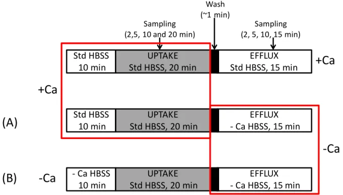
At least two potential schemes exist for the design of efflux studies in SCH (Figure 1.3). One is to pre-incubate and/or perform the uptake phase exclusively in standard (+Ca2+) Hanks’ Balanced Salt Solution
(HBSS), followed by a brief wash and efflux in ±Ca2+ HBSS [Fig. 1.3, (A)](181; 182). A potential
limitation of this approach is that substrate accumulation in the bile networks during the uptake phase may not be washed away completely before initiating the efflux phase in -Ca2+ HBSS. The time required
34
incubate in ±Ca2+ HBSS long enough to complete opening/dumping of bile spaces in -Ca2+ (~5-6 min),
and then replace buffer again and observe efflux. However, it may be difficult to detect cellular content for compounds that are rapidly excreted (e.g. taurocholate)(186). Another potential experimental design for efflux studies in SCH involves maintaining tight junction modulation throughout the study period by pre-incubating in ±Ca2+ HBSS, then performing an uptake phase in +Ca2+ HBSS (<30 min) to provide
relief from -Ca2+/EGTA , followed by a brief wash and efflux in ±Ca2+ HBSS [Fig. 1.3, (B)]. A limitation
with this method is potential re-sealing of the tight junctions during Ca2+ repletion in the uptake phase.
This has been characterized previously(186), and is of concern for compounds with extensive biliary excretion (BEI), such as taurocholate(186). However, imaging with CDF suggests that re-sealing of the tight junctions allowing substrate accumulation in the bile networks is minimal over a 20-min
loading/uptake phase (Figure 1.4). Another potential issue relates to cell integrity in prolonged and repeated -Ca2+ conditions; this does not appear to be an issue in rat SCH as judged by LDH release (<5%
of total cellular content over the study period). The advantages and limitations associated with these two approaches suggest that maintaining tight junction modulation throughout the study period [Fig. 1.3, (B)] is the most appropriate method for conducting efflux studies in the SCH system. It should be noted that a modeling and simulation approach to data analysis is required in order to differentiate basolateral and biliary efflux from flux out of bile spaces in +Ca2+ conditions. Using this approach, it is possible to
elucidate the relative contribution of basolateral and biliary efflux to the total hepatocellular elimination of probe drugs.
Whole Tissue and Animal Models
35
established approach to measure basolateral efflux directly(26; 190). However, when coupled with an appropriate experimental design and pharmacokinetic modeling of generated data, recirculating and single-pass IPLs are both useful for investigating hepatobiliary transport. In theory, liver tissue can be used from any species or strain, including those with natural or targeted disruption of genes encoding transport proteins, as described in further detail below.
A common paradigm for evaluating the role of transport proteins in the hepatobiliary disposition of drugs is to use a combination of single- and multiple-knockout animal models, as discussed above. Although there exist a number of naturally-occurring mutant animal strains in which transporter genes are disrupted, such as Abcc2-/Mrp2-deficient rats (TR- Wistar or EHBR Sprague-Dawley), targeted knockout
of transporter genes has been limited primarily to mice. A recent development has been the application and marketing of zinc-finger nuclease technology, allowing efficient, targeted editing of the genome in a variety of preclinical species. This technology is offered from Sigma (St. Louis, MO) under the SAGE (Sigma Advanced Genetic Engineering) line of products. These models have been adopted for a variety of biomedical research applications, including ADME and drug transport, with various single- and multiple-knockout models available. Abcb1-/-, Abcg2-/- and Abcc2-/- SAGE rat models were characterized
recently using loperamide, paclitaxel, sulfasalazine and carboxydichlorofluorescein.(191) One limitation to knockout animal models is that compensatory regulation of other transport mechanisms may confound the interpretation of generated data. Another challenge in distinguishing the role of hepatic excretion in vivo as the definitive mechanism responsible for DDIs and altered pharmacokinetics is that many of the same transport proteins are present at other epithelial barriers, most notably the apical border of the intestine and kidney. For example, Abcb1-/- and Abcg2-/- rats, along with chemical inhibitors
36
depending on clearance pathways and compound properties.(193) These studies highlight the fact that transport proteins contributing to hepatocellular efflux exist in other tissues, and altered function may be global (germline genetic variation) or local (DDIs, tissue-specific disease states or epigenetic regulation), with implications for hepatic or systemic exposure and alternative clearance pathways. In addition, since transport proteins are responsible for the disposition of many endogenous and exogenous substances, and vectorial transport from blood to bile is often a result of overlapping substrate specificity and reliance on multiple mechanisms, a complex network of cellular regulation is often perturbed along with transport function, leading to compensatory/additional changes in disposition mechanisms.
General Strategies
While animal models are useful and convenient for evaluating transporter activity, translating these data to humans can be challenging and must be done with caution, due to potential species differences in substrate and/or inhibitor affinity. A common strategy for strengthening the extrapolation of animal data to human conditions is to perform studies in systems (e.g., membrane vesicles, transfected cell lines) containing the human-specific homologues/isoforms that were shown to be important in animals(151; 152). A common paradigm for exploring the involvement of multiple transporters is a combination of genetic knockout and chemical “knockdown” in cultured hepatocytes, isolated livers and whole animals. Perhaps the most common application is the use of naturally-occurring Abcc2-/Mrp2-deficient rats (TR
37 Future Challenges
Tools to Study Xenobiotic Transport
One of the complexities and challenges of studying drug transport is the overlapping substrate specificity and redundant transporter activity in a given tissue. As a result, there remain no truly selective substrates or inhibitors to study the role of individual transporters in complex in vitro or in vivo models, including clinical DDI studies in humans(173). In addition, there is an urgent need for probes and methods to measure and characterize definitively the consequences of altered transport function on organ and tissue exposure of chemicals in vivo in humans(173). For these reasons, efforts must continue to further characterize existing and prospective probe substrates and inhibitors for their potential to interact with the panoply of transporters and mechanisms of regulation.
In Vitro-In Vivo Correlations
Scaling of in vitro transporter data into accurate in vivo predictions remains a significant challenge. It is widely accepted that one of the primary gaps currently precluding successful extrapolation is knowledge concerning the quantitative differences in transport protein expression amongst the in vitro systems used to evaluate transport protein function, and between these in vitro models and whole tissue expression in vivo(194). Mass spectrometry-based quantitative proteomics efforts are underway by a number of investigators to fill this gap for the major proteins involved in the hepatobiliary disposition of drugs. These efforts have been advanced greatly by the work of Terasaki and colleagues(195-199) in an academic setting, as well as Lai and colleagues(177; 178; 200-202) in the pharmaceutical industry. This work has focused primarily on whole-cell or -tissue expression(178; 198), often following isolation of crude membrane fractions(177; 202) in order to reduce background
38
relationship for single molecule-transporter interactions. Further characterization might include subcellular transporter localization in the vesicular network, which may play a role in the cellular accumulation of some drugs and their ultimate excretion by vesicle trafficking and plasma membrane fusion, as discussed above. Additionally, the role of post-translational modifications of transport proteins as they migrate through the antero- and retrograde vesicle pathways may be elucidated by the sensitive capabilities of mass spectrometry. One of the challenges associated with characterizing the subcellular distribution of transporter expression is assessing and correcting for the efficiency of the methods used to isolate various subcellular components. Although these methods have been established and incorporated into other quantitative proteomics efforts, the application of this technology to drug transporters remains in an early stage.
Emerging transporters
pathway(34-39
37). These are just the latest transporters to be identified and characterized, but other proteins are likely to emerge as important players in hepatic efflux of xenobiotics.
Conclusions
40
Table 1.1. Summary of hepatic efflux transporter expression changes in disease states.
Transporter
PBC NAFLD/ NASH HCV Cirrhosis HCV/ (mixed etiology) HCC PFIC3 ABCC1/MRP1
mRNA (109) (31) (114) (114) (116; 117) (118)
Protein (31)
ABCC2/MRP2
mRNA (108; 109) (31) (114) (114) (116; 117)
Protein (107) (31) (118)
ABCC3/MRP3
mRNA (109) (31) (113) (114) (114) (109; 116) (117) (118) Protein (107) (31)
ABCC4/MRP4
mRNA (107) (31) (113; 114) (114) (116) (118)
Protein (107) (31) (114) (118)
ABCB1/P-gp
mRNA (109) (31) (113; 114) (114) (109; 116; 117) (118) Protein (107) (31)
ABCG2/BCRP
mRNA (31) (113; 114) (114) (116)
41
Figure 1.1. Localization of Human Transport Proteins Involved in Hepatocellular Disposition of Drugs. (Adapted from Chandra and Brouwer, Pharm Res, 21:719, 2004)
bile
bile
sinusoidal membrane sinusoidal membrane ‐40mVblood flow blood flow
2 K+
3 Na+
ATP
ATP
BCRP
(ABCG2)
MRP2
(ABCC2) ATP
MDR1
(ABCB1)
MDR3
(ABCB4)
ATP
ABCG5
ABCG8
ATP ATP ATPBSEP
(ABCB11)
MATE1
(SLC47A1)
H+
MRP1
‐
6
(ABCC1‐6) ATP
OATP1B1,
1B3,
2B1
(SLCO1B1,1B3, 2B1)
OAT2
(SLC22A7)
OCT1
(SLC22A1)
NTCP
(SLC10A1) Na+
OST
α
/
β
42
Figure 1.2. Fold change in exposure as a function of fe at various [I]/KI ratios as described by eq. 1.
43
Figure 1.3. Scheme depicting potential experimental uptake/efflux protocols in the sandwich-cultured hepatocyte (SCH) model. Gray shading represents inclusion of substrate in the Hanks’ Balanced Salt Solution (HBSS) buffer during the uptake phase.
Std HBSS
10 min
UPTAKE
Std HBSS, 20 min
EFFLUX
Std HBSS, 15 min
UPTAKE
Std HBSS, 20 min
EFFLUX
‐
Ca HBSS, 15 min
Wash (~1 min) Sampling
(2,5, 10 and 20 min)
Sampling
(2, 5, 10, 15 min)
+Ca
‐
Ca HBSS
10 min
UPTAKE
Std HBSS, 20 min
EFFLUX
‐
Ca HBSS, 15 min
‐
Ca
+Ca
‐
Ca
Std HBSS
10 min
44
Figure 1.4. Fluorescence intensity in bile networks observed following administration of 1 μM carboxydichlorofluorescein diacetate (CDFDA) according to the schemes depicted in Figure 1.4 for the uptake (A.) and efflux (B.) phase.
A.

![Figure 1.2. Fold change in exposure as a function of f e at various [I]/K I ratios as described by eq](https://thumb-us.123doks.com/thumbv2/123dok_us/8318227.2204299/42.918.119.579.198.546/figure-fold-change-exposure-function-various-ratios-described.webp)