Christopher Lee Gibson
A thesis submitted to the faculty of the University of North Carolina at Chapel Hill in partial fulfillment of the requirements for the degree of Master of Arts in the Department of
Exercise & Sport Science in the College of Arts & Sciences (Athletic Training).
Chapel Hill 2013
Approved by:
William E. Prentice, PhD, ATC, PT Darin A. Padua, PhD, ATC
© 2013
ABSTRACT
CHRISTOPHER LEE GIBSON: Alterations in Foot Plantar Pressures and Contact Area with Plantar Electrical Stimulation
(Under the direction of William E. Prentice, PhD, ATC, PT)
Hyperpronation is a risk factor associated with medial tibial stress syndrome, patellar femoral pain syndrome, and ACL injuries. Previous research with site-specific electrical stimulation to the plantar sole has shown significant alterations in activity of muscles associated with those sites. The purpose of this study was to examine the effects of sensory-level electrical stimulation to the medial longitudinal arch on foot contact pressure-time-integral (PTI) and contact area associated with hyperpronation during two barefoot
ACKNOWLEDGEMENTS
I wish to thank Bill Prentice, Darin Padua, Lizzie Hibberd, and especially Becky Begalle for all the tireless help I have received through the process of this thesis. I could not have completed this task without you. Thank you to my family for always supporting me, and helping me to believe it is never too late to go back for more education. Thank you to all those in my graduating class for all the laughs and sticking together through the long hours, late nights, and early mornings; Ally Pierce, Jackie Harpam, Molly Smith, Reid Jones, Sarah Bell, Matt Mills, Dan Adelman, John Manor, and Wyatt Little; you have all become family. Thank you to my clinical supervisors Nicole Fava, Scott Oliaro, Scott Trulock, Doug
TABLE OF CONTENTS
LIST OF FIGURES ... x
LIST OF TABLES ... xi
CHAPTER I ... 1
INTRODUCTION ... 1
RESEARCH QUESTIONS ... 3
VARIABLES ... 4
OPERATIONAL DEFINITIONS ... 5
ASSUMPTIONS ... 6
DELIMITATIONS ... 6
LIMITATIONS ... 6
CHAPTER II: REVIEW OF LITERATURE ... 7
HYPERPRONATION BASICS ... 7
BASIC ANATOMY ... 7
The shank ... 7
The Ankle ... 8
Plantar Sole Afferent Innervation ... 12
HYPERPRONATION RISK FACTORS ... 13
Forefoot Varus ... 13
Rearfoot Varus Deformity ... 14
Ankle Equinus ... 14
HYPERPRONATION ASSOCIATED LOWER EXTREMITY INJURIES ... 14
Medial Tibial Stress Syndrome ... 15
Patellofemoral Pain ... 16
Anterior Cruciate Ligament ... 17
CURRENT TREATMENTS FOR HYPERPRONATION ... 18
Orthotics ... 18
Short Foot ... 19
ELECTRICAL STIMULAITON ... 20
Waveform and Electrodes ... 21
INSTRUMENTATION ... 22
Assessment of Hyperpronation ... 22
Plantar Contact Area and Plantar Pressure Measurement ... 23
TASK ... 24
Posture & Balance ... 24
CHAPTER III: METHODOLOGY ... 26
PARTICPANTS ... 26
Inclusion Criteria ... 26
Exclusion Criteria ... 26
INSTRUMENTATION ... 26
Matscan Pressure Measurement Device ... 26
LABORATORY SET-UP ... 27
CONSENT & SCREENING FORMS ... 27
PROCEDURE ... 28
Foot Posture Index-6 ... 28
Condition Counter-Balancing ... 29
Warm-Up ... 29
Single-Leg Balance Instruction ... 30
Conditions ... 30
DATA REDUCTION & ANALYSIS ... 32
CHAPTER IV: MANUSCRIPT ... 33
Context: ... 33
Objective: ... 33
Design: ... 33
Main Outcome Measures: ... 34
Results: ... 34
Conclusions: ... 34
Key words: ... 34
INTRODUCTION ... 35
METHODS ... 37
Participants ... 37
Laboratory Set-Up ... 38
Warm-Up ... 39
Single-Leg Balance Task ... 39
Conditions ... 40
DATA REDUCTION & ANALYSIS ... 41
RESULTS ... 42
DISCUSSION ... 42
Electrical Stimulation ... 43
Electrode Pad Size ... 44
Single-Leg Balance & Foot Variability ... 45
CONCLUSION ... 46
FIGURE 3.1: LABORATORY SET-UP ... 47
TABLE 3.1: FPI-6 INTRA-RATER RELIABILITY ... 49
TABLE 4.1: SUBJECT DEMOGRAPHICS ... 50
TABLE 4.2: DATA TABLE ... 51
APPENDIX 2.1: FOOT POSTURE INDEX MANUAL ... 52
APPENDIX 3.1: DEMOGRAPHICS/DATA COLLECTION SHEET ... 53
APPENDIX 3.2: SCREENING FORM ... 54
LIST OF FIGURES
FIGURE 3.1: LABORATORY SET-UP………....47
LIST OF TABLES
TABLE 3.1: FPI-6 INTRA-RATER RELIABILITY……….…...49
TABLE 4.1: SUBJECT DEMOGRAPHICS………...50
CHAPTER I
INTRODUCTION
Pronation is a normal and necessary component of foot motion involving calcaneal eversion, as well as talar plantarflexion and adduction, which helps make the foot more mobile when contacting the ground (Root 1977). These normal motions allow for adaptation to uneven terrain and help absorb ground reaction forces (Manter 1941; Delacerda 1980). While a mobile foot is useful during contact, it is designed to become more rigid for more efficient during push off when we move. This change from mobile to rigid involves a motion called resupination in which pronation is reduced/eliminated. Abnormal pronation, also known as hyperpronation, happens if the pronation is (1) too great, (2) begins too early, and/or (3) lasts too long, not allowing for correctly timed or a sufficient amount of
resupination before push off (Root 1977; Delacerda 1980; Donatelli, Hurlburt et al. 1988). Hyperpronation is associated with (1) medial tibial stress syndrome, or MTSS
(Messier and Pittala 1988; Bennett, Reinking et al. 2001; Yates and White 2004), (2) patellar femoral pain syndrome, or PFPS (Boling, Padua et al. 2009), and (3) ACL injuries (Beckett, Massie et al. 1992). Because of this, methods to reduce, or eliminate hyperpronation are important tools for rehabilitation and injury prevention clinicians. One method employed by clinicians is through the use of foot orthotics. Orthotics can mechanically reduce or
to produce correctly for a specific foot, which can make them expensive to purchase. Another method implemented is the short-foot concept. The short-foot can help decrease hyperpronation by teaching the individual to shorten the foot lengthwise and widthwise (Liebenson 2006). A down side of this method is it can require intensive clinician
participation, as he/she must passively create the foot position until the person can perform without assistance.
The short-foot concept is intended to stimulate the proprioceptive system, which has an important role in posture and equilibrium, by increasing signals from afferent receptors on the plantar aspect of the foot to the central nervous system (Liebenson 2006). Under most circumstances (Hilton’s Law), nerves which supply efferent signals to muscles which act on one side of a joint also receive afferent signals from articular and the cutaneous receptors on the same side (Gray, Standring et al. 2005). Since afferent nerves in the foot contain
Increasing sensory signals to the central nervous system through electrical stimulation without eliciting muscle activity could assist passive short-foot modeling by the clinician, and even reduce the time needed to learn the technique. The purpose of this investigation is study the effects of electrical stimulation during a static balancing task, and its effects on rearfoot and midfoot plantar pressures associated with pronation. If the results suggest the electrical stimulation can positively alter these pressures across a greater application time than that of Nakajima (Nakajima, Sakamoto et al. 2006), then it could act as a simple and practical adjunct to current rehabilitation techniques.
RESEARCH QUESTIONS
Research Question 1: What is the effect of plantar electrical stimulation on foot plantar
pressures and contact area during a single-leg balance (SLB) task?
Research Question 1a: What is the effect of plantar electrical stimulation on midfoot
pressure time integral (PTI) during a SLB task?
Research Question 1b: What is the effect of plantar electrical stimulation on midfoot
contact area during a SLB task?
Research Question 1c: What is the effect of plantar electrical stimulation on
Lateral-to-Medial heel ratio (L:M) PTI during a SLB task?
Research Hypothesis 1: Plantar electrical stimulation will induce statistically significant
changes in foot plantar pressures and contact area during a SLB task
Research Hypothesis 1a: pad+stim condition will have significantly less midfoot PTI
Research Hypothesis 1b: pad+stim condition will have significantly less midfoot
contact area than all other conditions. All other conditions will have non-significant differences in midfoot contact area.
Research Hypothesis 1c: pad+stim condition will have significantly greater L:M PTI
than all other conditions. All other conditions will have non-significant differences in L:M PTI.
Statistical Hypothesis 1
Statistical hypothesis 1a:
o H1ao: µps = µpo = µB o H1aa: µps < µpo = µB
Statistical Hypothesis 1b:
o H1bo: µps = µpo = µB o H1ba: µps < µpo = µB
Statistical Hypothesis 1c:
o H1co: µps = µpo = µB o H1ca: µps > µpo = µB VARIABLES
Independent Variables
One factor (condition) with 3 levels counter-balanced:
o Barefoot, Pad-only, and Pad+stim
Dependent Variables
Midfoot plantar pressure time integral (PTI) (N/cm2)
Midfoot contact area (cm2)
Lateral-to-Medial heel ratio (L:M) PTI
OPERATIONAL DEFINITIONS
Barefoot Condition (B): Condition in which no electrode pads are attached to the foot
Dominant leg: The leg the participant uses to kick a ball for maximal distance
Hyperpronation: measure of hyperpronation determined by Foot Posture Index-6 of greater
than or equal to 8
Lateral-to-Medial heel ratio pressure time integral: The pressure time integral ratio
between the lateral and medial zones of the calcaneus
Pad-Only (PO): A sham condition where participants are informed the electrical
stimulation intensity is set to sub-sensory threshold, which he/she cannot feel. Physically active: Self-report of consistent participation of at least thirty minutes of
physical activity, three times per week, for the past six months
Pressure time integral: The cumulative effect of midfoot pressure over the time frame
collected
Sensory stimulation (Pad+Stim, or PS): Asymmetrical constant-current biphasic
waveform (factory programmed) set to 100 µs and 2 Hz, with intensity just below the ability of the researcher to visibly detect motor activity
Single-leg balance task: Participant will begin with both hands on his/her hips, and lift the
placed on a wall 3.66 m away from him/her and 1.52 m above the floor (Gribble, Tucker et al. 2007)
ASSUMPTIONS
The testing environment will be consistent for all participants
All participants will be truthful in reporting previous history of injury and activity
level
All subjects will give maximal effort during trials
DELIMITATIONS
All participants prescreened for hyperpronation using the foot posture index
All participant will be UNC Chapel Hill students, faculty, and staff
All testing will take place in UNC Chapel Hill’s Sports Medicine Research
Laboratory LIMITATIONS
Weak core, hip, and leg muscles for stabilization may affect the balance of
individuals during a SLB task
Intrinsic joint laxity may be a confounding variable
Participant may self-alter their mechanics
Variability of foot motion during 20 second trials may increase the standard
CHAPTER II
REVIEW OF LITERATURE
HYPERPRONATION BASICS
Lower extremity compensations are normal and important components of lower extremity movement as well as stationary stance. When the subtalar and/or midtarsal joints must compensate for a structural or positional deviation of another lower extremity
component, the compensation is considered abnormal (Root 1977; Delacerda 1980).
Abnormal pronation, or hyperpronation, occurs if the normal subtalar joint pronation motion (1) is too great, (2) begins too early, or (3) lasts too long not allowing for normal
resupination during lower extremity movement (i.e., gait) (Root 1977; Delacerda 1980; Donatelli, Hurlburt et al. 1988). Because this abnormality can alter the mechanics of other bones and joints (kinetic chain), it can result in altered movement and injury to segments such as the foot, ankle and knee (Tiberio 1987).
BASIC ANATOMY
The shank
The Ankle
The ankle is comprised of two joints, the distal tibiofibular and talocrural joints. Distal Tibiofibular Joint: The distal tibiofibular joint is considered a syndesmosis
comprised of the concave fibular notch of the tibia and the convex fibula. The joint is held together by the anterior and posterior tibiofibular ligaments, the interosseous tibiofibular ligament, and the inferior transverse ligament (Ebraheim, Taser et al. 2006). These
ligaments create a strong connection between the distal tibia and fibula allowing these bones to form a mortis, or socket, for the trochlear dome of the talus.
Talocrural Joint: The talocrural joint consists of the articulation between the talar
trochlea and the distal tibiofibular joint. The superior aspect of the talar trochlea, or dome, articulates with inferior most portion of the tibia, also called the tibial plafond (Neumann 2010). Medially and laterally the talus articulates with medial and lateral malleoli
the transverse plane within the frontal plane (Isamn 1969). This oblique axis creates an abduction of the talus as the talocrural joint dorsiflexes, and adduction of the talus when it plantarflexes.
Subtalar joint
The subtalar joint is formed by the inferior aspect of the talus and the superior aspect of the calcaneus, and is divided into two compartments. The posterior compartment is comprised of the posterior articular facets of the talus and calcaneus, and is often called the talocalcaneal joint. The anterior compartment typically has two articulations, anterior and middle, which are referred to collectively as the talocalcaneonavicular joint. A tunnel runs between the two compartments formed by the sinus tarsi of the talus and the canalis tarsi of the calcaneous, which is called the tarsal sinus (Neumann 2010). Non-contractile restraints for the subtalar joint involve the two separate joint capsules of the anterior and posterior compartments, as well as multiple ligaments. The posterior capsule is supported by the medial and lateral talocalcaneal ligaments (Gray, Standring et al. 2005; Neumann 2010). The medial talocalcaneal ligament blends with the deltoid ligament, running from the medial talar tubercle to the medial surface of the calcaneus. The lateral talocalcaneal ligament is short and flat, running obliquely from the lateral talar process to the lateral calcaneal surface (Gray, Standring et al. 2005). Located in the tarsal sinus between the posterior and anterior compartments, the interosseous talocalcaneal ligament originates at the sulcus tali and splits into two bands, both of which insert along the calcaneal sulcus. Lateral to the interosseous talocalcaneal, the cervical ligament runs from the inferolateral tubercle on the talar neck to the lateral aspect of the tarsal sinus (Gray, Standring et al. 2005). The interosseous
(Neumann 2010). The subtalar joint’s axis of rotation runs obliquely from posterolateral to anteromedial and from inferior to superior. Some variation in this axis exists from person to person with it approximating 42 degrees from the transverse plane and 16 degrees from the sagittal plane (Manter 1941). Due to the directionality of the subtalar joint axis the talus and the calcaneus, when viewed in the sagittal plane, will counter-rotate on each other like two gears spinning. The triplanar motions of the axis combine to create pronation and supination. Pronation is the combination of dorsiflexion eversion and abduction, while supination is a combination of plantar flexion inversion and adduction (Root 1977). Movement of the calcaneus and talus to create subtalar pronation and supination changes however, depending on whether the person is in a weight-bearing or non-weight-bearing position. The talus has very little movement within the mortis of the distal tibiofibular joint, so while non-weight-bearing most of the motion at the subtalar joint is in the calcaneus. During weight-non-weight-bearing subtalar motion on the other hand, the calcaneus is mostly fixed to the ground with the body weight on top of it. While this allows significant calcaneal movement in the frontal plane, it does limit movement in the sagittal and transverse planes (Root 1977). Calcaneal eversion (frontal plane) still happens, but now the talus must compensate for the limited sagittal and transverse plane motions by plantarflexing and adducting respectively (Root 1977).
Conversely, subtalar supination involves calcaneal inversion, with dorsiflexion and abduction of the talus (Root 1977).
Midtarsal joint
Calcaneocuboid Joint: The lateral component of the midtarsal joint, the
calcaneocuboid, is formed by the anterior aspect of the calcaneous and the posterior aspect of the cuboid. This is a saddle joint comprised of concave and convex sections on both articular surfaces. Dorsal and lateral stability is due to the joint capsule thickened by the calcaneocuboid ligament, while the dorsal aspect is additionally stabilized by the lateral band of the bifurcated ligament (Neumann 2010). The stem of the bifurcated ligament originates at the anterior portion of the superior calcaneus and splits into two sections, with the lateral section attaching to the dorsomedial aspect of the cuboid (Gray, Standring et al. 2005). Plantar reinforcement comes from the short and long plantar ligaments. The short plantar ligament runs from the anterior calcaneal tubercle to the plantar aspect of the cuboid (Gray, Standring et al. 2005). Superficial to the short plantar ligament, the long plantar ligament runs from the calcaneus anterior to the calcaneal tuberosity, and to the cuboid with its deep fibers. The superficial fibers continue distally along the foot to the base the lateral metatarsals (Gray, Standring et al. 2005).
Talonavicular Joint: The talonavicular joint is the midtarsal component of the
talocalcaneonavicular joint. The talonavicular is formed by the convex talus and the concaved navicular articular surfaces (Neumann 2010). In addition to the joint capsule, dorsal reinforcement is from the dorsal talonavicular ligament, and medially by the
Counter-Rotation: The counter-rotation action of the calcaneus and talus during
supination makes the calcaneocuboid and naviculocalcaneal joint axes more oblique “winding up” the transverse tarsals, and resulting in a more rigid midfoot (Manter 1941); However, a more correct term might be a “binding up” of the tarsals. Pronation on the other hand changes the axes of the calcaneocuboid and naviculocalcaneal joints to a more parallel orientation “unwinding” the transverse tarsals, resulting greater midfoot mobility (Manter 1941). It is this constant change between mobile and rigid that allows the foot to act as both a shock absorber during ground contact and a rigid-lever during push-off.
Plantar Sole Afferent Innervation
combines information from visual, vestibular, and proprioceptive sense organ to maintain an upright stance (Lee and Aronson 1974).
HYPERPRONATION RISK FACTORS
Risk factors for hyperpronation include forefoot varus, rearfoot varus deformity, and ankle equinus (Root 1977)
Forefoot Varus
If the terrain under the right foot is sloped in the frontal plane such that the forefoot must invert to maintain full contact, this is considered normal; however, if there is a
pathology of the foot which causes “full-time” forefoot inversion, this is abnormal. For the forefoot to contact the ground completely, the rearfoot at the midtarsal joint must evert. Eversion at the rearfoot occurs at the calcaneus, and calcaneal eversion forces the talus to planterflex and adduct. The talar adduction transfers into the shank as internal rotation, which in turn creates torsional stresses at the knee (Root 1977). In the example with the sloped terrain the compensation is temporary. However, repetitive stresses to the multiple structures of the lower extremity caused by abnormalities can cause soft tissue and skeletal tissue breakdown and possible failure. One suggested reason for the forefoot varus
means if the subtalar joint does not begin resupinating early enough to lock-down the cuboid pulley and stabilize the hypermobile first ray, the first ray could in turn hold the subtalar joint in pronation longer, thus maintaining the first ray instability.
Rearfoot Varus Deformity
Rearfoot varus deformity is a malformation in the subtalar joint causing an inverted calcaneal orientation with respect to the talus (Hammer 1999). During infancy the tibia and/or calcaneus does not straighten from the inverted position (Hammer 1999). It is suggested that walking too early during infancy leads improper pressure of the physis, leading to the deformation (Kling 1987). When the calcaneus touches down (i.e., heel strike) ground reaction forces are transmitted through its lateral aspect. It is suggested that extreme lateral contact can force the subtalar joint the snap medially into pronation (Hammer 1999). Ankle Equinus
In the absence of a bony deformity, ankle equinus is typically due to over activity, or contracture of the gastrocsoleus complex (DiGiovanni and Langer 2007). If any component of the complex, the gastrocnemius or the soleus, is overactive, the Achilles tendon will become tight too early limiting dorsiflexion range of motion. If muscle over activity is enough to lift the heel early during the gait cycle, the subtalar joint can supinate at the correct time, the gastrocsoleus complex is a supinator. If the over activity and/or contracture is not enough to lift the heel early, hyperpronation must compensate for the reduced
dorsiflexion (Root 1977; DiGiovanni and Langer 2007).
HYPERPRONATION ASSOCIATED LOWER EXTREMITY INJURIES
White 2004), (2) patellar femoral pain syndrome, or PFPS (Boling, Padua et al. 2009), and (3) ACL injuries (Beckett, Massie et al. 1992). As such, methods to reduce, or eliminate hyperpronation are important clinical tools for rehabilitation as well as injury prevention. Medial Tibial Stress Syndrome
Medial tibial stress syndrome, or MTSS, is an overuse injury resulting in pain over the distal two-thirds of the posteromedial tibial border (Detmer 1986; Messier and Pittala 1988; Batt, Ugalde et al. 1998; Yates and White 2004). Although there is no consensus on the etiology of MTSS, two current hypotheses involve microfractures to the tibia which develop when osteoclast activity is greater than osteoblast activity, and a chronic peristaglia caused by tensile forces from posterior chain musculature (Michael and Holder 1985; Detmer 1986; Fredericson, Bergman et al. 1995; Batt, Ugalde et al. 1998).
Chronic MTSS Type I: Chronic MTSS Type I involves injury to the tibia only due
to stresses placed on it during activities such as running on hard surfaces or increasing running distance too rapidly and exceeding the tibia’s ability to remodel fast enough. Type I MTSS is typically seen along the medial edge (Detmer 1986), and clinically this presents as tenderness to palpation of the tibia itself. Type I MTSS is comprised of two subcategories. Type I-A involves a discrete stress fracture which may present clinically with point
Chronic MTSS Type II: Chronic MTSS type II (Mubarak, Gould et al. 1982;
Detmer 1986), also called chronic periostalgia, is often seen in explosive style running and ballistic athletes like sprinters, hurdlers, gymnastics, basketball, and dancers. With Type II the tibial periosteum is separated from the bone by tensile forces from a muscle, such as the soleus (Detmer 1986; Anderson, Ugalde et al. 1997), or bone trauma leading to
inflammation deep to the periosteum. This particular type of MTSS would be more susceptible to ankle equinus as a tight/overactive triceps surae group could apply greater tensile forces to the bone.
Patellofemoral Pain
individuals with PFP. Powers (2002) found no significant differences in pronation, or tibial internal rotation between participants with and without patellofemoral pain (Powers, Chen et al. 2002). The one significant result from this study was actually that those with PFP showed greater femoral external rotation than those without (Powers, Chen et al. 2002). It is likely that these individuals would represent the cadaver knees from the Lee study (Lee, Anzel et al. 1994) with increased medial patellofemoral contact pressures, as the external rotation would shift quadriceps line of pull medially. This can still fall within the realm of PFP. Another aspect to consider is that pronation does not only include talar internal rotation, but calcaneal eversion, measured as rear foot frontal plane angle as well. The rear foot motion leads to abduction moments at the knee, effectively increasing the Q-angle. Levinger
(Levinger, Gilleard et al. 2006) compared rear foot eversion and medial knee displacement – abduction and internal rotation moments – in individuals with PFP to those without . The results suggested those with PFP exhibited greater eversion and medial knee displacement than participants without (Levinger, Gilleard et al. 2006). While they could not determine whether the pronation caused the knee displacement, or the knee displacement caused the pronation, the association between the joints in this movement pattern is enough to warrant further investigation (Levinger, Gilleard et al. 2006). PFP and MTSS are not the only pathologies in which excessive tibiofemoral rotation and abduction moments are a risk factor for.
Anterior Cruciate Ligament
United States tear their ACL’s annually (Griffin, Agel et al. 2000; Uhorchak, Scoville et al. 2003). Because of the importance of the ACL to the knee, and the lengthy rehabilitation required when an ACL is surgically replaced, many researchers have looked for possible risk factors to reduce the risk of first time ACL injury as well as re-tears. Many of the risk factors associated with ACL injuries are the very same as with PFP. Tibial internal rotation and/or shank abduction (valgus) in concert with femoral internal or external rotation are the common mechanisms for ACL injury (Ferretti, Papandrea et al. 1992; Ireland 1999). The orientation of the ACL is such that tibial internal rotation or femoral external rotation, as they apply similar stress, can apply enough of a tensile load to tear the ACL. Similarly a sufficient valgus, or medial knee displacement, can also tear the ACL with tensile force. Tibial external, or femoral internal, rotation on the other hand produces a shear force on the ACL, with the distal most aspect of the femur and the proximal most aspect of the tibia act like the cutting edges from a pair of scissors. As hyperpronation is associated with tibial internal rotation and medial knee displacement, it can be considered a risk factor for ALC tear along with PFP.
CURRENT TREATMENTS FOR HYPERPRONATION
Orthotics
A mechanical method employed by clinicians to reduce hyperpronation is through the use of foot orthotics; orthotics can mechanically correct forefoot and rearfoot
neutral position. Orthotics however, are not effective for everybody, and some are not compliant with orthotic use as they may not want anything inside their shoes. Producing orthotics also often requires special equipment and training which can make them expensive to purchase.
Short Foot
lengthens the plantar sole to stimulate the muscles involved with the arches. With active assistive modeling, the individual attempts to move the metatarsal heads closer to the heel while gently pushing the toes into the ground, while the practitioner passively corrects faulty patterns, especially overly flexing the toes. Active modeling begins with consecutive
shortening and relaxing of the plantar sole without practitioner involvement. Once this is performed correctly the individual progresses from performing the technique seated, to double leg stance, to single leg stance, and eventually to walking (Janda 2006).
ELECTRICAL STIMULAITON
Although electrical stimulation is not often thought of in the rehabilitation of
anterior and excited the peroneus longus, soleus, and medial gastrocnemius. This would lift the heel. By changing the placement of the cutaneous stimulus to the medial longitudinal arch under the navicular it might be possible to elicit excitation of the tibialis anterior and peroneus longus, as both have the effect of stabilizing the first ray and lifting the medial longitudinal arch. Additionally, it might inhibit the soleus and medial gastrocnemius, as both are related to talocrural dorsiflexion and ankle equinus. While the overall change in muscle activity might not be enough to positively alter the risk factors for hyperpronation
significantly with electrical stimulation alone (i.e., no muscle activity), it could have an effect when the muscles are more active (Bagheri 1994), such as during a single-leg balance task. Additionally, electrical stimulation to the medial longitudinal arch could work as an adjunct modality to current exercises like the short foot in patients who have difficulty with active modeling. Since these patients may only have access to a clinician a few times per week, the addition of plantar electrical stimulation might help reinforce the proper short-foot technique.
Waveform and Electrodes
Nakajima used 0.5 cm electrodes with an inter-pole distance of 2 cm, and the
cathode always placed distally (Nakajima, Sakamoto et al. 2006). Although Bagheri did not specify pad size, they placed the cathode to the plantar surface of the great toe (distal) and the anode to the dorsum of the foot (proximal) (Bagheri 1994). Due to the inability of this researcher to find production surface electrodes of 0.5 cm, 3.18 cm round surface electrode were chosen. While it is possible this may not allow for “true” site-specific stimulation, and thus confound results, it was decided this still worth the risk to test for the possibility of significant results with easy to purchase a pads size.
INSTRUMENTATION
Assessment of Hyperpronation
The six-item version of the foot posture index (Appendix 2.1), or FPI-6, is a clinical tool developed by Redmond et al. (Redmond, Crosbie et al. 2006). Used to classify foot type as either supinated neutral or pronated foot type, the FPI combines scores from six different assessments of the foot and ankle. (Redmond, Crosbie et al. 2006). The areas of the foot and ankle scored are (1) talar head palpation, (2) supra and infra-malleolar curvature, (3)
arch and abduction/adduction of the forefoot on the rearfoot are measures of forefoot motion. (Chuter 2010).
The FPI was originally developed as an eight factor assessment, but two (Helbing’s sign and lateral border congruence) were dropped due to low reliability scores (Redmond, Crosbie et al. 2006). When compared to static (double limb stance) and dynamic measures (self-selected walking pace) captured using a Fastrack electromagnetic tracking system, the FPI predicted 64% of the static and 41% of the dynamic variance (Redmond, Crosbie et al. 2006). At 41% this measure showed stronger associations than other static assessment measures (McPoil and Cornwall 1996b; Cashmere, Smith et al. 1999; Redmond, Crosbie et al. 2006). The FPI has been used previously with high intrarater reliability in a population of average age 26±4.8 (ICC 0.75 – 0.99) (Cornwall, McPoil et al. 2008). While the FPI cannot replace quality measures of dynamic foot posture such as electromagnetic tracking, it does provide a valid and reliable (intrarater) assessment, which is inexpensive and easy to administer (Redmond, Crosbie et al. 2006; Cornwall, McPoil et al. 2008).
Plantar Contact Area and Plantar Pressure Measurement
must be an increase to the medial longitudinal arch, which should be accompanied by a compensatory calcaneal dorsiflexion moment. As the subtalar joint is triplanar, calcaneal dorsiflexion may also create a compensatory calcaneal inversion moment, which the Matscan may reveal as an increase in heel pressure laterally and a decrease in heel pressure medially. While a subtalar adduction moment cannot be assessed through midfoot and rearfoot pressure changes, the presence of calcaneal dorsiflexion and inversion may create an obligatory adduction moment of the calcaneus, and thus reduce pronation.
TASK
Posture & Balance
Posture is the ability of the body to support itself against the force of gravity, and is controlled by reflex mechanisms connected to the central nervous system (Widmaier 2004). In addition to keeping an upright posture, the human body must also remain in balance which is maintenance of the center of gravity within its base of support – in this case the feet (Widmaier 2004). If the center of gravity shifts outside the base of support the body will fall over unless that base (the foot) is moved beyond the center of gravity quickly. Clinically this means if a person leans too far forward he/she must move a foot forward to prevent falling. This task of balance is made possible by postural reflexes. There are three afferent postural reflex pathways to the central nervous system: (1) vestibular, (2) vision, and (3)
one second) pulsed electrical stimulation of the plantar cutaneous sole can briefly excite and inhibit muscles which effect hyperpronation while participants were seated, double-leg stance, and walking (Bagheri 1994; Nakajima, Sakamoto et al. 2006; Nakajima, Kamibayashi et al. 2008). However, for electrical stimulation to become an effective modality for treating those with hyperpronation, it must show the effects can last for the rehabilitation period, and that stimulation throughout the rehabilitation time frame can produce the desired results over that period of time.
SUMMARY
Previous research has shown it is possible to positively affect the muscles associated with hyperpronation with plantar cutaneous electrical during short time frames (Nakajima, Sakamoto et al. 2006). It did not however study possible mechanical alterations to the foot and ankle based on those changes in muscular activity. If electrical stimulation can
CHAPTER III
METHODOLOGY
PARTICPANTS
This study recruited 28 participants of ages 18 to 30, selected from a larger group of individuals who volunteered for this study. All selected participants were undergraduate and graduate students, faculty and staff from the University of North Carolina at Chapel Hill. Participant age, height, weight gender, and dominant leg were recorded (Appendix 3.1). Inclusion Criteria
Eligible participants self-reported (Appendix 3.2) being (1) physically active, defined as consistent participation of at least thirty minutes of physical activity three times per week for the past six months, and (2) a foot posture index 6 (FPI-6) score of + 8 or greater. Exclusion Criteria
Participants were excluded if they self-reported (1) a history of any lower extremity (LE) surgical procedure, (2) an LE injury in the past six months that resulted in the inability to participate in physical activity for three consecutive days, (3) any known condition which negatively affected balance coordination and/or proprioception, or (4) a history of visual disorders that could not be corrected by glasses.
INSTRUMENTATION
Matscan Pressure Measurement Device
Matscan instrument is a 43.6 x 36.9 cm platform 0.5 cm thick, which consists of 2288 sensing cells, or sensels, situated between two mating surfaces. When combined with the Matscan software these sensels measure pressure differentials between the mating surfaces, or more generically, between the foot and the ground beneath the platform. The Matscan allows researchers and clinicians the ability to quantify cumulative pressure over time (pressure time integral) and contact area. The Matscan has shown high linearity (R2 = 0.995, %RMSE = 14.3 ± 10.1) and moderate spatial variability (0.1299 ± 0.0348 kg/cm2)
(Giacomozzi 2010). It has also shown good reliability in the midfoot (0.95 ICC; 0.89 - 0.97 at 95% CI), and heel (0.87 ICC; 0.75 - 0.94 at 95% CI) across 3 trials (Zammit, Menz et al. 2010).
LABORATORY SET-UP
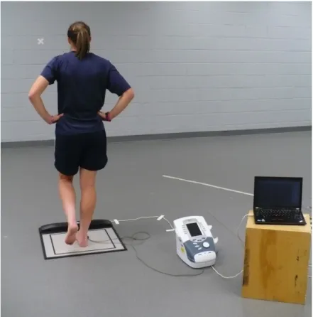
Participants stood 3.66 m from a non-descript wall with a white 0.10 m “X” made from 0.02 m athletic tape placed 1.52 m above the floor directly in front of the unit (Gribble, Tucker et al. 2007). The Matscan device was positioned so the participant’s foot was
centered on the device (Figure 3.1). The electrical stimulation unit (Chattanooga Model #2761) was placed on the ground out of sight of the participant (Figure 3.1). The stimulation unit was set to a continuous current asymmetrical biphasic waveform (factory programmed) with a 100 µs phase duration, and 2 Hz. The laptop computer connected to the Matscan device was positioned diagonally behind the participant out of sight-line. All wiring except for the electrode leads will be secured to the ground using athletic tape.
CONSENT & SCREENING FORMS
approved consent form, and completed the screening form which included the participant self-report questions regarding inclusion and exclusion criteria (Appendix 3.2). Those whose screening forms met study requirements had their dominant foot assessed for the pronated foot type using the FPI-6. Leg dominance was determined by the foot her/she would kick a ball for distance with.
PROCEDURE
Foot Posture Index-6
The FPI-6 was be used to classify the pronated foot type. Participants were instructed to stand barefoot, with feet a comfortable width apart and pointing straight forward, then march in place ten times. Participants were advised when finished marching to simply stop and not reposition either foot. Once finished the researcher inspected (1) talar head
palpation, (2) supra and infra-malleolar curvature, (3) calcaneal frontal plane position, (4) bulging in the talonavicular joint (TNJ) region, (5) height and congruence of the MLA, and (6) abduction/adduction of the forefoot on the rearfoot (Redmond, Crosbie et al. 2006). Scoring was consistent with that of the Foot Posture Index-6 manual (Appendix 2.1) Individual factor and composite scores were recorded using a demographics and data collection sheet. Only participants with composite a score +8 or greater were retained for further studying.
3.1). Talar head palpation showed a substantial level of agreement, while all others showed excellent levels of agreement.
Condition Counter-Balancing
All participants underwent all three conditions of the single-leg balance task with the two “padded” conditions counter-balanced. Each participant chose one sealed envelope from a group of identical envelopes placed on a table containing the order he or she would
perform the conditions. One-half the participants completed the sequence barefoot, pad-only, pad+stim. The remaining followed the sequence barefoot, pad+stim, pad-only. Warm-Up
Participants began the warm-up by riding a stationary bicycle for 5 minutes at a self-selected pace, followed by stretching of the quadriceps hamstrings and triceps surae (calves) muscle groups (Olmsted, Carcia et al. 2002). All stretches were demonstrated by the primary research before each participant performed them.
Quadriceps: Each participant stood on the leg not stretched, flexed the knee of the
stretched stretch leg to grab the ankle, and pulled it toward the buttocks of the same leg until there is a moderate stretch felt in the quadriceps. The stretch is held for 30 seconds with both knees in contact with one another. This stretch was performed on both legs.
Hamstrings: Each participant lay supine with the end most loop of a stretch strap
Calves: The participant assumed a lunge stance, front foot roughly 1 foot from a
wall. With the rear foot pointing straight forward he/she learned forward with a straight rear knee, while keeping the rear heel on the ground, until a moderate stretch was felt in the calf. If the participant’s body touched the wall before the stretch occurs, he/she restarted the process further from the wall. This stretch was performed on both legs and held for 30 seconds.
Single-Leg Balance Instruction
All participants received instruction on the SLB while standing on the Matscan platform for familiarization to the instrument. Each participant began the SLB with the tested great toe placed just behind piece of 0.02 m athletic tape placed on the Matscan to ensure consistent positioning in the middle of the device. With the dominant foot pointing straight forward, both hands placed on his/her hips, and looking at the “X” on the wall, he/she lifted the non-tested foot off the device into a comfortable position. Additionally, participants were instructed that (1) removing one or both hands from the hips, (2) touching the raised foot to the platform, surrounding floor or his /her person would result in the need to repeat the trial. All conditions would include 3 trials of the single-leg balance task for 20 seconds, with 30 seconds rest between trials and 5 minutes rest between conditions.
Conditions
Barefoot (B): After the instruction period participants of both groups will perform an
initial barefoot condition in which nothing was attached to the person.
Electrode Pad Placement: Electrode pad sites for the pad-only and pad+stim
The proximal electrode was positioned so its center bisected the navicular tuberosity in the sagittal plane, and was at the medial most aspect of the plantar sole. The distal electrode was positioned so its center was also at the medial most aspect of the plantar sole, with a gap of 1 cm between the two pads. Electrode leads were secured to the participant’s shank with athletic pre-wrap and tape for strain-relief. As the pad-only and pad+stim conditions were counter-balanced no additional changes were made to pad placement once set, no matter which condition was first.
Pad-Only (PO): The pad only condition was a sham, or placebo, condition in which
participants were told the electrical stimulation unit was set to a sub-sensory voltage
intensity which he/she could not feel. The sham condition was chosen for two reasons. (1) to help determine whether it was the electrical stimulation causing possible changes to the dependent variables, and not the pads acting as tactile cues or the placebo effect on the part of the participant; (2) the pad placed on the plantar sole could increase the midfoot contact area and confound results suggesting the electrical stimulation caused the foot to pronate even more.
Pad with Electrical Stimulation (Pad+Stim, or PS): Pad placement for this condition
negatively impact the data collection, and thus if it became painful at any time he/she must inform the researcher immediately.
Barefoot 2: Once the two pad conditions were completed the electrode pads and
wiring were removed, and all participants repeated the single-leg balance trials one last time. While this condition was not one of our objectives for this thesis it did allow us to assess for possible carry-over-effect.
DATA REDUCTION & ANALYSIS
Midfoot PTI and contact area, as well as lateral and medial PTI were processed and exported via Matscan Research software version 6.51 (Tekscan Inc.; Boston, MA). While each participant completed 20 second trials, only trial seconds 5 to 20 were recorded for analysis. Data was imported into a custom Microsoft Excel (Microsoft; Redmond, WA) spreadsheet to calculate mean and standard deviation values for midfoot PTI, lateral and medial heel PTI, and midfoot contact area across the 3 trials for each condition. Individual trial L:M PTI was calculated using individual trial lateral PTI divided by the medial PTI values, then the mean and standard deviation across trials was calculated.
CHAPTER IV
MANUSCRIPT
Context: Hyperpronation is a risk factor associated with medial tibial stress syndrome,
patellar femoral pain syndrome, and ACL injuries. Previous research with site-specific electrical stimulation to the plantar sole has shown significant alterations in activity of muscles associated with those sites. It is possible that a sensory-level electrical stimulation to the medial longitudinal arch may help reduce hyperpronation.
Objective: To examine the effects of sensory-level electrical stimulation on foot contact
pressure-time integral (PTI) and contact area associated with hyperpronation during single leg balance during a barefoot condition, one electrode pad only and one pad with electrical stimulation condition.
Design: Repeated measures design
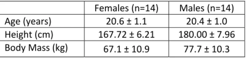
Participants: 14 females (age = 20.6 ± 1.1 years, height 167.72 ± 6.21 cm, mass = 67.1 ± 10.9 kg), and 14 males (age = 20.4 ± 1.0 years, height 180.00 ± 7.96 cm, mass = 77.7 ± 10.3 kg) volunteered.
Interventions: Independent variable was condition, with 3 levels: barefoot (B), electrode
Main Outcome Measures: Dependent variables were determined using a Matscan (Tekscan
Inc.; South Boston, MA) pressure measurement device (PMD). Variables included lateral-to-medial heel pressure-time integral ratio (L:M PTI) as a measure of change in calcaneal inversion and eversion, as well as midfoot pressure-time integral (N/cm2) and midfoot contact area (cm2) as a measures of change in medial longitudinal arch height.
Results: No significant differences were found between B, PO, and PS for L:M PTI
(F(1.55,41.88) = 0.188, p = 0.773) and midfoot PTI (F(2,54) = 0.428, p = 0.654). Significant differences were found for midfoot contact area (F(2,54) = 14.616, p < 0.001). Post hoc analysis revealed significantly greater contact area for PO (mean diff = 0.87 cm2) and PS (mean diff = 1.52 cm2) compared to B, but not between PO and PS (mean diff = 0.65 cm2) although it trended to significance; Tukeys critical value (Fcrit = 0.685 cm2).
Conclusions: Electrical stimulation as performed in this study had no significant effect of
altering calcaneal inversion and eversion as measured by L:M PTI. Electrical stimulation also had no significant effect on midfoot PTI, suggesting no changes in medial longitudinal arch height. A significant increase in midfoot contact area with pad-only and pad+stim could suggest a decreased arch, but more likely an increased contact area with the PMD due to electrode pad size and thickness.
Key words: Hyperproantion, medial tibial stress, ACL injury, pressure-time-integral,
INTRODUCTION
Pronation is a normal and necessary component of foot motion involving calcaneal eversion, as well as talar plantarflexion and adduction, which helps make the foot more mobile when contacting the ground (Root 1977). These motions allow for adaptation to uneven terrain and help absorb ground reaction forces (Manter 1941; Delacerda 1980). While a mobile foot is useful during contact, it is designed to become more rigid during push off when we move. Abnormal pronation, also known as hyperpronation, happens if the pronation is (1) too great, (2) begins too early, and/or (3) lasts too long, not allowing for correctly timed or a sufficient amount of resupination before push off (Root 1977; Delacerda 1980; Donatelli, Hurlburt et al. 1988).
Hyperpronation is associated with (1) medial tibial stress syndrome (Messier and Pittala 1988; Bennett, Reinking et al. 2001; Yates and White 2004), (2) patellar femoral pain syndrome (Boling, Padua et al. 2009), and (3) ACL injuries (Beckett, Massie et al. 1992). Because of this, methods to reduce, or eliminate hyperpronation are important tools for rehabilitation and injury prevention clinicians. One method involves the use of foot orthotics which mechanically brings the ground up to the foot, instead of the foot dropping down to the ground, thus keeping them in a more neutral position. However, orthotics are not
effective for everybody, and more importantly compliance can be an issue as not everybody who might benefit from orthotics want to use them. In sports were shoe construction is kept to a minimum (i.e., soccer and cross country) the addition of orthotics may cause the foot to “pop out” of the shoe easier. With the increasing popularity of minimalist shoes the
muscular re-education called the short-foot concept. This method teaches the individual to actively shorten the foot lengthwise and widthwise, effectively creating a more supinated foot (Janda 2006). A down side of the short-foot is it can require extensive clinician participation, as he/she must passively re-create the foot position until it can be performed without assistance.
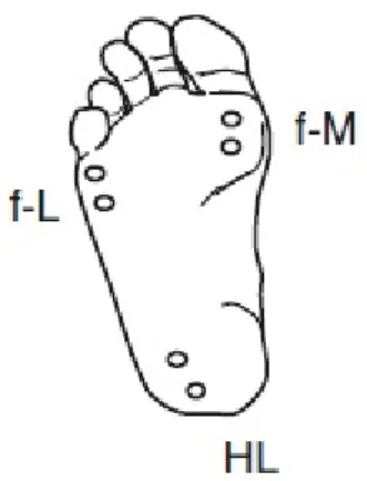
The short-foot concept is intended to stimulate the proprioceptive system, which has an important role in posture and equilibrium, by increasing signals from afferent receptors of the foot to the central nervous system (Janda 2006). Nakajima et al. (2006) selectively applied a brief, less than one second, mild electrical stimulation to different medial and lateral sections of the plantar sole to study muscle activity of the tibialis anterior, peroneus longus, soleus, and medial gastrocnemius (Nakajima, Sakamoto et al. 2006). When the stimulation was applied to the sole of the 1st metatarsophalangeal joint (f-M) activity of the peroneus longus, soleus, and medial gastrocnemius presented with inhibition, while the tibialis anterior presented with excitation (figure 4.1). Conversely, when the stimulation was applied to the sole of the heel (HL), activity of the tibialis anterior presented with inhibition, while the peroneus longus, soleus, and medial gastrocnemius presented with excitation (figure 4.1). The results suggest the neuromuscular system activity could result in reduced contact pressure at the site of stimulation (Nakajima, Sakamoto et al. 2006).
provide information regarding weight shifts associated with calcaneal inversion and eversion. If there is increased pressure laterally it could represent a calcaneal inversion moment associated with supination. From a rehabilitation standpoint sensory-level stimulation could act as an adjunct for clinicians when teaching the short-foot concept. Increasing sensory signals to the central nervous system through electrical stimulation without eliciting muscle activity could assist passive short-foot modeling by the clinician, and even reduce the time needed to learn the technique. The purpose of this investigation is study the effects of electrical stimulation during a static balancing task, and its effects on rearfoot plantar pressure as well as midfoot plantar pressures and contact area associated with pronation.
METHODS
Participants
Twenty-eight participants (14 males, 14 females) were identified through a
screening process of a larger group of potential participants who volunteered for this study (Table 4.1). To be eligible for this study participants had to fill out a questionnaire
The Foot Posture Index (FPI-6) was utilized to identify participants with foot hyperpronation. Intra-rater reliability was established for each of the six factors (Table 3.1) before data collection began using Kappa measure of agreement. Talar head palpation showed a substantial level of agreement, while all others showed excellent levels of agreement.
Evaluation was performed by asking participants to march in place ten times with both feet pointing straight forward, and at a comfortable width apart. Once finished
participants simply stopped moving and did not readjust either foot, or move the body about, until after the FPI-6 screen was completed. The primary researcher assessed the six factors following the guidelines of the FPI-6 Manual (Appendix 2.1) (Redmond 1998). As we want to include those with more severe pronation, participants had to have a composite score of + 8 or greater on their dominant leg. Leg dominance was defined as the leg used to kick a ball for maximum distance. All participants read and signed consent forms for this study
approved by the Institutional Review Board of the University of North Carolina at Chapel Hill.
Laboratory Set-Up
sight-line (figure 3.1). All wiring except for the electrode leads were secured to the ground using athletic tape. was placed on the ground out of sight of the participant. Nakajima et al. used an asymmetrical monophasic current with 100 µs duration and 2 Hz using a constant current stimulator and a pulse regulation system (Nakajima, Sakamoto et al. 2006). We were unable to re-create the exact waveform with our instrumentation, therefore we chose to use a continuous current asymmetrical biphasic waveform (factory programmed) with a 100 µs phase duration, and 2 Hz. We were unable to find data as to why a monophasic waveform would be more effective than biphasic in the literature.
Warm-Up
Participants road a stationary bicycle for 5 minutes at a self-selected pace, followed by static-stretching of the quadriceps, hamstrings, and triceps surae (calves) muscle groups for 1 set of 30 seconds each.
Single-Leg Balance Task
All participants performed a single-leg balance task (SLB) under three conditions (barefoot, pad-only, pad+stim), with the pad conditions counter-balanced. Each participant chose one sealed envelope from a group of identical envelopes placed on a table containing the order he or she would perform the conditions. One-half of the participants completed the sequence barefoot, pad-only, pad+stim. The remaining followed the sequence barefoot, pad+stim, pad-only.
and looking at the “X” on the wall, he/she lifted the non-tested foot off the device into a comfortable position. Additionally, participants were informed that removing one or both hands from the hips, touching the raised foot to the platform, surrounding floor, or his/her person would result in the need to repeat the trial. All conditions included 3 trials of the single-leg balance task for 20 seconds, with 30 seconds rest between trials and 5 minutes rest between conditions. In keeping with previous research using this method no practice trials were allowed (Gribble, Tucker et al. 2007).
Conditions
Barefoot (B): After the instruction period all participants performed a barefoot
condition with nothing attached to the foot.
Electrode Placement: Once B was completed electrode pad sites for the pad-only and
pad+stim conditions were cleaned with a 70% isopropyl alcohol solution to reduce skin impedance. A pair of stimulus electrodes, poles of 3.18 cm were placed on the medial plantar aspect of the midfoot. The proximal electrode was positioned so its center bisected the navicular tuberosity in the sagittal plane, and was at the medial most aspect of the plantar sole. The distal electrode was positioned so its center was also at the medial most aspect of the plantar sole, with a gap of 1 cm between the two pads. Electrode leads were secured to the participant’s shank with athletic pre-wrap and athletic tape for strain-relief. As the pad-only and pad+stim conditions were counter-balanced no additional changes were made to pad placement once set, no matter which condition was first.
Pad-Only (PO): The pad only condition was a sham, or placebo, condition in which
participants were told the electrical stimulation unit was set to a sub-sensory voltage
help determine whether it was the electrical stimulation causing possible changes to the dependent variables, and not the pads acting as tactile cues or the placebo effect on the part of the participant; (2) the pad placed on the plantar sole could increase the midfoot contact area and confound results suggesting the electrical stimulation caused the foot to pronate even more.
Pad with Electrical Stimulation (Pad+Stim, or PS): The intensity of electrical
stimulation was set by increasing the stimulation unit intensity until a visible local muscle response was produced, then reducing it until (1) that response was eliminated and (2) the stimulation was not painful. In all participants, it was verbally confirmed that the intensity of the electrical stimulation was not painful. Participants were informed the intensity should not be painful as it could negatively impact the data collection, and thus if it became painful at any time he/she needed to inform the researcher immediately.
DATA REDUCTION & ANALYSIS
Midfoot PTI and contact area, as well as lateral and medial PTI were processed and exported via Matscan Research software version 6.51 (Tekscan Inc.; Boston, MA). While each participant completed 20 second trials, only trial seconds 5 to 20 were recorded then exported for further reduction and analysis. Data were imported into a Microsoft Excel (Microsoft; Redmond, WA) spreadsheet to calculate mean and standard deviation values for midfoot PTI, and midfoot contact area across the 3 trials for each condition. Individual trial L:M PTI was calculated using lateral PTI divided by the medial PTI values for each trial. L:M PTI Mean and standard deviation across trials was then calculated. Separate repeated measures analysis of variance (ANOVA) procedures were then performed for each
Chicago, IL). Tukey’s post-hoc analysis was utilized where significant differences were observed. While not part of the objective for this study, a second barefoot condition (identical to B) was collected 5 minutes after the last condition (PO, or PS) to assess for a possible carry-over effect. Paired t-test was performed between these barefoot conditions. RESULTS
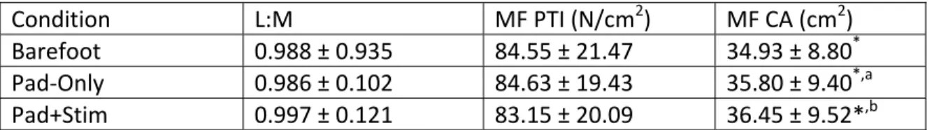
No significant differences were observed between B, PO, PS conditions for L:M PTI (F(1.55,41.88) = 0.188, p = 0.773) and midfoot PTI (F(2,54) = 0.428, p = 0.654). However, a significant difference was observed between B, PO, and PS for midfoot contact area (F(2,54) = 14.616, p < 0.001). Post hoc testing revealed a significantly larger contact area for PO (mean diff = 0.87 cm2) and PS (mean diff = 1.52 cm2) in comparison to B. No significant difference was observed between PO and PS (mean diff = 0.65 cm2), although it did trend towards significance; Tukeys critical value (Fcrit = 0.685) (table 4.2).
No significant differences were observed between the barefoot conditions for L:M PTI (t(27) = 1.038, p = 0.309), midfoot PTI (t(27) = 0.508, p = 0.615), and midfoot contact area (t(27) = 0.750, p = 0.460). This suggests there was no carry-over effect.
DISCUSSION
placed under the foot may have altered muscle activity enough to reduce the increased pressure over time (PTI) due to the increased amount of “foot” contact from the pads. Future analysis of other plantar sole zones, for instance the 1st metatarsal head, may shed light on it. The observed trend toward significance for an increase in contact area with the PS was not expected however. It is possible the electrical stimulation actually excited the posterior chain musculature creating a plantar flexion moment, but without a significant result with midfoot PTI it is difficult to suggest that. Once again, future analysis all plantar zones for possible relationships could should light on how plantar pressure are dispersed around the foot.
Electrical Stimulation
research with the electrical stimulation component could include comparing the waveform and EMG as Nakajima et al. used with a few waveforms that are more common place in the clinical setting such as high volt, or premodulation.
Additionally, setting the stimulation intensity above the motor threshold could elicit enough motor response to reduce hyperpronation, while still providing sensory input to the central nervous system. We chose not to use motor-level stimulation principally to observe if increased afferent signaling could elicit an increased motor response during the task in the absence of (1) conscience effort (i.e., instruction) to do so, and (2) an assisted motor
response via the stimulation itself. Khaslavskaia et al. observed MEP’s of the tibialis anterior and soleus before, during, and up to 60 minutes after cessation of two back-to-back 15 minute treatments of electrical stimulation to the common peroneal nerve at 2-3 times the motor threshold (Khaslavskaia, Ladouceur et al. 2002). Their results did suggest tibialis anterior MEP rose significantly out to about 60 minutes post treatment (Khaslavskaia, Ladouceur et al. 2002). Solues MEP however, showed no significant changes (Khaslavskaia, Ladouceur et al. 2002). A future study involving the greater motor-level stimulation, but retaining the medial plantar sole site could yield useful results.
Electrode Pad Size
Using larger electrodes increases the chances of stimulating multiple sensory zones (i.e., medial plantar, lateral plantar can calcaneal), which may provide conflicting signals to the central nervous system. Previous research used 5 mm electrode pads (Nakajima,
even increased posterior chain muscle activity leading the near significant difference in midfoot contact area between the PO and PS conditions. The results of previous site-specific stimulation to the lateral plantar sole suggested that a lateral boundary approximating the 5th tarsometatarsal joint for which the soleus and tibialis anterior EMG would reverse could also help to explain this (Nakajima, Sakamoto et al. 2006). Additionally, using the larger pad increases the likelihood of stimulating a motor plate with a lower intensity which could also limit the amount of sensory signals to the central nervous system before eliciting a motor response from the stimulation itself.
Single-Leg Balance & Foot Variability
In Vladimir Janda’s Sensory motor training progression once the active short-foot concept is learned the patient begins with easier tasks, such as double-leg weigh shifts before the single-leg balance (Janda 2006). The single-leg balance may have been too difficult a task to accomplish without the large amount of foot motion variability seen in some participants. Anecdotally several participants were visually assessed as having poor 1st ray stability at the 1st metatarsal head. These individuals used the great toe more for stability, which lifted the metatarsal head off the Mastcan. It is possible that both pad conditions acted to stabilize the 1st ray through the metatarsal head and thus increased the contact area. For future research analyzing center of mass in addition to zones could help answer this question.
electrical stimulation to the plantar sole could help individuals perform the short-foot actively with reduced clinician involvement.
CONCLUSION
The application of sensory-level electrical stimulation to the plantar sole as performed in this study appears to have no beneficial use clinically for reducing
hyperpronation. While it does not provided useful clinical value it does however, provide multiple directions for future research.
FIGURE 3.1: LABORATORY SET-UP
FIGURE 4.1: NAKAJIMA ET AL. (2006) STIMULATION SITES
TABLE 3.1: FPI-6 INTRA-RATER RELIABILITY
n=24 Talar Head Palpation
Malleolar Curvature
Calcaneal Position
TNJ Bulging MLA Congruence
TABLE 4.1: SUBJECT DEMOGRAPHICS
TABLE 4.2: DATA TABLE
Condition L:M MF PTI (N/cm2) MF CA (cm2)
Barefoot 0.988 ± 0.935 84.55 ± 21.47 34.93 ± 8.80* Pad‐Only 0.986 ± 0.102 84.63 ± 19.43 35.80 ± 9.40*,a Pad+Stim 0.997 ± 0.121 83.15 ± 20.09 36.45 ± 9.52*,b
Mean ± SD. * indicates statistical significance (p < 0.001). a,b
indicate statistical significance from barefoot‐1 based on Tukeys post hoc
APPENDIX 3.1: DEMOGRAPHICS/DATA COLLECTION SHEET
Test Date: ____________________
Participant Number:____________________
Test Group ( AB / BA )
Sex: □ Male □ Female
Age: ____________________
Weight (kg): ____________________
Height (cm):____________________
FPI‐6 scores Rearfoot
Talar head palpation‐‐‐‐‐‐‐‐‐‐‐‐‐‐______ Lateral malleolus curvature‐‐‐‐‐‐______ Calcaneal inversion/eversion‐‐‐‐______ Forefoot
TNJ region prominence‐‐‐‐‐‐‐‐‐‐______ MLA congruence‐‐‐‐‐‐‐‐‐‐‐‐‐‐‐‐‐‐______ Forefoot Abd/Add‐‐‐‐‐‐‐‐‐‐‐‐‐‐‐‐‐______
Composite score:______
Dominant leg: □ Right □ Le
APPENDIX 3.2: SCREENING FORM
Participant Number:______ Dominant Leg: __________
1). Have you ever had a lower extremity surgery? Yes / No If “yes” which extremity? R / L
2). Have you had a significant lower extremity injury in the past Yes / No 6 months?
If “yes” which extremity? R / L
If “yes” did it keep you from being physical activity Yes / No for more than 3 consecutive days
3). Have you participated in at least 30 minutes of exercise 3 Yes / No times per week for the last 6 months
4). Do you have a visual disorder that cannot be corrected by glasses? Yes / No 5). Do you currently have a respiratory tract infection, Yes / No inner ear infection, or head cold?
REFERENCES
Anderson, M. W., V. Ugalde, et al. (1997). "Shin splints: MR appearance in a preliminary study." Radiology 204(1): 177-180.
Bagheri, H., Baxendale, R.H. (1994). The Effect of Increasing The Number of Stimuli and Intensity of Muscle Contraction on The Cutaneomuscular Reflexes in Tibialis Anterior in Man. Alpha and Gamma Motor Systems. A. Taylor, Gladden, M.H., Durbaba, R.
Batt, M. E., V. Ugalde, et al. (1998). "A prospective controlled study of diagnostic imaging for acute shin splints." Med Sci Sports Exerc 30(11): 1564-1571.
Beckett, M. E., D. L. Massie, et al. (1992). "Incidence of Hyperpronation in the ACL Injured Knee: A Clinical Perspective." J Athl Train 27(1): 58-62.
Bennett, J. E., M. F. Reinking, et al. (2001). "Factors contributing to the development of medial tibial stress syndrome in high school runners." J Orthop Sports Phys Ther 31(9): 504-510.
Boling, M. C., D. A. Padua, et al. (2009). "A prospective investigation of biomechanical risk factors for patellofemoral pain syndrome: the Joint Undertaking to Monitor and Prevent ACL Injury (JUMP-ACL) cohort." Am J Sports Med 37(11): 2108-2116.
Brown, A. G. (2001). Nerve Cells and Nervous Systems: An introduction to neuroscience. London, UK, Springer-Verlag.
Cashmere, T., R. Smith, et al. (1999). "Medial longitudinal arch of the foot: stationary versus walking measures." Foot Ankle Int 20(2): 112-118.
Chuter, V. H. (2010). "Relationships between foot type and dynamic rearfoot frontal plane motion." J Foot Ankle Res 3: 9.
Cornwall, M. W., T. G. McPoil, et al. (2008). "Reliability of the modified Foot Posture Index." J Am Podiatr Med Assoc 98(1): 7-13.