An Integrated Chemical Cytometry Method: Shining a Light on
Akt Activity in Single Cells
Emilie R. Mainz,
Department of Chemistry, University of North Carolina, Chapel Hill NC 27599 (USA)
Prof. Qunzhao Wang,
Department of Chemistry, Division of Chemical Biology and Medicinal Chemistry, and Department of Pharmacology, University of North Carolina, Chapel Hill, NC 27599 (USA)
Prof. David S. Lawrence, and
Department of Chemistry, Division of Chemical Biology and Medicinal Chemistry, and Department of Pharmacology, University of North Carolina, Chapel Hill, NC 27599 (USA)
Prof. Nancy L. Allbritton
Department of Chemistry and Pharmacology, University of North Carolina, Chapel Hill, NC 27599 (USA), Joint Department of Biomedical Engineering, University of North Carolina and North Carolina State University, Chapel Hill and Raleigh, NC (USA)
Nancy L. Allbritton: [email protected]
Abstract
Tools to evaluate oncogenic kinase activity in small clinical samples have the power to guide precision medicine in oncology. Existing platforms have demonstrated impressive insights into the activity of protein kinases, but these technologies are unsuitable for the study of kinase behavior in large numbers of primary human cells. To address these limitations, we developed an integrated analysis system which utilizes a light-programmable, cell-permeable reporter deliverable en masse to many cells. The reporter's ability to act as a substrate for Akt, a key oncogenic kinase, was masked by a 2-4,5-dimethoxy 2-nitrobenzyl (DMNB) moiety. Upon exposure to ultraviolet light and release of the masking moiety, the substrate sequence enabled programmable reaction times within the cell cytoplasm. When coupled to automated single-cell capillary electrophoresis, statistically significant numbers of primary human cells were readily evaluated for Akt activity.
Keywords
biosensor; photochemistry; biological activity; peptides
Appropriate prescription of targeted therapeutics for cancer treatment relies on the accurate identification of aberrant signaling pathways, most frequently those involving protein kinases. However, patient selection for pathway inhibition is mostly empirical or reliant
HHS Public Access
Author manuscript
Angew Chem Int Ed Engl
. Author manuscript; available in PMC 2017 October 10.Published in final edited form as:
Angew Chem Int Ed Engl. 2016 October 10; 55(42): 13095–13098. doi:10.1002/anie.201606914.
A
uthor Man
uscr
ipt
A
uthor Man
uscr
ipt
A
uthor Man
uscr
ipt
A
uthor Man
uscr
upon sequencing to identify mutations within the genome. A targeted selection process revealing only those patients whose cells possess increased kinase activity would lessen costs as well as forego drug toxicity in individuals not likely to be responsive to pathway inhibition. Thus, kinase-activity sensors and their attendant analytical platforms to identify aberrant kinase activities in primary cells independent of genomic mutation status would be of high value.[1] However, the very small sample size of most clinical specimens and limited throughput of analysis has precluded the routine measurement of kinase activity in single human primary cells.[2] Requirements for these measurements are twofold: first; a sensor which is selective for the kinase of interest, deliverable en masse to single cells, and resistant to phosphorylation until the desired assay start time, and second; a platform which can analyze statistically relevant numbers of single cells.
Fluorescent peptide sequences are utilized as reporters to directly assay kinase activity, and stand to bridge the gap between small molecules which are poor substrates of limited specificity and genetically engineered proteins that are difficult to transfect uniformly into cells.[3] Peptide reporters phosphorylated by kinases within cells are highly sensitive, quantitative, and possess great multiplexing potential when coupled to capillary
electrophoresis (CE). To date, delivery of these reporters into the cell interior has relied on low throughput methods such as microinjection or on cell-penetrating peptides (CPPs) which largely deliver cargo to endosomal compartments.[4] Additionally, reporters delivered by CPPs are acted upon during the delivery process, thus the reaction time in the cell is unknown. Delivering a reporter into the cytosol of large numbers of cells while retaining control of assay initiation would dramatically improve overall throughput when coupled to an automated single-cell assay platform. The latter would enable sampling from sufficient numbers of cells for statistical analyses and identification of cellular subpopulations.
We addressed these opportunities by integrating chemical and instrumentation innovations. Specifically, we have developed a cell permeable, photoactivatable reporter that is
chemically masked (Scheme 1) and integrated into an automated analysis platform. The active reporter acts as a substrate for the protein kinase Akt, a kinase which plays critical roles in tumor formation and progression.[5] The native substrate (NS) Akt peptide reporter 6FAM-GRP-(R)-AFTF-(A)-Amide (R and A represent N-methylated versions of amino acids) has favorable phosphorylation kinetics, specificity, and a demonstrated resistance to intracellular degradation.[6]
NS possesses a single hydroxyl group that, when coupled to a photoremovable DMNB
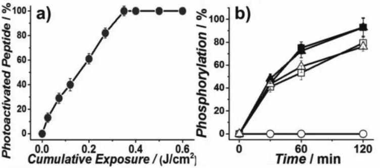
group, should block phosphorylation by Akt and thus silence the reporter as a substrate (Figure 1(b)). Figure 1(a) & S1 demonstrate the photochemical conversion of inactive to active reporter. Importantly, the active reporter 2 is phosphorylated at a rate indistinguishable from that of NS, both in vitro and in cell lysates (Figure 1(b)). The inactive Akt reporter 1 is not phosphorylated nor spontaneously converted to its active counterpart in the dark during the assays (Figure S6).
Having demonstrated that 1 is photoactivated and subsequently phosphorylated by Akt, we explored the delivery options for this reporter. Since NS is excluded from PANC-1 cells, prior work required microinjection of the reporter into cells, a protracted process. Many
A
uthor Man
uscr
ipt
A
uthor Man
uscr
ipt
A
uthor Man
uscr
ipt
A
uthor Man
uscr
CPPs have been reported, but most are taken up via endosomal pathways, resulting in significant peptide degradation and little to no delivery to the cytosol, the primary location of Akt.[7] These challenges were manifested in our initial attempts to render NS cell permeable, with multiple CPPs[8] appended to the reporter resulting in punctate uptake indicative of a non-cytoplasmic location (Figure S4). However, NS, when modified with DMNB (1), is sufficiently hydrophobic to enter into the cytosol when incubated with PANC-1 cells (Figure 2a, S5). The diffuse (rather than punctate) appearance of the fluorescence after loading peptide into the cells (Figure S5) combined with the observed phosphorylation of the reporter (post photoactivation) (Figure 2d) suggests delivery into cytosol rather than a cytosolic subcompartment. Quantification of intracellular peptide by CE revealed a correlation (R2 = 0.79) between the total moles of fluorescent reporter (ntotal = ncaged + nphosphorylated + ndegraded) and the cell diameter (Figure 2e). NS modified with a DMNB-Ser moiety (rather than DMNB-Thr) is likewise cell permeable (Figure S7), suggesting that the addition of DMNB may be competent to render peptides of similar hydrophobicity cell permeant.
We next evaluated the ability of this reporter to sense Akt activation after photolysis within intact PANC-1 cells. Akt is upstream of DNA repair mechanisms and is known to be a stress-activated kinase. Thus, we first assessed whether the UV exposure needed for photoactivation might cause DNA damage and/or activate Akt. CE-analysis revealed that UV illumination (0.4 J/cm2) of cells transforms 96 ± 2.2% of 1 into active 2. UV exposure under these conditions does not induce cytotoxicity or DNA pyrimidine lesions (Figures S8, S9). Using these conditions, we assessed Akt activation and inhibition in single PANC-1 cells (Figure 2 d,f). After photolysis, the contents of single cells were assayed by CE and the phosphorylated sensor quantified. Without endogenous stimuli, minimal Akt activity was detected (Figure 2f), which suggests that the illumination protocol does not activate Akt.[5] We also examined Akt pathway stimulation or inhibition in response to TNFα, or TNFα +LY294002, respectively. Significant heterogeneity in reporter phosphorylation in response to TNFα stimulation (0–100%) was observed, consistent with reports of mosaic Akt activation in individual cells of a cell population.[9] The PI3K inhibitor, LY294002, blocked reporter phosphorylation in response to TNFα, as expected since PI3K is downstream of the TNFα receptor and upstream of Akt. Having produced a cell permeable and
photoactivatable Akt reporter, we sought to harness these unique properties to enhance the measurement rate of Akt activity in single cells. Previous work describing microinjection of peptide-based protein kinase sensors offered a very limited throughput of 0.5 cell h−1 which is not biomedically useful.[6] A recently reported automated single-cell CE system
quantified kinase activity as rapidly as 3.5 cells min−1, rivaling microfluidic single-cell electrophoresis systems and offering excellent (10−20 mol) detection limits.[10] A limitation for the automated system, however, is that reporters are acted upon during the loading process into the cell so that the reaction time in the cell is unknown. These challenges are uniquely addressed by the reporter 1, which is membrane permeant with a programmable light-triggered reaction initiation.
We conducted a pilot study in which the unique properties of compound 1 were combined with the automated single-cell CE system. All cells in a population were simultaneously
A
uthor Man
uscr
ipt
A
uthor Man
uscr
ipt
A
uthor Man
uscr
ipt
A
uthor Man
uscr
photo-activated to assess Akt activity as a function of time. Cells were loaded with compound 1, illuminated, and serially analyzed (n =109) (Figure 3). The photoactivation efficiency is 97 ± 2.8% within intact cells with 42.4 ± 42.7 amol reporter loaded per cell. The amount of reporter (amol) detected correlated with the cell volume (Figure 2e) and appeared to be independent of dwell time (R2 = 0.018, Figure S10), suggesting that the reporter is not exported by the cell over the analysis period. An oft overlooked commonality among peptides, even when engineered for stability, is their eventual degradation within the intracellular context. Phosphorylation, degradation, and other intracellular alterations or environments can have similar effects on sensor fluorescence properties, creating a challenge for many sensors relying solely on fluorescence properties as a proxy for sensor
phosphorylation.[11] In contrast, the single-cell CE system measures intact and degraded reporter as well as that phosphorylated to accurately quantify phosphorylation rate (Figure 3b). When peptide standards of known concentration are also paired with single-cell CE, the moles of the different peptide species are readily quantified (Figure 3c).
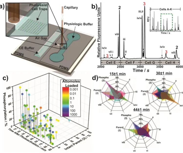
Rather than simply increasing as a function of reporter incubation time, the kinetics of Akt reporter phosphorylation in single cells was highly variable (0.0 – 0.024 zmol pg−1 s−1) at all times measured (Figure 3c,S9). While we and others have demonstrated heterogeneous Akt activity in PANC-1 and other cell lines at individual endpoints[12], the reporter 1 enabled time-resolved studies of this heterogeneity, suggesting that peptide phosphorylation may reach an equilibrium determined by the balance of kinase and opposing phosphatase activity, with the equilibrium point unique for each cell (Figure S11). As Akt is a major promoter of cell survival[13], this type of heterogeneity has been suggested to promote survival of a subset of cells under stressful conditions and/or confer resistance to chemotherapeutics.[14] The integrated analysis system also delivers insight into how peptides are modified over time within single cells (Figure 3d; fragment naming in Table S2), and the formation of key fragments can be monitored (Supplementary Information S.4).
The cell-permeable, photoactivated reporter, combined with an integrated platform, enabled analysis (7.2 cells h−1) of kinase activity in a population of cells substantially faster than previous studies. The protein kinase reporter is rapidly loaded into the cytoplasm of cells and converted, on demand, into the active reporter. We expect that outfitting peptide sensors with DMNB or related moieties to properly tune the amphiphilic properties of peptide-based protein kinase reporters, will prove useful in revealing aberrant kinase activity in small samples of clinical interest, especially when combined with automated analysis systems.
Supplementary Material
Refer to Web version on PubMed Central for supplementary material.
Acknowledgments
We thank the NIH (CA203032 and CA177993) for financial support.
A
uthor Man
uscr
ipt
A
uthor Man
uscr
ipt
A
uthor Man
uscr
ipt
A
uthor Man
uscr
References
1. Rubakhin SS, Romanova EV, Nemes P, Sweedler JV. Nat. Methods. 2011; 8:S20–S29. [PubMed: 21451513]
2. Turner NC, Reis-Filho JS. Lancet Oncol. 2012; 13:178–185.
3. Damayanti NP, Parker LL, Irudayaraj JMK. Angew. Chem. Int. Ed. Engl. 2013; 52:3931–3934. [PubMed: 23450802]
4. Veldhuyzen WF, Nguyen Q, McMaster G, Lawrence DS. J. Am. Chem. Soc. 2003; 125:13358– 13589. [PubMed: 14583022]
5. Garcia-Echeverria C, Sellers WR. Oncogene. 2008; 27:5511–5526. [PubMed: 18794885] 6. Proctor A, Herrera-Loeza G, Wang Q, Lawrence DS, Yeh JJ, Allbritton NL. Anal. Chem. 2014;
86:4573–4780. [PubMed: 24716819]
7. Räägel H, Säälik P, Pooga M. BBA - Biomembranes. 2010; 1798:2240–2248. [PubMed: 20170627] 8. a) Montrose K, Yang Y, Sun X, Wiles S, Krissansen GW. Sci. Rep. 2013; 3:1661. [PubMed:
23588666] b) Cruz J, Mihailescu M, Wiedman G, Herman K, Searson PC, Wimley WC, Hristova K. Biophys. J. 2013; 104:2419–2428. [PubMed: 23746514]
9. a) Proctor A, Herrera-Loeza G, Wang QZ, Lawrence DS, Yeh JJ, Allbritton NL. Anal. Chem. 2014; 86:4573–4780. [PubMed: 24716819] b) Yuan TL, Wulf G, Burga L, Cantley LC. Curr. Biol. 2011; 21:173–183. [PubMed: 21256021]
10. Dickinson AJ, Armistead PM, Allbritton NL. Anal. Chem. 2013; 85:4797–4804. [PubMed: 23527995]
11. Van, TNN.; Morris, MC. Fluorescent Sensors of Protein Kinases: From Basics to Biomedical Applications. Academic Press; 2013. p. 315
12. a) Proctor A, Herrera-Loeza SG, Wang Q, Lawrence DS, Allbritton NL. Anal. Chem. 2014; 86:4573–4580. [PubMed: 24716819] b) Mainz ER, Serafin DS, Nguyen TT, Tarrant TK, Sims CE, Allbritton NL. Anal. Chem. 2016; 86:7786–7792.c) Yuan TL, Wulf G, Burga L, Cantley LC. Curr. Biol. 2011; 21:173–183. [PubMed: 21256021] d) Meyer R, D’Alessandro LA, Kar S, Kramer B, She B, Kaschek D, Hahn B, Wrangborg D, Karlsson J, Kvarnstrom M, Jirstrand M, Lehmann WD, Timmer J, Hofer T, Klingmuller U. Front. Physiol. 2012; 3:451-. [PubMed: 23226133]
13. Datta SR, Brunet A, Greenburg ME. Genes Dev. 1999; 13:2905–2927. [PubMed: 10579998] 14. a) Yuan TL, Cantley LC. Oncogene. 2008; 27:5497–5510. [PubMed: 18794884] b) Schlieman MG,
Fahy BN, Ramsamooj R, Beckett L, Bold RJ. Br. J. Cancer. 2003; 89:2110–2115. [PubMed: 14647146] c) Mortenson M, Galante JM, Schlieman M, Bold RJ. Cancer Therapy. 2004; 2:227.
A
uthor Man
uscr
ipt
A
uthor Man
uscr
ipt
A
uthor Man
uscr
ipt
A
uthor Man
uscr
Figure 1. In vitro characterization of caged AKT reporter
Photolysis of 1 (10 µM) in a) extracellular buffer (ECB). b) Phosphorylation assays with recombinant Akt. Squares indicate assays performed with peptide NS, while circles and triangles were performed with peptides 1 and 2, respectively. Closed symbols: in vitro assays in buffer, open symbols: experiments performed in PANC-1 lysates (S3.5).
A
uthor Man
uscr
ipt
A
uthor Man
uscr
ipt
A
uthor Man
uscr
ipt
A
uthor Man
uscr
Figure 2. Intracellular characterization of peptide 1
Images of cells incubated at 4 °C with a) 1, b) 2 c), or without peptide. d) Single-cell Akt activity measured by chemical cytometry with the electrophoretic peaks labeled as 1, 2, and
3 as denoted in Scheme 1. 6FAM-labeled proteolytic products of 1 are marked as i–viii
(Table S2). The STND trace shows the migration time of a standard solution of the peptides. Those below are from individual PANC-1 cells which were deprived of serum (basal), stimulated with 100 ng/mL TNFα, or treated with 10 µM LY294002 + 100 ng/mL TNFα. e) The attomoles of reporter loaded as a function of cell diameter (R2 = 0.79). f) Akt activity for a population of single cells analyzed 20-min post-photolysis. Prior to loading with 1, photolysis and analysis, cells were treated as described in d).
A
uthor Man
uscr
ipt
A
uthor Man
uscr
ipt
A
uthor Man
uscr
ipt
A
uthor Man
uscr
Figure 3. Automated single cell analysis
a) The microfabricated integrated system used for automated single-cell CE analysis. Cell traps (45 µM deep, inset) were loaded with PANC-1 cells and the peptide within the cell was photoactivated. Cells were then assayed. b) Separation of intracellular contents from consecutively analyzed cells. Inset: 11 cells (A–K), expanded region: cells E–H. c) Reporter metabolism in PANC-1 cells. Each colored data point represents a single cell with the color denoting the moles loaded into the cell. d) Reporter metabolism at various time points after photoactivation. Each overlay (color) represents the metabolism from a single cell.
A
uthor Man
uscr
ipt
A
uthor Man
uscr
ipt
A
uthor Man
uscr
ipt
A
uthor Man
uscr
Scheme 1.
Native substrate (NS) was chemically caged with DMNB to generate 1, which is delivered
intracellularly. Photolysis releases the active sensor 2 (identical to NS) which is phosphorylated (3) and measured in single cells by automated single-cell, capillary electrophoresis.