THREE DIMENSIONAL TREATMENT OUTCOMES IN CLASS
II PATIENTS TREATED USING HERBST: A PILOT STUDY
Megan L. LeCornu, DMD
A thesis submitted to the faculty of the University of North Carolina at Chapel Hill in partial fulfillment of the requirements for the degree of Master of Science in the School
of Dentistry (Orthodontics).
Chapel Hill 2013
Approved by:
Tung Nguyen DMD, MS
iii
ABSTRACT
MEGAN L LECORNU: Three dimensional treatment outcomes in class II patients treated using Herbst
(Under the direction of Dr. Tung Nguyen)
iv
ACKNOWLEGEMENTS
I would like to thank the following for their contributions to this project:
Dr. Tung Nguyen, for his invaluable mentorship. His support, passion for teaching and encouragement throughout the development and completion of this project is unmatched. While, I am grateful to have such a dedicated advisor to guide me through this process, I am more grateful for his role as a teacher and friend through my professional development.
Dr. Lucia Cevidanes, for her insight, expertise, and advice. Not only was her expertise instrumental to completing this project, her guidance as a teacher will never be forgotten.
Ms. Hsiao-han Chen and Mr. John Paul Zermeno, for their time and assistance
with data processing.
Dr. Hong Tu Zhu and Dr. Chih-Da Wu for their invaluable assistance with statistical analysis.
v
TABLE OF CONTENTS
LIST OF TABLES ... vi
LIST OF FIGURES ... vii
I. CHAPTER I - LITERATURE REVIEW ... 1
II. CHAPTER II - MANUSCRIPT... 20
A. INTRODUCTION ... 21
B. MATERIALS AND METHODS ... 23
C. RESULTS... 26
D. DISCUSSION ... 28
E. CONCLUSIONS ... 33
vi
LIST OF TABLES
vii
LIST OF FIGURES
Figure A. The Original Herbst Design ... 13
Figure B. The Acrylic Herbst Design ... 13
Figure C. The Cantilever Herbst Design... 14
Figure 1. 3D Mandibular Landmark Identification... 35
Figure 2. Herbst T1 Semitransparency ... 38
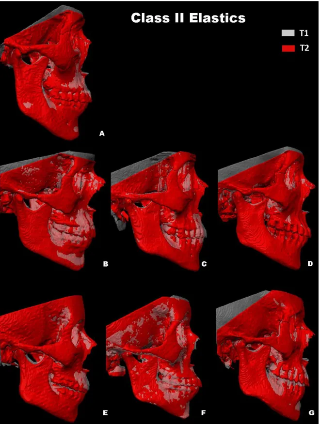
Figure 3. Class II Elastic T1 Semitransparency ... 39
Figure 4. Box Plot of Maxillary Skeletal Changes ... 40
Figure 5. Herbst T2 Semitransparency ... 41
Figure 6. Class II Elastic T2 Semitransparency ... 42
Figure 7. Box Plot of Mandibular Skeletal Changes ... 43
Figure 8. Box Plot of Gonial Angle and Condylar Flexure Skeletal Changes ... 43
Figure 9. Condyle Semitransparencies ... 44
Figure 10. Glenoid Fossa Color Maps ... 45
Figure 11. Box Plot of Condyle and Glenoid Fossa Skeletal Changes ... 46
1
CHAPTER I
LITERATURE REVIEW
Class II skeletal relationships are commonly encountered in orthodontic practices in the United States1. The etiology includes a prognatic maxilla, a retrognathic mandible or a combination of both. A study by McNamara in 1981 revealed that a majority of class II patients have some component of mandibular deficiency underlying the skeletal class II discrepancy 2. Ideally, the skeletal discrepancy needs to be addressed for optimal
treatment results3.
2
comorbidities including paraesthesia, anaesthesia, paralysis and potentially death. Because of these potential complications, patients are often reluctant to go through surgical treatment. In fact, from 1984 to 1996, only 42% of the patients seen at the Dentofacial clinic at the University of North Carolina for surgical correction of a class II skeletal problem accepted and completed surgical treatment4. Alternatively, if the patient is intercepted when there is inherent growth remaining, growth modification can be attempted to correct the skeletal discrepancy.
Numerous human and animal orthopedic investigations have established the optimal time for class II growth modification is during the pubertal growth spurt5-11. This treatment window is during the peak pubertal growth spurt, which corresponds to CVM stage of CS3-CS411. We know a majority of class II patients have mandibular deficiency, thus, utilizing growth modification treatment modalities that target the jaw at fault is ideal. Functional appliances are purported to increase mandibular projection6, 8, 12-16. Orthodontic treatment with appliances like the Herbst, bionator, twin block, or headgear can effectively achieve ideal overjet and class I dental relationships, however a
systematic review by Cozza and Baccetti published in 2006 revealed that the Herbst appliance is the most effective at increasing mandibular projection8 . Thus it is no wonder the Herbst is the most commonly employed functional appliance for the correction of a class II malocclusion17, 18.
3
favor. The Herbst appliance was forgotten until the late 1970’s when Pancherz began to revisit the treatment method17.
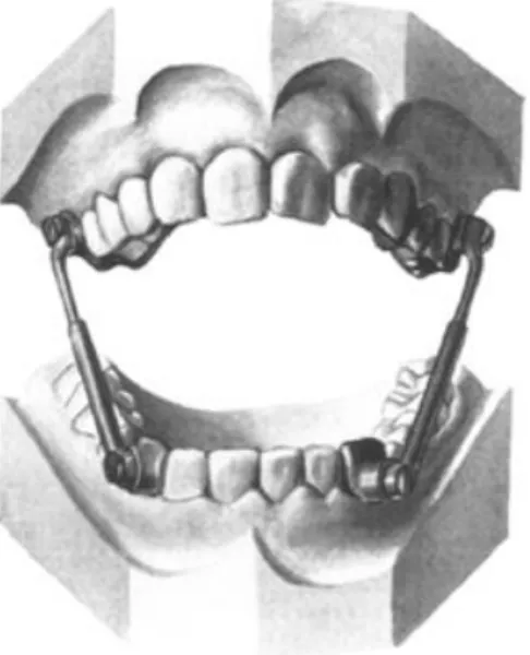
Appliance Design
Herbst appliance design has evolved over the past 100 years; however, the basic mechanism has remained unchanged. The device includes bilateral telescope mechanisms that guide the mandible into an anterior position during rest, and all functional
movements17. Original appliance design employed by Emile Herbst included crowns on the upper first molars, and crowns on the lower canines with curved telescoping
mechanisms that were designed to mimic the Curve of Spee (Figure A)17.
4
Herbst (Allesee Orthodontic Appliances, Sturtevant, WI) has been produced. This appliance allows the provider to place fixed appliances on the upper and lower arches from the second premolars. However, because of the short arm, undesirable vertical side effects may occur. Further development on this design is warranted.
Adverse effects of cantilever Herbst and design modifications: The cantilever
Herbst design requires extra consideration. Because of the long anterior arm extension, the distance of the force to the center of rotation is very large and can lead to significant mesial tipping of the mandibular molars. For this reason, an occlusal rest that extends from the mesial of the mandibular molar to the occlusal of the 1st premolar is
recommended. In addition, a rest from the distal of the mandibular first molars to the occlusal of the mandibular second molars helps to prevent eruption of the second molar. A lower lingual holding(LLHA) arch is often included in the design of the cantilever Herbst in order to prevent mesial crown tip of the mandibular molars.
In the maxilla, occlusal rests are extended from the distal of the first molars to the occlusal of the second molars. This also helps to control distal tipping of the first molars and prevents extrusion of the second molar.
Proclination of the lower incisors can be prevented in the cantilever Herbst with labial wires that add negative root torque of 10 degrees. Adding brackets to the lower incisors can also help to control the cantilever forces exhibited on the molars by
increasing anchorage. In addition, the archwire tubes on the terminal ends of the Herbst appliance can be placed gingivally in order to help correct deep bites with lower incisor intrusion. Conversely, the archwire tubes can be placed occlusally to help in the
5
University of North Carolina (UNC) graduate orthodontic department commonly utilizes the Mini Scope cantilever design (Allesee Orthodontic Appliances, Sturtevant, WI) to improve treatment efficiency without the unwanted vertical side effects. The rest of this discussion will focus on the Miniscope cantilever Herbst design since it is the most widely used. The purpose of this paper is to review the literature on the Herbst appliance and investigate the biomechanical effects leading to dentoalveolar and skeletal changes.
Dentoalveolar effects
Dentoalveolar effects of the Herbst provide large changes leading to class II correction. In general, mandibular molars will move mesially (often tipping) between 0.5 and 5-5mm. Maxillary molars may have up to 1 mm of intrusion, and distalize between 0.6 and 3.0 mm19. Distal tipping of the maxillary molars between 5.6° and 6.4° are also observed19. The mandibular and maxillary 2nd molars often extrude because
overcorrection of the OJ to an end-to end or negative overjet causes posterior
disocclusion. The lower incisors will Procline between 5.4° and 10.8° and will move mesially between 0.2mm and 4.0 mm19. The occlusal plane rotates in a clockwise direction due to intrusion of maxillary molars between 1.1° -5.5°.
Skeletal Effects
6
many studies, and the effect has been shown to be statistically similar to the effect produced by headgear16, 25-29. Meanwhile, some studies suggest the skeletal headgear effect displayed by the Herbst is negligible. 6, 30, 31 Ultimately, available data which examines the extent of skeletal verse dentoalveolar adaptation in that lead to the class II correction when using the Herbst is controversial. 24, 28, 30, 32 The skeletal component of class II correction has been reported to extend from 13% to 85%. 5, 7, 14, 28-30, 30, 31, 33-35 Maxillary changes:
When considering the variance in the literature, it is important to understand the various methodologies employed to measure changes in A point. The method developed by Pancherz utilizes a reference grid constructed from the occlusal line (OL) and the occlusal line perpendicular (OLp)14. Maxillary measurements using this method are subject to patient positioning errors. Many studies use SNA to examine maxillary changes 5, 25, 26. However, increases in the vertical dimension as seen with growth will mask the anterior-posterior change when using these angular measurements8. Skeletal changes observed at A point, undeniably depend on the methodologies used.
7
reported 19, 25, 30, 34. It is important to understand many studies found no difference in the anterior-posterior projection of the maxilla. 6, 30, 31
Mandibular changes:
Alteration of anterior-posterior projection of the mandible can be attributed to 1) changes in mandibular growth, 2) changes in the direction of growth and/ or 3) condylar/ fossa positional changes. Previous studies report conflicting results with some showing increased mandibular length with Herbst treatment 6, 8, 14, 14, 16, 25-27, 27-29. While other studies show no significant increase in mandibular length 30, 35. Deviations in patient positioning, as well as differences in magnification ratios between the left and right sides of the mandible can effect 2-D measurements of mandibular corpus length and ramus height.
Currently, most of the literature that evaluates mandibular growth following functional appliance therapy use condylion, an arbitrary condylar point, or a proxy- point such as articulare 6, 9, 14, 25, 27-29, 31, 33, 36-39. Condylion landmark identification is associated with low reliability due to obstruction of the overlying temporal bone40. Utilizing an arbitrary condylar point, as in the method described by Creekmoor, and used by Pancherz
8
errors will also affect vertical measurement error. Lastly, using articulare as a proxy condylar point is going to present significant measurement error. The position of
articulare is dependent on vertical and anterio-posterior changes of the glenoid fossa and condyle. Because articulare is dependent on growth, it does not suit well as a proxy point for condylion in longitudinal growth studies 38.
9
In addition to increased length, alterations in growth pattern will also impact anterior-posterior projection of the mandibular base. Opening of the gonial angle and posterior flexure of the condyle are anatomical changes that can lead to more anterior mandibular positioning. Animal studies have shown mild opening of the gonial angle with mandibular advancement 45, 46. And some human studies have made similar conclusions 14, 28. Initial placement of the Herbst causes the condyle to be placed anteriorly onto the articular eminence. After 6-12 weeks, the condyles showed a more posterior position in the glenoid fossa, and the posterior superior aspect of the condyle showed increased signal intensity on MRI 39. Condylar osteogenesis during Herbst treatment has also been shown in animal studies45-49. Sagittal condylar growth has been reported to occur between 1.8 and 3.8 mm14, 15, 19, 38, 43. The condyle moves between 1.5-3.1 mm superiorly and 2.1- 4.0 mm posteriorly14, 15, 19. Interestingly, the direction of condylar osteogenesis occurs in the direction of tension from the stretch of disc fibers on the condyle and glenoid fossa. 47
It is important to realize that measurements used to evaluate changes in gonial angle and condylar flexure in human studies all rely on reliable identification of condylion, thus the findings from these studies need to be interpreted cautiously.
10
studies 39. Pancherz et al looked at the size of the joint space pre- and post Herbst treatment. They found that there was no statistical difference in the condylar position. However, there was great variation among patients. It was revealed that post treatment condylar positions were on average slightly more anterior than pretreatment positions. Glenoid Fossa:
Translation of the glenoid fossa, has been shown to contribute to mandibular positional changes post Herbst treatment in animal studies 45-49. However, 2D imaging techniques used in human studies are greatly flawed when assessing for remodeling of the glenoid fossa. Human studies often rely on an unchanged condyle-fossa relationship because they utilize the method described by Buschang and Santos-Pinto 50, 51. Ruf and Pancherz conducted and MRI study to evaluate effective condylar growth in Herbst patients 39. They noted increase uptake in the T2-weighted sequences in the glenoid fossa and condyle. This was interpreted to be definitive areas of condyle and fossa remodeling. However, because the incidence of capsulitis rises during Herbst treatment up to 100%, virtually all patients would be expected to have increased T2 signal due to the amplified inflammatory process 52, 53. Differentiating inflammatory processes from the cellular cascade of skeletal remodeling is difficult. Additionally, techniques to register and superimpose MRI scans to evaluate changes critically from T1 to T2 have not been developed for the cranial base. Therefore, MRI scans cannot be used to adequately examine skeletal adaptations until a proper registration and superimposition technique is developed.
11
also evident that cellular responses to mandibular advancement were the most evident in the posterior glenoid fossa of rats47. Animal studies have clearly shown the adaptive potential of the glenoid fossa in response to functional appliance therapy 10, 45-49, 54, 55. Studies in monkeys reveal similar adaptive potential of the glenoid fossa45, 46, 48, 49, 54, 55. In fact Voudouris et. al detected reversal lines in the genoid fossa in cyanomologous monkeys (Macaca fasicularis), that are associated with the redirection of growth45, 46. He extended these findings to conclude the natural downward and backward growth of the glenoid fossa from the sella-nasion plane during facial growth might have the backward component of this natural growth pattern restricted by the Herbst appliance45, 46. Human studies have suggested remodeling may occur. However, these studies use condylion or articulare as a proxy point to approximate the position of the fossa. Those conclusions were not absolute due to imaging limitations and measurement errors 38, 39, 50. After examining all of the condylar and fossa changes, they concluded overall the “effective condylar growth” during Herbst treatment resulted in six-times more horizontal growth and four-times more vertical growth when compared to Bolton Standards. 39
The literature reveals tremendous variation in the amount of skeletal adaptation leading to improvement in the class II profile. This variation stems from the limitations of 2D imaging. Further research needs to be conducted using novel three dimensional
12
Figure A: Original Herbst Design
Pancherz et al. “History, background and development of the Herbst appliance”Semin Orthod. 2003; 9(1): 3-11.
13
Figure B: The Acrylic Herbst Design
Pancherz et al. “History, background and development of the Herbst appliance”Semin Orthod. 2003; 9(1): 3-11.
Figure C: The Cantilever Herbst Design
14
REFERENCES
1. Proffit WR, Fields HW,Jr, Moray LJ. Prevalence of malocclusion and orthodontic treatment need in the United States: estimates from the NHANES III survey. Int J Adult Orthodon Orthognath Surg. 1998;13(2):97-106.
2. McNamara JA,Jr. Components of class II malocclusion in children 8-10 years of age. Angle Orthod. 1981 Jul;51(3):177-202.
3. Proffit WR. Contemporary orthodontics. Fields HW, Proffit WR,.donor., Sarver DM, editors. St. Louis, Mo.: Mosby Elsevier; 2007.
4. Bell WH, 1927-. Surgical correction of dentofacial deformities. Proffit WR and Wilson JK,.donor., editors. Philadelphia: Saunders; 1980.
5. Ruf S, Pancherz H. Herbst/multibracket appliance treatment of Class II division 1 malocclusions in early and late adulthood. a prospective cephalometric study of consecutively treated subjects. Eur J Orthod. 2006 Aug;28(4):352-60.
6. Ruf S, Pancherz H. Dentoskeletal effects and facial profile changes in young adults treated with the Herbst appliance. Angle Orthod. 1999 Jun;69(3):239-46.
7. Bock N, Pancherz H. Herbst Treatment of Class II division 1 Malocclusions in Retrognathic and Prognathic Facial Types. Angle Orthod. 2006 11/01;
2011/05;76(6):930-41. Available from: http://dx.doi.org/10.2319/100605-352.
8. Cozza P, Baccetti T, Franchi L, De Toffol L, McNamara Jr JA. Mandibular changes produced by functional appliances in Class II malocclusion: A systematic review. American Journal of Orthodontics and Dentofacial Orthopedics. 2006
5;129(5):599.e1,599.e12.
9. Hagg U, Pancherz H. Dentofacial orthopaedics in relation to chronological age, growth period and skeletal development. An analysis of 72 male patients with Class II division 1 malocclusion treated with the Herbst appliance. Eur J Orthod. 1988 Aug;10(3):169-76. 10. Hinton RJ, McNamara JA,Jr. Effect of age on the adaptive response of the adult temporomandibular joint. A study of induced protrusion in Macaca mulatta. Angle Orthod. 1984 Apr;54(2):154-62.
11. Baccetti T, Franchi L, McNamara JA,Jr. An improved version of the cervical vertebral maturation (CVM) method for the assessment of mandibular growth. Angle Orthod. 2002 Aug;72(4):316-23.
15
13. McNamara JA,Jr, Howe RP, Dischinger TG. A comparison of the Herbst and Frankel appliances in the treatment of Class II malocclusion. Am J Orthod Dentofacial Orthop. 1990 Aug;98(2):134-44.
14. Pancherz H. The mechanism of Class II correction in Herbst appliance treatment. A cephalometric investigation. Am J Orthod. 1982 Aug;82(2):104-13.
15. Pancherz H. The Herbst appliance--its biologic effects and clinical use. Am J Orthod. 1985 Jan;87(1):1-20.
16. Wigal TG, Dischinger T, Martin C, Razmus T, Gunel E, Ngan P. Stability of Class II treatment with an edgewise crowned Herbst appliance in the early mixed dentition: Skeletal and dental changes. Am J Orthod Dentofacial Orthop. 2011 Aug;140(2):210-23. 17. Pancherez H. History, background and development of the Herbst appliance. Semin Orthod. 2003;9(1):3-11.
18. Keim RG, Gottlieb EL, Nelson AH, Vogels DS,3rd. 2009 JCO Orthodontic Practice Study. Part 1 Trends. J Clin Orthod. 2009 Oct;43(10):625-34.
19. Papadopoulos M. Orthodontic treatment of the Class II noncompliant patient: Current principles and techniques. Elsevier Health Sciences. 2006;34:35-56.
20. Mayes J. Improving appliance efficiency with the cantilever Herbst--A new answer to old problems. Clinical Impressions. 1994;3:2,2-5; 17-19.
21. Siara-Olds N, Pangrazio-Kulbersh V, Berger J, Bayirli B. Long-Term Dentoskeletal Changes with the Bionator, Herbst, Twin Block, and MARA Functional Appliances. Angle Orthod. 2010 01/01; 2011/05;80(1):18-29. Available from:
http://dx.doi.org/10.2319/020109-11.1.
22. von Bremen J, Pancherz H. Efficiency of early and late Class II Division 1 treatment. Am J Orthod Dentofacial Orthop. 2002 Jan;121(1):31-7.
23. Obijou C, Pancherz H. Herbst appliance treatment of Class II, Division 2
malocclusions. American Journal of Orthodontics and Dentofacial Orthopedics. 1997 9;112(3):287-91.
24. Serbesis-Tsarudis C, Pancherz H. “Effective” TMJ and Chin Position Changes in Class II Treatment. Angle Orthod. 2008 09/01; 2011/05;78(5):813-8. Available from:
http://dx.doi.org/10.2319/082707-391.1.
25. Baccetti T, Franchi L, Stahl F. Comparison of 2 comprehensive Class II treatment protocols including the bonded Herbst and headgear appliances: A double-blind study of consecutively treated patients at puberty. American Journal of Orthodontics and
16
26. Manfredi C, Cimino R, Trani A, Pancherz H. Skeletal Changes of Herbst Appliance Therapy Investigated With More Conventional Cephalometrics and European Norms. Angle Orthod. 2001 06/01; 2011/05;71(3):170-6. Available from:
http://www.angle.org/doi/abs/10.1043/0003-3219%282001%29071%3C0170%3ASCOHAT%3E2.0.CO%3B2.
27. Valant JR, Sinclair PM. Treatment effects of the Herbst appliance. Am J Orthod Dentofacial Orthop. 1989 Feb;95(2):138-47.
28. Pancherz H. The effects, limitations, and long-term dentofacial adaptations to treatment with the Herbst appliance. Semin Orthod. 1997 Dec;3(4):232-43.
29. VanLaecken R, Martin CA, Dischinger T, Razmus T, Ngan P. Treatment effects of the edgewise Herbst appliance: a cephalometric and tomographic investigation. Am J Orthod Dentofacial Orthop. 2006 Nov;130(5):582-93.
30. Barnett GA, Higgins DW, Major PW, Flores-Mir C. Immediate skeletal and
dentoalveolar effects of the crown- or banded type Herbst appliance on Class II division 1 malocclusion. Angle Orthod. 2008 Mar;78(2):361-9.
31. Nelson B, Hansen K, Hägg U. Class II correction in patients treated with Class II elastics and with fixed functional appliances: A comparative study. American Journal of Orthodontics and Dentofacial Orthopedics. 2000 8;118(2):142-9.
32. Baltromejus S, Ruf S, Pancherz H. Effective temporomandibular joint growth and chin position changes: Activator versus Herbst treatment. A cephalometric
roentgenographic study. The European Journal of Orthodontics. 2002 December 01;24(6):627-37.
33. Pancherz H. The nature of Class II relapse after Herbst appliance treatment: a cephalometric long-term investigation. Am J Orthod Dentofacial Orthop. 1991 Sep;100(3):220-33.
34. Pancherz H, Anehus-Pancherz M. The headgear effect of the Herbst appliance: a cephalometric long-term study. Am J Orthod Dentofacial Orthop. 1993 Jun;103(6):510-20.
35. Burkhardt DR, McNamara JA,Jr, Baccetti T. Maxillary molar distalization or mandibular enhancement: a cephalometric comparison of comprehensive orthodontic treatment including the pendulum and the Herbst appliances. Am J Orthod Dentofacial Orthop. 2003 Feb;123(2):108-16.
17
37. LaHaye MB, Buschang PH, Alexander RG“, Boley JC. Orthodontic treatment changes of chin position in Class II Division 1 patients. American Journal of Orthodontics and Dentofacial Orthopedics. 2006 12;130(6):732-41.
38. Pancherz H, Fischer S. Amount and Direction of Temporomandibular Joint Growth Changes in Herbst Treatment: A Cephalometric Long-Term Investigation. Angle Orthod. 2003 09/01; 2011/05;73(5):493-501. Available from:
http://www.angle.org/doi/abs/10.1043/0003-3219%282003%29073%3C0493%3AAADOTJ%3E2.0.CO%3B2.
39. Ruf S, Pancherz H. Temporomandibular joint growth adaptation in Herbst treatment: a prospective magnetic resonance imaging and cephalometric roentgenographic study. Eur J Orthod. 1998 Aug;20(4):375-88.
40. Adenwalla ST, Kronman JH, Attarzadeh F. Porion and condyle as cephalometric landmarks— An error study. American Journal of Orthodontics and Dentofacial Orthopedics. 1988 11;94(5):411-5.
41. Creekmore TD. Inhibition or stimulation of the vertical growth of the facial complex, its significance to treatment. Angle Orthod. 1967 Oct;37(4):285-97.
42. Karthi M, Shankar K, Venkatesan A. Reliability assessment of condylion and
articulare for mandibular length measurements- A cephalometric study. SRM University Journal of Dental Sciences. 2011;2(3):195.
43. Pancherz H, Michailidou C. Temporomandibular joint growth changes in hyperdivergent and hypodivergent Herbst subjects. A long-term roentgenographic cephalometric study. Am J Orthod Dentofacial Orthop. 2004 Aug;126(2):153,61; quiz 254-5.
44. Nelson B, Hagg U, Hansen K, Bendeus M. A long-term follow-up study of Class II malocclusion correction after treatment with Class II elastics or fixed functional
appliances. Am J Orthod Dentofacial Orthop. 2007 Oct;132(4):499-503.
18
48. McNamara Jr. JA, Carlson DS. Quantitative analysis of temporomandibular joint adaptations to protrusive function. Am J Orthod. 1979 12;76(6):593-611.
49. McNamara Jr. JA. Neuromuscular and skeletal adaptations to altered function in the orofacial region. Am J Orthod. 1973 12;64(6):578-606.
50. Pancherz H, Stickel A. Position changes of mandibular condyle in Herbst treatment. Radiographic study. Inf Orthod Kieferorthop. 1989;21(4):515-27.
51. Woodside DG, Metaxas A, Altuna G. The influence of functional appliance therapy on glenoid fossa remodeling. American Journal of Orthodontics and Dentofacial
Orthopedics. 1987 9;92(3):181-98.
52. Pancherz H, Michailidou C. Temporomandibular joint growth changes in hyperdivergent and hypodivergent Herbst subjects. A long-term roentgenographic cephalometric study. American Journal of Orthodontics and Dentofacial Orthopedics. 2004 8;126(2):153-61.
53. Buschang PH, Santos-Pinto A. Condylar growth and glenoid fossa displacement during childhood and adolescence. Am J Orthod Dentofacial Orthop. 1998
Apr;113(4):437-42.
54. Ruf S, Pancherz H. Does bite-jumping damage the TMJ? A prospective longitudinal clinical and MRI study of Herbst patients. Angle Orthod. 2000 Jun;70(3):183-99.
55. Ruf S, Pancherz H. Orthognathic surgery and dentofacial orthopedics in adult Class II Division 1 treatment: Mandibular sagittal split osteotomy versus Herbst appliance.
20
CHAPTER II
MANUSCRIPT
INTRODUCTION
Treatment of Class II malocclusions are a common challenge amongst
orthodontists in the United States. Approximately one third of all patients have a Class II, Division 1 malocclusion. Mandibular retrognathism serves as the primary etiologic factor in a majority of those cases1, 2. Functional appliances have been shown effective in correcting class II malocclusions by decreasing overjet and achieving Angle class I canine and molar relationships 1-5. Eliminating patient compliance factors and delivering continuous forces give fixed functional appliances a distinct treatment advantage
compared to removable appliances. Specifically, many studies have reported greatest anterior-posterior improvements in mandibular projection when using the fixed Herbst functional appliance 1, 2, 5-10.
Functional appliances, such as the Herbst, have been purported to improve mandibular projection, consequently improving the underlying skeletal discrepancy 5, 6, 8, 11
. However, available data which examines the extent of skeletal verse dentoalveolar adaptation in class II correction with functional appliances is controversial 3, 4, 11, 12. The skeletal component of class II correction has been reported to extend from 13% to 85% 3, 3, 9, 12-19
21
from physiologic and anatomic inconsistencies in study subjects, to limitations in study methodologies.
Skeletal adaptation depends on physiologic factors, such as skeletal maturation and growth potential. It is established most efficient treatment with the Herbst appliance is conducted during the pubertal growth spurt 6, 20, 20-22. Yet, studies focusing on Herbst treated patients treated during the peak of pubertal growth, exhibit vast inconsistencies in the extent of skeletal verse dentoalveolar adaptation1, 6, 11, 15, 20, 23, 24. The differences in treatment timing alone do not account for ambiguity reported in the literature. Studies suggest that anatomical factors, such as facial type and gonial angle may have an impact on the extent of skeletal adaptation 1, 9, 15, 20. However, literature focusing on these factors is limited and most studies include well matched control subjects thus nullifying these anatomic factors. Ultimately, it is impossible to accurately assess the extent of skeletal adaptation, let alone examine how anatomic factors affect these adaptations, with the limitations of current methodologies.
Condylion is used in several studies to evaluate mandibular length changes 1, 3, 9, 13, 20, 25, 26
22
mandibular projection with Herbst treatment, factors leading to these changes remain elusive due to limitations in 2D cephalometric imaging.
Two-dimensional imaging is subject to magnification, distortion, patient positioning errors and obstruction of critical landmarks by overlapping anatomical structures. Additionally, there is inherent examiner bias in the registration process if examiners are not blinded. All of these factors reduce measurement accuracy, which influences our ability to accurately report skeletal changes resulting from the Herbst appliance. Shortcomings of 2D cephalometric imaging can also account for discord in the literature regarding skeletal effects of this appliance. 3D imaging techniques overcome these inadequacies. Studies by Cevidanes et. al. demonstrate accurate superimposition of CBCT scans in growing patients 38-40. The protocol uses a voxel based registration technique which eliminates examiner bias in the registration process. Maxillary and mandibular adaptive and positional changes can be accurately examined and measured relative to the anterior cranial base using these 3D superimposition techniques 38-40. This method gives us more accurate and detailed information when assessing for skeletal changes.
23
maxillary positional changes, differences in mandibular growth and mandibular positional changes will be evaluated.
MATERIALS AND METHODS
Adolescent patients near the pubertal growth spurt (determined by cervical vertebral maturation method stages CS3-CS4) with class II skeletal relationships (ANB> and Class molar relationships seen at the University of North Carolina Department of Orthodontics were evaluated for Herbst appliance therapy41. Seven consecutive patients, who met the inclusion criteria, were enrolled in this prospective pilot study (Table 1). Seven control subjects (treated with Class II elastics) were obtained from the University of Minnesota database. Approval from the University of North Carolina institutional review board was obtained for this study.
Herbst appliance design included mini-scope telescoping arms with cantilever and occlusal rests second molars and first premolars (Allesee Orthodontic Appliances,
Sturtevant, WI). The appliance was initially advanced to Class I molar position. Fixed appliances were placed on maxillary and mandibular incisors and canines and tied back to the molar crown after alignment was achieved. Herbst appliance was advanced at 2 mm increments to an overcorrected position (OJ= 0 to -1 mm). The duration of
24
CBCT scans were taken pre-treatment for both Herbst and Control patients (T1H and T1c) and post Herbst removal (T2H) and post- treatment for control patients (T2C). Herbst patients’ scans were taken using the New Tom 3G (Aperio Services LLC, Sarasota, FL) with a 12 inch field of view (FOV). Control subject CBCT scans were taken using an iCat machine (Imaging Sciences Interation, Hatfield, Pa) with a 16x22cm FOV. The dicom scans were downsized to 0.5x0.5x0.5 mm and de-identified using Imagine http://www.ia.unc.edu/dev/download/imagine/index.htm). ITK SNAP
(www.itksnap.org) was used to construct virtual 3D surface models 38. T1 and T2 scans were registered on the anterior cranial fossa using a fully automated voxel-wise ridged registration technique described by Cevidanes 38-40. Boundaries for the anterior cranial base registration were defined anteriorly by inner cortical layer of the frontal bone, posteriorly by the anterior wall of sella, and laterally including the lesser wings of the sphenoid bone and frontal bone marking the superior boundary of the orbits. This region includes the cribiform plate and the superior aspect of the ethmoid bone. These structures are known to complete growth by the age of seven, and are thus considered stable
landmarks 43-45.
25
corpus at the point where it starts to curve to from the angle of the mandible, identified from a sagittal view with the functional occlusal plane parallel to the floor. For all measurements, positive values indicated an anterior displacement and negative values indicated a posterior displacement relative to time 1. Cephalometric landmark placement on 3D volumes has been shown to be accurate and reproducible 46, 47
All measurements were repeated two times by the same examiner (ML) at one week interval to assess intraexaminer reliability for landmark identification, point to point and ICP measurements.
Statistical Analysis:
Data analysis was conducted using SPSS statistical software package. Means, standard deviations and ranges were calculated for the Herbst and Control subjects to describe the samples. Statistical differences were assessed using analysis of variance (ANOVA). Wilcoxon rank test was employed to assess differences in displacement between Herbst and Control subjects. Intraobserver reliability was evaluated for repeated measures using intraexaminer correlation coefficient (ICC) test. Statistical significance was tested at P<0.05.
RESULTS
26
Intraexaminer correlation coefficient revealed high correlation for all
measurements. ICC was above 0.90 for all ICP, point-to-point and angular measurements indicating high reliability for landmark identification.
Maxillary Skeletal Changes:
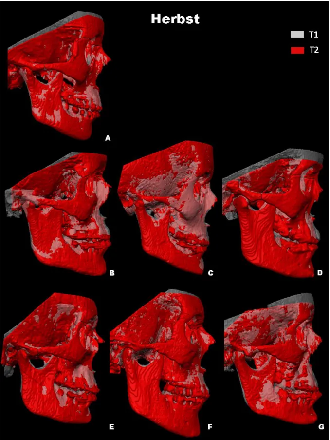
Qualitative assessment of skeletal changes is best conducted using a
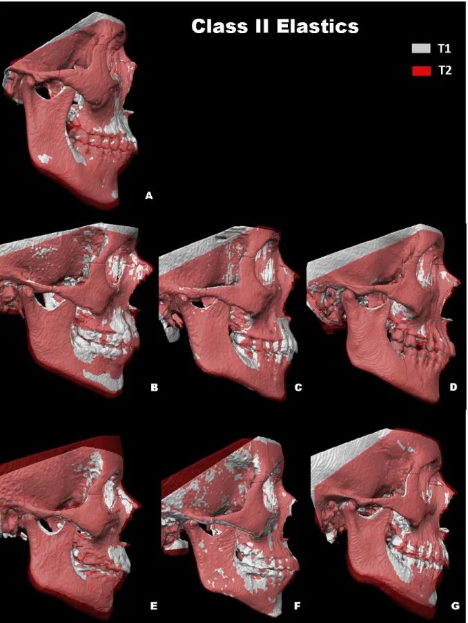
semitransparent overlay of the superimpositions (Figures 2, 3, 5, 6 and 9). For structures that are obstructed from view, alternating the transparency of the T1 and T2 images allow for better visualization. Maxillary displacement changes are shown in Figure 2 and Figure 3. All Herbst patients, except for subject A, demonstrated maxillary restraint. Herbst subjects B, C, D and G displayed largest maxillary displacements. Retroclination of upper incisors is evident in Control subjects C and E, and maxillary restraint can be noted in control subject A (Figure 3).
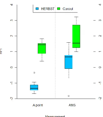
Quantitative assessments of maxillary changes are reported in Table II and Figure 4. More anterior projection of A point and ANS (1.2mm and 1.96mm respectively) was demonstrated by treated controls, when compared to Herbst subjects (-1.22 and 0.26mm respectively) (P<0.01).
Mandibular Skeletal Changes:
27
flexure reveal no statistical difference between Herbst and control subjects (Table II, Figure 8).
Condylar/ Glenoid Fossa Changes:
Mean condylar and glenoid fossa displacement is shown in Table II and Figure 11. In general, Herbst patients showed forward displacement of the condyles while control subjects exhibited posterior displacement. The mean difference in displacement of the condyle between the two groups is approximately 2.5-2.9mm when measured from the anterior surface (P<0.001) and 1.74-1.35mm when measured from the posterior surface of the condyles (P< 0.05).
In addition, point-to-point linear changes were evaluated for condylion (Table II, Figure11). Box plots in figure 11 depict net anterior displacement of condylion in Herbst patients (right: 0.38mm, left: 0.56mm). Conversely, a net posterior displacement of condylion was observed in the control group (right: -0.88mm, left:-1.16mm). These changes in condylar position are less than those found using ICP (right: 1.26mm, left: 1.72 mm), but remain statistically significant (P>0.01).
Mean changes for fossa remodeling are shown in Table II and Figure 7. Herbst patients showed resorption at the anterior wall (right: 1.69mm and left: 1.43mm) with deposition at the posterior wall of the glenoid fossa (right: 0.59mm, left: 0.79mm) (Figure10). Conversely, the control group showed boney apposition on the anterior wall (right: -1.51mm, left: -1.31mm) with resorption at the posterior wall (right: -1.24mm, left: -1.41mm). This corresponds with the direction of condylar displacement within the respective groups. (Figure 11).
28
Figure 12 shows the composite of individual color maps which demonstrates global changes computed using iterative closest point algorithms. Although maxillary,
mandibular, condylar and glenoid fossa positional changes in Herbst patients show statistical differences when compared to the control subjects, considerable variation as to the magnitude and direction of these skeletal changes are seen when examining color maps of individual cases (Figure 12).
DISCUSSION
Past literature examining functional appliances often use samples from the Bolton-Brush or Michigan Growth Studies to obtain their untreated class II controls 11, 13, 26
. Unfortunately no such 3-D sample exists today. An ethical issue regarding not treating class II malocclusion during the pubertal growth spurt, a time associated with optimal treatment response for class II correction, prevents us from obtaining 3-D scans from untreated class II patients to serve as control. Class II elastics have been shown to act primarily through dentoalveolar movements with no skeletal enhancement.18 Nelson et al reported skeletal contribution to reduction in overjet was only 4% in control subjects treated with elastics compared to 51% in the Herbst subjects. Therefore, using class II subjects treated solely with class II elastics, as control subjects, can be substantiated.
The first aim of this study was to evaluate maxillary positional changes in Herbst subjects and compare these changes to controls. Numerous studies report a maxillary restraining effect, comparable to headgear, produced by the Herbst treatment1, 12, 13, 26, 28, 51
29
employed to measure changes in A point. The method developed by Pancherz utilizes a reference grid constructed from the occlusal line (OL) and the occlusal line perpendicular (OLp)15. Maxillary measurements using this method are subject to patient positioning errors. Many studies use SNA to examine maxillary changes 1, 14, 28. However, increases in the vertical dimension as seen with growth will mask the anterior-posterior change when using these angular measurements6. Skeletal changes observed at A point, undeniably depend on the methodologies used. Our 3-D study showed the anticipated forward and downward growth pattern of the maxilla in the majority of our class II control subjects. However the Herbst group showed a mild maxillary restraining effect.
Alteration of anterior-posterior projection of the mandible can be attributed to 1) changes in mandibular growth, 2) changes in the direction of growth and/ or 3) condylar/ fossa positional changes. Previous studies report conflicting results with some showing increased mandibular length with Herbst treatment 1,12, 15, 28, 51,, while other studies show no significant increase in mandibular length 3, 19. Deviations in patient positioning, as well as differences in magnification ratios between the left and right sides of the
mandible can effect 2-D measurements of mandibular corpus length and ramus height. In addition, the conflicting findings regarding mandibular length were addressed by
Voudouris et. al. who noted that in pre-adolescent cyanomologous monkeys (Macaca fasicularis), the condylar growth response was increased with Herbst treatment however,
30
Perhaps skeletal maturity may have a larger and more directed influence on skeletal response to the Herbst appliance than we previously understood. While our findings suggest no statistically significant difference for mandibular length between Herbst and control groups, it is important to recall the difference in observation times for these two groups. The control group had an additional 5 months of observation time which would increase the perceived mandibular growth (Co-Gn) when compared to Herbst subjects. In additional to growth, mandibular directional growth changes, such as opening of the gonial angle and posterior condylar flexure, will impact anterior-posterior
projection of the mandibular base. Animal studies have shown mild opening of the gonial angle with mandibular advancement 36, 37. And some human studies have made similar conclusions 12, 15. Our study, along with others 1, 9, 28, showed no difference in gonial angle or condylar flexure between Herbst and control subjects. It is worth noting that Herbst subjects in our study had lower mandibular plan angle. A previous study by Pancherz et al. examined skeletal changes in hyperdivergent and hypodivergent facial types 29. Their results found hyperdivergent subjects demonstrated more posteriorly directed condylar growth compared to hypodivergent subjects. Posteriorly directed condylar growth would lead to an opening of the gonial angle, and increased condylar flexure. It is possible that the larger number of hypodivergent subjects in this study may effect our results on gonial angle and condylar flexure changes.
condyle-31
fossa relationship because they utilize the method described by Buschang and Santos-Pinto 7, 54. Furthermore, these studies use condylion or articulare as a proxy point to approximate the position of the fossa. Ruf and Pancherz conducted an MRI study to evaluate effective condylar growth in Herbst patients 24. They noted increase uptake in the T2-weighted sequences in the glenoid fossa and condyle. This was interpreted to be definitive areas of condyle and fossa remodeling. However, because the incidence of capsulitis rises during Herbst treatment up to 100%, virtually all patients would be expected to have increased T2 signal due to the amplified inflammatory process 55, 56. Differentiating inflammatory processes from the cellular cascade of skeletal remodeling is difficult. In addition, MRIs lack detailed information regarding bony structure and may not be the best tool to evaluate fossa remodeling. With 3D cone beam computed tomography scans, and current registration and superimposition techniques, we were able to accurately analyze skeletal changes occurring at the glenoid fossa (figure 10). We found resorption of the anterior wall of the glenoid fossa with deposition at the posterior wall in Herbst patients. This is in direct contrast to findings in the control subjects who exhibited posteriorly directed remodeling of the fossa. Posterior repositioning of the glenoid fossa we observed in the control group has been well documented in class II subjects, and represents the expected class II growth pattern43, 54, 57-60. Our findings suggest the Herbst appliance is altering the growth pattern of the glenoid fossa resulting in a more anteriorly positioned fossa and therefore more anteriorly position mandible.
32
displacements are occurring in unison. This supports conclusions made in both animal and human studies suggesting that the condyle- glenoid fossa relationship remains relatively unchanged with Herbst treatment 24, 33, 61. This is the first 3-D study to clearly demonstrate anterior repositioning of the fossa and condyle in response to class II functional appliance therapy in humans.
This study was designed as a pilot study to determine whether skeletal differences between Herbst subjects and patients treated with class II elastics could be surmised. As a pilot study, limitations in the sample size are inherent. Additional weaknesses of this study sample arise from differences in observation time between Herbst subjects and control patients. This confounder will have an effect on the statistical comparison for treatment differences. In essence, having a control group enables us to differentiate skeletal changes due to treatment verse growth. Since the control group had an average of 5 months longer observation time, they are anticipated to exhibit larger changes due to growth. Most likely this difference might underestimate the skeletal changes resulting from Herbst treatment. A larger study, which can further evaluate changes we observed for these patients, and a long- term follow up study are recommended. Relapse potential for patients treated with the Herbst appliance is well documented; however the
33
Clearly, follow up studies using 3D imaging techniques to address the true nature of the relapse of Herbst subjects are indicated.
CONCLUSIONS
3D imaging and superimposition techniques revealed the following skeletal adaptations:
1. Herbst treatment produced anterior displacement of the condyles with adaptive remodeling of the glenoid fossa while Class II controls exhibited distal displacement of the TMJ complex.
2. The Herbst group showed more maxillary restraint compared to the controls. 3. No significant difference in mandibular corpus and ramal growth, condylar
flexure and gonial angle change were observe between the two groups.
34
35
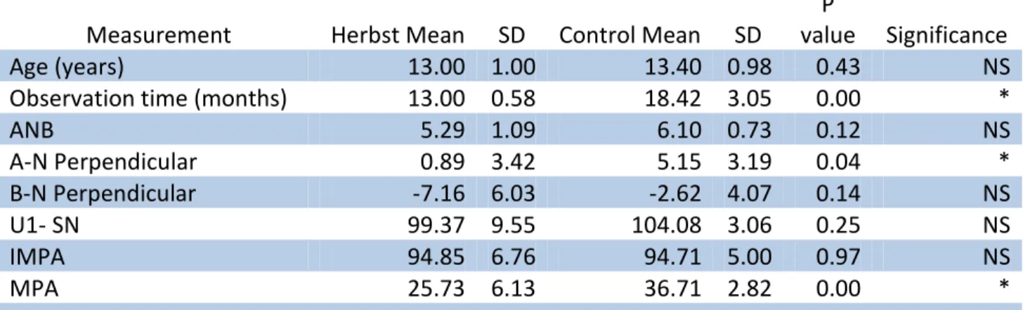
Table I. Demographics and statistical comparison for Herbst subjects and class II control subjects
Measurement Herbst Mean SD Control Mean SD
P
value Significance
Age (years) 13.00 1.00 13.40 0.98 0.43 NS
Observation time (months) 13.00 0.58 18.42 3.05 0.00 *
ANB 5.29 1.09 6.10 0.73 0.12 NS
A-N Perpendicular 0.89 3.42 5.15 3.19 0.04 *
B-N Perpendicular -7.16 6.03 -2.62 4.07 0.14 NS
U1- SN 99.37 9.55 104.08 3.06 0.25 NS
IMPA 94.85 6.76 94.71 5.00 0.97 NS
MPA 25.73 6.13 36.71 2.82 0.00 *
36
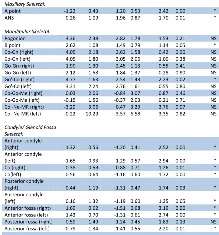
Table II. Difference between T1 and T2 skeletal changes for Herbst subjects and class II controls
Measurement
Herbst
Mean SD
Control
Mean SD Difference
P
value Significance
Maxillary Skeletal:
A point -1.22 0.43 1.20 0.53 2.42 0.00 *
ANS 0.26 1.09 1.96 0.87 1.70 0.01 *
Mandibular Skeletal:
Pogonion 4.36 2.38 2.82 1.78 1.53 0.21 NS
B point 2.62 1.08 1.49 0.79 1.14 0.05 *
Co-Gn (right) 4.05 2.18 3.62 1.58 0.42 0.90 NS
Co-Gn (left) 4.05 1.80 3.05 2.06 1.00 0.38 NS
Go-Gn (right) 1.90 1.30 2.45 1.13 0.55 0.41 NS
Go-Gn (left) 2.12 1.58 1.84 1.37 0.28 0.90 NS
Go'-Co (right) 4.77 1.63 2.54 1.43 2.23 0.02 *
Go'-Co (left) 3.31 2.24 2.76 1.61 0.55 0.80 NS
Co-Go-Me (right) 0.03 2.06 -0.84 3.07 0.87 0.46 NS
Co-Go-Me (left) -0.15 1.56 -0.37 2.03 0.21 0.71 NS
Co'-Ne-MR (right) -3.29 3.06 0.47 3.29 3.76 0.07 NS
Co'-Ne-MR (left) -0.22 10.29 -3.57 6.58 3.35 0.82 NS
Condyle/ Glenoid Fossa Skeletal:
Anterior condyle
(right) 1.32 0.56 -1.20 0.41 2.52 0.00 *
Anterior condyle
(left) 1.65 0.93 -1.29 0.57 2.94 0.00 *
Co (right) 0.38 0.59 -0.88 0.71 1.26 0.01 *
Co(left) 0.56 0.64 -1.16 0.60 1.72 0.00 *
Posterior condyle
(right) 0.44 1.19 -1.31 0.47 1.74 0.03 *
Posterior condyle
(left) 0.16 1.32 -1.19 0.60 1.35 0.05 *
Anterior fossa (right) 1.69 0.62 -1.51 0.68 3.19 0.00 *
Anterior fossa (left) 1.43 0.70 -1.31 0.61 2.74 0.00 *
Posterior fossa (right) 0.59 1.49 -1.24 0.45 1.83 0.13 NS
Posterior fossa (left) 0.79 1.34 -1.41 0.55 2.20 0.01 *
37
38
39
40
41
42
Figure 7. Box plots of mandibular skeletal changes for Herbst and control subjects.
43
Figure 9. Semitransparencies of left condyle for one Herbst and control subject from both sagittal and axial views. T1 and T2 3D volumes were registered at the anterior cranial base. Left condyles were isolated from adjacent structures for improved
44
45
46
47
REFERENCES
1. Baccetti T, Franchi L, Stahl F. Comparison of 2 comprehensive Class II treatment protocols including the bonded Herbst and headgear appliances: A double-blind study of consecutively treated patients at puberty. American Journal of Orthodontics and Dentofacial Orthopedics. 2009 6;135(6):698.e1,698.e10.
2. Shen G, Hägg U, Darendeliler M. Skeletal effects of bite jumping therapy on the mandible ? removable vs. fixed functional appliances. Orthodontics & Craniofacial Research. 2005;8(1):2-10.
3. Barnett GA, Higgins DW, Major PW, Flores-Mir C. Immediate skeletal and dentoalveolar effects of the crown- or banded type Herbst appliance on Class II division 1 malocclusion. Angle Orthod. 2008 Mar;78(2):361-9.
4. Baltromejus S, Ruf S, Pancherz H. Effective temporomandibular joint growth and chin position changes: Activator versus Herbst treatment. A cephalometric
roentgenographic study. The European Journal of Orthodontics. 2002 December 01;24(6):627-37.
5. Siara-Olds N, Pangrazio-Kulbersh V, Berger J, Bayirli B. Long-Term Dentoskeletal Changes with the Bionator, Herbst, Twin Block, and MARA Functional Appliances. Angle Orthod. 2010 01/01; 2011/05;80(1):18-29. Available from:
http://dx.doi.org/10.2319/020109-11.1.
6. Cozza P, Baccetti T, Franchi L, De Toffol L, McNamara Jr JA. Mandibular changes produced by functional appliances in Class II malocclusion: A systematic review. American Journal of Orthodontics and Dentofacial Orthopedics. 2006
5;129(5):599.e1,599.e12.
7. Pancherz H, Michailidou C. Temporomandibular joint growth changes in hyperdivergent and hypodivergent Herbst subjects. A long-term roentgenographic cephalometric study. American Journal of Orthodontics and Dentofacial Orthopedics. 2004 8;126(2):153-61.
8. Obijou C, Pancherz H. Herbst appliance treatment of Class II, Division 2
malocclusions. American Journal of Orthodontics and Dentofacial Orthopedics. 1997 9;112(3):287-91.
9. Bock N, Pancherz H. Herbst Treatment of Class II division 1 Malocclusions in Retrognathic and Prognathic Facial Types. Angle Orthod. 2006 11/01;
48
10. Lai M, McNamara J,James A. An evaluation of two-phase treatment with the herbst appliance and preadjusted edgewise therapy. Semin Orthod. 1998 3;4(1):46-58.
11. Serbesis-Tsarudis C, Pancherz H. “Effective” TMJ and Chin Position Changes in Class II Treatment. Angle Orthod. 2008 09/01; 2011/05;78(5):813-8. Available from:
http://dx.doi.org/10.2319/082707-391.1.
12. Pancherz H. The effects, limitations, and long-term dentofacial adaptations to treatment with the Herbst appliance. Semin Orthod. 1997 Dec;3(4):232-43.
13. VanLaecken R, Martin CA, Dischinger T, Razmus T, Ngan P. Treatment effects of the edgewise Herbst appliance: a cephalometric and tomographic investigation. Am J Orthod Dentofacial Orthop. 2006 Nov;130(5):582-93.
14. Ruf S, Pancherz H. Herbst/multibracket appliance treatment of Class II division 1 malocclusions in early and late adulthood. a prospective cephalometric study of consecutively treated subjects. Eur J Orthod. 2006 Aug;28(4):352-60.
15. Pancherz H. The mechanism of Class II correction in Herbst appliance treatment. A cephalometric investigation. Am J Orthod. 1982 Aug;82(2):104-13.
16. Pancherz H. The nature of Class II relapse after Herbst appliance treatment: a cephalometric long-term investigation. Am J Orthod Dentofacial Orthop. 1991 Sep;100(3):220-33.
17. Pancherz H, Anehus-Pancherz M. The headgear effect of the Herbst appliance: a cephalometric long-term study. Am J Orthod Dentofacial Orthop. 1993
Jun;103(6):510-20.
18. Nelson B, Hansen K, Hägg U. Class II correction in patients treated with Class II elastics and with fixed functional appliances: A comparative study. American Journal of Orthodontics and Dentofacial Orthopedics. 2000 8;118(2):142-9.
19. Burkhardt DR, McNamara JA,Jr, Baccetti T. Maxillary molar distalization or mandibular enhancement: a cephalometric comparison of comprehensive orthodontic treatment including the pendulum and the Herbst appliances. Am J Orthod Dentofacial Orthop. 2003 Feb;123(2):108-16.
20. Ruf S, Pancherz H. Dentoskeletal effects and facial profile changes in young adults treated with the Herbst appliance. Angle Orthod. 1999 Jun;69(3):239-46.
21. Tulloch JF, Proffit WR, Phillips C. Outcomes in a 2-phase randomized clinical trial of early Class II treatment. Am J Orthod Dentofacial Orthop. 2004 Jun;125(6):657-67. 22. von Bremen J, Pancherz H. Efficiency of early and late Class II Division 1
49
23. Pancherz H, Fischer S. Amount and Direction of Temporomandibular Joint Growth Changes in Herbst Treatment: A Cephalometric Long-Term Investigation. Angle Orthod. 2003 09/01; 2011/05;73(5):493-501. Available from:
http://www.angle.org/doi/abs/10.1043/0003-3219%282003%29073%3C0493%3AAADOTJ%3E2.0.CO%3B2.
24. Ruf S, Pancherz H. Temporomandibular joint growth adaptation in Herbst treatment: a prospective magnetic resonance imaging and cephalometric roentgenographic study. Eur J Orthod. 1998 Aug;20(4):375-88.
25. LaHaye MB, Buschang PH, Alexander RG“, Boley JC. Orthodontic treatment changes of chin position in Class II Division 1 patients. American Journal of Orthodontics and Dentofacial Orthopedics. 2006 12;130(6):732-41.
26. Wigal TG, Dischinger T, Martin C, Razmus T, Gunel E, Ngan P. Stability of Class II treatment with an edgewise crowned Herbst appliance in the early mixed dentition: Skeletal and dental changes. Am J Orthod Dentofacial Orthop. 2011 Aug;140(2):210-23.
27. Pancherez H. History, background and development of the Herbst appliance. Semin Orthod. 2003;9(1):3-11.
28. Manfredi C, Cimino R, Trani A, Pancherz H. Skeletal Changes of Herbst Appliance Therapy Investigated With More Conventional Cephalometrics and European Norms. Angle Orthod. 2001 06/01; 2011/05;71(3):170-6. Available from:
http://www.angle.org/doi/abs/10.1043/0003-3219%282001%29071%3C0170%3ASCOHAT%3E2.0.CO%3B2.
29. Pancherz H, Michailidou C. Temporomandibular joint growth changes in hyperdivergent and hypodivergent Herbst subjects. A long-term roentgenographic cephalometric study. Am J Orthod Dentofacial Orthop. 2004 Aug;126(2):153,61; quiz 254-5.
30. Adenwalla ST, Kronman JH, Attarzadeh F. Porion and condyle as cephalometric landmarks— An error study. American Journal of Orthodontics and Dentofacial Orthopedics. 1988 11;94(5):411-5.
31. McNamara Jr. JA, Carlson DS. Quantitative analysis of temporomandibular joint adaptations to protrusive function. Am J Orthod. 1979 12;76(6):593-611.
50
33. Woodside DG, Metaxas A, Altuna G. The influence of functional appliance therapy on glenoid fossa remodeling. American Journal of Orthodontics and Dentofacial
Orthopedics. 1987 9;92(3):181-98.
34. McNamara JA,Jr, Bryan FA. Long-term mandibular adaptations to protrusive function: an experimental study in Macaca mulatta. Am J Orthod Dentofacial Orthop. 1987 Aug;92(2):98-108.
35. Hinton RJ, McNamara JA,Jr. Effect of age on the adaptive response of the adult temporomandibular joint. A study of induced protrusion in Macaca mulatta. Angle Orthod. 1984 Apr;54(2):154-62.
36. Voudouris JC, Woodside DG, Altuna G, Kuftinec MM, Angelopoulos G, Bourque PJ. Condyle-fossa modifications and muscle interactions during herbst treatment, part 1. New technological methods. Am J Orthod Dentofacial Orthop. 2003 Jun;123(6):604-13.
37. Voudouris JC, Woodside DG, Altuna G, Angelopoulos G, Bourque PJ, Lacouture CY, et al. Condyle-fossa modifications and muscle interactions during Herbst
treatment, Part 2. Results and conclusions. Am J Orthod Dentofacial Orthop. 2003 Jul;124(1):13-29.
38. Cevidanes LHC, Heymann G, Cornelis MA, DeClerck HJ, Tulloch JFC. Superimposition of 3-dimensional cone-beam computed tomography models of growing patients. American Journal of Orthodontics and Dentofacial Orthopedics. 2009 7;136(1):94-9.
39. Cevidanes LHC, Oliveira AEF, Grauer D, Styner M, Proffit WR. Clinical Application of 3D Imaging for Assessment of Treatment Outcomes. Semin Orthod. 2011 3;17(1):72-80.
40. Cevidanes LHS, Styner MA, Proffit WR. Image analysis and superimposition of 3-dimensional cone-beam computed tomography models. American Journal of
Orthodontics and Dentofacial Orthopedics. 2006 5;129(5):611-8.
41. Baccetti T, Franchi L, McNamara JA,Jr. An improved version of the cervical vertebral maturation (CVM) method for the assessment of mandibular growth. Angle Orthod. 2002 Aug;72(4):316-23.
42. Chayanupatkul A, Rabie AB, Hagg U. Temporomandibular response to early and late removal of bite-jumping devices. Eur J Orthod. 2003 Oct;25(5):465-70.
51
44. Ghafari J, Engel FE, Laster LL. Cephalometric superimposition on the cranial base: A review and a comparison of four methods. American Journal of Orthodontics and Dentofacial Orthopedics. 1987 5;91(5):403-13.
45. Björk A, Skieller V. Normal and abnormal growth of the mandible. A synthesis of longitudinal
cephalomettic implant studies over a period of 25 years. European journal of orthodontics. 1983(5):1-46.
46. Park S, Yu H, Kim K, Lee K, Baik H. A proposal for a new analysis of craniofacial morphology by 3-dimensional computed tomography. American Journal of
Orthodontics and Dentofacial Orthopedics. 2006 5;129(5):600.e23,600.e34.
47. Periago DR, Scarfe WC, Moshiri M, Scheetz JP, Silveira AM, Farman AG. Linear accuracy and reliability of cone beam CT derived 3-dimensional images constructed using an orthodontic volumetric rendering program. Angle Orthod. 2008
May;78(3):387-95.
48. Hagg U, Pancherz H. Dentofacial orthopaedics in relation to chronological age, growth period and skeletal development. An analysis of 72 male patients with Class II division 1 malocclusion treated with the Herbst appliance. Eur J Orthod. 1988
Aug;10(3):169-76.
49. Bjork A, Skieller V. Growth and development of the maxillary complex. Inf Orthod Kieferorthop. 1984;16(1):9-52.
50. Bjork A, Skieller V. Facial development and tooth eruption. An implant study at the age of puberty. Am J Orthod. 1972 Oct;62(4):339-83.
51. Valant JR, Sinclair PM. Treatment effects of the Herbst appliance. Am J Orthod Dentofacial Orthop. 1989 Feb;95(2):138-47.
52. Creekmore TD. Inhibition or stimulation of the vertical growth of the facial complex, its significance to treatment. Angle Orthod. 1967 Oct;37(4):285-97. 53. McNamara Jr. JA. Neuromuscular and skeletal adaptations to altered function in the orofacial region. Am J Orthod. 1973 12;64(6):578-606.
54. Buschang PH, Santos-Pinto A. Condylar growth and glenoid fossa displacement during childhood and adolescence. Am J Orthod Dentofacial Orthop. 1998
Apr;113(4):437-42.
52
56. Ruf S, Pancherz H. Orthognathic surgery and dentofacial orthopedics in adult Class II Division 1 treatment: Mandibular sagittal split osteotomy versus Herbst appliance. American Journal of Orthodontics and Dentofacial Orthopedics. 2004 8;126(2):140-52. 57. Buschang PH, Gandini Junior LG. Mandibular skeletal growth and modelling between 10 and 15 years of age. Eur J Orthod. 2002 Feb;24(1):69-79.
58. Buschang P, Tanguay R, Demirjian A, LaPalme L, Turkewicz J. Mathematical models of longitudinal mandibular growth for children with normal and untreated Class I division 1 malocclusion. The European Journal of Orthodonticsan Journal of
Orthodontics YR 1988 FD February 01 VO 10 IS 1 SP 227 OP 234 DO
10.1093/ejo/10.1.227 UL http://ejo.oxfordjournals.org/content/10/1/227.abstract AB Two-level polynomial models a;10(1):227-34. Available from:
http://ejo.oxfordjournals.org/content/10/1/227.
59. Baccetti T, Franchi L, McNamara Jr. JA, Tollaro I. Early dentofacial features of Class II malocclusion: A longitudinal study from the deciduous through the mixed dentition. American Journal of Orthodontics and Dentofacial Orthopedics. 1997 5;111(5):502-9.
60. Bjork A, Skieller V. Normal and abnormal growth of the mandible. A synthesis of longitudinal cephalometric implant studies over a period of 25 years. Eur J Orthod. 1983 Feb;5(1):1-46.
61. Pancherz H, Stickel A. Position changes of mandibular condyle in Herbst treatment. Radiographic study. Inf Orthod Kieferorthop. 1989;21(4):515-27.
62. Nelson B, Hagg U, Hansen K, Bendeus M. A long-term follow-up study of Class II malocclusion correction after treatment with Class II elastics or fixed functional
appliances. Am J Orthod Dentofacial Orthop. 2007 Oct;132(4):499-503.
63. De Clerck H, Nguyen T, de Paula LK, Cevidanes L. Three-dimensional assessment of mandibular and glenoid fossa changes after bone-anchored Class III intermaxillary traction. American Journal of Orthodontics and Dentofacial Orthopedics. 2012 7;142(1):25-31.
64. Nguyen T, Cevidanes L, Cornelis MA, Heymann G, de Paula LK, De Clerck H. Three-dimensional assessment of maxillary changes associated with bone anchored maxillary protraction. American Journal of Orthodontics and Dentofacial Orthopedics. 2011 12;140(6):790-8.
65. Hansen K, Pancherz H. Long-term effects of Herbst treatment in relation to normal growth development: a cephalometric study. Eur J Orthod. 1992 Aug;14(4):285-95. 66. Ruf S. Short-and Long-Term Effects of the Herbst Appliance on
53