i
ELECTROCEUTICAL TECHNOLOGY: ANTI-INFLAMMATORY EFFECTS OF 40-160 T/S INDUCTIVELY COUPLED ELECTRICAL STIMULATION (ICES) IN THE ACUTE
INFLAMMATION MODEL
Devin Kerry Hubbard
A dissertation submitted to the faculty of the University of North Carolina at Chapel Hill in partial fulfillment of the requirements for the degree of Doctor of Philosophy in the
Department of Biomedical Engineering in the School of Medicine.
Chapel Hill 2013
ii ©2013
iii ABSTRACT
DEVIN KERRY HUBBARD: Electroceutical Technology: Anti-inflammatory Effects of 40-160 T/s Inductively Coupled Electrical Stimulation (ICES) in the Acute Inflammation Model
(Under Direction of Robert G. Dennis)
Electromagnetic therapies (sic: electroceuticals) have been studied and used for many years as a treatment for many ailments including chronic and acute pain, inflammation, muscle atrophy, non-union bone fractures, as well as peripheral and central neuropathies1. Presently we seek to explore the realm of inductively coupled electrical stimulation (ICES) which is a subclass of pulsed electromagnetic field therapies (PEMFs) that uses rapidly changing electromagnetic fields to induce current flows in tissues. Such fields are
hypothesized to act via various mechanisms. However, in the present we seek to clarify the often ambiguous and confusing literature regarding ICES mechanisms by conducting a scholarly review by which we then provide a dose reporting scheme for accurately describing the relevant parameters required to fully define ICES treatments. Based on our review and experience, we hypothesize that ICES requires very specific parameters to function appropriately. We seek to ascertain the efficacy of 40-160 Tesla/second (T/s) ICES stimulation as an anti-inflammatory therapy. A specific mechanism explored is the
iv
v
vi
ACKNOWLEDGEMENTS
It would be a great injustice indeed if I did not at least dedicate text to some of the most influential individuals in my life and scientific career. There is no possible way I could thank everyone who has positively influenced my life and decisions thus far—for each person mentioned herein, there are scores more who will not be mentioned by name. I am grateful to all of the people I have met throughout my life—I have made it a point from a very young age to take away positive experiences from each individual and situation I meet. Those not mentioned by name know who they are—I am very thankful for you because you have provided a guiding light in some manner or another for the decisions I have made thus far and those I will make in the future.
vii
My sister, Kara, has played a large role in my getting where I am today. Her
creativity is a source of inspiration to me as she always provides a fresh viewpoint for me on many things. The countless hours she and I spent together as kids moving and travelling around the world, playing games together and causing mischief comprise some of the most memorable times of my life. Having such a great sibling taught me the value of respect, sharing and cooperation. She was a close companion growing up and will remain a cherished member of my life forever.
Next I have to thank those particular instructors I feel have had a very poignant impact on my academic career (although most have also had an impact on my life outside of academia). Roderick Teh, a very influential musician and teacher, taught me the value of doing everything to the best of my abilities. Roderick held me to standards of perfection with regards to my musical career—the strong work ethic I practiced under his tutelage has
permeated into all of the work that I do.
Dr. Doty was the first person to introduce me to physics. I had been curious to
understand the mathematical constructs of science for a long time—but Dr. Doty’s course on the fundamentals of physics was my first exposure to the objective description of science. It was while taking Dr. Doty’s course that I began to understand how one could use the tools of mathematics and science to solve complex and novel problems—a mindset that has not changed since I discovered it in her class.
viii
scientist much younger than I would have been able to otherwise. The countless hours of one on one tutoring and guidance she gave me were invaluable and critical to my decision to pursue a career in science and academia. I credit her with some of the most fundamental excitement and important knowledge that I have about science. Her inspiring personality and enthusiasm about learning were such great examples for me in my pursuit of academia and teaching. I strive to become as inspirational and informative as Dr. Ross was for me.
Dr. Brian Hogan was the next individual to mirror the same type of enthusiasm for instruction and knowledge that Dr. Doty and Dr. Ross had. He was one of the greatest
reasons I pursued the course that I did—his course in biochemistry was so inspirational that it was the impetus behind my decision to major in biochemistry as an undergraduate.
While an undergraduate, I began working with Dr. Robert Dennis, and to him I am eternally indebted for introducing me to the world of applied sciences. Under Bob’s direction I can confidently say that I went from being a student in science to being a scientist and engineer. Bob taught me nearly everything about engineering and applied science—his ability to cleverly apply nearly every piece of knowledge he possesses is so impressive and inspiring that I can only hope to one day be able to emulate a fraction of his abilities.
ix
my life-long friends. I am confident that both will have very successful lives and contribute significantly to many people’s lives, just as they have contributed to my life.
x
TABLE OF CONTENTS
LIST OF FIGURES……….xv
LIST OF TABLES……….xvii
LIST OF ABBREVIATIONS ……….xviii
LIST OF SYMBOLS……….xxii
CHAPTER 1: ..………..1
Introduction………1
Background………5
Types of biologically relevant signals...………6
Current flowing into the coils (primary or first-level signal)………...….7
Magnetic flux produced by coils (secondary or second-level signal)………9
Induced electric field within tissues (Tertiary or third-level signal)………11
ICES as a biological signal………...14
ICES waveform shapes………....17
xi
Triangular/Trapezoidal/Square………....18
Asymmetric pulses………...19
PRF/Modulated signals………...19
Waveform parameters………..23
Amplitudes………...23
Frequency……….24
Slew rate………...25
The thermal noise threshold……….26
Review of past literature………..26
Bone studies……….26
Cell studies………...27
Soft tissue studies……….28
Nerve healing………...28
Inflammatory pathways………...29
Anti-inflammatory effects of ICES………..35
Possible mechanisms of inflammation reduction………36
xii
Possible cutaneous wound healing mechanisms………..48
Summary and the future of ICES……….50
CHAPTER 2: IN VITRO STUDIES………..……….54
Overview………..54
Introduction………..54
Materials and Methods……….56
Cell culture………...56
ICES stimulation protocol………57
Cell count and viability assay………..59
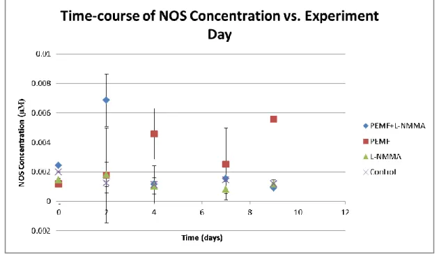
Nitric oxide assay……….60
Results………..60
Discussion………61
CHAPTER 3: ICES AS A MEANS OF ACUTE INFLAMMATION REDUCTION...63
Overview………..63
Introduction………..63
Materials and methods……….64
xiii
ICES instrumentation………..64
Treatment groups……….65
Carrageenan challenge/CFE induction………67
Plethysmometry measurements………...67
Circulating inflammatory factor measurements………...67
Results………..67
Plethysmometry………...67
Cytokine concentrations………...71
Discussion………74
CHAPTER 4: ICES DOSE-RESPONSE IN CFE REDUCTION ………..77
Overview………..77
Introduction………..77
Materials and methods………...81
Animals………81
ICES treatment units………83
Treatment groups……….84
xiv
Plethysmometry measurement……….86
Tissue collection………..86
Histology………..86
Results………..87
Plethysmometry………...87
Histology………..92
Discussion………97
CHAPTER 5: SUMMARY, DISCUSSION AND FUTURE WORK ……….102
xv
LIST OF FIGURES
Figure 1.1 – Induced electric field caused by Helmholtz coils……….………...10
Figure 1.2 – Cartoon depiction of induced current flow around cells………...13
Figure 1.3 – Representative ICES waveforms………...21
Figure 1.4 – Chemical structure of λ-carrageenan……….………...31
Figure 1.5 – Diagram of the arachadonic acid inflammatory pathways……...………...34
Figure 1.6 – Simplified illustration of central interactions between Aδ, Aβ and C fibers………...42
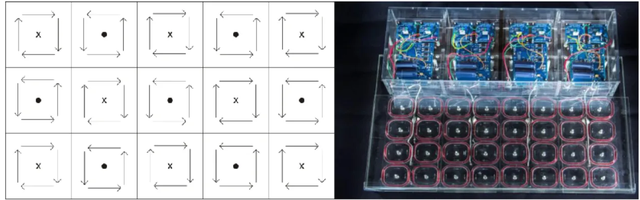
Figure 2.1 – Diagram showing the in vitro 6-well ICES stimulator………...………….58
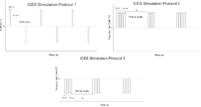
Figure 2.2 – Representative drawing of the ICES stimulation protocols used throughout the experiments presented herein……….…………..………...59
Figure 2.3 – Plot of nitrite vs. time for conditioned media from in vitro study………...61
Figure 3.1 – Illustration of the mat style ICES stimulation unit..……….………...65
Figure 3.2 – 8-hour CFE time-course summary plot of inflammation……….………...68
Figure 3.3 – 8-hour total disease burden for ICES CFE study……….………...70
Figure 3.4 – 8-hour disease suppression for ICES CFE study……….………....71
Figure 3.5 – 8-hour summary of IFN-γ concentration in CFE-induced rats….………...72
xvi
Figure 3.7 – 8-hour summary of IL-6 concentration in CFE induced rats…..………...74
Figure 4.1 – Image of the mat style ICES stimulation unit………..………...82
Figure 4.2 – Depiction of cage-insert style ICES stimulation unit…………...………...83
Figure 4.3 – Graphical representation of ICES stimulation protocol………...………...84
Figure 4.4 – 4-hour CFE time-course summary for ICES dose-response…..……….88
Figure 4.5 – 8-hour CFE time-course summary for ICES dose-response…..……….89
Figure 4.6 – 8-hour total disease burden summary for ICES dose-response..……….90
Figure 4.7 – 4-hour disease burden summary for ICES dose-response……..……….91
Figure 4.8 – Disease suppression summary for ICES dose-response……….……….92
Figure 4.9 – Representative histological section for dexamethasone positive control paw stained with H&E…..………...93
Figure 4.10 – Representative histological section of a PBS negative negative control paw stained with H&E .………94
Figure 4.11 – Representative histological section of a 40 T/s ICES treatment paw stained with H&E……….95
xvii
LIST OF TABLES
Table 1-1 – Summary of important descriptive ICES waveform parameters……..………....22
Table 3-1 – Dosing chart for ICES CFE study………....66
xviii
LIST OF ABBREVIATIONS
ANOVA – Analysis of variance
ARFI – Acoustic radiation force impulse
ATP – Adenosine triphosphate
BMP-2 – Bone morphogenic protein 2
Ca2+ – Calcium (2+) ion
CaM – Calmodulin
CFE – Carrageenan footpad edema
Cl- – Chloride
CLR – C-type lectin receptor
CNS – Central nervous system
DAMP – Damage-associated molecular pattern
DNA – Deoxyribonucleic acid
eNOS – Endothelial nitric oxide synthase
FBS – Fetal bovine serum
FGF-2 – Fibroblast growth factor 2
xix HMSC – Human Mesenchymal derived stem cell
ICES – Inductively coupled electrical stimulation
IFN-λ – Interferon gamma
IL1-β – Interleukin-1 Beta
IL-6 – Interleukin-6
iNOS – Inducible NOS
IU – International units
K+ – Potassium
L-NMMA – N-monomethyl-L-arginine
Na+ – Sodium
NASA – National Aeronautics and Space Administration
NLR – NOD-like receptor
nNOS – Neuronal NOS
NO – Nitric oxide
NOS – Nitric oxide synthase
NPIR – Non-phagocytic inflammatory response
xx PAMP – Pathogen-associated molecular pattern
PBS – Phosphate buffered saline
PEMF – Pulsed electromagnetic field
PET – Positron emission tomography
PIR – Phagocytic inflammatory response
PNS – Peripheral nervous system
PO – per os (by mouth)
PPS – pulses per second
PRF – Pulsed radio frequency
PRR – Pattern recognition receptor
rCIA – Rat collagen-induced arthritis
RLR – RIG-1-like receptor
RNA – Ribonucleic acid
rTMS – Repetitive transcranial magnetic stimulation
SAID – Steroid anti-inflammatory drug
TENS – Transcutaneous electrical nerve stimulation
xxi TVEMF – Time-varying electromagnetic fields
UNC TCF – University of North Carolina Tissue Culture Facility α-MEM – Alpha minimum essential medium
xxii
LIST OF SYMBOLS
μL – Microliter
μM – Micromolar
1
CHAPTER 1 : A HISTORY OF ICES Introduction
Over at least the past seventy years, many different forms of pulsed electromagnetic field (PEMF) therapy have been reported in the scientific literature. Among these, techniques that employ the use of inductively coupled electrical stimulation (ICES) therapy have been reported in peer-reviewed scientific literature as an effective means of reducing pain and inflammation in a wide variety of conditions while often promoting healing3,10–16. Other forms of PEMF therapy have also been reported as effective, such as transcutaneous
electrical nerve stimulation (TENS), however, the focus of this work will be on ICES rather than on other forms of PEMF treatment because of the many advantages of ICES.
One conceptual advantage of the use of ICES over other types of PEMF technology is that it does not require the assumption that magnetic fields themselves interact directly with living tissues by means of some form of magic, or a complex and poorly-understood
2
essentially be viewed as an air-core electrical transformer, the primary coil being the external ICES. Based on the well-understood Law of Induction, one of the four classical Maxwell Equations, electrical currents are induced in and around the cells within the living tissue within the conductive paths in and around cells. This takes the form of ions in solution being forced to move, driven by the induced fields1. Thus, ICES is essentially a means by which electromagnetic induction allows the electrical stimulation of deep living tissues without requiring the use of invasive electrodes. For this reason ICES has advantages over the much more common use of direct conductively coupled electrical stimulation, sometimes
3
The physics of ICES gives a clear and understandable mechanism by which electrical energy can be induced within deep tissues by the Law of Induction. It remains to be
elucidated how the induced fields are transduced into useful biological signals. Observations and mathematical models suggest that one of the primary anti-inflammatory mechanisms of ICES is via the Calcium-Calmodulin (Ca2+/CaM) dependent nitric-oxide synthase
pathway2,5,7–9,17–21. Specifically, it is hypothesized that electromagnetic pulses of appropriate parameters will preferentially induce calcium binding to CaM7. Regardless of the
mechanism, of utmost importance are the waveform parameters—with the most effective parameters reportedly falling within a range producing induced electrical fields on the order of 1 V/cm7,22.
Unfortunately, the majority of the PEMF and ICES literature fails the basic scientific requirement of repeatability. By our accounting, more than 90% of all published reports fail to include adequate waveform parameters to fully define the dosimetry of the applied
treatment. This shortcoming in the literature is very unfortunate as it tends to drive reputable clinicians and scientists away from the scientific study and clinical acceptance of PEMF in general and ICES specifically, even though there is strong evidence to suggest that ICES, when properly applied, is safe and can be very effective at reducing inflammation and pain while also accelerating healing of otherwise refractory injuries.
4
The concept of using pulsed electromagnetic fields (PEMF) has been explored as a clinical therapeutic since the 1950’s12–14,23–25
. Since then, PEMF has been used to treat
5
stimulation parameters can one successfully provide signals to which tissues will respond in a favourable way. Herein, we seek to provide objective evidence that ICES, when applied appropriately, can provide significant and repeatable anti-inflammatory effects in an acute inflammation animal model.
Background
Electromagnetic therapies have been in use for many years. Electrical stimulation of tissues has been studied since Galvani’s experiments using electricity to stimulate contraction of the muscles in dissected frog legs65. The systematic study of the effects of electrical and magnetic fields on living and dead tissues began with Galvani in the late 18th century, whose research led to the discovery that one of the primary methods of information transfer within nerve and muscle tissues is via electrical pathways. In the middle of the 20th century, it was discovered that bone is piezoelectric in nature, and therefore was hypothesized to also transduce information electrically23,24. Soon thereafter, many experiments demonstrated that directly-applied electrical currents can be employed to induce bone formation and
remodeling12–14,25. One problem with these early methods of direct electrical stimulation of bone tissue was that they required the implantation of electrodes into and around the bones to be stimulated. The deeply invasive nature of direct electrical stimulation of bone lead to the development of non-invasive methods, such as the use of induced electrical fields. These inductive methods employ magnetic fields from external magnets or solenoids that change over time to induce the desired electrical fields within the tissues, based on the well-understood Faraday’s Law of Induction66
6
non-depolarizing electromagnetic fields on tissues other than bone. Non-depolarizing electric fields are those which are too low to induce overt depolarization of the cell membrane as in the case of an action potential, but strong enough to presumably have other effects on molecular mechanisms within cells and in the extracellular space. Such mechanisms have been widely hypothesized to mediate the signals involved in functional adaptation of
musculoskeletal tissues, and are the subject of ongoing mainstream scientific research. These signals are generally thought to be very small in magnitude compared to action potentials in excitable tissues, and many competing mechanisms have been hypothesized.
Nerve regeneration is also thought to be subject to similar signaling mechanisms that induce accelerated healing and repair as it was shown that non-depolarizing electromagnetic pulses could improve nerve lesion healing. Further studies showed that inflammatory factors could be reduced in tissue inflammation in humans post operatively10,11. Pilla and colleagues developed a theory of interaction between pulsed radio frequency (PRF) waves and tissues which makes use of the frequency response of tissues and places lower bounds on waveform parameters based on the thermal noise threshold2,4,5,7,17–19,67,68. More recently, ICES has been studied in terms of behavioural modulations—specifically the effects of ICES on bipolar-disorder, autism spectral disorder (ASD), Alzheimer’s, and Parkinson’s disease29–37,46. Prior to discussing the effects of ICES on cells, tissues and systems, it is necessary to discuss the important parameters which govern how tissues will respond to electromagnetic radiation.
Types of biologically relevant signals
7
1. Current flowing into the coils from the stimulation unit. This is the original driving signal that is produced by the electronic circuit within the ICES device to drive the coil that will then produce the magnetic field.
2. The time-varying magnetic flux in and around the coils resulting from the electrical current driving the wire coils.
3. The induced electric field in the tissue volume resulting from the time-varying magnetic flux generated by the coils.
Based on our detailed review of the literature, we have determined that in most cases investigators report only a partial description of the original driving signal emanating from the electronic circuit (#1 above), but do not measure, calculate, report, or estimate the
resulting magnetic field vs. time (#2 above) or the electrical fields that are ultimately induced within the target tissues (#3 above). For the most part, the second level signal—magnetic flux—is the most relevant signal to specify because it is prone to deviate from theoretical values when calculated based upon the presumed driver circuit performance, it is readily measured using modern analog signal Hall effect sensors, and when measured accurately yields good estimates of the induced field within the tissues. It should be noted that it is the final signal—the electric field induced within the tissues—which is the hypothesized
mediator of the responses seen in vivo and in vitro, but that it is difficult to directly measure these induced fields within tissue.
Current flowing into the coils (primary or first-level signal)
8
discussion we will not consider “static” magnetic devices such as permanent magnets or solenoids driven by steady DC current. In static cases the magnetic fields are largely steady and non-varying over time, so their ability to induce electrical fields is essentially zero because the first time derivative of the magnetic flux in steady magnetic fields is by
definition equal to zero. That is not to say that such devices would have no biological effects, because they certainly may have effects through such mechanisms as the Hall Effect, in which charged particles (ions) ubiquitous in biological systems would be influenced as they move through the steady magnetic field. The induction of electrical fields within tissues requires magnetic fields that vary in time, and typically this is accomplished using a
computer or a microcontroller-based platform to drive current waveforms through solenoid coils. To induce the desired electrical fields it is essential to control the slew-rate (rate of change or first time derivative of the magnetic flux) of the signal. Thus, it is of utmost importance that the primary driving electronics have adequate dynamic performance.
9
consumption of the primary driving electronics provide a direct and convenient opportunity to measure and determine the upper boundary for power for the entire system.
Magnetic flux produced by coils (secondary or second-level signal)
10
curvature of the induced field drops to zero in the x-y plane. The inner conical surface is perhaps most relevant because it is the volume of tissue between or within the coils that generally is intended to undergo treatment with ICES.
11
Induced electric field within tissues (Tertiary or third-level signal)
Finally, it is necessary to discuss the induced electric field—specifically with regard to the tissue volumes of interest. The induced field can be calculated simply using equation 1 below:
( ) (1)
For example, if one considers a stimulation volume on the order of 10 µm (average cell diameter), then with a magnetic flux slew rate of 1,400,000 Gauss/second (=140 Tesla/second), a magnetic pulse will induce a peak electric field of approximately 3.5 x 10⁻⁴ V/m around the perimeter of a typical cell. If one considers thermal noise averaging, and cellular response, then the predicted threshold induced field for a measureable response is on the order of 10-3 – 10-5 V/m 69. Thus, a 140 T/s stimulus should cause a measurable
12
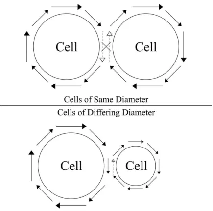
pathways, some circular, but most are not. Considering that the field strength in a plane varies with respect to the radius of interest, one can determine that if cells meet in locations where the cross sectional radii are not identical, then the currents where the cells meet will not cancel, and there will be a net flow of current around the larger radius of interest. However, if two cells meet at a location such that their cross-sectional areas are
approximately the same (and they are both relatively circular cross sections) then circular currents flow around each cell, and should approximately cancel where the cells meet— producing a conducting path around both cells (Figure 1.2). Because of these geometric effects, it is possible that amplification effects might be seen for signals that fall below theoretically calculated stimulation thresholds. Such circumstances may dominate the
13
Figure 1.2: A cartoon of the current flow induced around cells. Top.) Cells of equivalent radius have offsetting electric fields between them, resulting in a net current flow
around the perimeter of both cells, but not between them. Bottom.) Cells of different radii of intersection will have a net current flow around their perimeter and in the direction between them determined by the larger cell.
The above arguments generally hold true for the simplest of cell geometries: 10 micron diameter spherical cells. Though this assumption of simple geometry may be
adequate for many estimations of tissue properties, such as estimating the number of cells in a given volume of tissue, the spatial details of cell membrane geometry and receptor
distribution may well dominate when considering the mechanisms that relate to
14
known as the "assumption of a spherical cow", which takes the form of a joke based upon commonly encountered scientific over-simplifications than make calculations easier at the cost of ignoring the most important details of the system being studied. Many cells in the musculoskeletal system are known to have a complex surface structure containing thin filaments that stretch out into the space between cells. For example, osteocytes (cells in bone tissue) are known to have cytoplasmic processes which extend into canaliculi (tiny canals) in the hard bone matrix27,28. These thin extensions of the bone cell membrane are known to be involved in the collection of nutrients and elimination of waste, but it is hypothesized that osteocytes may detect mechanical loads through the detection of signaling that arises from the mechanically-induced flow of fluids and ions through the lacuno-canicular network surrounding each osteocyte27. It is one of our working hypotheses that ICES systems work at this pericellular level to emulate the mechanical signals in musculoskeletal tissue systems that would normally induce a functional adaptive response, such as bone growth and remodeling to increase bone density as a result of exercise. We further hypothesize that the emulation of these signals by ICES stimulators has the added benefit of employing the natural signal amplification systems within the musculoskeletal system without actually applying the mechanical loads to the tissues being stimulated, thus allowing musculoskeletal tissues to adaptively respond to the emulated signals without also being subjected to the structural micro damage that would otherwise occur from the mechanical loads.
ICES as a biological signal
15
and cellular responsiveness to low-level signals has evolved in many cases to become highly specific and responsive only to a very precisely defined signal, so as to prevent amplification of spurious background noise that might elicit inappropriate cellular or molecular response. Within the receptive bandwidth of such low-level biological signals the signal itself must have a high signal-to-noise ratio (SNR), with minimal energy being expended upon parameters of the signal that do not contribute to the intended message. This evolved SNR allows all other signals that fall outside of the receptive bandwidth to be essentially ignored. The evolutionary process tends to make good use of such highly selective and efficient processes once they have passed the test of natural selection, so it is reasonable to
hypothesize that a signal that might elicit a functional adaptive response in one tissue, for example bone, might also be employed by other tissues for similar purposes. This would be especially true for tissues within the same functional groups such as musculoskeletal tissues, cardiovascular tissues, nerve tissues, etc. On the basis of this reasoning we hypothesize that specific signals that induce tissue growth and regeneration in one tissue in musculoskeletal system might elicit the same general response in many or all other tissues of the
16
preferring instead to use a brute-force approach to coerce the target tissue toward the desired response rather than employing high fidelity signals that work with innate biological filters and amplifiers. The literature suggests that this latter brute-force approach, though crude, is in fact effective to a limited degree. However, such a crude approach has no basis from which to develop increasingly sophisticated, efficient, and effective ICES signals. Thus, most commercially-available ICES technologies simply are not improved over time. Once
treatments can be demonstrated to be statistically significant in their intended biological effects, the evolution of the ICES waveform protocols toward increasingly better signals generally does not occur. This process of accepting the first guess that “works” is
exacerbated by modern regulatory practices, such as those enforced by the FDA relating to medical devices. Once a medical device has been proven “safe and effective”, it is
exceedingly difficult and costly to make any changes to the stimulation parameters. The cost of making even a slight change to PEMF or ICES parameters on an approved medical device can rise as high as $180M and can take from 3 to 5 years to complete because the entire pre-market approval (PMA) process must be repeated for each change to any waveform
17
putting the “NO” firmly back into inNOvation. This helps to explain why health and medicine have generally not enjoyed the same pace of technological progress that other non-medical technologies have enjoyed for the past 4 decades, including computing,
communication, entertainment, transportation, safety equipment, and basically every other aspect of our lives.
The unintended consequence of this over-regulated, inefficient, and crude approach to the development of ICES waveforms has been that most ICES systems remain very
inefficient, bulky, costly, and they subject the target tissue to unnecessary levels of electromagnetic energy. More rational approaches to ICES waveform design are certainly possible.
ICES waveform shapes
Many different methods exist for inducing an electric field within tissues and all of these have been employed at various times by different ICES systems. Waveforms can be divided into four distinct waveform categories: pure sinusoidal,
triangular/sawtooth/trapezoidal/square, asymmetric pulses, and pulsed radio frequency (PRF)/Modulated signals. Stead (DC) magnetic fields will not be considered, though they are frequently employed, for the reasons stated previously. One must also keep in mind that there are three levels of signals, as discussed above. For the following discussion the signal
waveforms refer to signals in level #2: the magnetic field generated by the coils.
Sinusoidal waves
18
typically respond to radio frequencies (RF) from 0 Hz to 10 kHz—outside of this range, tissues and cells are essentially transparent (with the exception of PRF signals). The smallest wavelength of such signals in an electrolyte environment is on the order of 3000 meters— thus cells are unlikely to be acting as antennae at such frequencies. Furthermore, a frequency of approximately 30 THz would be required to induce resonance in a cell of size on the order of 10 µm in a saline solution. Interestingly, because tissues have been found to be responsive in such a low frequency range, one must consider the mechanisms by which cells or
molecules might transduce these signals. Much of the biological response is dependent upon the bulk electrical properties of tissues (direct and reactive impedances), which dictate how electrical energy is absorbed through a medium. In the case of a magnetic field, because the vast majority of mammalian tissues are not known to interact with magnetic fields, one must consider magnetically induced electric field pathways as the primary method in which magnetic fields can interact with tissues. Because the induced electric field is proportional to the rate of change of the magnetic field, the amplitude and frequency of the magnetic field dictates the strength of the cellular response. Thus, higher frequency and amplitude signals should be more effective in eliciting a response. It should be noted that there is significant theoretical evidence that suggests that there is a lower bound for frequencies as well due to the thermal noise threshold19,69,70. Interestingly, there have been a number of studies that find effects well below the theoretical frequency and amplitude limits predicted mathematically, suggesting either a placebo effect or an alternative transduction mechanism29–40,69,71.
Triangular/Trapezoidal/Square waves
19
that they are effective, it may equally be the case that their efficacy is due to the high slew-rates that can be produced. For practical purposes, pure square waves are impossible to create electronically: there is always a finite rise-time and fall-time for the primary electrical
signals, they cannot change instantaneously. Thus, this category of three waveforms can be collapsed into triangle and trapezoidal, which includes square waves which are actually trapezoids because their rising and falling slopes are not perfectly vertical. Both triangular and trapezoidal waveforms provide bipolar induced fields, which depend upon the slope of the sides of each trapezoidal waveform—the main difference being that there is a delay between positive and negative peaks in a trapezoidal pulse given by the length of the signal plateau.
Asymmetric pulses
Asymmetric pulses are typically triangular or trapezoidal in nature, but have a differing rising and falling edge. Such waveforms can be useful for inducing non-equal bipolar induced electric fields. Examples of asymmetric pulses include saw-tooth waves such as those shown in Figure 1.3.
PRF/Modulated signals
20
21
22
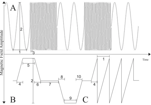
Table 1-1: Summary of important descriptive ICES waveform parameters. All numbered items are labeled in Figure 1.3 on their respective waveform type letter. Unlabeled components are those which cannot easily be drawn on a figure, however are absolutely necessary. Trapezoidal waveforms are assumed to be constructed of straight lights—if lines are curved, a function may be required to define the edge slopes. It should be noted that this table is not comprehensive, as more complicated waveforms may require additional information to fully define one full cycle of stimulation.
Summary of Important Secondary Wave Structures Necessary for Fully Defining ICES
Waveforms
Sinusoidal
(A)
PRF (A) Trapezoidal (B) Asymmetric Pulse
(C) Period (1) Amplitude (2) Peak Slope Bulk Pulse characteristics Carrier Period (3)
Amplitude (2) Encoded frequency
Peak Slope Pulse rate (pps)
Amplitude (2) Positive Rising Edge Slope Positive Rising Edge time (4)
Time at Max (5) Positive Falling Edge Slope Positive Falling Edge time (6)
Time at Zero (7) Negative Falling Edge Slope Negative Falling Edge time (8)
Time at Minimum (9) Negative Rising Edge Slope Negative Rising Edge time (10)
Peak Slope Pulse rate (pps)
Period (1) Amplitude (2) Rising Edge Slope Rising Edge time (4)
Falling Edge Slope Falling edge time (6)
23 Waveform parameters
All waveform categories and shapes are defined by a set of waveform parameters. These include amplitude, frequency, slew rate, and other parameters. Some waveforms are well described by only two parameters, such as continuous pure sine waves which can be defined by the two parameters amplitude and frequency. Other waveforms are more complex and may require six or more parameters for a complete description. An example of this is asymmetric trapezoidal waves that are generated in short bursts of pulses followed by periods of no stimulation. In this case the waveform would be fully defined by at least twelve
parameters: start time, initial slope, peak amplitude, duration (time) held at peak amplitude, final slope, terminal amplitude (can be zero or have opposite sign for bipolar pulses), duration of zero or opposite-sign plateau, return slope (if non-zero), time between pulses, number of pulses in each burst, dwell time between bursts, and at least one additional parameter to define the periodicity of the bursts of asymmetric trapezoidal pulses.
Amplitudes
The mechanism for the biological effects of ICES as they relate to magnetic flux peak amplitude, and thus the relative importance of this parameter, remains slightly ambiguous at this point because there is a large range of experimentally effective amplitudes that fall well below thermal noise limits. However, generally speaking, larger amplitudes are more
24
a level with significant thermal effects within the tissue. This effect is put to positive use in modern surgery when radio ablation is utilized to destroy tumors or other unwanted tissues. Assuming that the RF power is below a damaging level, we have noted in a wide variety of literature that induced electric fields on the order of 0.01 – 10 V/m appear to be most effective in treating chronic pain and inflammation. Generating such field strengths can be done using several magnetic waveforms. It should also be noted that much lower amplitude magnetic fields, on the order of picotesla, (10-12 T) have been reported to be clinically effective for treatment of multiple sclerosis and Parkinson’s patients29–40. So, one can
conclude that waveform amplitude certainly plays a role in both the efficacy and the potential risk involved in the use of ICES stimulators, but the precise role and the underlying
biological and electromagnetic mechanisms remain to be elucidated.
Frequency
25
electric field on the order of 1.75 V/m around the perimeter of a 35 mm disk. This peak field strength is approximately 100 times higher than the average field produced in a clinical MRI unit, which is a very large amplitude indeed. One tesla = 10,000 gauss, so a 100 T field = 1 MG, which is about 1.5 million times the average magnetic field strength of the Earth. At higher frequencies the calculus, a simple derivative of the sinusoidal waveform, indicates that significantly lower magnetic flux amplitudes could theoretically become biologically
effective. For example, by increasing the frequency from 1 Hz to 1 kHz, the required peak magnetic field becomes approximately 0.1 T, which is more reasonable and technically is much easier and less expensive to achieve—but it remains very large.
Slew rate
26
well below 0.1 T provided the pulse can be delivered in a short enough time (approximately 100 µs). Frequency modulated signals provide an alternative method for producing high slew-rate signals by encoding low frequency signals in high frequency (1-27.12 MHz) sinusoidal carrier waves.
The thermal noise threshold
An interesting and important discussion must be had regarding the thermodynamic effects of electric fields. Specifically, as one decreases the magnitude of the induced electric field, there comes a point where thermal fluctuations due to random motion within the sample can easily produce field strengths large enough to mask the applied signal. This masking is referred to as the thermal noise threshold and is on the order of 9x10⁻² V/m when signal averaging is not taken into account. Thus one would expect that any induced field below the thermal noise threshold should not induce a physiologic response. However, cells are able to integrate applied signals, which allows the theoretical noise threshold to fall even further to levels as low as 10-3 – 10-5 V/m69. Observations in literature support the claim that ICES efficacy can be observed orders of magnitude below the thermal noise threshold—a feat that is attributed to the ability of tissues to integrate and amplify very small signals.
Review of past literature
Bone studies
27
possibility that applied electromagnetic fields could drive other biological processes. Thus, much of the pioneering work done by Bassett, et al. laid the foundation for subsequent work in other tissues.
Cell studies
Effects of ICES on cells have been studied extensively in those cells of bone or cartilage-derived lineage. In vitro studies on both primary and immortalized cells have been conducted, and there is evidence to suggest that each responds differently to ICES72. Cell studies done on osteoblast-like cells have mainly focused on the nitric oxide synthase (NOS) pathway of cells such as MC3T3-E1 cells8,9. Proliferation in several different cell types has been extensively studied and found to be increased in the presence of low-magnitude ICES on the order of 0.002 V/m20,45,73–76. In addition to modulating proliferation, ICES has been implicated in the up regulation of DNA synthesis, IGF-2 (osteosarcoma)77,78, FGF-2
(endothelial cells)74 and BMP-2 mediated osteoblastic differentiation in human mesenchymal stem cells (HMSCs)79. In addition to the studies on bone, there have been questions as to the efficacy of ICES in nerve regeneration. In particular, a study conducted at NASA by
28
Soft tissue studies
To understand the effects of ICES therapy on a system level, we feel it is easiest to break the existing literature into the broader categories of nerve healing, and
anti-inflammatory studies. Because ICES is so well established as an effective treatment in bone-healing, we choose not to review that literature—however the reader should be aware that there is a vast literature concerning bone remodeling (A good reference with which to start is the 1974 Bassett reference).
Nerve healing
To understand the effects of ICES on nerve regeneration, we have broken the in vivo studies into three broad categories: peripheral, spinal cord and cortical studies. We have chosen to separate the cord from central and peripheral studies because it is the junction point for both central and peripheral nerves, and thus has the potential to affect both
simultaneously.
The focus of the majority of peripheral nerve studies has been to examine the ability of ICES to temper pain and stimulate regrowth. As previously mentioned, the studies performed at NASA by Goodwin et. al. indicated that neuronal proliferation could be significantly affected by low frequency pulses much lower in magnitude than the earth’s magnetic field. Studies performed by Raji et al. have shown that rat peroneal nerve regeneration can be enhanced by the use of ICES43,44.
29
Square wave pulses (~600 T/s magnetic flux rate), as studied by Sisken et al., seem effective in increasing sciatic nerve regeneration regardless of the orientation of the Helmholtz
stimulation coils47. However, Baptista et. al. showed that there was no significant effect from treating sciatic crush lesions in Swiss mice using a stimulation protocol that induced a 20 kT/s magnetic flux rate—a relatively large stimulus81.
Finally, it is important to discuss the potential cortical effects of ICES. Cortical effects should be considered from two different views: direct stimulation (ex: rTMS, low magnitude ICES, etc.) which stimulate the brain directly, and indirect stimulation that causes cortical remapping or modulation through plasticity by stimulating peripherally. Direct stimulation methods such as those used in the studies published by Sandyk et al. have indicated that very small induced fields may be effective in alleviating some of the difficulties associated with multiple sclerosis and Parkinson’s disease29–40
. However, it should be noted that the field strengths in question fall far below the thermal noise threshold and that the majority of these studies are case studies, not controlled laboratory studies. Unfortunately the literature regarding the central effects of peripherally applied ICES on central nerve function is rather sparse. Because peripheral neurons play a very large role via the feedback mechanism in cortical plasticity, it follows that if ICES affects these neural feedback loops, then fMRI and PET studies would reveal potentially significant effects of peripherally applied ICES on cortical plasticity.
Inflammatory pathways
30
follows a well-defined series of steps that are mediated by specific molecules and cells in the body. Acute inflammation can be distinguished from chronic inflammation by molecular and cellular differences (however, the transition from acute to chronic is still poorly defined).
The inflammatory process is initiated by a noxious stimulus—such as a burn, cut, blunt trauma, chemical exposure, auto-immune response, or infection. In all situations, the cascade begins when a pattern recognition receptor (PRR) on the surface of an immune cell detects the presence of a pathogen-associated molecular pattern (PAMP) or a damage-associated molecular pattern (DAMP). Molecular patterns are molecules that are released in response to a noxious stimulus. Upon detection of a pattern, the PRRs trigger the release of various inflammatory factors which lead to increased blood flow, vascular permeability, and hyperalgesia—the inflammatory factors released depend upon the type of stimulus as well as the duration of the stimulus (i.e. acute vs. chronic). These inflammatory factors cause the classic signs of inflammation: Rubor (redness), calor (heat), tubor (edema/swelleing), dolor (pain), and function laesa (loss of function).
Pattern recognition receptors are a family of receptors that are specifically tuned to respond to pathogens and damage-associated molecular patterns (PAMPs and DAMPs). A recent review highlights that there are four accepted PRR families: Toll-like receptors (TLRs), C-type lectin receptors (CLRs), Retinoic acid-inducible gene (RIG)-1-like receptors (RLRs), and NOD-like receptors (NLRs)82. These four families can be classified as
31
typically respond to fungal infections; NLRs respond to bacteria, and RLRs respond to RNA and DNA viruses82.
Pathogen and damage associated molecular patterns are the specific ligands that trigger a response in PRRs of the immune system. Lipopolysaccharides such as those found on bacteria, are examples of PAMPs that can trigger an immune response. Damage
associated molecular patterns are groups of molecules that are released in response to physical damage to cells and tissues. It should be noted that PAMPs and DAMPs have become a more recent focus of study, and at this point, are relatively poorly understood.
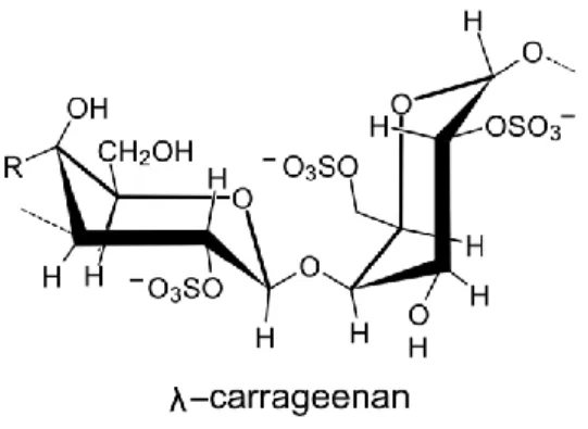
Figure 1.4: Chemical structure of λ-carrageenan. A representative chemical structure drawing of a single molecule in a polymer chain of the algal-derived sulfonated
32
A very well-established model for the study of the anti-inflammatory efficacy of new drugs is the carrageenan-induced footpad edema (CFE) model83,84. This model makes use of an injection of λ-carrageenan (an algal-derived sulfonated poly-saccharide, Figure 1.4) into the footpad of a rodent (typically rat or mouse). The CFE model is an acute model of
inflammation, and therefore typically resolves spontaneously within the course of 8-12 hours while displaying mostly acute inflammatory-associated cytokines and proteins. It has been shown that the CFE model displays two inflammatory responses: a non-phagocytic
inflammatory response (NPIR) and a phagocytic inflammatory response (PIR). The NPIR occurs first, and is initiated by the trauma associated with a subcutaneous injection, and is dose-independent for carrageenan. The second, more prolonged, PIR occurs approximately 60 minutes post-injection and involves recruitment of neturophils and macrophages84. Furthermore, it has been shown that pleural injection of carrageenan does not result in an NPIR, but rather a PIR only—thus implicating the damage associated with an injection into solid tissue as the cause for the NPIR83. To control for NPIR it is common to inject the contralateral (non-treatment) hind footpad with an equivalent volume of sterile PBS—this triggers NPIR but does not trigger a PIR, allowing one to subtract the swelling caused by NPIR from the CFE induced footpad.
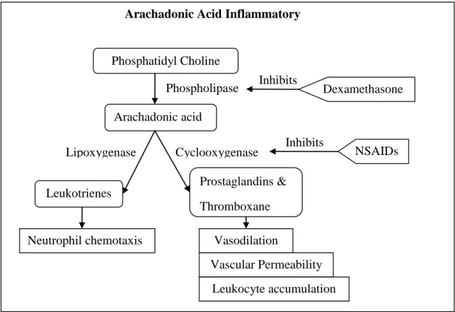
An important aspect of the CFE model is that it is strongly suppressed by the introduction of steroid anti-inflammatory agents such as dexamethasone. Dexamethasone inhibits production of arachadonic acid—a phospholipid derivative which is necessary for the production of leukotrienes, prostaglandins and thromboxanes. Figure 1.5 shows a
33
34
Figure 1.5: Diagram of the arachadonic acid inflammatory pathways. A bifurcation in the pathway explains the differences between dexamethasone anti-inflammatory
treatments and commonly available over the counter non-steroidal anti-inflammatories (NSAIDs). Dexamethasone-like drugs inhibit arachadonic acid formation, while
NSAIDs inhibit the cyclooxygenase pathways.
A final note should be added regarding two other important inflammatory pathways that are involved. First is the platelet activating factor pathway—a pathway which leads to a change in vascular permeability as well as contributing to leukocyte trapping. A second pathway is the nitric oxide pathway—which is responsible for changes in vasodilation. Specifically nitric oxide causes smooth muscle relaxation which leads to an increase in vasodilation. Because nitric oxide synthase pathways are activated in inflammatory
35
responses, swelling is (typically) increased as a result of edema caused by increased blood flow. The nitric oxide synthase pathway is a specific pathway of interest in the study of ICES as it has been implicated by several researchers as the primary mechanism by which ICES operates physiologically6,7,10,18,19,67.
Anti-inflammatory effects of ICES
There are two notable studies that shed significant mechanistic light on the anti-inflammatory and pain reducing effects of ICES: those of Per Hedén et al. and Christine Rohde et al10,11. Both studies examined the post-operative effects of ICES on breast augmentation and breast reduction patients respectively. In the former, a pilot study of patients undergoing breast augmentation, ICES (2-ms bursts of 27.12 MHz PRF, 3.2 V/m peak applied for 30 minutes every 4, 8 or 12 hours on different post-operative days) was shown to significantly reduce pain scores11. The second study, performed by Rohde et al. using similar ICES parameters showed significant pain reduction, and interestingly a drastic reductionin IL1-β levels in wound exudate as compared to sham groups10. Reduction in inflammatory factors suggests at least one possible biochemical mechanism—perhaps the Ca2+/CaM dependent NOS pathway suggested by Pilla et al7. It is interesting to note that although these reports and others have demonstrated very significant and repeatable
36
evidence indicating adverse effects—the use of ICES for any form of pain management remains outside even the fringe of standard medical practice.
Possible mechanisms of inflammation reduction
While there are many possible mechanisms by which ICES could influence cells, tissues, organs and whole systems—there only a few basic mechanisms that are adequately explored in the scientific literature.
For low-amplitude magnetic fields, a Larmor precession model is discussed which states that the Larmor precession behaviour of certain atoms or molecules (such as water) can be modulated in the presence of a magnetic field. In the case of water, modifying the Larmor precession can impact the ability of thermal fluctuations to drive chemical reactions—
shifting the amount of energy required by a ligand to displace water from a binding site on a target molecule5,17,85.
37
protein binding pockets could be modulated by induced EMFs or eddy current flow. A more specific proposed mechanism is that put forth by Pilla and his collaborators, which states that ICES of the appropriate waveform and pulse duration (specifically pulsed radio frequencies) is able to modulate the Michaelis-Menten binding kinetics of the Calcium-Calmodulin dependent nitric oxide synthases. The theory that ICES functions via modifying Ca/CaM binding has been supported in the literature for cell-free preparations, as well as in vitro and in vivo studies2,5,7,10–13,18,19,68,86. Modulating such a fundamental pathway could result in modulated levels of NO production and therefore have very drastic downstream effects in the body.
Another consideration to be made is that electrical stimuli are known to have well-understood effects on muscle, bone and nerve tissues. Muscles, bones and nerves all require electrical stimuli on various scales of time and signal intensity from millisecond time base curing events of excitation and action potential generation, to much lower levels signals that are hypothesized to be essential to the maintenance of homeostasis and functional
adaptation—thus it is not a far stretch to suppose that ICES acts to promote homeostasis or facilitate functional adaptive responses in cells/tissues that have been injured. Because the ICES protocols used in the following experiments are based on stimulation patterns found in healthy, adult-phenotype muscles and nerves, it may be that part or all of the effects seen for ICES are a result of an electrically-mediated restoring force towards physiological
38
that are man-made. Thus, it is very reasonable to assume that appropriately chosen
stimulation patterns should be interpreted by electrically responsive tissues as homeostatic in nature and could thus promote a reduction in inflammation (i.e. promote a lack of change from homeostasis).
A different viewpoint is that cells can be modeled as living finite state machines. Cells could find themselves locked into perpetual pathologic states even after the
39
Central and peripheral nervous system components should be considered as well. Pain is perceived through signals transmitted from the peripheral nervous system to the central nervous system. To some extent, pain transduction is poorly understood, but generally speaking, pain is detected by free nerve endings which sense changes in heat, pressure and chemicals. When inflammation occurs, local pressure gradients are changed, resulting in physical forces on nerves (pressure) which can cause irritation and pain. Furthermore, tissue damage releases cytosolic components that are known to irritate nerves and cause pain. Long-term activation of inflammatory pathways is known to lead to cortical remapping and chronic pain87,88.
These theories are far from complete or comprehensive; however, they serve as a good beginning to the development of an understanding of the basic mechanisms to elucidate the effects of ICES on the body.
Possible mechanisms of pain reduction
In order to understand the possible mechanisms by which ICES might modify pain in the body, it is important to understand the central and peripheral organization of pain.
Peripherally, pain is typically described in terms of the nerve types that carry the pain data. There are two classic peripheral pain pathways in the body: “Slow” pain, conducted by unmyelinated C-fibers; and “fast” pain, conducted by myelinated Aδ fibers.
40
carried to layer V, where it then travels up the paleospinothalamic pathway to the brain. In the brain, slow-fiber pain neurons terminate in the thalamus, reticular nuclei, tectal area and periaqueductal gray regions prior to transmission for higher-level processing in the
somatosensory cortex (3a)87.
Fast pain—typically described as shallow, sharp and well localized—is conducted peripherally through Aδ fibers that carry information at a rate of 2-30 m/s. From the periphery, primary Aδ fibers travel to the spinal cord where most synapse with secondary nerves in Lamina I of the dorsal horn. From Lamina I, the secondary nerves cross to the contralateral side in the anterior commissure prior to traveling superiorly through the neospinothalamic pathway to the thalamus. From the thalamus, the pain signal travels to SI area 1/3b for processing.
The central nervous system also has the ability to modulate the pain response peripherally through analgesic pathways. These pathways tend to originate from the
41
Additionally, it should be noted that neurons in laminae I and V likely receive
afferent input from non-nociceptive Aβ fibers. It has been shown that Aβ fibers projecting to lamina V have inhibitory effects on nociception by stimulating inhibitory neurons that project to lamina II. This inhibition tends to inhibit nociception. However, it should be noted that nociceptive inputs from Aδ and C fibers also have effects on the inhibitory interneurons in lamina II89—which is to say that Aδ and C fibers feed back on Aβ fibers and vice versa. Simply put, nociception can be inhibited by non-nociceptive (typically mechanical) stimulation, and mechanical perception can be inhibited by nociception.
Finally it is worth noting that there are various central pathways via which opiates and other chemical analgesics can affect pain, however since this discussion is mostly concerned with how ICES might interact with pain perception, we choose to simply mention that these pathways can be stimulated electrically and chemically, as well as inhibited by drugs such as Naloxone. Stress response in humans is a known factor that can cause activation of the opiate pain reduction pathways.
The general organization of pain typically regards the thalamus as the primary region through which the pain signal is relayed prior to higher-order processing in the
42
Figure 1.6: Simplified illustration of central interactions between Aδ, Aβ and C fibers. (Top left/blue) Activation of Aδ fibers increases activation in area 3b/1 of SI, while at the same time inhibiting input from peripheral Aβ fibers and inhibiting central area 3a. (Bottom left/yellow) Activation of non-nociceptive Aβ fibers inhibits peripheral Aδ and C fiber signals, while upregulating area 3b/1 and inhibiting area 3a of SI. (Bottom right/red) Activation of peripheral C fibers inhibits peripheral Aβ (non-nociceptive) input, while centrally activating area 3a and eventually inhibiting 3b/1 of SI.
43
1. Mechanical stimulus carried by Aβ fibers can modulate the perception of pain both centrally and peripherally.
2. Nociceptive stimuli carried by C fibers can modulate the perception of mechanical stimulus centrally and peripherally. C fiber stimulus only modulates perception of Aδ fiber input centrally by inhibiting area 3b/1 in SI.
3. Nociceptive stimuli carried by Aδ fibers can modulate the perception of mechanical stimuli centrally and peripherally; however Aδ activation only modulates nociception from C fibers centrally by activating area 3b/1 and inhibiting 3a.
In order to appropriately address the question of how external electromagnetic stimuli might modulate pain response, it is essential to ask the question: How is pain transduced? It is very well established that both C and Aδ fibers are free nerve endings (i.e. they do not specifically “connect” to anything like a Pacinian corpuscle, etc.). Furthermore, it is still debated as to the exact mechanism by which nociceptors can distinguish between the different types of pain, degrees of pain and qualities pain. It is well understood that C fibers are typically responsible for transmission of heat information, but they are also responsible for other types of slow pain such as aches, nausea and deep, poorly localized pain. Similarly, Aδ fibers have a similar problem: they seem to be able to describe several types of pain, yet they are all essentially identical. Recently, it has been proposed that one of the ways in which pain is transduced is by modifying the conditions in the cells surrounding the free nerve endings in question90. Specifically, it is hypothesized that levels of nucleotides (specifically ATP) can significantly affect the ability of nerves to respond to stimuli90. Specific receptors (such as TRPV1, as well as the P2X and P2Y families) are implicated directly in the
44
sensitive to extracellular ATP levels—allowing them to be capable of modulating nerve response. In addition to having nucleotide receptors that modulate nociceptive quality, it should be noted that all nociceptive stimuli are likely to elicit responses in surrounding non-nociceptive somatosensory afferents. Thus, when integrated in the CNS, perception of the quality of pain is likely the result of both nociceptive and non-nociceptive somatosensory input.
45
these modulating factors is a potential target of ICES, however only two of these four are likely, as discussed below.
46
body—leading to a modulation in the sensitivity of the neuron to firing. However, since a change of 0.1 mV is only about 0.1% change in the potential across the cell, it is unlikely that such a minute change would act directly on driving the membrane potential. Also note, the induced eddy currents would induce electrical potentials and ion flux transverse or parallel to the non-conducting membranes rather than across them, as membrane potential is defined. It is more likely that a small change in membrane potential would lead to a modulation in the permeability of membrane proteins responsible for developing the standing membrane potential. It is worth noting that one might expect a difference in the effects of ICES on cells based on whether or not they are myelinated. Since myelin is an insulator, it can act like a capacitor, thus an electric field developed across the myelin sheath could in fact cause
longer-lasting effects that those on a non-myelinated sheath. Additionally, myelin may shield the nerve from some of the electrical signal, reducing the efficacy of ICES on heavily
47
A slightly less obvious mechanism by which ICES may interact with cells (including neurons) is by modifying the ability of target ions to bind their appropriate receptor. There is a large amount of literature to suggest that calcium is one ion that can be easily modulated by ICES1,2,4,7,10,11,17–19,67,68,86,95. Specifically, calcium in the context of calmodulin activation has been studied—however the specific model developed by Markov and Pilla that predicts the interactions of calcium with its target calmodulin can be developed for nearly any ion and receptor in the body. Above a certain threshold stimulation intensity, the relevant parameters for modifying binding are frequency and pulse width7. Modifying the ability of ions to interact with their targets has obvious implications for both resting membrane potential as well as sensitivity to depolarization.
Modifying the availability of ions and neurotransmitters is unlikely to be a
mechanism by which ICES interacts with neurons—with the exception of perhaps modifying the availability of neurotransmitters produced by other cells (such as nucleotides, etc.
produced in non-nerve cells).
Modifying inputs from other nerves would likely be an indirect feedback mechanism caused by a direct interaction of ICES with a peripheral nerve. For example, if ICES
modulates the activity of a peripheral neuron that projects to an area of the CNS that provides a feedback response to peripheral nociceptive neurons (such as the central feedback
mechanisms described above), then pain could be amplified or inhibited by the interactions of ICES with peripheral nerves.
48
surrounding neurons should be modulated by ICES. Furthermore, if there are pericellular current flows within innervated tissues, one would expect a corresponding change in tissue metabolism1. Thus, if one modifies the conditions of the surrounding tissue using ICES, a neuron might be expected to behave differently given the same stimulus. This also implies that if nociception is typically proportional to tissue damage, that if inflammation is reduced by ICES, then the intensity of nociception should decrease10,11.
Possible cutaneous wound healing mechanisms
ICES has been implicated in wound healing in the literature, and may hold the potential to modify scar formation pathways. Scar formation is typically governed by a well understood mechanism involving fibroblasts laying down a well-organize collagen matrix. There are three well understood types of scarring—hypertrophic, keloid and atrophic. The typical scar is formed in the following steps:
1. A wound is created (cut, scratch, surgery, lesion, etc.)
2. Inflammatory mechanisms are activated (inflammatory factors released, vasodilation, increased blood flow, macrophage response, immune response, etc.)
3. Repair mechanisms are triggered to form a scar (fibroblasts and other repair cells start laying down new matrix)
Upon tissue damage, several events trigger fibroblast migration and differentiation. Chemical factors are released into the tissue surrounding the wound—cytokines from
49
wound is disrupted, the mechanical stimuli to which the cells are accustomed in homeostasis change. Evidence has been collected to demonstrate that fibroblasts normally experience very little average extracellular mechanical stress due to the amount of crosslinking in the
extracellular matrix in which they exist. In the presence of a wound (i.e. a source of high extracellular stress), fibroblasts are then hypothesized to undergo differentiation into myofibroblasts—a subset of fibroblasts that express α-smooth muscle actin (α-SMA)96–102. This mechano-differentiation hypothesis is supported by the fact that stress fibers readily form in vitro when fibroblasts are plated on hard-substrates (such as polystyrene), but do not form when grown in soft hydrogels such as collagen97. Once fibroblasts have moved into a wound, they begin expressing stress fibers in the form of cytoplasmic actins—this phase is called the “protomyofibroblast” stage102
. These stress fibers connect to both integrins of cell-matrix junctions as well as N-cadheren-type adherens junctions102. In vivo, these stress fibers are eventually replaced with α-SMA when the pseudomyofibroblasts are exposed to TGF-β1. As well as from the circulating fluids in a wound, TGF-β1 is released by the existing and newly formed ECM when the psudomyofibroblasts begin supplying a mechanical stress to the ECM103. A wound can then be pulled closed by the stronger pull of α-SMA before scar formation begins as myofibroblasts begin laying down thick extracellular matrix.