APC sets the Wnt tone necessary for
cerebral cortical progenitor development
Naoki Nakagawa,
1Jingjun Li,
1Keiko Yabuno-Nakagawa, Tae-Yeon Eom, Martis Cowles, Tavien Mapp,
Robin Taylor, and E.S. Anton
University of North Carolina Neuroscience Center, Department of Cell Biology and Physiology, University of North Carolina School of Medicine, Chapel Hill, North Carolina 27599, USA
Adenomatous polyposis coli (APC) regulates the activity ofβ-catenin, an integral component of Wnt signaling. However, the selective role of the APC–β-catenin pathway in cerebral cortical development is unknown. Here we genetically dissected the relative contributions of APC-regulatedβ-catenin signaling in cortical progenitor devel-opment, a necessary early step in cerebral cortical formation. Radial progenitor-specific inactivation of the APC–β -catenin pathway indicates that the maintenance of appropriateβ-catenin-mediated Wnt tone is necessary for the orderly differentiation of cortical progenitors and the resultant formation of the cerebral cortex. APC deletion de-regulatesβ-catenin, leads to high Wnt tone, and disrupts Notch1 signaling and primary cilium maintenance nec-essary for radial progenitor functions.β-Catenin deregulation directly disrupts cilium maintenance and signaling via Tulp3, essential for intraflagellar transport of ciliary signaling receptors. Surprisingly, deletion ofβ-catenin or in-hibition ofβ-catenin activity in APC-null progenitors rescues the APC-null phenotype. These results reveal that APC-regulatedβ-catenin activity in cortical progenitors sets the appropriate Wnt tone necessary for normal cerebral cortical development.
[Keywords: cortical progenitors; APC;β-catenin; cerebral cortical development; Wnt signaling; primary cilia; autism]
Supplemental material is available for this article.
Received May 26, 2017; revised version accepted August 21, 2017.
Radial glial cells (RGCs) in the inner and outer ventricular zones (VZs) are progenitors of the mammalian neocortex (Hansen et al. 2010; Evsyukova et al. 2013). Symmetric RGC divisions result in two daughter RGCs and the ex-pansion of the progenitor pool. Asymmetric divisions of RGCs can give rise to either a RGC and a daughter neuron or a transit-amplifying intermediate progenitor (IP). IPs subsequently divide symmetrically to generate more neu-rons. Ventricular RGCs are highly polarized, with a short apical process that forms an end foot attached to the ven-tricular surface and a long radial process attached to the pial basement membrane that serves as a scaffold to guide the radial migration of newborn neurons (Rakic 1988; Evsyukova et al. 2013). A subpopulation of RGCs, outer RGCs, does not have apical attachments to the ventricu-lar surface (Hansen et al. 2010). The appropriate organiza-tion, balance of self-renewal, and differentiation of radial progenitors are central to the construction of the cerebral cortex.
Multiple signaling pathways, including Wnts, GSK-3, CDC42, APC (adenomatous polyposis coli), Arl13b, Notch, retinoic acid, neuregulins, IGF, PDGF, and Sonic
Hedgehog (Shh), have been identified as regulators of radi-al progenitor development in the cerebrradi-al cortex (Chenn and Walsh 2002; Machon et al. 2003; Hirabayashi et al. 2004; Cappello et al. 2006; Mizutani et al. 2007; Rasin et al. 2007; Kim et al. 2009; Yokota et al. 2009, 2010; Kusek et al. 2012; Fang et al. 2013; Higginbotham et al. 2013; Imayoshi et al. 2013; Lui et al. 2014; Chen et al. 2015; Guo et al. 2015; Kong et al. 2015; Lun et al. 2015; Durak et al. 2016; Wang et al. 2016a,b). Recent evidence suggests that the primary cilium, a polarized microtubule (MT)-based organelle that projects from the cell mem-brane, functions as a signaling center in progenitors dur-ing development (Hildebrandt et al. 2011; Higginbotham et al. 2013). Components of the Shh pathway are enriched in primary cilia, and mutations that disrupt Shh signaling result in defective patterning and proliferation of neural progenitors (Wilson et al. 2012; Guo et al. 2015). A recip-rocal relationship between the primary cilium and Wnt signal transduction exists. Components of the noncanon-ical Wnt/planar cell polarity pathway are important for ciliogenesis, and primary cilium signaling can either
1These authors contributed equally to this work.
Corresponding author: [email protected]
Article published online ahead of print. Article and publication date are online at http://www.genesdev.org/cgi/doi/10.1101/gad.302679.117.
contextually restrain or activate canonical Wnt/β-catenin signaling (Lancaster et al. 2011; Kong et al. 2015). Further-more, a role for the primary cilium in Notch signaling and progenitor differentiation has also been identified (Ezratty et al. 2011). Importantly, ciliopathies caused primarily by defects in primary ciliary structure and/or function have pronounced neurodevelopmental deficits, including dis-rupted progenitor development (Hildebrandt et al. 2011; Guo et al. 2015). The development and differentiation of radial progenitors are controlled by the temporal and spa-tial integration of these various signaling pathways and centers. However, the underlying molecular and cellular mechanisms necessary to achieve this integration in neu-ral progenitor cells remain largely unknown.
Here, we demonstrate that APC–β-catenin-regulated Wnt signaling balance is required to integrate the develop-mental signaling pathways necessary for appropriate progenitor development and thus cerebral cortical forma-tion. APC deletion up-regulatesβ-catenin, causing loss of primary cilia, defective Shh and Notch signaling, and com-plete disruption of cerebral cortical formation. However, down-modulation ofβ-catenin and thus Wnt signaling in APC-null progenitors dramatically restores cortical pro-genitor development and cerebral cortical formation in APC-null brains. These results suggest that the APC–β -catenin pathway sets the appropriate Wnt tone in neuronal progenitors necessary for cerebral cortical formation.
Results
APC is essential for cortical progenitor proliferation and neurogenesis
To examine the role of APC in the proliferation and differentiation of cortical progenitors, we generated
APCLox/Lox; hGFAP-Cre (APC conditional knockout
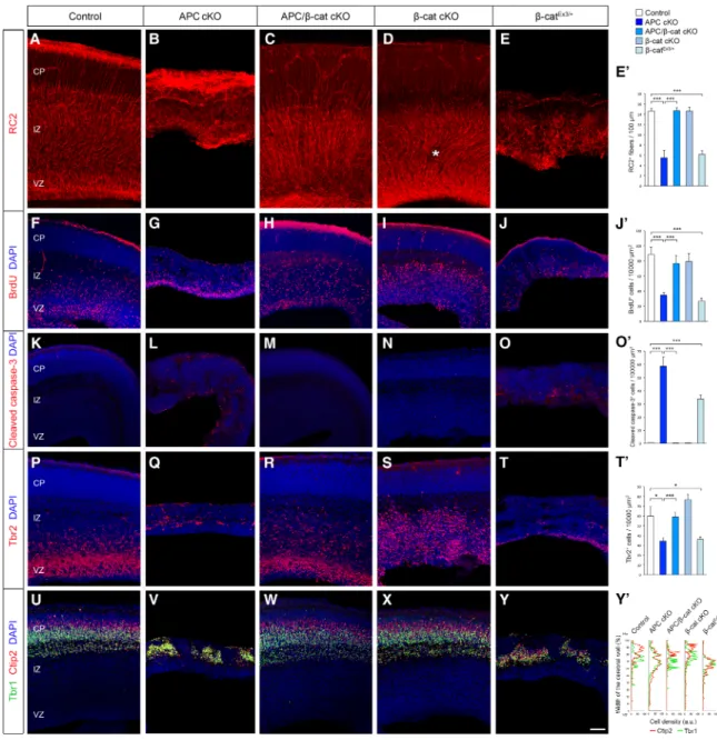
[cKO]) mice in which Cre-mediated inactivation of APC occurs in dorsal cortical progenitors from embryonic day 13.5 (E13.5) (Supplemental Fig. 1A; Zhuo et al. 2001; Yokota et al. 2009). Proliferating radial progenitors in S phase were pulse-labeled with bromodeoxyuridine (BrdU) and visualized by immunolabeling with anti-Pax6 antibodies. Loss of APC leads to reduction of BrdU+/ Pax6+radial progenitors (Fig. 1A–I). Consistently,
phos-phorylated histone H3-positive (PH3+) M-phase radial pro-genitors were also reduced in APC cKO (Supplemental Fig. 2A,B). Many of the APC-deficient radial progenitors were located ectopically in the cortical plate (CP) or inter-mediate zone (IZ) instead of in the VZ (Fig. 1D,H). We then determined the effect of altered proliferation of radial pro-genitors in the generation of IPs and neurons. Loss of APC in radial progenitors led to significantly fewer Tbr2+IPs, which also showed reduced proliferation (Fig. 1J–R). In spite of altered generation and proliferation of cortical pro-genitors, neurons were generated from these APC-defi-cient progenitors, albeit in fewer numbers (Fig. 1S,T). However, the emergence of neuronal identity in these newborn neurons is compromised. Unlike normal new-born neurons, a significant number of APC-deficient neu-rons coexpressed both the radial progenitor marker Pax6
and neuron-specific Tuj1 (Fig. 1U–Y). Furthermore, BrdU pulse followed 24 h later by Ki67 antibody colabeling indi-cates an increase in BrdU+/Ki67+progenitors in the APC
cKO cortex (Supplemental Fig. 2F,G,K,L), suggesting affected cell cycle progression in APC cKO progenitors. Examination of neurogenic niches such as the subventric-ular zone (SVZ) and hippocampus in the postnatal brains also indicates compromised proliferation of neural pro-genitors in APC cKO (data not shown). Together, these ob-servations indicate that APC is necessary for normal cortical progenitor proliferation, appropriate neurogene-sis, and the emergence of neuronal identity in the develop-ing cerebral cortex.
The role of APC in progenitor organization and function is dependent onβ-catenin
APC’s modulation of theβ-catenin signaling pathway is considered to be an essential component of its function (Nelson and Nathke 2013). However, the relative contri-bution of APC–β-catenin interactions to radial progenitor development in the cerebral cortex is unknown. To exam-ine how APC/β-catenin signaling regulates cortical pro-genitor development, we conditionally deleted APC and
β-catenin in radial progenitors by generating APCLox/Lox;
Ctnnb1Lox/Lox; hGFAP-Cre (APC/β-cat cKO) mice.
Dele-tion of APC and/orβ-catenin in APC cKO, Ctnnb1Lox/Lox;
hGFAP-Cre (β-cat cKO), or APC/β-cat cKO brains was con-firmed by immunoblotting analysis (Supplemental Fig. 1A). Loss of APC in APC cKO RGCs drastically disrupted RGC organization and function in the embryonic cortex (Fig. 2). APC cKO RGCs are short, have misoriented radial fibers (Fig. 2A,B), and do not proliferate appropriately (Fig. 2F,G;Supplemental Fig. 2A,B). Importantly, deletion ofβ -catenin in APC-deficient RGCs rescued the defects in APC cKO RGC organization. APC/β-catenin-deficient RGCs formed normally polarized RG scaffolds, and these progenitors proliferated similar to controls (Fig. 2C,H; Supplemental Fig. 2C), suggesting that APC-modulated
β-catenin signaling is essential for APC’s pivotal role in cortical radial progenitor development. APC deletion in RGCs eventually leads to apoptosis of these progenitors (Fig. 2K,L). This enhanced apoptosis was also rescued by
β-catenin deletion in APC-deficient RGCs (Fig. 2M). In contrast,β-catenin deletion alone did not severely affect the general organization of the RG scaffold, except for mi-nor ectopias within the SVZ/IZ (Fig. 2D).
RGCs function as neural precursors during corticogen-esis. RGCs divide asymmetrically and give rise to IPs and neurons. APC deletion in RGCs resulted in marked-ly reduced generation of Tbr2+IPs (Fig. 2P,Q) and Tuj1+
these observations indicate that APC plays an essential role in cortical radial progenitor organization and func-tion as neural precursors. These funcfunc-tions of APC in cortical progenitors depend on appropriate β-catenin signaling.
To further delineate the role of APC andβ-catenin in cortical progenitors, we activatedβ-catenin in normal cor-tical progenitors. This is analogous to the increasedβ -cat-enin activity that occurs in APC-deficient progenitors. We crossed Ctnnb1LoxEx3/LoxEx3mice, in which exon 3 of the
endogenousβ-cateningene is flanked by loxP sites (Har-ada et al. 1999), with hGFAP-Cre mice. The excision of exon 3 by Cre recombinase leads to the endogenous en-hancer/promoter-driven expression of a stabilized form ofβ-catenin lacking the N-terminal region necessary for phosphorylation and ubiquitination-mediated degrada-tion. The induction of stabilizedβ-catenin was confirmed by immunoblotting analysis (Supplemental Fig. 1B). We found that activation of β-catenin in RGCs in Ctnnb1LoxEx3/+; hGFAP-Cre (β-catEx3/+) mice phenocopied
APC deletion effects seen in APC cKO mice (Fig. 2).
Induc-tion of activeβ-catenin alone in normal radial progenitors leads to disrupted radial scaffold organization (Fig. 2E), de-fective RGC proliferation and cell cycle kinetics (Fig. 2J; Supplemental Fig. 2E,J,O), increased apoptosis (Fig. 2O), decreased generation of Tbr2+IPs (Fig. 2T), and loss of lam-inar organization of neurons (Fig. 2Y)—all phenotypes similar to that seen in APC cKO. These observations fur-ther support our findings that appropriate Wnt/β-catenin signaling is essential for APC-mediated progenitor devel-opment and function in the cerebral cortex.
processes attached to the pial surface (Fig. 3A–E). DNTCF and ICAT also rescued proliferation defects of APC-defi-cient radial progenitors (Fig. 3F,G). Together, these data clearly indicate that the defects observed in APC-deficient
They display severe hydrocephaly and are inactive ( Sup-plemental Movie 1). In contrast, APC/β-cat cKO mice with the rescued cortical phenotype survive to adulthood and show normal cage exploration activity (Supplemental Movie 1). Survival time and basal behavior of β-catEx3/+ mice are similar to those of APC cKO mice (Supplemental Movie 1).
Disrupted Notch1 and Shh signaling in APC-deficient cortical progenitors
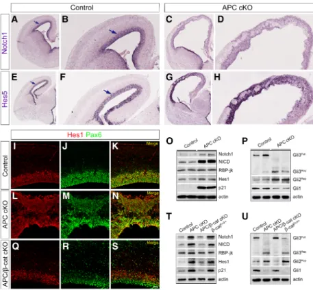
To define how APC might regulate appropriate radial pro-genitor development, we first examined the Notch signal-ing pathway, known to be essential for the development and identity of radial progenitors (Mizutani et al. 2007), Figure 3. Inhibition ofβ-catenin activity in APC-deficient RGCs rescues morphological and proliferation defects. (A) E14.5 cortices from
APCLox/+; hGFAP-Cre or APCLox/Lox; hGFAP-Cre mice were focally electroporated with BLBP-DsRed and DNTCF or ICAT and analyzed
in APC cKO cortices. Notch1 and its downstream effec-tors, Hes1 and Hes5, were significantly up-regulated and mislocalized in the APC cKO cortex (Fig. 4A–N). Pro-cessed Notch1, NICD, was also dramatically up-regulated (Fig. 4O). Although RBPJ, a constitutive repressor of Notch signaling, was not affected by APC deletion, the cell cycle inhibitor p21 was drastically up-regulated in the APC cKO cortex (Fig. 4O). These results suggest that deregulation of Notch1 activity in APC-deficient radial progenitors contributes to its disrupted development.
Notch1 activity is influenced by Shh signaling (Kong et al. 2015), and Shh is necessary for RGC development (Wang et al. 2016a). Transcriptional regulation in response to Shh is driven primarily via the Gli transcription factors 1, 2, and 3, which can be processed to Gli activator (GliA) or Gli repressor (GliR) forms. Shh signaling normally leads to an increase of GliA; in the absence of Shh ligand, GliR is increased, which inhibits Shh target gene transcription. We noticed a significant increase in repressor forms of Gli3 and Gli2 and a corresponding decrease in Gli1 in APC mutants (Fig. 4P), suggesting disrupted Shh signaling and primary cilium functions.
APC is necessary for primary cilium maintenance and signaling
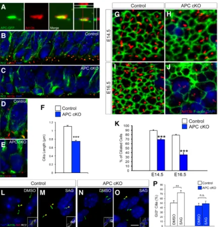
Since Shh signaling depends on primary cilia, we exam-ined primary cilia in APC-deficient progenitors. APC is coexpressed with Arl13b, a primary cilium marker (Fig. 5A). Deletion of APC disrupts the maintenance of primary cilia in progenitors, and APC mutant cilia are sig-nificantly shorter (Fig. 5B–F). As cortical development
proceeds, at late embryonic stages, APC-null cortical pro-genitors lose their primary cilia (Fig. 5G–K). To investi-gate whether the signaling function of cilia is disrupted in APC-deficient progenitors, we examined the require-ment for APC in Shh signaling. Stimulating Shh signaling with SAG, a Smo agonist, leads to the rapid accumulation of Gli3 at the tips of the primary cilia in cortical progeni-tors (Fig. 5L,M,P). In contrast, treatment of APC-deficient progenitors with SAG did not lead to Gli3 translocation to the tips of the cilia (Fig. 5N–P). Importantly, re-expression of APC in APC-deficient progenitors rescued the defects in cilium length, RGC proliferation, and morphology (Supplemental Fig. 3). These observations suggest that APC is essential to maintain primary cilium function in radial progenitors. Deletion of APC leads to aberrant pri-mary cilium signaling and maintenance, which may con-tribute to compromised radial progenitor development in APC cKO.
Aberrant APC–β-catenin signaling disrupts Notch activity and primary cilium function in cortical progenitors
Since our in vivo genetic analysis indicates that APC–β -catenin signaling is essential for radial progenitor develop-ment, we next examined whether APC-regulatedβ -cate-nin signaling underlies altered Notch activity and the primary cilium dysfunction in APC-deficient progenitors. Deletion of β-catenin in APC-deficient progenitors res-cued Notch signaling deficiency and the ciliary defects (Figs. 4Q–T, 6A–C). In RGCs without APC andβ-catenin, primary cilium length and number were similar to those
of controls (Fig. 6A–C). Activation ofβ-catenin alone in wild-type progenitors resulted in shorter cilia and cilium maintenance defects similar to those observed in APC-de-ficient progenitors (Fig. 6A–C). Consistent with these ob-servations, focal down-regulation ofβ-catenin signaling in APC-deficient progenitors using either DNTCF or ICAT rescued the defects in cilium length in APC-deficient pro-genitors (Fig. 6D–F). Furthermore, APC–β-catenin activity is essential for ciliary Shh signaling. Deletion ofβ-catenin improved the ability of APC-null RGCs to respond to SAG, whereas induction of constitutively activeβ-catenin in RGCs resulted in defective Shh signaling, similar to that observed in APC-deficient RGCs (Fig. 6G,H). Aber-rant processing patterns of the Gli family of transcription factors were also rescued in APC/β-catenin double-cKO cortices (Fig. 4U). Together, these data strongly suggest that deregulatedβ-catenin signaling underlies primary cil-ium defects in APC-deficient RGCs.
β-Catenin-mediated regulation of primary cilia
To identify cilium-related genes directly regulated by the APC–β-catenin pathway, we screened for ciliary genes that have high-affinityβ-catenin–TCF4/LEF-binding sites in their regulatory regions (Schuijers et al. 2014), com-pared them with the SysCilia database (http://syscilia. org) (van Dam et al. 2013), and identified 34 putative tar-gets ofβ-catenin–TCF4/LEF with known cilium functions (Fig. 7A). Of these genes, 24 encode proteins that are high-ly enriched in primary cilia and play key roles in primary cilium maintenance and signaling (Supplemental Table
1). To validate the relevant β-catenin ciliary targets in APC-deficient progenitors, we examined their expression in control and APC cKO cortices using quantitative RT– PCR. We observed a significant increase in the expression of Gpr98, Pard3, Pcdh15, and Tulp3 in APC cKO cortices (Fig. 7B). These genes were also up-regulated inβ -catenin-activated cortices (β-catEx3/+), while these increases were abolished in the APC/β-catenin-double-null cortex (Fig. 7C). Identification of these ciliary genes controlled byβ -catenin/TCF4 activity in APC-deficient cortices provides a potential mechanism by which APC-regulatedβ-catenin may directly modulate primary cilium structure and function.
Of the APC–β-catenin-regulated ciliary genes,Tulp3is known to play a critical role in cilium maintenance and targeting of signaling receptors to primary cilia (Badgandi et al. 2017). To examine whether the Tulp3 up-regulation in APC cKO cortices contributes to APC-null phenotypes of RGCs, we expressed validated shRNA against Tulp3 in APC-deficient RGCs (Supplemental Fig. 4). Knockdown of Tulp3 in APC-deficient RGCs increased the percentage of polarized RGCs with pial-attached basal processes (Fig. 7D–H) and ameliorated cilium defects (Fig. 7I–K). Down-regulation of Tulp3 activity also rescued proliferation de-fects in APC cKO radial progenitors (Fig. 7L–N). Coelec-troporation of shRNA-resistant human Tulp3 did not rescue APC cKO defects (Fig. 7). These results indicate that Tulp3 is a ciliary effector functioning downstream from the Wnt/β-catenin pathway, whose deregulation leads to ciliary defects associated with radial progenitor malformation in the APC-deficient neocortex.
Discussion
Here, we elucidated the functional contributions of APC-mediatedβ-catenin signaling in the development of corti-cal progenitors and cerebral corticorti-cal formation. Loss of APC in radial progenitors deregulatesβ-catenin activity and disrupts Notch pathway and primary cilium signal-ing, thus contributing to aberrant progenitor organization and differentiation. APC’s effects on cortical progenitors and the resultant cerebral cortical formation depend on precise regulation of β-catenin activity and associated Wnt tone.
APC/β-catenin signaling in cortical progenitor development
APC is essential for the formation and maintenance of po-larized radial progenitors during corticogenesis (Yokota et al. 2009). APC affects the outcome and patterns of pro-liferation of radial glia (Figs 1, 2;Supplemental Fig. 2).β -Catenin is a central component in the canonical Wnt pathway and acts downstream from APC. In the absence of Wnt ligand, the destruction complex, containing APC, phosphorylates and ubiquitinatesβ-catenin, leading to its proteasomal degradation. Wnt ligand binding leads
to the formation of a Frizzled receptor and low-density li-poprotein receptor-related protein (LRP) coreceptor com-plex, which stabilizes β-catenin and thus enables its translocation to the nucleus, where it acts as a transcrip-tional coactivator with members of the TCF/LEF family of transcription factors (Logan and Nusse 2004; Nelson and Nathke 2013). Deletion of APC and the resultant dis-ruption of the destruction complex leads to accumulation ofβ-catenin and unrestricted activation of Wnt signaling. APC in its role as an RNA-binding protein can also directly bind to the transcripts of several canonical Wnt signaling pathway members (Preitner et al. 2014). During cortical development, overexpression of activeβ-catenin in neuroepithelial cells promotes progenitor self-renewal (Chenn and Walsh 2002), whereas deletion ofβ-catenin de-pletes the basal progenitor pool and promotes premature neuronal differentiation (Machon et al. 2003). Increased
β-catenin levels in IPs promotes neurogenesis (Mutch et al. 2010; Munji et al. 2011). Thus, modulation of opti-malβ-catenin signaling is critical for the appropriate spa-tiotemporal balance of proliferation and differentiation of progenitors during corticogenesis. Precise context-depen-dent regulation of β-catenin signaling levels in different progenitors at different time points along embryonic de-velopment appears to be an essential feature of cerebral Figure 6. Primary cilium defects in APC-deficient cortices are dependent onβ -cate-nin signaling. (A) VZs of cortices (E18.5) from control, APC cKO, APC/β-cat cKO,
and β-catEx3/+ were labeled with
cortical formation. Considering the pivotal role played by APC inβ-catenin activity (Rusan et al. 2008; Nelson and Nathke 2013), we genetically dissected the effect of APC-regulatedβ-catenin signaling on cortical progenitor development. APC’s effect on cortical progenitor develop-ment depends on an appropriate balance of downstreamβ -catenin activity. In the absence ofβ-catenin, APC deletion did not significantly affect cortical progenitor develop-ment and cerebral cortical formation. APC appears to be crucial in setting the appropriate Wnt tone in cortical pro-genitors during cortical development. Increased β -cate-nin-mediated Wnt tone and the disrupted Notch and primary cilium signaling in the absence of APC appear to prevent the appropriate developmental differentiation of radial progenitors in the cerebral cortex, thus leading to disrupted corticogenesis in APC cKO.
APC–β-catenin versus APC–MT interactions during cortical development
APC is transported along MTs and associates with MT plus ends. APC regulates cytoskeletal dynamics as a MT plus end-tracking protein by binding and recruiting
β2B-tubulin mRNA to dynamic MTs and as a mediator of actin nucleation (Preitner et al. 2014). However, the res-cue of the APC cKO phenotype whenβ-catenin is deleted in APC-null progenitors (APCLox/Lox; Ctnnb1Lox/Lox;
hGFAP-Cre) suggests that APC’s direct effect on MTs in-dependently ofβ-catenin activity does not appear to be crucial for progenitor development. However, deregulated normal and active β-catenin levels in distinct cellular compartments (e. g., centrosomes, adhesion contacts, and nuclei) may indirectly affect MTs. Furthermore,β -cat-enin regulates adherens junctions between progenitors in the VZ, and the integrity of cell contacts between progen-itors in the ventricular surface is disrupted in APC cKO but rescued in APC/β-cat cKO. Deregulated distribution of β-catenin in different cellular compartments (e.g., adhesion contacts vs. nuclei) in APC cKO may have fur-ther destabilized the adherens junctions between radial progenitors that are necessary for their function and the integrity of the ventricular surface (Rasin et al. 2007).
β-Catenin signaling and primary cilium function in cortical progenitors
Early radial progenitor development and adult neural progenitors are dependent on primary cilium function (Higginbotham et al. 2013; Tong et al. 2014; Guo et al. 2015). Primary cilium signaling regulates major mental pathways important for cerebral cortex develop-ment, including Shh and Wnt/β-catenin signaling (Chenn and Walsh 2002; Houart et al. 2002; Hirabayashi et al. 2004; Logan and Nusse 2004; Shimogori et al. 2004; Wang et al. 2016a). Shh, Wnts, and other ligands necessary for progenitor development and cortical pat-terning are present in the embryonic brain cerebrospinal fluid (CSF) and cerebral wall (Lun et al. 2015; Wang et al. 2016b). We found that primary cilium maintenance and
function are severely disrupted in APC-deficient progeni-tors with deregulatedβ-catenin signaling. Down-regula-tion ofβ-catenin activity is sufficient to rescue primary cilium deficits in APC-deficient progenitors. Further-more, we show that ectopic induction of activeβ-catenin in progenitors, similar to that seen in APC-null cells, im-pairs Shh signaling in radial progenitors. Primary cilium and Shh signaling is thought to promote appropriate cell cycle exit (Phua et al. 2017) and generation and renewal of outer RGCs and IPs (Wang et al. 2016a). Excision of cil-ium tips and the resultant cilcil-ium disassembly may drive cell cycle (Phua et al. 2017). Furthermore, the asymmetric inheritance of primary ciliary membrane and thus earlier appearance of a primary cilium in a daughter cell during asymmetric cortical progenitor division may help it to re-tain stem cell characteristics (Paridaen et al. 2013). To-gether, these observations suggest that defective primary cilium/Shh signaling may contribute to the proliferation and differentiation defects observed in APC-deficient progenitors.
Wnt responses (Gerdes et al. 2007; Corbit et al. 2008), whereas inhibition of other cilium-related genes, such as ChibbyandSeahorse, attenuatesβ-catenin activity (Take-maru et al. 2003). Our observations underscore the direct regulation of cilium maintenance and signaling via APC-modulatedβ-catenin in cortical progenitors. This pathway contributes to the appropriate patterns of radial progenitor proliferation and organization necessary for the formation of the cerebral cortex.
APC–β-catenin signaling and neurodevelopmental disorders
Dominant mutations in APC have been known to cause fa-milial adenomatous polyposis (FAP), characterized by in-testinal polyposis and colorectal cancer (Nelson and Nathke 2013; Dow et al. 2015). Extracolonic manifesta-tions of FAP are referred to as Gardner syndrome or Turcot syndrome, and neurological outcomes include brain tu-mor polyposis type 2 and autism (Zhou et al. 2007; Gómez García and Knoers 2009; Mohn et al. 2014; Song et al. 2014). Recent analysis of autism spectrum disorder (ASD) risk gene networks implicate canonical Wnt signal-ing as a pathway disrupted in ASD. In mice,Dvl1andDvl3 deficiency deregulates a transcriptional program involving
β-catenin, Brn2, and Tbr2, causing an expansion of the IP cell population and ASD-related aberrant social behavior (Belinson et al. 2016). Treatment of these mice with a GSK3βantagonist, lithium, which activatesβ -catenin-re-lated canonical Wnt signaling, rescued these defects. De-regulated β-catenin levels and cellular localization also impact its interactions with axin, which acts as an ampli-fication to the differentiation switch in IPs (Fang et al. 2013). Inhibition of axin degradation in IPs with a tankyr-ase inhibitor, XAV939, leads to excessive production of up-per-layer neurons and autism-like behaviors (Fang et al. 2014). Individual ASD risk genes such asCHD8(Durak et al. 2016), PTEN(Chen et al. 2015), UBE3A(Yi et al. 2017), DIXDC1,DDX3X, andCTNND2 and chromatin remodeling genes overrepresented in ASD risk (e.g., ARID1B) can also modulateβ-catenin-mediated Wnt sig-naling. Importantly, several Wnt/β-catenin pathway loci are shared risk factors for schizophrenia, bipolar disorder, and ASD, includingCHD8,DISC1,TCF4, andCTNNB1 (Ernst 2016; Mulligan and Cheyette 2017). Other Wnt genes, including the secreted Wnt antagonistsDKK1and DKK4and Wnt receptorsKREMEN1, are also implicated in schizophrenia. Furthermore, genomic analyses of bipo-lar disorder have identified disruptions in Wnt/β-catenin pathway genes, includingWNT7A,WNT2B,DISC1, and TCF4 (Ernst 2016; Mulligan and Cheyette 2017). These and other neuropsychiatric disorder-associated genes may be differentially regulated (up or down) by deviations of normal Wnt signaling in the developing brain, thus lead-ing to different disease outcomes.
Disrupted Wnt signaling pathway in progenitors is emerging as a shared convergent route to schizophrenia, bipolar disorder, and ASD. (Ernst 2016; Packer 2016; Hazlett et al. 2017; Mulligan and Cheyette 2017). Our studies demonstrating the essential role of the APC–β
-cat-enin pathway in setting the right Wnt signaling tone nec-essary for cortical progenitor development provide evidence that changes in this process can lead to disrup-tions in cerebral cortical circuit formation that underlie these neurodevelopmental disorders.
Materials and methods
Animals
Mice were cared for according to animal protocols approved by the University of North Carolina. APC was conditionally inactivated in radial progenitors by mating mice carrying the APC allele flanked by LoxP sites (Shibata et al. 1997) with hGFAP-Cre mice. Littermate APCLox/+; hGFAP-Cre mice served as controls. A similar mating strategy with Ctnnb1Lox/Lox or Ctnnb1LoxEx3/LoxEx3was used to inactivate or activateβ-catenin in radial progenitors. APCLox/Lox; Ctnnb1Lox/Lox mice were crossed with APCLox/+; Ctnnb1Lox/+; hGFAP-Cre mice to generate
APCLox/Lox; Ctnnb1Lox/Lox; hGFAP-Cre mice in whichβ-catenin
was deleted in APC-deficient radial progenitors. The effect of the recombination in APC cKO, Ctnnb1 cKO, APC/Ctnnb1 cKO, or Ctnnb1LoxEx3/+ cortices was assessed by immunoblot analysis as described previously (Yokota et al. 2009).
Immunohistochemistry
Immunohistochemical labeling of embryonic brain sections and isolated cortical progenitors was performed as described earlier (Higginbotham et al. 2013; Guo et al. 2015). The following prima-ry antibodies were used: Arl13b (1:1000; NeuroMabs, 75-287), APC (1:500 [Santa Cruz Biotechnology, C-20] and 1:500 [gift from Dr. I. Nathke, University of Dundee]) (Yokota et al. 2009), β-catenin (Sigma; C2206; 1:500), BrdU (BD Biosciences; 347580; 1:100), cleaved-caspase3 (Cell Signaling; 9661; 1:400), Ctip2 (1:500; Abcam, ab18465), GFP (1:1000; Abcam, ab13970), Gli3 (1:500; R&D Systems, AF3690), Hes1 (1:500; gift from Dr. R. Kageyama, Kyoto University), Pax6 (1:500; BioLegend, 901301), PH3 (1:500; Upstate Biotechnology), RC2 (1:3; Iowa Hy-bridoma Bank), RFP (1:500; Abcam, ab28664), Tbr1 (1:2500; Milli-pore, ab2261), Tbr2 (1:500; Abcam, ab23345), and Tuj1 (1:1000; Covance, MMS-435). Appropriate Cy2, Cy3, or Alexa dye-conju-gated secondary antibodies (Jackson ImmunoResearch, Molecu-lar Probes) were used to detect primary antibody binding. DAPI (Invitrogen) was used as nuclear counterstain. For routine BrdU labeling, dams were injected intraperitoneally with 50 mg of BrdU per kilogram of bodyweight (Sigma, B5002) 1 h prior to sac-rifice. To assay for cell cycle pattern, BrdU was injected 24 h be-fore tissue harvesting. Cortical sections were then colabeled with anti-BrdU and Ki67 antibodies. Ratio of Ki67−, BrdU+/BrdU+cells was quantified and used as the cell cycle exit index. Quantifica-tion of BrdU-, PH3-, Caspase3-, Tbr2-, Tuj1-, and Arl13b-labeled cells was performed as described previously (Yokota et al. 2009; Higginbotham et al. 2013).
Immunoblot analysis
with one of the following primary antibodies: APC (1:500; Santa Cruz Biotechnology, C-20),β-actin (1:1000; Abcam, ab8226),β -catenin (1:1000; Sigma, C2206), Gli1 (1:500; Cell Signaling, 2534), Gli2 and Gli3 (1:200; gift from Dr. B. Wang, Cornell Univer-sity), Hes1 (gift from Dr. R. Kageyama, Kyoto University; 1:500), NICD (1:500; Abcam, ab8925), Notch1 (1:500; Santa Cruz Bio-technology, C-20), p21 (1:200; Santa Cruz BioBio-technology, F-5), or RBP-jk (1:1000; Millipore, AB2284). Immunoblots were developed using horseradish peroxidase-conjugated goat anti-mouse or goat anti-rabbit antibodies and detected with enhanced chemiluminescence.
RT–PCR and quantitative PCR
RNA was extracted from dorsal cortex at E18.5 using RNeasy minikit (Qiagen) and cleared of genomic DNA using Turbo DNase (Life Technologies). cDNA was synthesized using iScript (Bio-Rad). Real-time PCR was performed in triplicates using Ap-plied Biosystem Power SYBR Green PCR master mix and the ap-propriate forward and reverse primers (seeSupplemental Table 2). For quantification of gene expression changes, three replicates from three independent experiments were averaged. Samples were normalized againstβ-actin expression.
Shh assay for ciliary signaling
Isolated cortical radial progenitors from E14.5 control and APC cKO cortices were maintained in DMEM/N2/B27 for 1 d prior to treatment with 200 nM SAG (Calbiochem) for 24 h. Cells were then immunolabeled with RC2, Arl13b, and Gli3 antibod-ies. The percentage of radial progenitor cilia with Gli3 at the tips was quantified from three independent experiments.
In utero electroporation
E14.5 embryos were electroporated as described previously (Yokota et al. 2009; Higginbotham et al. 2013). The following constructs were coelectroporated with pBLBP-DsRed: pcDNA-DNTCF4, pcDNA-FLAG-ICAT, pCIG2-GFP, and pAPC-GFP. Radial progenitors in electroporated cortices were analyzed 48 h later.
Tulp3 shRNA
Tulp3 shRNA was obtained from the University of North Caroli-na Gene Therapy Center. shRNA target sequences used were as follows: CCTACAGTGTACCTGAACTTA (1), CCGACA GATTTGTCTCGTGAA (2), GTGGAGAATTTAGAGGACTTT (3), CGTGTTCACACTGGACTATAA (4), and GTGTAAAG TAACCAGGGATAA (5). Two pools (1.5 µg/µL) of shRNAs [shTulp3 #1 (1–3) and shTulp3 #2 (4–5)] containing equivalent amounts of different shRNAs were used for in utero electropora-tion. Nonsilencing scrambled shRNA (pLKO.1 shRNA vector; University of North Carolina Gene Therapy Center) was used as control. In shRNA + cDNA rescue experiments, human TULP3 cDNA (University of North Carolina Gene Therapy Cen-ter) was used at 1 µg/µL concentration.
Acknowledgments
We thank Jiami Guo and Charlotte Plestant for helpful com-ments. This research was supported by National Institutes of Health grant MH060929 to E.S.A., and the confocal imaging core of a National Institute of Neurological Disorders and Stroke
institutional center core grant (5P30NS045892-12). N.N. is sup-ported by the Uehara Memorial Foundation research fellowship. E.S.A., N.N., J.L., K.Y-N., M.C., T.M., and T.-Y.E. designed the ex-periments. N.N., J.L., K.Y-N., T.-Y.E., M.C., and R.T. conducted the experiments and analyzed the data. N.N., J.L., K.Y-N., T.-Y.E., M.C., and E.S.A. wrote the manuscript.
References
Badgandi HB, Hwang S, Shimada IS, Loriot E, Mukhopadhyay S. 2017. Tubby family proteins are adapters for ciliary trafficking of integral membrane proteins.J Cell Biol216:743–760. Belinson H, Nakatani J, Babineau BA, Birnbaum RY, Ellegood J,
Bershteyn M, McEvilly RJ, Long JM, Willert K, Klein OD, et al. 2016. Prenatalβ-catenin/Brn2/Tbr2 transcriptional cas-cade regulates adult social and stereotypic behaviors.Mol Psy-chiatry21:1417–1433.
Cappello S, Attardo A, Wu X, Iwasato T, Itohara S, Wilsch-Bräu-ninger M, Eilken HM, Rieger MA, Schroeder TT, Huttner WB, et al. 2006. The Rho-GTPase cdc42 regulates neural pro-genitor fate at the apical surface.Nat Neurosci9:1099–1107. Chen Y, Huang W-C, Sejourne J, Clipperton-Allen AE, Page DT. 2015. Pten mutations alter brain growth trajectory and alloca-tion of cell types through elevatedβ-catenin signaling.J Neu-rosci35:10252–10267.
Chenn A, Walsh CA. 2002. Regulation of cerebral cortical size by control of cell cycle exit in neural precursors.Science297: 365–369.
Corbit KC, Shyer AE, Dowdle WE, Gaulden J, Singla V, Reiter JF. 2008. Kif3a constrains β-catenin-dependent Wnt signalling through dual ciliary and non-ciliary mechanisms.Nat Cell Biol10:70–76.
Dow LE, O’Rourke KP, Simon J, Tschaharganeh DF, van Es JH, Clevers H, Lowe SW. 2015. Apc restoration promotes cellular differentiation and reestablishes crypt homeostasis in colorec-tal cancer.Cell161:1539–1552.
Durak O, Gao F, Kaeser-Woo YJ, Rueda R, Martorell AJ, Nott A, Liu CY, Watson LA, Tsai L-H. 2016. Chd8 mediates cortical neurogenesis via transcriptional regulation of cell cycle and Wnt signaling.Nat Neurosci19:1477–1488.
Ernst C. 2016. Proliferation and differentiation deficits are a ma-jor convergence point for neurodevelopmental disorders. Trends Neurosci39:290–299.
Evsyukova I, Plestant C, Anton ES. 2013. Integrative mechanisms of oriented neuronal migration in the developing brain.Annu Rev Cell Dev Biol29:299–353.
Ezratty EJ, Stokes N, Chai S, Shah AS, Williams SE, Fuchs E. 2011. A role for the primary cilium in Notch signaling and epider-mal differentiation during skin development. Cell 145: 1129–1141.
Fang W-Q, Chen W-W, Fu AKY, Ip NY. 2013. Axin directs the am-plification and differentiation of intermediate progenitors in the developing cerebral cortex.Neuron79:665–679. Fang W-Q, Chen W-W, Jiang L, Liu K, Yung W-H, Fu AKY, Ip
NY. 2014. Overproduction of upper-layer neurons in the neo-cortex leads to autism-like features in mice. Cell Rep 9: 1635–1643.
Garcia-Gonzalo FR, Phua SC, Roberson EC, Garcia G, Abedin M, Schurmans S, Inoue T, Reiter JF. 2015. Phosphoinositides reg-ulate ciliary protein trafficking to modreg-ulate hedgehog signal-ing.Dev Cell34:400–409.
function and perturbs intracellular Wnt response.Nat Genet 39:1350–1360.
Gómez García EB, Knoers NV. 2009. Gardner’s syndrome (famili-al adenomatous polyposis): a cilia-related disorder. Lancet Oncol10:727–735.
Guo J, Higginbotham H, Li J, Nichols J, Hirt J, Ghukasyan V, An-ton ES, Guemez-Gamboa A, Coufal NG, Gleeson JG, et al. 2015. Developmental disruptions underlying brain abnormal-ities in ciliopathies.Nat Commun6:7857.
Hansen DV, Lui JH, Parker PRL, Kriegstein AR. 2010. Neurogenic radial glia in the outer subventricular zone of human neocor-tex.Nature464:554–561.
Harada N, Tamai Y, Ishikawa T, Sauer B, Takaku K, Oshima M, Taketo MM. 1999. Intestinal polyposis in mice with a domi-nant stable mutation of the β-catenin gene. EMBO J 18: 5931–5942.
Hazlett HC, Gu H, Munsell BC, Kim SH, Styner M, Wolff JJ, Eli-son JT, SwanEli-son MR, Zhu H, Botteron KN, et al. 2017. Early brain development in infants at high risk for autism spectrum disorder.Nature542:348–351.
Higginbotham H, Guo J, Yokota Y, Umberger NL, Su C, Li J, Verma N, Hirt J, Ghukasyan V, Caspary T, et al. 2013. Arl13b-regulated cilia activities are essential for polarized ra-dial glial scaffold formation.Nat Neurosci16:1000–1007. Hildebrandt F, Benzing T, Katsanis N. 2011. Ciliopathies.N Engl J
Med364:1533–1543.
Hirabayashi Y, Itoh Y, Tabata H, Nakajima K, Akiyama T, Masuyama N, Gotoh Y. 2004. The Wnt/β-catenin pathway di-rects neuronal differentiation of cortical neural precursor cells.Development131:2791–2801.
Houart C, Caneparo L, Heisenberg C, Barth K, Take-Uchi M, Wil-son S. 2002. Establishment of the telencephalon during gastru-lation by local antagonism of Wnt signaling. Neuron 35: 255–265.
Imayoshi I, Isomura A, Harima Y, Kawaguchi K, Kori H, Miyachi H, Fujiwara T, Ishidate F, Kageyama R. 2013. Oscillatory con-trol of factors determining multipotency and fate in mouse neural progenitors.Science342:1203–1208.
Kim W-Y, Wang X, Wu Y, Doble BW, Patel S, Woodgett JR, Snider WD. 2009. GSK-3 is a master regulator of neural progenitor homeostasis.Nat Neurosci12:1390–1397.
Kong JH, Yang L, Dessaud E, Chuang K, Moore DM, Rohatgi R, Briscoe J, Novitch BG. 2015. Notch activity modulates the re-sponsiveness of neural progenitors to sonic hedgehog signal-ing.Dev Cell33:373–387.
Kusek G, Campbell M, Doyle F, Tenenbaum SA, Kiebler M, Tem-ple S. 2012. Asymmetric segregation of the double-stranded RNA binding protein Staufen2 during mammalian neural stem cell divisions promotes lineage progression.Cell Stem Cell11:505–516.
Lancaster MA, Schroth J, Gleeson JG. 2011. Subcellular spatial regulation of canonical Wnt signalling at the primary cilium. Nat Cell Biol13:700–707.
Logan CY, Nusse R. 2004. The Wnt signaling pathway in develop-ment and disease.Annu Rev Cell Dev Biol20:781–810. Lui JH, Nowakowski TJ, Pollen AA, Javaherian A, Kriegstein AR,
Oldham MC. 2014. Radial glia require PDGFD–PDGFRβ sig-nalling in human but not mouse neocortex. Nature 515: 264–268.
Lun MP, Monuki ES, Lehtinen MK. 2015. Development and func-tions of the choroid plexus-cerebrospinal fluid system.Nat Rev Neurosci16:445–457.
Machon O, van den Bout CJ, Backman M, Kemler R, Krauss S. 2003. Role ofβ-catenin in the developing cortical and hippo-campal neuroepithelium.Neuroscience122:129–143.
Mizutani K, Yoon K, Dang L, Tokunaga A, Gaiano N. 2007. Dif-ferential Notch signalling distinguishes neural stem cells from intermediate progenitors.Nature449:351–355. Mohn JL, Alexander J, Pirone A, Palka CD, Lee S-Y, Mebane L,
Haydon PG, Jacob MH. 2014. Adenomatous polyposis coli protein deletion leads to cognitive and autism-like disabili-ties.Mol Psychiatry19:1133–1142.
Mulligan KA, Cheyette BNR. 2017. Neurodevelopmental per-spectives on Wnt signaling in psychiatry.Mol Neuropsychia-try2:219–246.
Munji RN, Choe Y, Li G, Siegenthaler JA, Pleasure SJ. 2011. Wnt signaling regulates neuronal differentiation of cortical inter-mediate progenitors.J Neurosci31:1676–1687.
Mutch CA, Schulte JD, Olson E, Chenn A. 2010.β-Catenin signal-ing negatively regulates intermediate progenitor population numbers in the developing cortex.PLoS One5:e12376. Nelson S, Nathke IS. 2013. Interactions and functions of the
ade-nomatous polyposis coli (APC) protein at a glance.J Cell Sci 126:873–877.
Packer A. 2016. Neocortical neurogenesis and the etiology of au-tism spectrum disorder.Neurosci Biobehav Rev64:185–195. Paridaen JTML, Wilsch-Bräuninger M, Huttner WB. 2013. Asym-metric inheritance of centrosome-associated primary cilium membrane directs ciliogenesis after cell division.Cell155: 333–344.
Patterson VL, Damrau C, Paudyal A, Reeve B, Grimes DT, Stew-art ME, Williams DJ, Siggers P, Greenfield A, Murdoch JN. 2009. Mouse hitchhiker mutants have spina bifida, dorso– ventral patterning defects and polydactyly: identification of Tulp3 as a novel negative regulator of the Sonic hedgehog pathway.Hum Mol Genet18:1719–1739.
Phua SC, Chiba S, Suzuki M, Su E, Roberson EC, Pusapati GV, Setou M, Rohatgi R, Reiter JF, Ikegami K, et al. 2017. Dynamic remodeling of membrane composition drives cell cycle through primary cilia excision.Cell168:264–279.
Preitner N, Quan J, Nowakowski DW, Hancock ML, Shi J, Tcher-kezian J, Young-Pearse TL, Flanagan JG. 2014. APC is an RNA-binding protein, and its interactome provides a link to neural development and microtubule assembly. Cell 158: 368–382.
Rakic P. 1988. Specification of cerebral cortical areas.Science 241:170–176.
Rasin M-R, Gazula V, Breunig JJ, Kwan KY, Johnson MB, Liu-chen S, Li H, Jan LY, Jan Y-N, Rakic P, et al. 2007. Numb and Numbl are required for maintenance of cadherin-based adhe-sion and polarity of neural progenitors. Nat Neurosci 10: 819–827.
Rusan NM, Akong K, Peifer M. 2008. Putting the model to the test: are APC proteins essential for neuronal polarity, axon outgrowth, and axon targeting?J Cell Biol183:203–212. Schuijers J, Mokry M, Hatzis P, Cuppen E, Clevers H. 2014.
Wnt-induced transcriptional activation is exclusively mediated by TCF/LEF.EMBO J33:146–156.
Shibata H, Toyama K, Shioya H, Ito M, Hirota M, Hasegawa S, Matsumoto H, Takano H, Akiyama T, Toyoshima K, et al. 1997. Rapid colorectal adenoma formation initiated by condi-tional targeting of the Apc gene.Science278:120–123. Shimogori T, Banuchi V, Ng HY, Strauss JB, Grove EA. 2004.
Em-bryonic signaling centers expressing BMP, WNT and FGF pro-teins interact to pattern the cerebral cortex. Development 131:5639–5647.
Tago K, Nakamura T, Nishita M, Hyodo J, Nagai S, Murata Y, Adachi S, Ohwada S, Morishita Y, Shibuya H, et al. 2000. Inhi-bition of Wnt signaling by ICAT, a novelβ-catenin-interacting protein.Genes Dev14:1741–1749.
Takemaru K-I, Yamaguchi S, Lee YS, Zhang Y, Carthew RW, Moon RT. 2003. Chibby, a nuclear β-catenin-associated antagonist of the Wnt/Wingless pathway. Nature 422: 905–909.
Tong CK, Han Y-G, Shah JK, Obernier K, Guinto CD, Alvarez-Buylla A. 2014. Primary cilia are required in a unique sub-population of neural progenitors.Proc Natl Acad Sci 111: 12438–12443.
van Dam TJ, Wheway G, Slaats GG, Huynen MA, Giles RH, Giles RH. 2013. The SYSCILIA gold standard (SCGSv1) of known ciliary components and its applications within a systems biol-ogy consortium.Cilia2:7.
Wang L, Hou S, Han Y-G. 2016a. Hedgehog signaling promotes basal progenitor expansion and the growth and folding of the neocortex.Nat Neurosci19:888–896.
Wang W, Jossin Y, Chai G, Lien W-H, Tissir F, Goffinet AM. 2016b. Feedback regulation of apical progenitor fate by imma-ture neurons through Wnt7–Celsr3–Fzd3 signalling. Nat Commun7:10936.
Wilson SL, Wilson JP, Wang C, Wang B, McConnell SK. 2012. Pri-mary cilia and Gli3 activity regulate cerebral cortical size.Dev Neurobiol72:1196–1212.
Yi JJ, Paranjape SR, Walker MP, Choudhury R, Wolter JM, Fragola G, Emanuele MJ, Major MB, Zylka MJ. 2017. The autism-linked UBE3A T485A mutant E3 ubiquitin ligase activates the Wnt/β-catenin pathway by inhibiting the proteasome.J Biol Chem292:12503–12515.
Yokota Y, Kim WY, Chen Y, Wang X, Stanco A, Komuro Y, Snider W, Anton ES. 2009. The adenomatous polyposis coli protein is an essential regulator of radial glial polarity and construction of the cerebral cortex.Neuron61:42–56.
Yokota Y, Eom T, Stanco A, Kim W, Rao S, Snider WD, Anton ES. 2010. Cdc42 and Gsk3 modulate the dynamics of radial glial growth, inter-radial glial interactions and polarity in the de-veloping cerebral cortex.Development137:4101–4110. Zhou X-L, Giacobini M, Anderlid B-M, Anckarsäter H, Omrani D,
Gillberg C, Nordenskjöld M, Lindblom A. 2007. Association of adenomatous polyposis coli (APC) gene polymorphisms with autism spectrum disorder (ASD).Am J Med Genet B Neuropsychiatr Genet144B:351–354.