Journal of Global Pharma Technology
Available Online at:
www.jgpt.co.in
RESEARCH ARTICLE
Insilico and Electron Microscope Study about Attachment of
Mesenchymal Stem Cell Derived from Stromal Vascular Fraction
to Scaffold (Bovine Bone Cancellous, Hydroxyapatite-Calcium
Sulphate, Calcium Phosphate)
Panji Sananta
1, 2*,
Rusendi Hidayat
2,
Marvin Anthony Putera
2,
Sri Andarini
3,
Umi Kalsum
4, Respati Suryanto Dradjat
21. Doctoral Program of Medical Sciences, Faculty of Medicine, Universitas Brawijaya.
2. Orthopaedic and Traumatology Department, Faculty of Medicine, Universitas Brawijaya.
3. Public Health Department, Faculty of Medicine, Universitas Brawijaya.
4. Pharmacology Department, Faculty of Medicine, Universitas Brawijaya.
*Corresponding Author: Panji Sananta
Abstract
Objective: Insilico study using molecular docking with Hex had already surpassed all of the other previous approach in assessment of molecular binding to scaffold. The combination of insilico study to investigate the molecular binding to scaffold and electron microscope to assess the population density of adipocyte derivative stem cells by measuring the number of cluster cells could show the inter cell communication between adipocyte derived stem cell and scaffold. The aim of this study is to prove the attachment of mesenchymal stem cell (MSC) from stromal vascular fraction (SVF) to scaffold. Methods: The research is true experimental research using molecular docking with HEX version 8.0. Electron microscope used to measure the density and number of cluster cells of adipocyte derivative stem cells. Results: The strongest bound to scaffold is between MSC from SVF to hydroxyapatite in both receptor which was Integrin Alpha V to Hydroxyapatite which need a total energy of -89.24 (J/Mol) and Integrin Beta 2 to Hydroxyapatite which need a total energy of -177.8 (J/Mol). The highest impact obtained from hydroxyapatite-calcium phosphate with an average value of 12.66 cluster cells counting per 100 µm2 scaffold material observed by electron microscope. Conclusion: This study showed that MSC from SVF could attach to scaffold with stronger binding between Integrin Alpha V and Integrin Beta 2 to hydroxyapatite. From microscope electron study we can prove that MSC can make a cluster cell in those scaffolds with hydroxyapatite-calcium sulphate having the biggest cluster cell counting.
Keywords: Hydroxyapatite, Integrin Alpha V, Integrin Beta 2, Mesenchymal stem cell, Scanning electron microscope, Stromal vascular fraction.
Introduction
Tissue Engineering (TE) point to
improvement, restoration, and maintenance of damaged tissues caused by diverse factors such as injury, disease, or congenital disabilities. Tissue regeneration and healing in conventional method is using the auto graft method and mainly dependent on the presence of donor tissues, coupled with other unwanted effects such as pain and risks to patients such as morbidity of donor tissue and infectious diseases. Nowadays artificial scaffolds have been applied and used as a supporting structure for cell cultures and
domination of cell growth in repair of defective tissues or organs. While the cell
regenerated, the scaffold temporarily
But with an increasing complexity of design, there are bigger challenges to translation. For example, receiving regulatory approval
for an acellular scaffold requires
substantially less time and fewer resources than does a drug-eluting scaffold that has been pre-seeded with stem cells. Scaffolds in TE are cytocompatible biomaterials that cells can adhere to and/or replace with extracellular matrix to generate native tissues. Whether a simple morselized autologous bone or complexes inject able, thermally responsive synthetic hydro gels capable of mineralizing in situ could be used as a scaffold.
Signals in the TE paradigm could be defined
as internally or externally derived
environmental factors that can affect the regeneration of tissues. Same as scaffold, these signals can be further deranged into
subcategories including mechanical,
biological, chemical and electrical cues. Cells, in order to create living tissues, as well as integrate living engineered tissue with native host tissues, must be present. Cells can be recruited into an implanted scaffold by methods such as chemokines release, cell ligands attachment to scaffold, or scaffolds containing cells which could be implanted into a defect.
Unlike other TE fields, there is less controversy in regard to stem cell type in orthopedic TE. Until this day, mesenchymal stem cell (MSC) is the most commonly utilized cell type [2].Stromal vascular fraction (SVF) is a heterogeneous, versatile, and clinically relevant cell system.
SVF is known to have MSC, fibroblasts, smooth muscle cells, mural cells, blood cells, macrophages, and a whole cadre of other stem cell phenotype. Interaction of all these cell types contributes to SVF’s overall therapeutic potential [3].Electron microscope is subcategorized into transmission electron
microscopes (TEM), scanning electron
microscopes (SEM) and low energy electron microcopy (LEEM).
All of these serve in purpose of a significant demand exist for the development of novel
techniques capable of imaging
nanostructures, macromolecules, and
surfaces to provide analytical capabilities with sub-nanometer resolution [4]. In this paper we want to make a study of insilico and
electron microscope about attachment
MSCderived from SVF to scaffold.
Materials and Methods
Insilico study: Molecular Docking with HEX version 8.0
He x is an interactive protein docking and molecular superposition program, written by Dave Ritchie. Hex understands protein and DNA structures in PDB format, and it can also read small-molecule SDF files [5].
Sample:
Integrin Alpha V (PDB ID 1JV2)
Integrin Beta 2 (PDB ID 3K6S)
Nanoparticle hydroxyapatite (PUBCHEM ID 18986957)
Hydroxyapatite (PUBCHEM ID 14781)
Calcium Sulphate (PUBCHEM ID 24497)
Calcium Phosphate (PUBCHEM ID 24456)
Electron microscope: Assessment of
population density of adipocyte derivative stem cells by measuring the number of cluster cells seen in imaging scanning electron microscope / SEM to reduce measurement bias, then measured the number of cluster cells by two observers, namely researchers and expert analysts in the field of stem cells. In addition, cells measured for density on SEM imaging were performed under a light microscope to prove that cells imaged in SEM were adipocyte derivative stem cells [6].
Results
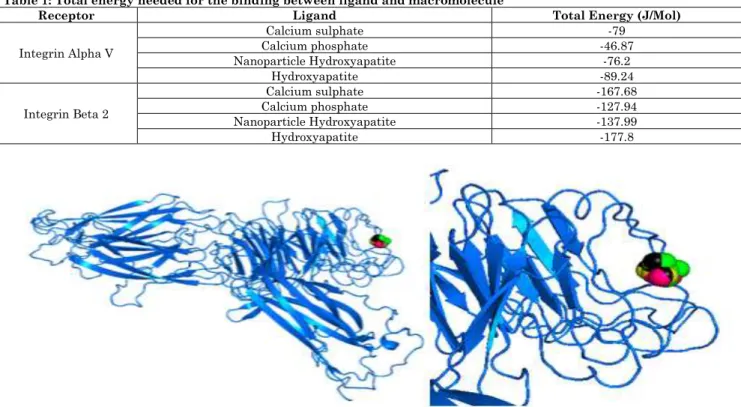
From the insilico study we have obtained a result from the docking of MSC from SVF to scaffold. Integrin Alpha V and Integrin Beta 2 were chosen as a macromolecule because more than 80% of MSC’s will express Integrin Beta 2 and 20-50% will express Integrin Alpha V. Both of these proteins will have a huge role on attachment and adhesion.
Table 1: Total energy needed for the binding between ligand and macromolecule
Receptor Ligand Total Energy (J/Mol)
Integrin Alpha V
Calcium sulphate -79
Calcium phosphate -46.87 Nanoparticle Hydroxyapatite -76.2
Hydroxyapatite -89.24
Integrin Beta 2
Calcium sulphate -167.68 Calcium phosphate -127.94 Nanoparticle Hydroxyapatite -137.99
Hydroxyapatite -177.8
Figure 1: Interaction between Integrin Alpha V and Nanoparticle
Table 2: Interaction between Integrin Alpha V and Nanoparticle shown by color
Ligand Color
Calcium sulphate Calcium phosphate Nanoparticle Hydroxyapatite
Hydroxyapatite black
Figure 2: Interaction between Integrin Beta 2 and Nanoparticle
Table 3: Interaction between Integrin Beta 2 and Nanoparticle shown by color
Ligand Color
Calcium sulphate Calcium phosphate Nanoparticle Hydroxyapatite
Hydroxyapatite black
From electron microscope study it had been proved that MSC from SVF could attach to scaffold and develop a cluster cells.
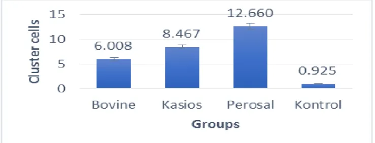
The cluster cells were counted and the result was showed in Table 4.
Table 4: Descriptive analysis of Cluster cells counting per 100 µm2based on each scaffold
Group N Mean Std. Deviation
Bovine bone cancellous 6 6.008 0.085 HA-Calcium Sulphate 6 12.660 0.413 Calcium Phosphate 6 8.467 0.322
Based on the results of the descriptive analysis in Table 4 it can be seen that the number of cluster cells per 100 µm2 is described as follows:
Observations of Bovine bone cancellous
from 6 replications obtained an average value of 6.008 and a standard deviation of 0.085.
Observations of HA-Calcium sulphate
(Perosal) from 6 replications obtained an average value of 12.66 and a standard deviation of 0.413.
Observations of Calcium phosphate
(Kasios) from 6 replications obtained an
average value of 8,467 and a standard deviation of 0,322.
Observations of control from 6 replications obtained an average value of 0.925 and a standard deviation of 0.085.
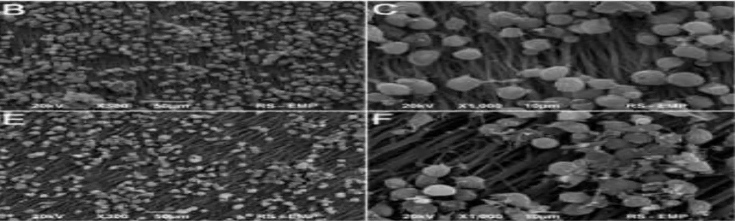
Through the results above, it can be said that the administration of Scaffold material in ADSC cells has a significant effect, and the administration of HA-Calcium sulphate (Perosal) material is higher than other groups. Figure 3 until 5 each shows the imaging of the scanning electron microscope and the descriptive analysis graph of cell cluster counting.
Figure 3: Imaging Scanning electron microscope of adipose derived stem cellin bonegraft medium (A) control, (B)
adipose derived stem cell in bovine cancellous graft medium, (C) adipose derived stem cell in calcium phosphate
medium, and (C) adipose derived stem cell in hydroxapatite-calcium sulfate. Magnification 1000x
Figure 5: Imaging Scanning electron microscope of adipose derived stem cell
Discussion
These days, various protein docking programs have been made available as web servers. These range from rapid Patch Dock server to much more computationally intensive approaches incorporating models of flexibility such as Rosetta Dock and Haddock. Several FFT-based docking programs have also been made available as web servers. Similar as the geometric hashing approach, the FFT-based approaches assume that the proteins to be docked are rigid, but they sample densely all possible rigid-body orientations in the 6D search space.
But despite all of that assumption, Cartesian grid-based FFT docking algorithms are inherently overpriced. To counter those limitations of the Cartesian FFT approaches, the ‘Hex’ spherical polar Fourier (SPF) was developed using rotational correlations which significantly reduces execution times. Thus, Hex algorithm obtained an enormous speed-up by exploiting the huge computational power of modern graphics processor units (GPU) using the CUDA (Common Unified Device Architecture) development tools. A typical Hex docking calculation is up to two orders of magnitude faster than conventional
Cartesian grid-based FFT docking
approaches. The Hex SPF algorithm has been validated in CAPRI (Critical Assessment of
Predicted Interactions) blind docking
experiment and Hex prediction has been found commonly within the top 100 orientations in recent CAPRI scoring sections.
Therefore, Hex Server set a very fast and simple way to generate a high quality
docking predictions for subsequent
refinement With the Hex we can prove the attachment between MSC from SVF to scaffold, and showed that the strongest bound is between MSC from SVF to
hydroxyapatite in both receptor which was Integrin Alpha V to Hydroxyapatite which need a total energy of -89.24 (J/Mol) and Integrin Beta 2 to Hydroxyapatite which need a total energy of -177.8 (J/Mol) [7].The scanning electron microscope (SEM) study made it possible to control porosity percentage and the pores size. Study conducted by Mahmood et al, 2017 show that SEM could show the superficial and internal structure of scaffolds. With this method it was possible to control the porosity percentage and the pores size. SEM analysis showed excellent micro pores structures with sizes varying from 9 to 526 m [8].
In this study SEM imaging was also carried out in each treatment with a magnification of
7000x. Although not yet analyzed
qualitatively, the imaging results suggest that adipocyte derived stem cell (ADSC) cell populations are denser in all three scaffolds than controls, cell density has been shown to affect cell-cell interactions and as an important factor, controlling subsequent cell proliferation and gene expression profiles. In addition, Cell proliferation is strongly influenced by the surface area of cell attachment and inhibition of contact between adjacent cells. So it is estimated that the relationship / inter-cell communication from ADSC increases after being combined with scaffold [6].
Conclusion
Acknowledgement
The authors of this study want to give thank you to all of the support from the colleagues
from Orthopaedic and Traumatology
Department, Faculty of Medicine,
Universitas Brawijaya.
References
1. Eltom Abdalla, Zhong Gaoyan,
Muhammad Ameen (2019) Scaffold
Techniques and Designs in Tissue Engineering Functions and Purposes: A Review. Advances in Materials Sciences and Engineering, 1-13.
2. Tatara Alexander M, Mikos Antonios G
(2016) Tissue Engineering in
Orthopaedics. The Journal of Bone and Joint Surgery, 98: 1132-9.
3. Ramakrishnan VM, Boyd NL (2018) The
Adipose Stromal Vascular Fraction as a Complex Cellular Source for Tissue Engineering Applications. Tissue Eng. Part B Rev., 24(4): 289-299.
4. Mankos M, Shadman K, Persson HHJ,
N’Diaye ATN, Schmid AK, Davis RW (2014) A Novel Low Energy Electron Microscope for DNA Sequencing and Surface Analysis. Ultra microscopy, 145: 36-49.
5. Ritchie DW (2008) Recent progress and
future directions in protein-protein
docking. Curr Prot Pep Sci., 9(1): 1-15.
6. Kim K, Dean D, Mikos AG, Fisher JP
(2009) Effect of initial cell seeding density on early osteogenic signal expression of rat bone marrow stromal cells cultured on cross-linked poly (propylene fumarate) disks. Biomacromolecules, 10:1810-17.
7. Macindoe Gary, Mavridis Lazaros,
Venkatraman Vishwesh, Devignes Marie-Dominique, and Ritchie David W (2010) Hex Server: an FFT-based protein docking server powered by graphics processors. Nucleic Acids Research, 38: W445-W449.
8. Mahmood Saffanah Khuder, Zakaria Md