R E S E A R C H
Open Access
Regulation by FSH of the dynamic
expression of
retinol-binding protein 4
in
the mouse ovary
Yanwen Jiang
†, Yun Zhao
†, Shuxiong Chen, Lu Chen, Chunjin Li
*and Xu Zhou
*Abstract
Background:Ovarian retinoid homeostasis plays an important role in the physiological function of the ovary. Retinol-binding protein 4 (RBP4) acts as the mediator for the systemic and intercellular transport of retinol and is heavily involved in cellular retinol influx, efflux, and exchange. However, the expression patterns and regulatory mechanisms ofRbp4in the ovary remain unclear.
Methods:The expression pattern of ovarianRbp4was examined in immature mice during different developmental stages and in adult mice during different stages of the estrous cycle. The potential regulation and mechanisms of ovarianRbp4expression by estrogen and related gonadotropins in mouse ovaries were also investigated.
Results:The present study demonstrated that the ovarian expression ofRbp4remained constant before puberty and increased significantly in the peripubertal period. In adult female mice, the expression ofRbp4increased at proestrus and peaked at estrus at both the mRNA and protein levels. The protein distribution of RBP4 was mainly localized in the granulosa cell and theca cell layer in follicles. In addition, the expression ofRbp4was significantly induced by follicle-stimulating hormone (FSH) or FSH + luteinizing hormone (LH) in combination in immature mouse (3 weeks old) ovaries in vivo and in granulosa cells cultured in vitro, both at the mRNA and protein levels. In contrast, treatment with LH or 17β-estradiol did not exhibit any observable effects on ovarianRbp4expression. Transcription factors high-mobility group AT-hook 1 (HMGA1), steroidogenic factor 1 (SF-1), and liver receptor homolog 1 (LRH-1) (which have been previously shown to be involved in activation ofRbp4transcription), also responded to FSH stimulation. In addition, H-89, an inhibitor of protein kinase A (PKA), and the depletion of HMGA1, SF-1, and LRH-1 by small interfering RNAs (siRNAs), resulted in a dramatic loss of the induction ofRbp4expression by FSH at both the mRNA and protein levels.
Conclusions:These data indicate that the dynamic expression ofRbp4 is mainly regulated by FSH through the cAMP-PKA pathway, involving transcriptional factors HMGA1, SF-1, and LRH-1, in the mouse ovary during different stages of development and the estrous cycle.
Keywords:Follicle-stimulating hormone, Retinol, Ovary, Granulosa cells
* Correspondence:[email protected];[email protected] †Equal contributors
College of Animal Science, Jilin University, 5333 Xian Road, Changchun, Jilin 130062, China
Summary sentence
Ovarian Rbp4 expression remains constant before pu-berty, increases significantly around puberty in imma-ture mice, and peaks at estrus in adult mice, which is mainly regulated by FSH through the cAMP-PKA path-way partly and involves transcriptional factors HMGA1, SF-1, and LRH-1.
Background
Retinol (vitamin A) and its derivatives, collectively known as retinoids, play crucial roles in ovarian develop-ment and normal physiological function [1]. Retinol is not biologically active per se, and within cells can be oxi-dized to retinal and retinoic acid (RA) by dehydroge-nases. Most of the cellular actions of retinoids can be accounted for by the transcriptional regulatory activity of RA through their nuclear receptors, known as RA re-ceptors (RARs) and retinoid X rere-ceptors (RXRs), which associate with RA response elements (RAREs) within the promoters of retinoid-responsive genes [1]. RA in ovar-ian antral follicles enhanced FSH-mediated ovarovar-ian follicular cell differentiation and female fertility, and vitamin A deficiency inhibited oocyte development and decreased ovulated oocytes in mice [2, 3]. RA also plays a crucial role in both nuclear and cytoplasmic matur-ation of mouse and bovine oocytes [4, 5] and can also stimulate steroidogenesis, such as testosterone produc-tion in human theca cells and estradiol producproduc-tion in mouse granulosa cells [1,6]. In addition, ovarian retinoid levels vary with the estrous cycle [7], and the concentra-tion of retinol is greater in the follicular fluids of the dominant follicles than that of small follicles [8, 9]. However, the regulatory mechanisms of ovarian retinoid homeostasis have not yet been fully understood.
The data from our laboratory suggest that FSH en-hances retinol uptake, accumulation, and metabolism in the mouse ovary (unpublished data), but the mecha-nisms remain unclear. Retinol-binding protein 4 (RBP4), which acts as the mediator for the systemic and intercel-lular transport of retinol, plays an important role in cellular retinol influx, efflux, and exchange [10]; and seems to play an important role in retinol intercellular transport and accumulation in follicular fluids of the dominant follicles. Evidence shows that the RBP4 immu-nostaining was observed in the layers of theca and gran-ulosa cells of antral follicles with the most intense staining noted in the cells of large and healthy follicles. Furthermore, the levels of RBP4 and retinol in the fluids of large follicles were higher than those in the fluids of medium or small follicles [8]. High RBP4 levels are also observed in the serum of women with polycystic ovary syndrome (PCOS) and in the fluids from swine follicular cysts [11,12]. Based on these data [8–12], the regulation of Rbp4 expression during follicular development
remains an interesting and important point of study and would provide an explanation for the possible mecha-nisms involved in changing ovarian retinoid levels dur-ing follicular development.
The regulatory mechanisms of follicular development and ovarian function are primarily realized through neu-roendocrine activities in the hypothalamus–pituitary– ovary (HPO) axial, although early stage occurs inde-pendently of the HPO axis. Follicle-stimulating hormone (FSH) or FSH+ luteinizing hormone (LH), which are re-leased by the pituitary gland, principally control follicu-lar development and ovulation by regulating estradiol (E2) production and the functions of granulosa and theca cells. FSH and LH exert their actions by activating their membrane receptors (namely FSHR and LHR, G-protein coupled receptors) thereby resulting in an increase in intracellular cyclic AMP (cAMP), a second messenger involved in the transduction of hormonal or growth sig-nals that regulate cell functions such as proliferation, dif-ferentiation, and metabolism [13, 14]. Elevated cAMP activates protein kinase A (PKA), which phosphorylates certain transcription factors and regulates downstream gene transcription. However, it is not until puberty (at approximately 4 weeks of age in mouse) that gonado-tropin pulsatility is sufficient to stimulate fully-grown mature follicles and induce ovulation.
In the adult female mouse (after 6–8 weeks old), under the influence of reproductive hormones (FSH, LH and E2), ovarian activities (follicular growth, ovulation, and corpus luteum formation and regression) alternate peri-odically during the estrous cycle (averages 4–5 days: pro-estrus for 9–18 h, estrus for about 12 h, metestrus for 24–48 h, and diestrus for 48–72 h) [15,16]. During pro-estrus, some recruited follicles in the ovary start to grow rapidly under the stimulation of FSH [17]. During the estrus, the dominant follicles mature and ovulate under the influence of FSH, LH, and estradiol [17–19]. After ovulation, the corpus luteum starts to form under the influence of LH and mice enter metestrus [20]; and dur-ing diestrus, the mature corpus luteum releases proges-terone which inhibits LH and FSH release by the anterior pituitary and inhibits follicular development [17]. Metestrus terminates with the regression of the corpus luteum under the influence of the pulses of pros-taglandin (PGF2α) released from the uterus [20,21].
liver receptor homolog 1 (LRH-1) to form an activation complex and induceRbp4transcription in Hepa1 mouse hepatoma cells [22].
The present study investigated the expression patterns ofRbp4during different stages of development and the es-trous cycle. We also explored the potential regulatory mechanisms governing ovarianRbp4expression by estro-gen and related gonadotropins in mouse ovaries and ex-amined the involvement of HMGA1, LRH-1, and SF-1. The results of this study are expected to provide mechan-istic evidence for the fluctuations in ovarian retinoid levels during follicular development and estrous cycle.
Methods
Animals
Immature (3 weeks old, the same below) and adult (8 weeks old, the same below) BALB/c mice were ob-tained from the Medical Department of Jilin University (Changchun, China). The mice were raised in an envir-onment with controlled temperature (22–24 °C) and hu-midity (60–70%) in a 12 h light/dark cycle and provided with food and water ad libitum. Vaginal smear tests were performed on adult female mice (8 weeks old), and ovar-ies were collected from the mice at various stages of the estrous cycle. Some of the adult female mice (8 weeks old) were mated with male mice, and the ovaries of new-born female mice were collected at different develop-mental stages (postnatal 1, 2, 3, 4 and 5 weeks). To avoid the effect of endogenous gonadotrophins and E2, we choose immature female mice (3 weeks old) to inves-tigate hormonal effects on ovarian Rbp4 expression. They were injected intraperitoneally with a single dose of FSH (10 IU/mouse; Ningbo Second Hormone Factory, Ningbo, China), LH (10 IU/mouse; Ningbo Second Hor-mone Factory, Ningbo, China), or 17β-estradiol (1 μg/ mouse/day in corn oil; Sigma, USA) [23]. Mice were anesthetized with isoflurane and sacrificed via cervical dislocation at 24 or 48 h after injection, and ovarian samples used for RNA isolation were rapidly placed in EZNATM RNA safe stabilizer reagent (Omega, USA) and then stored at−80 °C. All animal studies were con-ducted in accordance with the protocol approved by the Animal Care and Use Committee of Jilin University.
Vaginal smear tests
To retrieve ovarian samples from adult mice (8 weeks old) during each of the four stages of the estrous cycle, we performed the vaginal smear tests to identify the spe-cific estrous cycle stages. Briefly, we placed the tip of a plastic pipette filled with 40μl of phosphate buffered sa-line after (PBS; Hyclone, USA) into the mouse vagina, and gently flushed the vagina 3–5 times. Vaginal con-tents (cells and vaginal fluids) from each mouse were collected into a PCR tube and then smeared onto a glass
slide. The evaluation of the vaginal smear images was performed under a microscope IX71 system (Olympus, Japan). Proestrus was characterized by the mostly nucle-ated and some cornified epithelial cells in the smear; es-trus by cornified squamous epithelial cells without visible nuclei; metestrus by the predominance of leuco-cytes and a few nucleated epithelial and/or cornified squamous epithelial cells; and diestrus was characterized by a predominance of leukocytes [24,25]. The reader is directed to further citations for a full description [24,
25]. Vaginal smear tests were performed at 08:00 and 20:00 each day. When the stages were identified, the mice were sacrificed via cervical dislocation and their ovaries were collected.
Immunohistochemistry
Intact ovaries collected from immature mice (3 weeks old) that were that were untreated controls or treated with FSH for 48 h were fixed in 4% paraformaldehyde (Boster, Wuhan, China) for 12 h at 4 °C. After washed with PBS and dehydrated, these tissues then were em-bedded in paraffin wax (Thermo, USA); and paraffin-embedded tissue sections (5 μm) were deparaffinized. After washing with PBS, the sections were treated with 10 mM citric acid buffer (pH 6.0) (Boster, Wuhan, China) for antigen activation for 15 min in boiling water. The sections were washed with PBS and blocked by 5% (v/v) BSA in PBS (Boster, Wuhan, China). Sections were incubated overnight at 4 °C with mouse monoclonal antibody specific to RBP4 at a 1:200 dilution in PBS (Proteintech, USA). In order to validate the specificity of RBP4 antibody, 5% nonimmune goat serum [26–29] or PBS [30] was used as negative controls instead of RBP4 antibody. After washed by 0.3% (v/v) Triton X-100 in PBS, sections were incubated with goat-anti-mouse sec-ond antibody (Boster, Wuhan, China) for 1 h at room temperature, stained using ABC kits, and counterstained with hematoxylin.
Follicular granulosa cell isolation and culture in vitro
were removed and adherent cells were treated with FSH (100 IU/L), LH (100 IU/L), 8-Br-cAMP (500μM; Sigma, USA), or H-89 (10μM; PKA inhibitor; Beyotime, Beijing, China).
RNA interference
siRNA oligonucleotides specific for mouse Hmga1, Sf-1, and Lrh-1 were designed and synthesized by Gene-Pharma (Shanghai, China). The sequences that provided successful knockdown were Hmga1 siRNA, 5′-GAGT
CAGAAAGAGCCCAGT-3; Sf-1 siRNA, 5′- GCCTCG
ATGTGAAATTCCT-3′; and Lrh-1 siRNA, 5′-GCAG AAGAAAGCCCTCATT-3′. The negative control (NC) siRNA was 5′-TTCTCCGAACGTGTCACGT-3′. Cells were transfected at 50–70% confluency with 50 nM of siRNA duplexes using the FuGENE HD transfection re-agent (Roche Applied Science, USA) in accordance with the manufacturer’s instructions. FSH was added to the culture medium 24 h after siRNA transfection and cells were treated for an additional 24 h.
Total RNA extraction and real-time quantitative PCR assay
Total RNA from ovarian tissue and granulosa cells cul-tured in vitro were extracted using an RNAprep pure
Micro Kit (Tiangen, Beijing, China) and reverse tran-scribed into cDNA using a PrimeScript RT reagent kit (Takara, Japan) according to the manufacturer’s instruc-tions. Real-time PCR was performed on a sequence de-tection system (Agilent Technologies, USA) using a SYBR Premix Ex TaqII kit (Takara, Japan). The gene-specific primers (forward and reverse, respectively) that we used for real-time quantitative PCR amplification were as follows: Rbp4 (NM_011255.3), 5′-AGTCAAG
GAGAACTTCGACAAGG-3′, 5′-CAGAAAACTCAGC
GATGATGTTG-3′; Hmga1 (NM_016660.2), 5′-GCAG
GAAAAGGATGGGACTG-3′, 5′-AGCAGGGCTTCCA
GTCCCAG-3′; Sf-1 (NM_139051.3), 5′-CCAGACCTT
TATCTCCATTGTCG-3′, 5′-AGTGTCATCTGGTCAG
CCACCT-3′); Lrh-1 (NM_030676.3), 5′-TCATGCTG
CCCAAAGTGGAGA-3′, 5′-TGGTTTTGGACAGTTC
GCTT-3′. The expression level of mouse β-actin (NM_007393.3), 5′-TCTGGCACCACACCTTCTA-3′,
5′-AGGCATACAGGGACAGCAC-3′, was used as
an internal reference. The relative gene expression levels were calculated using the 2−ΔΔCt method. All primers were obtained from Sangon Biotech (Shanghai, China), all experiments were repeated at least three times.
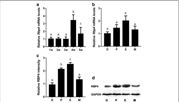
Fig. 1Dynamic expression ofRbp4in mouse ovaries during the different stages of development and the estrous cycle. (A) Real-time qPCR
analyses ofRbp4mRNA levels in mouse ovaries during different developmental stages. Data are presented as means ± SEM,n= 8. (B) Relative
Rbp4mRNA levels in mouse ovaries during diestrus (D), proestrus (P), estrus (E), and metestrus (M). Data are presented as mean ± SEM, n = 8.
RBP4 protein levels in the ovaries of mice during the estrous cycle were determined via Western blot analyses (D), and blots were quantified via
densitometry (C). The protein levels were normalized to GAPDH. Data are presented as means ± SEM,n= 3. Means with different letters represent
Western blot analyses
Protein samples were obtained by homogenizing whole ovaries and by lysing granulosa cells in lysate buffer (Beyotime, Beijing, China) with a 10μg/ml protease and phosphatase inhibitor cocktail (Thermo, USA). Samples were then centrifuged at 13,000 g at 4 °C. Tissue and cell extracts were normalized to the sample protein concen-tration, as determined by a protein assay kit (Beyotime, Beijing, China). Proteins (40 μg) were separated via so-dium dodecyl sulfate polyacrylamide gel (12%) electro-phoresis and transferred to polyvinylidene difluoride membranes. The membranes were then blocked in Tris-buffered saline with Tween 20 (TBST) containing 5% non-fat instant milk and incubated overnight at 4 °C with mouse monoclonal antibodies specific to RBP4 or GAPDH (Proteintech, USA); and rabbit monoclonal anti-bodies specific to HMGA1, SF-1, or LRH-1 (Abcam, USA) each at 1:5000 dilution. After washing with TBST, the membranes were incubated with horseradish peroxidase-conjugated secondary antibody (Proteintech, USA) for 1 h. Enhanced chemiluminescence (ECL) detection was
performed using an ECL system according to the specifi-cations of the manufacturer (Beyotime, Beijing, China). Protein levels were normalized to GAPDH and quantified via densitometry using a Tanon gel imaging system (Tanon, Shanghai, China).
Statistical analyses
The statistical analyses of the data were conducted via one-way ANOVA followed by Tukey’s multiple-range test. Differences were considered significant atP < 0.05. All the statistical analyses were performed using SPSS 22.0 for Windows (StatSoft, USA).
Results
Variable expression ofRbp4in mouse ovaries during the different stages of development and the estrous cycle
We first performed experiments to investigate the expres-sion pattern ofRbp4in whole mouse ovaries during differ-ent developmdiffer-ental stages. Rbp4 mRNA expression in mouse ovaries remained constant at 1 to 3 weeks postna-tally and notably increased at 4 weeks (i.e., peripuberpostna-tally),
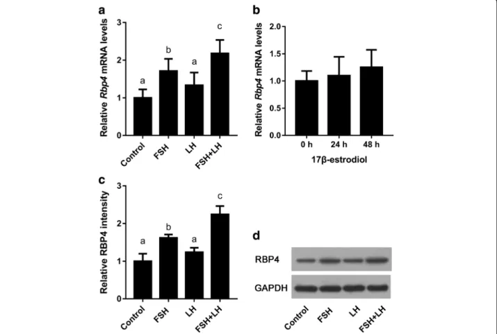
Fig. 2Effects of gonadotropins and ovarian steroids on the expression ofRbp4in the 3-week-old mouse ovary. (A)Rbp4mRNA expression levels
in the ovaries of mice treated with FSH, LH, or FSH + LH for 48 h. Data are presented as means ± SEM, n = 8. (B)Rbp4mRNA expression levels in
the ovaries of mice treated with 17β-estradiol for 24 or 48 h. Data are presented as mean ± SEM, n = 8. RBP4 protein levels in the ovaries of mice
treated with FSH or LH were determined using Western blot analyses (D), and blots were quantified via densitometry (C). The protein levels were
and dropped at 5 weeks (Fig. 1A). We also investigated the expression patterns of Rbp4 in whole mouse ovaries during the estrous cycle. The expression ofRbp4mRNA in the ovaries of adult mice (8 weeks old) having normal cycles increased at estrus (Fig.1B), and the levels of RBP4 protein in the ovaries of normally cycling adult mice in-creased at proestrus and peaked at estrus (Fig.1C and D). These data imply a potential relationship between Rbp4 expression and follicular growth [8].
Effects of gonadotropins and ovarian steroids onRbp4 expression in mouse ovaries
Follicular growth is mainly controlled by gonadotropins and estrogen. Therefore, we determined the effects of these hormones on Rbp4 expression in mouse ovaries. First, immature mice (3 weeks old) were treated with FSH, LH, or FSH + LH. The results showed that the ex-pression levels of Rbp4 in whole ovarian samples in-creased significantly after treatment with FSH (Fig.2A) or FSH + LH (Fig. 2A), but not with LH alone (Fig.
2A), and the induction with combined FSH + LH was greater than that with FSH alone (Fig. 2A). The levels of RBP4 protein in the ovaries of mice treated with FSH and/or LH in proportion to the mRNA levels (Fig.
2C and D). 17β-Estradiol showed no evident effect on ovarian Rbp4 expression (Fig. 2B). Thus,Rbp4 expres-sion in mouse ovaries appeared to be predominantly regulated by FSH.
Localization of RBP4 protein in mouse ovary
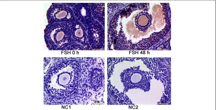
The localization of RBP4 protein in mouse ovaries was analyzed using immunohistochemical staining with spe-cific antibody (Fig.3). Positive signals for RBP4 were de-tected in the layers of granulosa and theca cells, and marked staining was also observed in the follicular an-trum of large antral follicles in the ovaries of mice treated with FSH for 48 h (Fig.3).
FSH stimulates the expression ofRbp4in mouse granulosa cells
Granulosa cells are one of the two most important types of gonadotropin-sensitive somatic cells in the mamma-lian ovary. We examined the effect of FSH and FSH + LH on the expression of Rbp4 using an in vitro culture model of granulosa cells, and our results showed that the expression of Rbp4 was induced by both FSH and FSH + LH treatments, and that the induction by com-bined FSH + LH treatment was greater than that by FSH alone (Fig.4A, B and C).
cAMP-PKA pathway mediates FSH-inducedRbp4 expression
As previously reported, FSH and LH perform their actions by activating their membrane receptors, the FSH receptor (FSHR) and the LH receptor (LHR), both of which belong to the G-protein coupled receptors [13, 14]. Stimulation of these receptors results in an increase in intracellular cAMP [13, 14], which promotes the rapid activation of
Fig. 3Localization of RBP4 protein in the ovaries of the immature mice (3 weeks of age) treated with or without FSH for 48 h. Negative controls (NC), which were incubated with 5% goat serum (NC1) or PBS (NC2) instead of the primary antibody, were used to validate the specificity of
protein kinase A (PKA) [33]. To determine whether the cAMP-PKA pathway was involved in FSH-induced Rbp4 expression in mouse granulosa cells, we first treated the cells in vitro with 500μM 8-Br-cAMP (a cAMP agonist) or 10 μM H-89 (a PKA inhibitor). If the cAMP-PKA
pathway was involved in FSH induction, 8-Br-cAMP would show similar induction effects and H-89 would pre-vent FSH induction. As expected, the treatment of mouse granulosa cells with 500μM 8-Br-cAMP for 24 h resulted in a dramatic increase inRbp4 expression (Fig. 5A), and
Fig. 5PKA mediates the FSH induction ofRbp4in mouse granulosa cells. (A) Activation of PKA by cAMP and FSH increasedRbp4mRNA levels.
Data are presented as means ± SEM, n = 4. (B) The PKA inhibitor H-89 abrogated FSH inducedRbp4mRNA expression. Granulosa cells were
pre-incubated with 10μM H-89 for 1 h, followed by incubation with 100 IU/L FSH for 24 h. Data are presented as means ± SEM, n = 4. (C)
Densitometric quantification of Western blots for RBP4 (D). The protein levels were normalized to GAPDH. Data are presented as means ± SEM, n = 3. Means with different letters represent a significant difference at the 0.05 level
Fig. 4Effects of gonadotropins on the expression ofRbp4in mouse granulosa cells. (A) RelativeRbp4mRNA levels in mouse granulosa cells
treated with FSH and LH for 24 h. Data are presented as means ± SEM,n= 4. RBP4 protein levels in mouse granulosa cells treated with FSH and
LH for 24 h were determined using Western blot analyses (C), and blots were quantified via densitometry (B). The protein levels were normalized
the induction ofRbp4by FSH was prevented with 10μM H-89 (Fig. 5B, C and D). Thus, the cAMP-PKA pathway likely mediates FSH-inducedRbp4expression.
HMGA1, SF-1, and LRH-1 mediate FSH stimulation ofRbp4
HMGA1 can bind to the promoter ofRbp4 and recruit SF-1 and LRH-1 to regulate the transcriptional activity ofRbp4 [22]. The present study examined the effects of FSH on the expression of these transcription factors, and the results showed that FSH significantly stimulated the expression of these genes in granulosa cells. More-over, the PKA inhibitor H-89 significantly inhibited FSH induction of these genes (Fig. 6A, B, and C). These data suggest that FSH stimulated the expressions of HMGA1, SF-1, and LRH-1 via PKA. To determine whether HMGA1, SFI, and LRH-1 mediated the FSH induction of Rbp4, we sought to knock down the corresponding genes using specific siRNAs. As expected, the expres-sions of these genes at the mRNA and protein levels were diminished to approximately 50% in cells trans-fected with 50 nM siRNA compared with controls (Fig. 7A, B, C, D, E, and F). After 24 h of transfection, the cells were treated with FSH for an additional 24 h. The cells were harvested and Rbp4 mRNA and protein levels were examined. Rbp4 expression was significantly reduced in cells transfected withHmga1siRNA (Fig.7G and H); while, knockdown of Lrh-1 or Sf-1 also pre-vented FSH induction of Rbp4 (Fig. 7G, I, and J). Thus,
the transcription factors HMGA1, SF-1, and LRH-1 are likely to be involved in the induction ofRbp4expression by FSH.
Discussion
Firstly, the present study presented the dynamic expres-sion patterns forRbp4in mouse ovaries during different stages of development and the estrous cycle. The expres-sion of ovarianRbp4 remained constant at 1 to 3 weeks postnatally, notably increased at 4 weeks (i.e., peripuber-tally), and dropped at 5 weeks. Numerous studies have supported the concept that the gonadotropin-releasing hormone (GnRH) neuronal network generates pulse and surge modes of gonadotropin secretion that are critical for puberty [34], and stimulate the secretion of a mass of local ovarian factors and follicular growth in females. This modulation in Rbp4 expression may be related to the pituitary gonadotropin wave at the onset of puberty. After puberty, some follicles in ovary start to grow and ovulate irregularly under the influence of serum gonado-tropins, and at 5 weeks of age, some mice may be in the follicular-growth phase and some in the post ovulatory period. The different levels of gonadotrophins in these mice may constitute a possible explanation for the drop inRbp4expression at 5 weeks. Furthermore, the expres-sion of Rbp4 in the ovaries of adult mice (8 weeks old) having normal cycles was found to increase at proestrus and peaked at estrus, the phase with higher serum levels
of FSH and LH. In a previous study, higher RBP4 pro-tein concentrations were observed in the follicular fluids of dominant follicles relative to small follicles [8,
9]. The dynamic expression patterns ofRbp4in mouse ovaries during different stages of development and the estrous cycle motivated us to pose an alternative hy-pothesis: that the expression of ovarian RBP4 is affected by gonadotropins and gonadal steroids and is related to follicular growth.
We then investigated the potential regulatory effects of gonadotropins and estrogen on the dynamic expression ofRbp4in the mouse ovary. The expression ofRbp4was significantly stimulated by administration of FSH and combined FSH + LH, but unaffected by administration of LH or 17β-estradiol. In addition, intense immuno-staining was observed in granulosa cell layers of large antral follicles in the ovaries of mice treated with FSH. To determine whether the induction of Rbp4 by FSH Fig. 7HMGA1, SF-1, and LRH-1 are involved in the FSH induction ofRbp4in mouse granulosa cells. mRNA (A) and protein (D) levels ofHmga1in
mouse granulosa cells transfected with 50 nMHmga1siRNA. The mRNA (B) and protein (E) levels ofSf-1in mouse granulosa cells transfected with
50 nMSf-1siRNA. The mRNA (C) and protein (F) levels ofLrh-1in mouse granulosa cells transfected with 50 nMLrh-1siRNA. The mRNA levels
were determined using real-time PCR. Data are presented as means ± SEM, n = 4. The protein levels were determined using Western blot analyses, and blots were quantified via densitometry. The protein levels were normalized to GAPDH. Data are presented as means ± SEM, n = 3.
*p < 0.05, **p< 0.01. (G) The mRNA levels ofRbp4in mouse granulosa cells transfected with NC siRNA,Hmga1siRNA,Sf-1siRNA, orLrh-1siRNA
and FSH + LH occurred due to increased expression of Rbp4in granulosa cells, we examined primary granulosa cells cultured in vitro. Our results showed that the ex-pression of Rbp4 was induced by FSH and combined FSH + LH treatments, and that the induction by com-bined FSH + LH treatment was stronger than that by FSH alone. The administration of LH alone did not exert an effect. Because the primary granulosa cells were iso-lated from the immature female mouse (3 weeks old) ovaries without FSH or equine chorionic gonadotropin (eCG) pretreatment and with no or low LHR expression, the cells were mainly immature granulosa cells without LHR expression [35]. This may be the reason as to why LH alone manifested no obvious effects on granulosa cells and immature ovaries in the present study. How-ever, LHR in granulosa cells can be induced by FSH [35], and during late maturation of granulosa cells in antral follicles, LH exerts a relatively more notable effect than FSH on cAMP formation [14, 36, 37], suggesting that LHR density was relatively greater than FSHR density or that LHR was more effectively coupled to cAMP gener-ation. These findings may explain the stronger induction of Rbp4 expression by FSH + LH than by FSH alone, and the higher levels of RBP4 in the follicular fluids of the large antral follicles [8].
To determine whether the induction of Rbp4 by FSH was mediated by the cAMP-PKA pathway, we examined the effects of the cAMP analog 8-Br-cAMP and the PKA inhibitor H-89 on the induction of Rbp4 in granulosa cells. The treatment of cells with 500 μM 8-Br-cAMP showed a dramatic increase inRbp4 expression, whereas 10 μM H-89 significantly prevented the induction of Rbp4by FSH. Thus, the cAMP-PKA pathway likely par-ticipates in the induction ofRbp4by FSH.
The cis-regulatory sequences of the mouse Rbp4gene contain a bipartite promoter, i.e., a proximal region ne-cessary for basal expression and a distal segment that contains several binding sites for the structural HMGA1 proteins that are structural components of chromatin [22, 38]. HMGA1 binds to the adenine thymine (AT)-rich regions of DNA, changes the structure of DNA and recruit transcription factors to the promoter, and facili-tates gene transcription [39]. There are several AT-rich motifs homologous to the binding sites of HMGA1 in the promoter region of mouse Rbp4, and HMGA1 pro-teins can bind upstream sequences of the Rbp4 pro-moter. In the present study, Hmga1 was sensitive to FSH and this response was prevented by the PKA inhibi-tor H-89; whereas the knockdown ofHmga1with siRNA resulted in a dramatic loss of FSH-inducedRbp4 expres-sion. In addition, HMGA1 can interact with SF-1 and LRH-1 and recruit them to the complex [22]. In the present study, the depletion of SF-1 and LRH-1 resulted in a dramatic loss of FSH-inducedRbp4expression. SF-1
was shown to be expressed at higher levels in theca cells, and also found in granulosa cells; whereas LRH-1 ex-pression was found to be abundant and highly restricted to the granulosa cells of developing follicles [40, 41]. Consistent with our data, the two transcription factors were shown to be induced in granulosa cells by FSH/ cAMP through the activation of PKA [40,41], as cAMP signals are mediated largely via PKA [33] and can be ele-vated in granulosa cells by FSH [42, 43] or LH [44]. Thus, the expression of ovarian Rbp4 was likely regu-lated by FSH via the cAMP-PKA pathway, involving HMGA1, SF-1, and LRH-1.
Conclusion
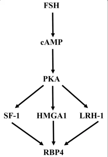
In conclusion, the present study showed a dynamic ex-pression pattern of Rbp4in the mouse ovary during dif-ferent stages of development and the estrous cycle, which was predominantly regulated by FSH. Further-more, the cAMP-PKA pathway and transcription factors HMGA1, SF-1, and LRH-1 were likely involved in the induction of Rbp4 by FSH (Fig. 8). This work expands
Fig. 8Schematic overview of the proposed pathway involved in the
induction ofRbp4by FSH. FSH binds to its receptor (FHR) and elevates
cAMP levels, which activates PKA. PKA then stimulates the expression
our understanding of the mechanisms underlying the regulation ofRbp4expression patterns in the ovary dur-ing different stages of development and the estrous cycle. Although further research needs to be conducted in this area, our data are important for the understand-ing of the molecular regulation of retinol transport and accumulation in the follicular fluids during ovarian fol-licular growth.
Abbreviations
8-Br-cAMP:8-Bromoadenosine 3′:5′-cyclic monophosphate; FSH: Follicle-stimulating hormone; GAPDH: Glyceraldehyde 3-phosphate dehydrogenase; HMGA1: High-mobility group AT-hook 1; LH: Luteinizing hormone; LRH-1: Liver receptor homolog 1; PKA: Protein kinase A; RBP4: Retinol-binding protein 4; SF-1: Steroidogenic factor 1
Acknowledgments
We would like to thank LetPub (www.letpub.com) for providing linguistic
assistance during the preparation of this manuscript.
Funding
This work was supported by the National Natural Science Foundation of China (grants numbers 31372308, 31672417 and 31301969).
Availability of data and materials
Data sharing was not applicable to this article as no datasets were generated or analyzed during the current study.
Authors’contributions
YJ, CL, and XZ conceived and designed the study; YJ performed the experiments; YJ and YZ analyzed the data; YJ, SC, and LC wrote the manuscript. All authors read and approved the final manuscript.
Ethics approval
The animal protocols used herein were conducted according to the Guide for the Administration of Laboratory Animals (Directive 86/609/EEC on the Protection of Animals Used for Experimental and Other Scientific Purposes, 1986), and were approved by the Institutional Animal Care and Use Committee (IACUC) of Jilin University, China.
Consent for publication Not applicable
Competing interests
The authors declare that they have no competing interests.
Publisher’s Note
Springer Nature remains neutral with regard to jurisdictional claims in published maps and institutional affiliations.
Received: 8 November 2017 Accepted: 15 March 2018
References
1. Jiang YW, Li CJ, Chen L, Wang FG, Zhou X. Potential role of retinoids in
ovarian physiology and pathogenesis of polycystic ovary syndrome. Clin
Chim Acta. 2017;469:87–93.
2. Kipp JL, Golebiowski A, Rodriguez G, Demczuk M, Kilen SM, Mayo KE. Gene
expression profiling reveals Cyp26b1 to be an activin regulated gene involved in ovarian granulosa cell proliferation. Endocrinology. 2011;152: 303–12.
3. Kawai T, Yanaka N, Richards JS, Shimada M. De novo-synthesized retinoic
acid in ovarian antral follicles enhances FSH-mediated ovarian follicular cell
differentiation and female fertility. Endocrinology. 2016;157:2160–72.
4. Ikeda S, Kitagawa M, Imai H, Yamada M. The roles of vitamin a for
cytoplasmic maturation of bovine oocytes. J Reprod Dev. 2005;51:23–35.
5. Tahaei LS, Eimani H, Yazdi PE, Ebrahimi B, Fathi R. Effects of retinoic acid on
maturation of immature mouse oocytes in the presence and absence of a
granulosa cell co-culture system. J Assist Reprod Genet. 2011;28:553–8.
6. Wickenheisser JK, Nelson-DeGrave VL, Hendricks KL, Legro RS, Strauss JF,
McAllister JM. Retinoids and retinol differentially regulate steroid
biosynthesis in ovarian theca cells isolated from normal cycling women and women with polycystic ovary syndrome. J Clin Endocrinol Metab. 2005;90: 4858–65.
7. Haliloglu S, Baspinar N, Serpek B, Erdem H, Bulut Z. Vitamin a and
beta-carotene levels in plasma, corpus luteum and follicular fluid of cyclic and
pregnant cattle. Reprod Domest Anim. 2002;37:96–9.
8. Brown JA, Eberhardt DM, Schrick FN, Roberts MP, Godkin JD. Expression of
retinol-binding protein and cellular retinol-binding protein in the bovine
ovary. Mol Reprod Dev. 2003;64:261–9.
9. Schweigert FJ, Zucker H. Concentrations of vitamin a, beta-carotene and
vitamin E in individual bovine follicles of different quality. J Reprod Fertil. 1988;82:575–9.
10. Kawaguchi R, Zhong M, Kassai M, Ter-Stepanian M, Sun H. Vitamin a
transport mechanism of the Multitransmembrane cell-surface receptor
STRA6. Membranes (Basel). 2015;5:425–53.
11. Jia J, Bai J, Liu Y, Yin JN, Yang P, Yu SQ, et al. Association between
retinol-binding protein 4 and polycystic ovary syndrome: a meta-analysis. Endocr J.
2014;61:995–1002.
12. Sun YL, Ping ZG, Li CJ, Sun YF, Yi KL, Chen L, et al. Comparative proteomic
analysis of follicular fluids from normal and cystic follicles in sows. Reprod
Domest Anim. 2011;46:889–95.
13. Richards JS. Hormonal control of gene expression in the ovary. Endocr Rev.
1994;15:725–51.
14. Richards JS, Russell DL, Robker RL, Dajee M, Alliston TN. Molecular
mechanisms of ovulation and luteinization. Mol Cell Endocrinol. 1998;145: 47–54.
15. McLean AC, Valenzuela N, Fai S, Bennett SA. Performing vaginal lavage,
crystal violet staining, and vaginal cytological evaluation for mouse estrous cycle staging identification. J Vis Exp. 2012;67:e4389.
16. Cora MC, Kooistra L, Travlos G. Vaginal cytology of the laboratory rat and
mouse: review and criteria for the staging of the estrous cycle using stained
vaginal smears. Toxicol Pathol. 2015;43:776–93.
17. Parkening TA, Collins TJ, Smith ER. Plasma and pituitary concentrations of
LH, FSH, and prolactin in aging C57BL/6 mice at various times of the
estrous cycle. Neurobiol Aging. 1982;3:31–5.
18. Kumar TR, Wang Y, Lu N, Matzuk MM. Follicle stimulating hormone is
required for ovarian follicle maturation but not male fertility. Nat Genet. 1997;15:201.
19. Montgomery V, Loutradis D, Tulchinsky D, Kiessling A. FSH-induced
ovulation in intact and hypophysectomized mice. J Reprod Fertil. 1988;84:1–6.
20. McCracken JA, Custer EE, Lamsa JC. Luteolysis: a neuroendocrine-mediated
event. Physiol Rev. 1999;79:263–323.
21. Stocco C, Telleria C, Gibori G. The molecular control of corpus luteum
formation, function, and regression. Endocr Rev. 2007;28:117–49.
22. Bianconcini A, Lupo A, Capone S, Quadro L, Monti M, Zurlo D, et al.
Transcriptional activity of the murine retinol-binding protein gene is regulated by a multiprotein complex containing HMGA1, p54(nrb)/NonO, protein-associated splicing factor (PSF) and steroidogenic factor 1 (SF1)/liver
receptor homologue 1 (LRH-1). Int J Biochem Cell Biol. 2009;41:2189–203.
23. Wang C, Dehghani B, Li YX, Kaler LJ, Proctor T, Vandenbark AA, et al.
Membrane estrogen receptor regulates experimental autoimmune encephalomyelitis through up-regulation of programmed death 1. J
Immunol. 2009;182:3294–303.
24. Byers SL, Wiles MV, Dunn SL, Taft RA. Mouse estrous cycle identification tool
and images. PLoS One. 2012;7:e35538.
25. Caligioni CS. Assessing reproductive status/stages in mice. Curr Protoc
Neurosci. 2009; Appendix 4:Appendix 4I
26. Mankhey RW, Bhatti F, Maric C. 17beta-estradiol replacement improves renal
function and pathology associated with diabetic nephropathy. Am J Physiol
Renal Physiol. 2005;288:F399–405.
27. Chen H, Guan R, Lei Y, Chen J, Ge Q, Zhang X, et al. Lymphangiogenesis in
gastric cancer regulated through Akt/mTOR-VEGF-C/VEGF-D axis. BMC Cancer. 2015;15:103.
28. Mhawech-Fauceglia P, Wang D, Samrao D, Godoy H, Pejovic T, Liu S, et al.
Pair-box (PAX8) protein-positive expression is associated with poor disease
29. Di Somma S, Marotta M, Salvatore G, Cudemo G, Cuda G, De Vivo F, et al. Changes in myocardial cytoskeletal intermediate filaments and myocyte contractile dysfunction in dilated cardiomyopathy: an in vivo study in
humans. Heart. 2000;84:659–67.
30. Ronkainen H, Soini Y, Vaarala MH, Kauppila S, Hirvikoski P. Evaluation of
neuroendocrine markers in renal cell carcinoma. Diagn Pathol. 2010;5:28.
31. Liang N, Xu YL, Yin YM, Yao GD, Tian H, Wang GS, et al. Steroidogenic
Factor-1 is required for TGF-beta 3-mediated 17 beta-estradiol synthesis in
mouse ovarian granulosa cells. Endocrinology. 2011;152:3213–25.
32. Yao GD, Yin MM, Lian J, Tian H, Liu L, Li X, et al. MicroRNA-224 is involved in
transforming growth factor-beta-mediated mouse granulosa cell proliferation and granulosa cell function by targeting Smad4. Mol
Endocrinol. 2010;24:540–51.
33. Hunzicker-Dunn M, Maizels ET. FSH signaling pathways in immature
granulosa cells that regulate target gene expression: branching out from protein kinase a. Cell Signal. 2006;18:1351–9.
34. Herbison AE. Control of puberty onset and fertility by
gonadotropin-releasing hormone neurons. Nat Rev Endocrinol. 2016;12:452–66.
35. Camp TA, Rahal JO, Mayo KE. Cellular localization and hormonal regulation
of follicle-stimulating hormone and luteinizing hormone receptor
messenger RNAs in the rat ovary. Mol Endocrinol. 1991;5:1405–17.
36. Yong EL, Baird DT, Hillier SG. Mediation of gonadotrophin-stimulated
growth and differentiation of human granulosa cells by adenosine-3′,5′
-monophosphate: one molecule, two messages. Clin Endocrinol. 1992;37:51–8.
37. Yong EL, Baird DT, Yates R, Reichert LE Jr, Hillier SG. Hormonal regulation of
the growth and steroidogenic function of human granulosa cells. J Clin
Endocrinol Metab. 1992;74:842–9.
38. Jessen KA, Satre MA. Induction of mouse retinol binding protein gene
expression by cyclic AMP in Hepa 1-6 cells. Arch Biochem Biophys. 1998; 357:126–30.
39. Bustin M, Reeves R. High-mobility-group chromosomal proteins:
architectural components that facilitate chromatin function. Prog Nucleic
Acid Res Mol Biol. 1996;54:35–100.
40. Falender AE, Lanz R, Malenfant D, Belanger L, Richards JS. Differential
expression of steroidogenic factor-1 and FTF/LRH-1 in the rodent ovary.
Endocrinology. 2003;144:3598–610.
41. Hinshelwood MM, Repa JJ, Shelton JM, Richardson JA, Mangelsdorf DJ,
Mendelson CR. Expression of LRH-1 and SF-1 in the mouse ovary: localization in different cell types correlates with differing function. Mol Cell
Endocrinol. 2003;207:39–45.
42. Mukherjee A, Park-Sarge OK, Mayo KE. Gonadotropins induce rapid
phosphorylation of the 3′,5′-cyclic adenosine monophosphate response
element binding protein in ovarian granulosa cells. Endocrinology. 1996;137: 3234–45.
43. Salvador LM, Park Y, Cottom J, Maizels ET, Jones JC, Schillace RV, et al.
Follicle-stimulating hormone stimulates protein kinase A-mediated histone H3 phosphorylation and acetylation leading to select gene activation in
ovarian granulosa cells. J Biol Chem. 2001;276:40146–55.
44. Wang YK, Hao XQ, Yang J, Li J, Zhang MJ. CREB activity is required for
luteinizing hormone-induced the expression of EGF-like factors. Mol Reprod
Dev. 2016;83:1116–27.
• We accept pre-submission inquiries
• Our selector tool helps you to find the most relevant journal • We provide round the clock customer support
• Convenient online submission • Thorough peer review
• Inclusion in PubMed and all major indexing services • Maximum visibility for your research
Submit your manuscript at www.biomedcentral.com/submit