Dectin-2 is a primary receptor for NLRP3
inflammasome activation in dendritic cell
response to Histoplasma capsulatum
Tzu-Hsuan Chang1, Juin-Hua Huang1, Hsiu-Chao Lin1, Wen-Yu Chen1, Yu-Hsiang Lee1, Li-Chung Hsu2, Mihai G. Netea3, Jenny P.-Y. Ting4,5, Betty A. Wu-Hsieh1*
1 Graduate Institute of Immunology, National Taiwan University College of Medicine, Taipei, Taiwan, 2 Graduate Institute of Molecular Medicine, National Taiwan University College of Medicine, Taipei, Taiwan, 3 Department of Internal Medicine and Radboud Center for Infectious Diseases, Radboud University Medical
Center, Nijmegen, The Netherlands, 4 Department of Microbiology and Immunology, University of North Carolina, Chapel Hill, North Carolina, United States of America, 5 Lineberger Comprehensive Cancer Center, University of North Carolina, Chapel Hill, North Carolina, United States of America
Abstract
Inflammasome is an intracellular protein complex that serves as cytosolic pattern recogni-tion receptor (PRR) to engage with pathogens and to process cytokines of the interleukin-1 (IL-1) family into bioactive molecules. It has been established that interleukin-1β(IL-1β) is important to host defense against Histoplasma capsulatum infection. However, the detailed mechanism of how H. capsulatum induces inflammasome activation leading to IL-1β pro-duction has not been studied. Here, we showed in dendritic cells (DCs) that H. capsulatum triggers caspase-1 activation and IL-1βproduction through NLRP3 inflammasome. By reciprocal blocking of Dectin-1 or Dectin-2 in single receptor-deficient DCs and cells from
Clec4n-/-, Clec7a-/-, and Clec7a-/-Clec4n-/-mice, we discovered that while Dectin-2 operates as a primary receptor, Dectin-1 serves as a secondary one for NLRP3 inflammasome. In addition, both receptors trigger Syk-JNK signal pathway to activate signal 1 (pro-IL-1β syn-thesis) and signal 2 (activation of caspase-1). Results of pulmonary infection with H.
capsu-latum showed that CD103+DCs are one of the major producers of IL-1βand Dectin-2 and Dectin-1 double deficiency abolishes their IL-1βresponse to the fungus. While K+efflux and cathepsin B (but not ROS) function as signal 2, viable but not heat-killed H. capsulatum trig-gers profound lysosomal rupture leading to cathepsin B release. Interestingly, cathepsin B release is regulated by ERK/JNK downstream of Dectin-2 and Dectin-1. Our study demon-strates for the first time the unique roles of Dectin-2 and Dectin-1 in triggering Syk-JNK to activate signal 1 and 2 for H. capsulatum-induced NLRP3 inflammasome activation.
Author summary
Histoplasma capsulatumis a dimorphic fungal pathogen. The microconidia and hyphal
elements are breathed in and transform to become yeasts in the lungs. Histoplasmosis occurs worldwide and endemic in mid-western United States. The infection is primarily a1111111111 a1111111111 a1111111111 a1111111111 a1111111111 OPEN ACCESS
Citation: Chang T-H, Huang J-H, Lin H-C, Chen
W-Y, Lee Y-H, Hsu L-C, et al. (2017) Dectin-2 is a primary receptor for NLRP3 inflammasome activation in dendritic cell response to Histoplasma
capsulatum. PLoS Pathog 13(7): e1006485. https://doi.org/10.1371/journal.ppat.1006485
Editor: Anita Sil, University of California, San
Francisco, UNITED STATES
Received: February 23, 2017
Accepted: June 21, 2017
Published: July 3, 2017
Copyright:©2017 Chang et al. This is an open access article distributed under the terms of the
Creative Commons Attribution License, which permits unrestricted use, distribution, and reproduction in any medium, provided the original author and source are credited.
Data Availability Statement: All relevant data are
within the paper and its Supporting Information files.
Funding: This work was supported by research
grants 101-2320-B-002-030-MY3 and 105-2811-B-002-092 from the Ministry of Science and Technology (http://www.most.gov.tw) and Thematic Research Program AS-105-TP-B08-3 from Academia Sinica (https://www.sinica.edu.tw/
index.shtml). MGN was supported by an ERC
in the lungs that can become disseminated and cause fatal disease when left untreated. It was reported that IL-1βis important to host defense againstH.capsulatuminfection, but the detailed mechanism of how myeloid cells respond to this fungal pathogen and which receptor(s) is involved to induce IL-1βproduction is largely unknown. We demonstrate in this study thatH.capsulatum-induced caspase-1 activation leading to IL-1βproduction is NLRP3-dependent. NLRP3-deficienct mice succumb to otherwise sublethalH. capsula-tuminfection. Although the role of Dectin-1 in fungus-induced NLRP3 inflammasome is well-established, we found that Dectin-2 serves as a primary receptor and Dectin-1 plays a secondary role in inducing Syk-JNK signaling to mediate NLRP3 inflammasome in response toH.capsulatum. In addition, while both K+efflux and cathepsin B function as signal 2, the viabilityH.capsulatumaffects the amounts of cathepsin B release. Our study is the first to reveal the roles of Dectin-2 and Dectin-1 and the downstream signaling events in fungal pathogen-induced NLRP3 inflammasome.
Introduction
Inflammasome is a large intracellular multimeric protein platform which is activated upon infection or stress [1]. The function of inflammasome is to drive the maturation of proinflam-matory cytokines of the IL-1 family, most importantly IL-1βand IL-18 and induction of inflammatory cell death [2]. Among all identified inflammasome complexes, NLRP3 inflam-masome is well-characterized. It is generally accepted that NLRP3-driven processing and secretion of IL-1βand IL-18 in macrophage and DC require two signals [3]. Signal 1 is induced by engagement of pathogen-associated molecular patterns (PAMPs) with pattern recognition receptors (PRRs) leading to gene transcription and synthesis of NLRP3, inactive pro-IL-1β
and pro-IL-18 [4]. Signal 2 induces the assembly of inflammasome complex and activates cas-pase-1 to facilitate pro-IL-1βand pro-IL-18 cleavage into their mature forms, and is induced by intracellular events including reactive oxygen species (ROS) production, potassium (K+) efflux, cathepsin B release, calcium influx and mitochondrial destabilization [5–9]. There are multiple PAMPs on a single fungal pathogen. It is of interest to determine the complex interac-tion between a fungus and the host cell and how the interacinterac-tion triggers either signal 1 or 2 or both for inflammasome activation.
Histoplasma capsulatumis a dimorphic fungal pathogen. The microconidia and mycelial
fragments ofH.capsulatumspread in the air and infect humans through inhalation [10,11].
H.capsulatumstimulates mouse dendritic cell (DC) to secrete pro-inflammatory cytokines
such as IL-1β, IL-18, TNF and IL-6 [12]. Human DC phagocytosesH.capsulatumyeasts through fibronectin receptor VLA-5 and kills the organism via phagolysosomal fusion [13,
14]. A recent study showed that CD103+conventional DC in the lungs produces IFN-I to restrict the growth ofH.capsulatumduring pulmonary infection [15]. These studies point to a crucial role of DC in secreting cytokines and killingH.capsulatumduring early phase of infec-tion [13–15]. There is still much to be learned about the detailed mechanisms of cytokine pro-duction by DC through interaction withH.capsulatum.
The fungal pathogensCandida albicans,Aspergillus fumigatus,Cryptococcus neoformans,
Microsporum canisandMalasseziaspp. induce inflammasome activation [16–21]. In a
sys-temicC.albicansinfection model, NLRP3 or caspase-1 deficiency leads to increased fungal burdens and higher mortality [16]. In protection against mucosal candidiasis, NLRC4 func-tions at the level of mucosal stroma and NLRP3 at both the hematopoietic and stromal compartments [21]. AIM2 and NLRP3 are both required for mice that are treated with
from the Netherlands Organization for Scientific Research (http://www.nwo.nl/en). The funders had no role in experiment design, data analysis, decision to publish or preparation of the manuscript.
Competing interests: The authors have declared
immunosuppressive agents to confineA.fumigatusin inflammatory foci after intranasal inoc-ulation with conidia [18]. In pulmonary infection with acapsular form ofC.neoformans, NLRP3 inflammasome activation results in immune cell infiltration and effective fungal clear-ance [17]. While elevated IL-1βin the lungs of mice infected withH.capsulatumplays a critical role in host defense againstH.capsulatum[22], it has never been determined which mecha-nisms are involved in inflammasome activation and IL-1βproduction.
Hyphal forms ofC.albicansrecognized by either TLR-2 or Dectin-1 on bone marrow-derived macrophage triggers the synthesis of pro-IL-1βthrough Syk kinase [23]. In addition to triggering pro-IL-1βaccumulation, Syk signaling is also involved in ROS production and cas-pase-1 activity in bone marrow-derived dendritic cells (BMDCs) stimulated byC.albicans
[16]. Interestingly, in contrast to stimulation by hyphal forms, stimulation of BMDC by yeast form ofC.albicansfor IL-1βproduction is mediated by Dectin-2 through MAPKs signaling [24,25]. Both CR3 and Dectin-1 are involved in macrophage TNF and IL-6 response toH.
cap-sulatumthrough activation of Syk-JNK-AP-1 pathway [26]. In studying vaccine immunity,
Wanget al. reported that Dectin-1 and Dectin-2 fusion proteins separately bind toH.
capsula-tumand that CARD9 signaling is important for development of Th17 cells and adaptive immunity againstH.capsulatum[27].
In this study, we demonstrated thatH.capsulatuminduced NLRP3 inflammasome for IL-1βproduction in BMDCs. Dectin-2 was the primary receptor that mediated both signal 1 and 2 for NLRP3 inflammasome. Results of reciprocal blocking of 1 or 2 in Dectin-2- and Dectin-1-deficient cells and that of using single- and double-deficient cells showed that Dectin-1 played a role as a secondary receptor. Both Dectin-2 and Dectin-1 activated the Syk/ JNK signaling pathway but the role of Dectin-1 was less prominent than Dectin-2. Pulmonary infection results showed that CD103+DCs are one of the major sources of IL-1β, and Dectin-2 and Dectin-1 together mediated the IL-1βresponse of CD103+DC toH.capsulatuminfection. Both K+efflux and cathepsin B release but not ROS functioned as signal 2 forH.capsulatum -induced NLRP3 inflammasome. While Dectin-2 did not affect K+efflux, signals from Dectin-1 and Dectin-2 cooperatively regulated cathepsin B release. Our work revealed the roles of Dec-tin-2 and Dectin-1 in inducing signals for activation of NLRP3 inflammasome during fungal infection.
Results
H. capsulatum-induced IL-1
β
production in dendritic cells is
caspase-1-and NLRP3-dependent
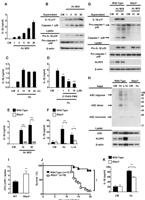
Fig 1. H. capsulatum induces NLRP3-dependent inflammasome activation. (A, B and C) BMDCs from wild type
mice were stimulated with live H. capsulatum at (A and B) different MOI and (C) at MOI of 20 for 6, 12, 18 and 24 h. (D) BMDCs were pretreated with caspase-1 inhibitor (Z-YVAD-FMK) at different concentrations before stimulation with H.
capsulatum for another 18 h. (E, G and H) BMDCs and (F) sorted MHCII+CD11c+splenic DCs from wild type and
While the absence of NLRP3 did not affect pro-IL-1βand pro-caspase-1 p45, the secretion of IL-1βp17 and caspase-1 p20 was completely abrogated inNlrp3-/-cells (Fig 1G). A protein-crosslinking experiment showed thatH.capsulatuminduced robust NLRP3-dependent ASC oligomerization, which reflected inflammasome assembly (Fig 1H).Nlrp3-/-mice had greater fungal burden (Fig 1I) and poorer survival (Fig 1J) compared to wild type mice after intrave-nous infection withH.capsulatum. It appears that higher fungal burden caused by NLRP3 deficiency may be one of the contributing factors to poor survival. In addition, splenic DCs
fromNlrp3-/-infected-mice produced lower IL-1βcompared with cells from infected wild type
mice (Fig 1K). These results together show thatH.capsulatuminduces IL-1βresponse through activation of both signal 1 and signal 2 of an NLRP3-dependent inflammasome in DCs and that NLRP3 is important to protection against histoplasmosis.
Dectin-2 mediates inflammasome activation in H. capsulatum-stimulated
BMDCs
We then used blocking antibodies to explore surface receptor(s) that is involved in inflamma-some activation byH.capsulatum. Results show that BMDCs treated with anti-Dectin-2, but not anti-CR3, -Dectin-1 or -TLR-2 blocking antibody significantly reducedH.capsulatum -induced pro-IL-1βp35, IL-1βp17 and caspase-1 p20 expressions (Fig 2A and 2B) and Dectin-2 blocking antibody dose-dependently reduced pro-IL-1βp35, IL-1βp17 and caspase-1 p20 production (Fig 2C and 2D). Thus, it appears that Dectin-2 is involved in pro-IL-1βsynthesis, IL-1βmaturation and caspase-1 activation in response toH.capsulatum. Results inFig 2E and 2Fshow that Dectin-2 deficiency (Clec4n-/-cells) exhibited reduction of IL-1βsecretion, which is comparable with wild type BMDCs treated with anti-Dectin-2 antibody. Sorted MHCII+CD11c+splenic DCs fromClec4n-/-mice also produced lower IL-1βcompared with their wild type counterparts (Fig 2G). Since Dectin-2 signal is transduced through Fc receptor
γchain (FcRγ), we studied inflammasome activation inFcRγ-/-BMDCs. We found that FcRγ deficiency, like Dectin-2 deficiency, significantly reduced IL-1βand caspase-1 p20 (S2 Fig). These results together indicate that through interaction with Dectin-2,H.capsulatumtriggers both signal 1 and signal 2 for inflammasome activation in BMDC, demonstrating the impor-tance of Dectin-2 inH.capsulatum-induced inflammasome activation in DC.
Syk and its downstream ERK and JNK regulate inflammasome
activation after stimulation by H. capsulatum
It has been reported that Dectin-2 coupling to Syk leads to downstream activation of MAPKs in response toC.albicans[25]. Whether Syk-MAPK signaling pathway is involved inH.
capsulatum-induced inflammasome activation is still unclear. We analyzed Syk, JNK, ERK
and p38 phosphorylation in BMDCs after stimulation withH.capsulatum. Western blotting showed that phosphorylation of Syk, JNK, ERK and p38 occurred as early as 10 min afterH. inflammasome components were analyzed in cell pellets and cell lysates, respectively. Stimulation with LPS (500 ng/ ml, 6 h) plus ATP (5 mM, 30 minutes) (L+A) was used as a positive control for NLRP3-dependent IL-1βinduction. (I and J) Wild type and NLRP3-deficient mice were intravenously infected with H. capsulatum (1×10⁷). (I) Fungal burden in the spleen was determined on day 11 after infection. It is shown as CFU per organ (n = 6). (J) Survival was analyzed by log-rank test. (K) Wild type and NLRP3-deficient mice were intravenously infected with 2.5×10⁵of H. capsulatum.
MHCII+CD11c+splenic DCs were sorted from mice on day 5 after infection and stimulated with live H. capsulatum at MOI of 1 for 18 h. (A, C, D, E, F and K) IL-1βin cell-free supernatants were quantified by ELISA (n = 3). One representative of three (A, B, C, D and E) or two (F, G, H, I and J) independent experiments is presented. ND, not detectable. CM, complete medium.*p<0.05,**p<0.01,***p<0.001 [one-way ANOVA with Tuckey post-hoc analysis (A, B and D); 2-tailed t-test (E, F, I and K); log-rank test (J)].
Fig 2. Dectin-2 mediates inflammasome activation upon H. capsulatum stimulation. (A) BMDCs were
pretreated with anti-CR3, -Dectin-1, -Dectin-2 and -TLR-2 (2μg/ml) and (C) different concentrations of anti-Dectin-2 blocking antibodies at 2, 5, and 10μg/ml for 1 h before stimulation with H. capsulatum for another 18 h. IgG2a was used as an isotype control. Cell-free supernatants and cell lysates were analyzed by Western blotting with indicated antibodies. (B and D) Relative intensity of secreted IL-1βp17, caspase-1 p20, pro-IL-1β
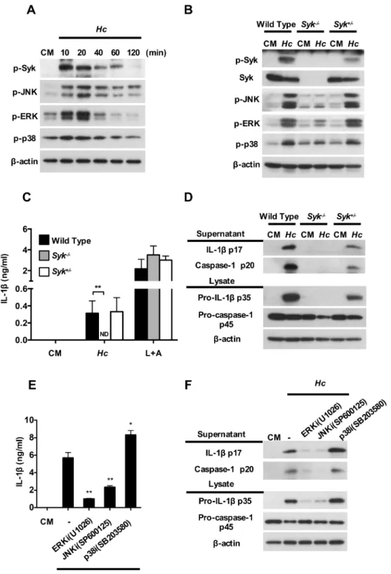
Dectin-capsulatumstimulation (Fig 3A). The role of Syk inH.capsulatum-induced MAPKs and inflammasome activation was validated in Syk-deficient fetal liver-derived dendritic cells (FLDCs). Results showed that phosphorylation of JNK, ERK and p38 was largely diminished in Syk-deficient cells uponH.capsulatumstimulation (Fig 3B), which demonstrates that MAPKs activation is downstream of Syk. While Syk-deficiency completely abolished FLDCs IL-1βsecretion uponH.capsulatumstimulation, it did not affect IL-1βresponse to LPS plus ATP stimulation showing the specificity of this pathway (Fig 3C). Western blotting analysis also showed that in the absence of Syk, pro-IL-1βexpression was abolished, which is congruent with the absence of mature IL-1βp17 production and indicates that Syk was required for signal 1 priming (Fig 3D). In addition, pro-caspase-1 p45 was partially reduced but caspase-1 p20 was completely absent in Syk deficiency, suggesting that Syk is likely necessary for optimal signal 2 activation (Fig 3DandS3 Fig). Pharmacological inhibition of either ERK or JNK dra-matically reduced the production of pro-IL-1βp35, IL-1βp17 and caspase-1 p20 uponH.
cap-sulatumstimulation (Fig 3E and 3F). Interestingly, inhibition of p38 increased pro-IL-1β
expression that led to increase IL-1βsecretion (Fig 3E and 3F). Again, these inhibitors greatly diminished signal 1 by reducing pro-IL-1βproduction, but also affected signal 2 by reducing the conversion of pro-caspase-1 to active caspase-1. Together, these data demonstrate thatH.
capsulatum-induced inflammasome activation is through Syk and its downstream ERK and
JNK activation.
Dectin-1 deficiency further reduces inflammasome activation in the
absence of functional Dectin-2 through Syk-JNK signal pathway
Our data in Figs2and3demonstrated that while Syk deficiency completely abolished IL-1β
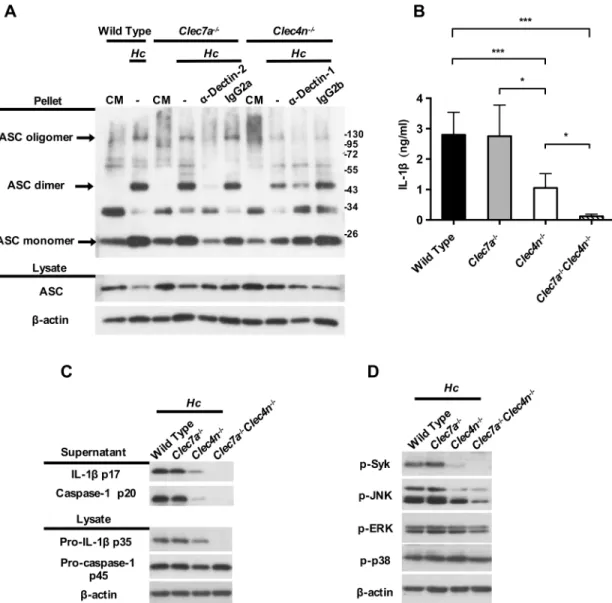
production, Dectin-2 deficiency reduced IL-1βproduction by only ~50%. It is reported that blocking Dectin-2 in Dectin-1-deficient BMDC leads to further reduction in TNF and IL-10 production in response toC.albicanscompared to Dectin-1 deficiency alone [25]. Flow cyto-metric analysis results showed that Dectin-1 and Dectin-2 single deficiency did not affect the expression of the other receptor nor the uptake ofH.capsulatum(S4andS5Figs). We used blocking antibodies and receptor single- and double-deficient BMDCs to study whether Dec-tin-1 works together with Dectin-2 to transduce downstream signaling for inflammasome activation. Western blotting analysis and ELISA results showed that blocking Dectin-1 and Dectin-2 in Dectin-2-deficient and Dectin-1-deficient, respectively, reduced not only the secretion of mature IL-1βp17 (S6A and S6B Fig), but also the synthesis of pro-IL-1βp35, the active form of caspase-1 p20, and ASC oligomerization, compared to wild type cells and cells with either receptor deficiency alone (S6B FigandFig 4A). Dectin-1 and Dectin-2 double deficiency completely abolished IL-1βproduction (Fig 4B and 4C) and pro-IL-1βp35, cas-pase-1 p20 (Fig 4C). It appears that, in the presence of Dectin-2, Dectin-1 does not function to respond toH.capsulatumfor inflammasome activation. Yet in the absence of functional Dectin-2, recognition ofH.capsulatumby Dectin-1 is involved in triggering both signal 1 and signal 2.
2-deficient (Clec4n-/-) mice were treated with or without blocking antibody against Dectin-2 before stimulation
with H. capsulatum. Stimulation by LPS plus ATP (L+A) was used as a positive control for IL-1βinduction. Cell-free supernatants were collected at 18 h after stimulation. Secreted IL-1βwas quantified by ELISA (n = 3). Error bars indicate standard deviation of the mean. One representative of three independent experiments is presented.*p<0.05,**p<0.01,***p<0.001 [one-way ANOVA with Tuckey post-hoc analysis (B and D); 2-tailed t-test (E, F and G)].
Fig 3. Syk-ERK/JNK pathway regulates inflammasome activation in response to H. capsulatum. (A)
Cell-Interestingly, while Dectin-2 (but not Dectin-1) deficiency significantly reduced p-Syk and p-JNK, Dectin-1 and Dectin-2 double deficiency and blockade of Dectin-2 in Dectin-1-defi-cient cells further diminished Syk and JNK phosphorylation (Fig 4DandS7,S8A and S8BFigs). Blockade of Dectin-1 in Dectin-2-deficient cells also reduced Syk and JNK phosphorylation free supernatants and cell lysates were analyzed for inflammasome components by Western blotting.β-actin was used as an internal control. Error bars indicate standard deviation of the mean. One representative of three (A, C, D and E) or two (B and F) independent experiments is presented.*p<0.05,**p<0.01 [2-tailed t-test (C); one-way ANOVA with Tuckey post-hoc analysis (E)].
https://doi.org/10.1371/journal.ppat.1006485.g003
Fig 4. Dectin-1 deficiency further reduces inflammasome activation in the absence of functional Dectin-2 through Syk-JNK signal pathway. BMDCs from wild type, Dectin-2-deficient (Clec4n-/-), Dectin-1-deficient (Clec7a-/-),
and Dectin-1 and Dectin-2 double-deficient (Clec7a-/-Clec4n-/-) mice were pretreated with (A) or without (B, C and D) anti-Dectin-1 or -Dectin-2 blocking antibody (2μg/ml) for 1 h before stimulation with H. capsulatum. (A, C and D) Cell-free supernatants, cell lysates and cell pellets were subjected to Western blotting to analyze ASC oligomerization (A), inflammasome components (C) and signaling molecules (D). IgG2a and IgG2b were used as isotype controls. (B) Cell culture supernatants were collected at 18 h after stimulation. Secreted IL-1βwas quantified by ELISA (n = 5). Error bars indicate standard deviation of the mean. One representative of two independent experiments is presented.*p<0.05, **p<0.01,***p<0.001 [one-way ANOVA with Tukey post-hoc analysis (B)].
(S8A Fig), although it did not reach statistical significance (S8B Fig). Thus, Dectin-2 activates both Syk and JNK to transduce signals for inflammasome activation upon interaction withH.
capsulatum. Dectin-1 is also involved in inflammasome activation, although less prominently
than Dectin-2, in triggering Syk and JNK signaling pathway.
Both Dectin-1 and Dectin-2 are important for IL-1
β
production by
CD103
+DC in pulmonary H. capsulatum infection
We found that CD103+DCs, Siglec-F+F4/80+alveolar macrophages and Ly6G+CD11b+ neu-trophils were three major cell populations in the lungs before and after intratrachealH.
capsu-latuminfection (S9 FigandFig 5A). While infection did not change the percentages of
Fig 5. Both Dectin-1 and Dectin-2 are important for IL-1βproduction by CD103+DC in pulmonary H.
capsulatum infection. (A, B and C) Wild type, Clec4n-/-and Clec7a-/-Clec4n-/-mice were intratracheally
infected with or without 2.5×10⁵H. capsulatum. Mice were killed on day 7 after infection. Lungs were digested
and CD103+DCs, CD11b+DCs, alveolar macrophages (AM) and neutrophils were sorted based on the following markers: CD103+DCs (CD11c+CD103+CD11b-), CD11b+DCs (CD11c+CD103-CD11b+), alveolar macrophages (Siglec-F+F4/80+), neutrophils (CD11b+Ly6G+). Sorted cells were subjected to mRNA extraction. Il1b mRNA expression was analyzed by RT-qPCR (n = 3). Error bars indicate standard deviation of the mean.
*p<0.05,**p<0.01,***p<0.001, NS, not significant [two-way ANOVA with Sidak post-hoc analysis (A & B) or Tukey post-hoc analysis (C)].
CD103+DCs (13–14%), CD11b+DCs (2–3%), alveolar macrophages (11–16%), and neutro-phils (10–12%) in the lungs (Fig 5A), there was a 10-fold induction ofIl1bmRNA in CD103+ DCs, 2-fold induction in CD11b+DCs, 1.5-fold induction in alveolar macrophages and 2-fold induction in neutrophils after infection (Fig 5B). These data showed that CD103+DCs are one of the major producers of IL-1βin the lungs upon pulmonaryH.capsulatuminfection. Inter-estingly, the levels ofIl1bmRNA in CD103+DCs from infected Dectin-2-deficient mice were lower than cells from infected wild type mice although the difference did not reach statistical significance (Fig 5C). Dectin-1 and Dectin-2 double deficiency significantly reducedIl1b
mRNA expression in all four cell populations (Fig 5C). Together, these data demonstrate that CD103+DC being one of the major sources of IL-1βin pulmonaryH.capsulatuminfection, both Dectin-2 and Dectin-1 play an important role in DC response toH.capsulatum.
Cathepsin B and K
+efflux, but not ROS synthesis, function as signal 2 in
H. capsulatum-stimulated inflammasome activation
Next, we analyzed the mechanisms that are required for inflammasome activation byH.
capsu-latumand addressed the downstream signaling events triggered by Dectin-2. Results showed
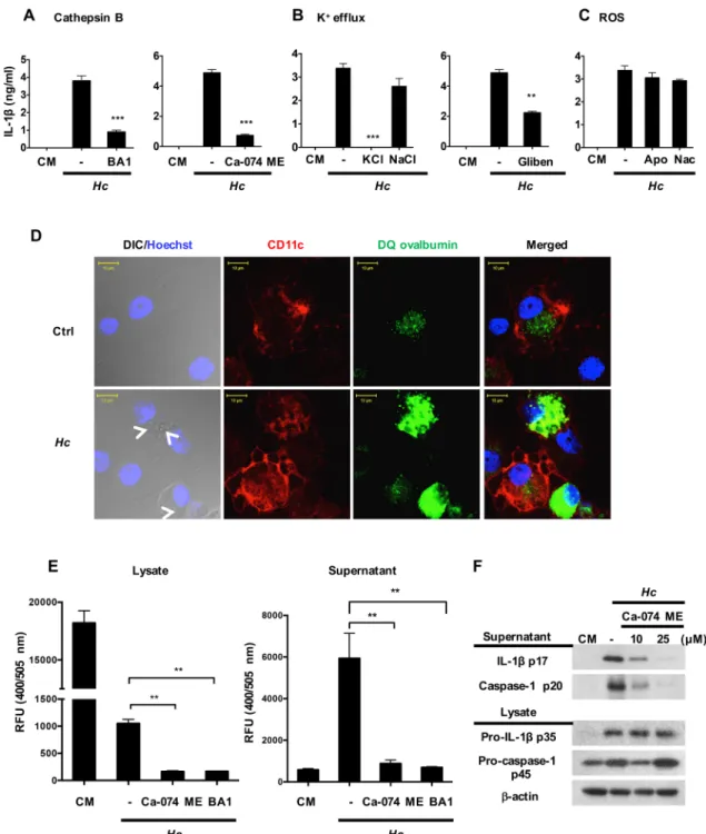
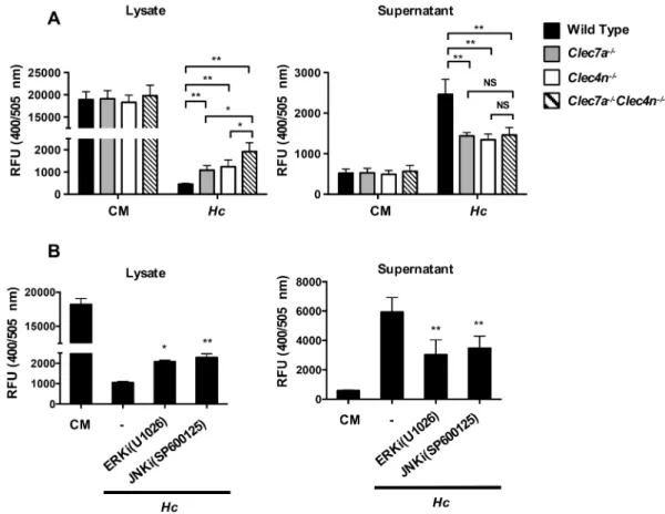
that blocking cathepsin B activity or K+release, but not inhibition of ROS, reduced the produc-tion of IL-1βin BMDC response toH.capsulatum(Fig 6A, 6B and 6C). We used DQ ovalbu-min fluorescence imaging to investigate whetherH.capsulatuminduces lysosomal rupture in BMDCs. WithoutH.capsulatumstimulation, DQ ovalbumin ingested by BMDCs was located in endosome/lysosome (Fig 6Dupper panel). After stimulation withH.capsulatum, endo-some/lysosome became enlarged and leaky (Fig 6Dlower panel). Cathepsin B activity assay also showed that activated cathepsin B was released to culture supernatants after stimulation (Fig 6E). Inhibiting cathepsin B activity or phagosome acidification reduced the release of acti-vated cathepsin B (Fig 6E). In addition, inhibition of cathepsin B activity reduced the secretion of caspase-1 p20 and IL-1βp17, but not the level of pro-IL-1β, confirming the role of cathepsin B as an upstream regulator of signal 2 during inflammasome activation (Fig 6F). Together, these data suggest that uptake ofH.capsulatuminduces dendritic cell lysosomal rupture and cathepsin B release that trigger inflammasome activation.
Dectin-2 and Dectin-1 singly and cooperatively induce cathepsin B
release
Cathepsin B activity was quantified in cells with Dectin-1, Dectin-2 single or double deficiency. Data showed that Dectin-1-deficient, Dectin-2-deficient, and double-deficient cells retained acti-vated cathepsin B in the cytosol and double-deficient cells retained significantly more than single-deficient cells (Fig 7Aleft panel). Single- and double-deficient cells reduced the release of acti-vated cathepsin B to culture supernatants although the differences between single- and double-deficient cells were not significant (Fig 7Aright panel). Additionally, pharmacological inhibition of either ERK or JNK reduced the release of active cathepsin B (Fig 7B). Interestingly, Dectin-2 deficiency, did not make a difference in the kinetics of the drop of intracellular K+levels com-pared to wild type cells (S10 Fig). These data indicate that, while Dectin-2 does not play a role in K+efflux, Dectin-1 and Dectin-2 singly and cooperatively regulate activated cathepsin B release.
The viability of H. capsulatum is critical for signal 2 in inflammasome
activation
Fig 6. Cathepsin B and K+efflux both function as signals 2 in H. capsulatum-stimulated inflammasome activation.
Wild type BMDCs were pretreated with (A) cathepsin B activity inhibitor (Ca-074 ME, 25μM), phagosome acidification inhibitor (bafilomycin A1 (BA1), 250 nM), (B) extracellular KCl (50 mM) and NaCl (50 mM), K+channel inhibitor
showed that heat inactivation and proteinase K digestion ofSchistosomaegg antigen (SEA) abolish its ability to induce signal 2 [30]. We found that viableH.capsulatuminduced higher levels of IL-1βthan their heat-killed counterparts while the viability of the organism did not affect the levels of TNF production (Fig 8A). Western blot analysis also showed that live organ-ism induced higher levels of caspase-1 p20 and IL-1βp17 (Fig 8B) than killed microorganism. It is worth noting that the viability ofH.capsulatumdid not affect the levels of pro-IL-1βp35 or NLRP3 (Fig 8B). These data showed that the viability ofH.capsulatumaffects signal 2, but not signal 1. Acridine orange was used to analyze lysosome swelling and fluorescent cathepsin B peptide substrate and activity assay to study activated cathepsin B release. Results showed that stimulation with viable but not heat-killedH.capsulatumled to lysosome swelling (Fig 8C) and cathepsin B release from cytosol (Fig 8D and 8E). Therefore, it appears that viableH.
capsulatumefficiently triggers signal 2 by inducing lysosomal rupture and greater cathepsin B
release.
three (A, B and C) and two (D, E and F) independent experiments is presented.*p<0.05,**p<0.01,***p<0.001 [2-tailed t-test (A, B and C); one-way ANOVA with Tuckey post-hoc analysis (E)].
https://doi.org/10.1371/journal.ppat.1006485.g006
Fig 7. Both Dectin-2 and Dectin-1 and downstream signals regulate dendritic cell release of cathepsin B.
BMDCs from wild type, Clec7a-/-, Clec4n-/-and Clec7a-/-Clec4n-/-mice. (A) Cells were stimulated with H. capsulatum. Cell lysates and supernatants were harvested 18 h later and subjected to cathepsin B activity assay
(n = 3). (B) Cells were treated with or without ERK (U1026) and JNK (SP600125) inhibitor before stimulation with H.
capsulatum for 18 h. Cell lysates and supernatants were harvested and subjected to cathepsin B activity assay
(n = 3). Error bars indicate standard deviation of the mean.*p<0.05,**p<0.01,***p<0.001, NS, not significant [one-way ANOVA with Tuckey post-hoc analysis (A) or Sidak post-hoc analysis (B)].
Fig 8. The viability of H. capsulatum is critical to signal 2 in inflammasome activation. (A and B) BMDCs
were stimulated with live or heat-killed H. capsulatum for 18 h. (A) Secreted IL-1βand TNF in culture supernatants were quantified by ELISA (n = 3). (B) Culture supernatants and cell lysates were subjected to Western blotting. (C and D) Acridine orange and Magic Red®were added 1 h before (C) or after (D) stimulation with live or heat-killed H.
Discussion
Pathogens interacting with PRRs is the first step in inflammasome activation [28].
Micro-sporum canis,Malasseziaspp. andC.albicansrecognized by C-type lectin receptors transduce
signals to induce NLRP3 inflammasome activation in macrophages and DCs [19,20,23].M.
canisinteracting with Dectin-1 induces pro-IL-1βtranscription in THP-1 cells [20].
Malasse-ziaspp. triggers IL-1βproduction in human monocyte-derived DCs through Dectin-1 [19]. TLR-2 and Dectin-1 are both involved in induction of pro-IL-1βtranscription by hyphal forms ofC.albicansin mouse BMDM [23]. However, Dectin-2 is solely responsible for cyto-kine induction in mouse BMDC including that of IL-1βby yeast form ofC.albicans[24]. Dec-tin-2 deficiency affects, but does not completely abolish, cytokine induction by hyphal form of
C.albicans[24]. Ritteret al. showed in BMDC thatSchistosomaegg antigen (SEA) engagement
with Dectin-2 provides signal 2 after triggering signal 1 with Pam3Cys for NLRP3 inflamma-some [30]. Thus, it appears that the interaction of fungal pathogens with receptors to initiate inflammasome activation is complex. It varies according to cell types, the type of pathogens, the different morphological forms and the triggering of signal 1 or signal 2. Data in this study show that Dectin-2 plays a role as a primary receptor that mediates both signal 1 and 2 for NLRP3 inflammasome inH.capsulatum-stimulated BMDC. In the presence of Dectin-2, Dec-tin-1 does not respond toH.capsulatum. In the absence of Dectin-2, however, recognition of
H.capsulatumby Dectin-1 does take place, although less prominently, and it can trigger both
signal 1 and 2.
Triggering Dectin-2 downstream signaling by agonistic anti-Dectin-2 antibody results in phosphorylation of Syk, ERK, p38 and JNK [25]. Stimulation of Dectin-2 byC.albicansyeasts activates Syk downstream CARD9-dependent MAPKs signaling and cytokine production in BMDC [24,25]. The hyphal form ofC.albicansactivates PLC-γ2 through Dectin-2, and it is critical for ROS production in BMDM [31]. Our finding showing thatH.capsulatum stimula-tion activates Dectin-2 downstream Syk, ERK, JNK and p38 signaling is consistent with that is reported forC.albicans[24,25]. We further demonstrated that Syk deficiency or pharmaco-logical inhibition of ERK and JNK phosphorylation inhibits pro-IL-1β, caspase-1 activation and IL-1βproduction. It is worth noting that inhibiting p38 significantly increases pro-IL-1β expression, suggesting that p38 negatively regulatesIl1bgene transcription. In a study of malaria hemozoin stimulation of macrophages, Shioet al. showed that both ERK and PI3K are involved in activation of NLRP3 inflammasome through Syk and Lyn kinases, whereas p38 plays no role [32]. Thus, MAPK molecules triggered by different stimuli may have distinct functions even on a specific pathway like that results in IL-1βproduction. ERK, JNK and p38 are activated upon Dectin-2 engagement withH.capsulatum. While Syk-JNK/ERK signaling positively regulates NLRP3 inflammasome, Syk-p38 serves as a negative regulator.
Growing evidence shows that Dectin-2 collaborates with Dectin-1 to induce cytokine response upon fungal stimulation [25,33–35]. Blocking Dectin-2 in Dectin-1-deficient BMDC stimulated withC.albicansnearly abrogates TNF and IL-10 production compared to wild type and Dectin-1-deficient cells [25]. Blocking both Dectin-1 and Dectin-2 completely abolishes the expression ofIl1bmRNA in human primary monocyte-derived DC responding toC. albi-canscompared to blocking each receptor separately [35]. Dectin-1 and Dectin-2 double-defi-cient BMDC fails to secrete IL-1βin response to the fungal pathogenTrichophyton rubrum
DIC/Hoechst field. Cells were viewed under confocal microscope. (E) Collected cell lysates and supernatants from live or heat-killed H. capsulatum stimulated cells were subjected to cathepsin B activity assay. Error bars indicate standard deviation of the mean. One representative of three (A) or two (B and E) independent experiments is presented.*p<0.05,***p<0.001 [2-tailed t-test (A and E)].
[34]. These studies indicate that Dectin-1 and Dectin-2 together mediate cytokine production in response to fungal pathogens. However, the relation between the signaling pathway(s) downstream of Dectin-1 and Dectin-2 has not been fully investigated. Results of our study show a collaborative relationship between Dectin-2 and Dectin-1 in activation of Syk-MAPKs pathway for NLRP3 inflammasome activation. Dectin-2 dominates as a receptor to transduce downstream Syk-JNK signaling in triggering NLRP3 inflammasome even in the presence of Dectin-1. When Dectin-2 is absent, Dectin-1-mediated recognition takes over, although responding less prominently, and activates the same signaling pathway. When both Dectin-2 and Dectin-1 are absent, Syk-JNK signaling becomes almost null. Thus, it appears that while Dectin-2 is the primary receptor that recognizesH.capsulatum, Dectin-1 takes its place in its absence for triggering Syk-JNK signaling for NLRP3 inflammasome in BMDC response toH.
capsulatum.
Coadyet al. showed in pulmonaryH.capsulatuminfection that IL-1R-/-mice survive intra-nasal infection with 1.8×10⁴ofH.capsulatum[36]. However, Deepeet al. reported that while IL-1R-/-mice survive infection with low dose ofH.capsulatum(1×10⁴and 2×10⁵), high dose
ofH.capsulatum(2×10⁶) causes IL-1R-/-mice to die [22]. These results together demonstrate
that IL-1βis protective when mice are challenged with high but not low dose ofH.capsulatum. We observed that when mice were infected with lower dose (2×10⁶) ofH.capsulatum(S11 Fig), there was no difference in survival betweenNlrp3-/-and wild type mice. Infection with high dose of the fungus either intratracheally or intravenously,Nlrp3-/-mice had significantly less survival than wild type mice (Fig 1JandS12 Fig). Ourin vitrodata show that stimulation of cells with higher MOI of fungus dose-dependently elicits greater inflammasome response. It appears that high dose ofH.capsulatumtriggers greater NLRP3 inflammasome response and higher IL-1βproduction that are protective against lethalH.capsulatumchallenge.
It is reported that CD103+conventional DCs in the lungs produce IFN-I through TLR7/9
uponH.capsulatuminfection [15]. CD103+DC IL-2 response toA.fumigatusis mediated by
Dectin-1 and the downstream Ca++-calmodulin-dependent NFAT signaling pathway [37]. We provide evidence to show that CD103+DCs are one of the major producers of IL-1βin the lungs inH.capsulatuminfection. Infection induces upregulation of pro-IL-1βin CD103+and CD11b+DCs as well as in neutrophils. The induction was much higher in CD103+DC than in CD11b+DCs and neutrophils, showing that CD103+DCs are more active in producing IL-1β than other two cell types in response to infection. In addition, Dectin-2 singly and in collabo-ration with Dectin-1 are involved in CD103+DC IL-1βresponse. Interestingly, while Dectin-2 single deficiency does not affect IL-1βresponse in CD11b+DCs, alveolar macrophages and neutrophils, Dectin-2 and Dectin-1 double deficiency almost completely abolish IL-1β produc-tion in all cell types. It appears that both Dectin-2 and Dectin-1 are important in host IL-1β response toH.capsulatumpulmonary infection.
caspase-1, more lysosomal swelling and increased cathepsin B release than heat-killed organism. It has been previously shown that human monocyte-derived DC is capable of killing intracellularH.
capsulatumby pronounced phagolysosomal fusion [14] and that phagosomal acidification is an
early event preceding lysosomal rupture [6]. We speculate that viableH.capsulatum-induced lysosomal rupture leading to cathepsin B release takes place after phagolysosomal fusion.
The relationship between receptor(s) and its downstream signaling that mediates signal 2 activation has not been very well-established. Recognition ofC.albicansyeast by Dectin-1 induces ROS production in BMDMs [39]. Hyphal form ofC.albicanstriggers Dectin-2 down-stream PLCγ-2 signaling that activates ROS production [31].C.albicansyeast has also been shown to activate Syk to induce ROS production leading to inflammasome activation in BMDC [16]. Compared to ROS, less is known about the receptor(s) and signaling pathway(s) that lead to cathepsin B release. Shioet al. reported that malarial hemozoin activates Syk-medi-ated cathepsin B activation in THP-1 cells [32]. In an arthritis mouse model, intra-articular injection of zymosan induces cathepsin release in a Dectin-1- and NOD2-dependent manner [40]. Interestingly, cathepsin B release is Syk-dependent in B cell receptor-mediated apoptosis [41]. In this study we provide the first direct evidence that C-type lectins, Dectin-2 and Dec-tin-1, collaboratively regulate cathepsin B release in BMDC.
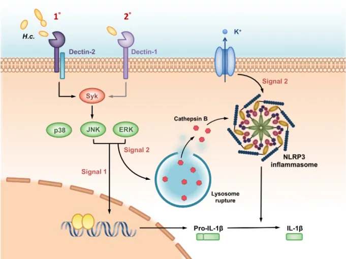
Based on our findings demonstrated in this study, we propose a working model as depicted inFig 9of the roles of Dectin-2 and Dectin-1 and their downstream signals inH.capsulatum -induced NLRP3 inflammasome activation in BMDC. Dectin-2 serves as a primary receptor and Dectin-1 as a secondary one in response toH.capsulatum. Dectin-2 and Dectin-1 down-stream signals Syk-JNK trigger both signal 1 to induce pro- IL-1βexpression and signal 2 to regulate cathepsin B release for NLRP3 inflammasome activation and IL-1βrelease. While ROS is not involved, K+efflux also functions as signal 2 but is independent of receptor regula-tion. Our data are the first to provide insight into the roles of Dectin-2 and Dectin-1 in signal-ing events for NLRP3 inflammasome activation byH.capsulatum.
Materials and methods
Ethics statement
All animal experiments were undertaken in accordance with the Guidebook for the Care and Use of Laboratory Animals, 3rd Ed., 2007, published by The Chinese-Taipei Society of Laboratory Animal Sciences, approved by the Institutional Animal Care and Use Committee (IACUC, Permit number: 20140533) of National Taiwan University, College of Medicine.
Mice
C57BL/6 wild type mice (originally from the Jackson Laboratory, Bar Harbor, Maine, USA),
Nlrp3-/-[42],Clec7a-/-(from Dr. Gordon Brown, University of Cape Town, Cape Town, South
Africa),Clec4n-/-[43],Clec7a-/-Clec4n-/-,Syk+/-andFcRγ-/-(originally from Dr. Clifford Lowell, University of California, San Francisco, CA, USA) were bred and maintained in the Labora-tory Animal Center, National Taiwan University, College of Medicine.Clec7a-/-Clec4n-/-mice were obtained from crossingClec7a-/-andClec4n-/-mice. All mice were housed in sterilized cages with sterilized bedding and filter cage tops and were fed with sterilized food and water. Mice at 8–12 weeks of age were used in all experiments.
Fungus
H.capsulatumstrain 505 was cultured at 37˚C on brain heart infusion agar [37 mg/ml; Becton
(BD) and 20% heat-inactivated certified FBS (Biological Industries). Fresh yeast cell suspen-sions were prepared in RPMI-1640 medium (Invitrogen) for each experiment. Heat-killed yeast cells were prepared by heating at 65˚C water bath for 2 h.
Cells
Bone marrow cells were flushed from mouse femurs and tibias. Cells were seeded in 24-well culture plate after RBC lysis, cultured in RPMI 1640 complete medium containing 10% heat-inactivated fetal bovine serum (FBS) and 15 ng/ml of GM-CSF. Culture medium was replen-ished on days 3 and 6. Non-adherent cells were harvested on day 7. About 75–80% of the cells were CD11c+as determined by FACS analysis. Cells were seeded in 96-well plate at 2×10⁵ cells /well or in 12-well plate at 2×10⁶cells / well in RPMI 1640 complete medium containing 10% heat-inactivated FBS. Cells were used for experiments 18–24 h later. To obtain Syk-/-cells, Syk-/-embryos were separated from Syk+/+and Syk+/-embryos after crossing Syk+/-mice by their exhibition of severe petechiae and confirmed by genotyping [44]. Single-cell suspensions from fetal liver tissues were cultured in RPMI 1640 complete medium containing GM-CSF for
Fig 9. A working model—Dectin-2/Dectin-1 and their downstream signals regulate H. capsulatum-induced NLRP3 inflammasome activation. Dendritic cells being one of the major IL-1βproducers in pulmonary H. capsulatum infection, the pathogen is recognized preferentially by Dectin-2 and less so by Dectin-1 in dendritic cells. The recognition triggers Syk-JNK signaling pathway that leads to IL-1βproduction. Syk-JNK signaling provides signal 1 to induce pro-IL-1βsynthesis as well as signal 2 to activate cathepsin B release. While K+efflux also functions as signal 2, it is independent of either receptor. Signal 1 and 2
7 days. Over 80% of the nonadherent cells were CD11C+which were identified as fetal liver-derived dendritic cells (FLDCs).
Splenic DCs isolation
Splenocytes were collected from wild type,Nlrp3-/-andClec4n-/-mice and stained with phyco-erythrin (PE)-conjugated MHC class II and allophycocyanin (APC)-conjugated anti-CD11c (eBioscience) antibodies. MHCII+CD11c+cells were sorted by FACSAria II cell sorter (BD). Sorted MHCII+CD11c+cells were seeded in 96-well plate in RPMI-1640 complete medium containing 10% heat-inactivated FBS and used in experiments immediately.
H. capsulatum systemic infection and fungal burden
Mice were intravenously infected with 1×10⁷ofH.capsulatumyeast cells and monitored for 18 days for survival. To determine fungal burdens, spleens were collected on day 11 and spleen homogenates were plated on glucose-peptone agar plates. Mycelial colonies were counted 10 days later.
Reagents and antibodies
Blocking antibodies anti-Dectin-1 (2A11), anti-Dectin-2 (D2.11E4) and anti-CR3 (5C6) were purchased from Serotec and anti-TLR-2 (6C2), IgG2a (eBR2a) and IgG2b (eB149/10H5) were from eBioscience. PE/Cy7-conjugated anti-CD45 (30-F11), APC-conjugated anti-F4/80 (BM8), Alexa 488-conjugated anti-CD103 (2E7) and Alexa 647-conjugated anti-Ly6G (1A8) were obtained from BioLegend. APC-conjugated CD11c (N418) and PE-conjugated anti-CD11b (M1/70) were from eBioscience and PE-conjugated anti-Siglec-F (E50-2440) from Pharmingen.
Z-YVAD-FMK (Caspase-1 inhibitor) was obtained from BioVision. U1026 (ERK inhibi-tor), SP600125 (JNK inhibiinhibi-tor), SB203580 (p38 inhibiinhibi-tor), N-acetyl-L-cysteine (ROS scaven-ger), Apocynin (NADPH-oxidase inhibitor), Ca-074 methyl ester (cathepsin B inhibitor), bafilomycin A1 (phagosomal acidification inhibitor) and glibenclamide (K+channel inhibitor) were obtained from Sigma-Aldrich.
IL-1
β
and TNF ELISA assays
BMDCs (2×10⁵/well) or splenic DCs (1×10⁵/well) were seeded in 96-well plates and cultured overnight before treatment with indicated reagents or blocking antibodies. Live or heat-killed
H.capsulatum(yeast:cell ratio of 10:1 or 20:1) were added 60 min later. Culture supernatants
were collected at different time points and stored at -80˚C. ELISA kits (eBioscience) were used to quantify IL-1βand TNF in the culture supernatants with 7.8125 pg/ml as the lowest limit of detection.
Western blotting
blocked with 5% non-fat milk and left in buffer containing anti-IL-1βp17 (R&D system), anti-Caspase-1 p20 (Adipogen), anti-NLRP3 (Adipogen), anti-ASC (Adipogen), anti-p-Syk (Abcam), anti-p-ERK (Cell Signaling), anti-p-JNK (Cell Signaling), anti-p-p38 (Cell Signaling) or anti-β-actin (GeneTex) antibody at 4˚C overnight. The membrane was washed with TBST before addition of HRP-conjugated anti-goat IgG (1:3000), anti-rabbit (1:20,000) or anti-rat IgG (1:20,000). ECL reagent (PerkinElmer Life Science, Merck Millipore and GE Healthcare) was used for detection.
ASC pyroptosome detection
BMDCs were seeded in 12-well plates (2×10⁶cells/well) and treated with liveH.capsulatum
or LPS (100 ng/ml, 6 h) plus ATP (5 mM, 30 minutes) for 18 h. Cells were centrifuged at 4500 rpm for 5 min. Cell pellets were resuspended in cold 0.3 ml buffer A (20 mM HEPES-KOH, pH 7.5, 10 mM KCl, 1.5 mM MgCl2, 1 mM EDTA, 1 mM EGTA, 320 mM sucrose) and prote-ase inhibitor cocktail. Cell lysate was obtained after passing the suspension through 29-G syringe for 15 times. Cell lysates were spun at 4500 rpm for 8 min to remove contaminating nuclei, unlysed cells andH.capsulatum. Supernatants were diluted with 1 volume of CHAPS buffer (20 mM HEPES-KOH, pH 7.5, 5 mM MgCl2, 0.5 mM EGTA and 0.1% CHAPS) to lyse residual organelles before centrifugation at 8000 rpm for 8 min. After centrifugation, superna-tants were discarded and the pellets were treated with disuccinimidyl suberate (DSS, 2 mM, Sigma-Aldrich) for 30 min at room temperature. The cross-linked pellets were re-suspended in 40μl SDS sample buffer and proteins were separated on 12.5% SDS polyacrylamide gel fol-lowed by immunoblotting with anti-ASC antibody (Adipogen) as previously described [45]
Phagocytosis assay
The detailed methodology has been previous described [46]. BMDCs (2×10⁶) from wild type,
Clec7a-/-andClec4n-/-mice were seeded in 12-well plates. The plates were cooled on ice for 20
min before the addition of FITC-labeledH.capsulatumat MOI of 20. After 60 min cooling on ice, the plates were moved to 37˚C CO2incubator for 60 min. Cells were collected and quenched FITC-labeled yeast by trypan blue for 5 min. After washing with dPBS, cells were fixed with 1% paraformaldehyde. The phagocytosis rate was determined by flow cytometry.
Isolation of lung cells
Wild type,Clec4n-/-andClec7a-/-Clec4n-/-mice were intratracheally infected with 2.5×10⁵H.
capsulatum. Lungs were collected on day 7 after infection and digested with 0.15 mg/ml of
Lib-erase TM (Roche) at 37˚C for 30 min on a rotatory agitator. Cell pellets were resuspended in 45% Percoll (GE Healthcare) and overlaid on 81% Percoll. After centrifugation, cells at inter-face (45/81% Percoll) were harvested and stained with antibodies before sorting by FACSAria II cell sorter (BD). CD45+cells were sub-gated to sort out different cell populations: CD103+ DCs (CD11c+CD103+CD11b-), CD11b+DCs (CD11c+CD103-CD11b+), alveolar macrophages (Siglec-F+F4/80+), neutrophil (CD11b+Ly6G+).
RT-qPCR
Quantitative real-time PCR was performed by using ABI7900 Real-time PCR detection sys-tem in total volume of 10μl per reaction, containing 2μl of cDNA with 0.2μM forward and reverse primers in 8μl Fast SYBR Green Master Mix (Applied Biosystems).Il1bmRNA was normalized againstActbmRNA. Primers forIl1bare 5’-GAA CTC AAC TGT GAA ATG CCA CC-3’(forward) and 5’-CCA CAG CCA CAA TGA GTG ATA CT-3’(reverse) and forActb 5’-TGT ATG AAG GCT TTG GTC TCC CT-3’ (forward) and 5’-AGG 5’-TGT GCA CTT TTA TTG GTC TCA A-3’ (reverse).
Lysosomal rupture
BMDCs (1×10⁵) were seeded in 96-well plate and treated with 10μM of DQ ovalbumin (Sigma-Aldrich) for 1 h before stimulation with or withoutH.capsulatumat MOI of 1. Cells were collected 1 h later and cytospun on microscope slides. The cytospun cells were fixed in 4% paraformaldehyde, blocked with 5% heat-inactivated FBS and stained with APC-conju-gated anti-CD11c antibody (eBioscience). Hoechst 33258 was used to stain cell nuclei. The slides were viewed under Zeiss LSM 510 META Confocal Microscope.
Cathepsin B activity assay
BMDCs (2×10⁶) were cultured in 12-well plate with phenol red-free RPMI complete medium (Invitrogen) and stimulated with or withoutH.capsulatumat MOI of 20. Cells and culture supernatants were collected separately 18 h after stimulation. Cell-free supernatants were con-centrated 10-fold by Vivaspin 500 (GE Healthcare). Cells were lysed with cathepsin B lysis buffer (Abcam, ab65300). Both cell lysates and concentrated supernatants were loaded unto 96-well plate (solid black, Corning) before addition of CB reaction buffer. CB substrate Ac-RR-AFC (final concentration of 200μM) was added and incubated at 37˚C for 1 h. Samples were read in SpectraMax M5 (Molecular Devices) at 400 nm excitation and 505 nm emission wavelengths.
Intracellular K
+concentration
BMDCs (2×10⁵) were seeded in 96-well plate (black with clear flat-bottom tissue culture plate, Corning) and were loaded with 2μM potassium-binding benzofuran isophthalate-AM (PBFI/AM, Molecular Probes, Thermo Fisher Scientific) in phenol red-free RPMI medium (Invitrogen) in the presence of 0.05% (w/w) Pluronic F-127 (Sigma-Aldrich) at 25˚C in the dark for 60 min. After one wash, cells were stimulated withH.capsulatumat MOI of 1. Fluo-rescence intensity of PBFI (excitation wavelength 340 nm, emission wavelength 500 nm) was recorded every min by SpectraMax M5 while the culture was maintained in 37˚C. Stimulation with ATP (5 mM) was used as a positive control.
Staining for lysosome and activated cathepsin B
ROS production
BMDCs (1.2×10⁶) were incubated in phenol red-free HBSS containing 10μM CM-H2DCFDA (Thermo Fisher Scientific) for 30 min at 37˚C. After replenishment with fresh phenol red-free RPMI 1640 complete medium, cells were stimulated withH.capsulatum(MOI = 20). Oxida-tive DCF was analyzed by flow cytometry.
Statistics
Differences between treatment groups were analyzed with the student 2-tailedttest (compar-ing two group) and one-way (compar(compar-ing multiple groups with one control) or two-way (com-paring multiple groups with two control groups) ANOVA followed by Tukey or Sidak post-hoc test. Log-rank test was used to analyze % survival. All statistics analyses were calculated by Prism 6 software. The level of statistical significance was defined asp<0.05. All results were expressed as mean±standard deviation of the mean.
Supporting information
S1 Fig. The absence of NLRP3 does not affectH. capsulatum-induced TNF production in
dendritic cells. (A) BMDCs and (B) sorted splenic DCs from wild type and NLRP3-deficient
(Nlrp3-/-) mice were stimulated withH.capsulatumat MOI of 10 and 20 for 18 h. Stimulation
with LPS (500 ng/ml, 6 h) plus ATP (5 mM, 30 minutes) (L+A) was used as a positive control for TNF induction. (A and B) TNF in the supernatants were quantified by ELISA (n = 3). Error bars indicate standard deviation of the mean. One representative of three independent experiments is presented. [2-tailedt-test].
(TIF)
S2 Fig.FcRγ-/-BMDCs produce lower levels of IL-1βand caspase-1 p20 uponH. capsula-tum stimulation. (A and B) BMDCs from wild type and Fc receptorγchain-deficient (FcRγ-/-) mice were stimulated with or withoutH.capsulatum. (A) IL-1βin cell-free supernatants were quantified by ELISA (n = 3). (B) Cell lysates and supernatants were subjected to Western blot-ting. Error bars indicate standard deviation of the mean.p<0.01 [2-tailedt-test (A)].
(TIF)
S3 Fig. Syk deficiency reduces expression of pro-caspase-1 p45 afterH. capsulatum
stimu-lation. Relative intensity of pro-caspase-1 p45 were quantified by ImageJ (n = 3) from data in Fig 3D. Data were pooled from three independent experiments.p<0.05 [one-way ANOVA with Tuckey post-hoc analysis].
(TIF)
S4 Fig. Surface expression of Dectin-1 and Dectin-2 on wild type, Dectin-1-deficient and Dectin-2-deficient BMDCs. Cells from wild type, Dectin-1-deficient (Clec7a-/-), and Dectin-2-deficient (Clec4n-/-) mice were stained with APC-anti-CD11c antibody, purified-antibody against Dectin-1 and Dectin-2 and Alexa 488-goat anti-rat IgG. IgG2a and IgG2b were used as isotype controls. Histograms show the fluorescence intensity of each receptor on CD11c+cells. (TIF)
S5 Fig. Neither Dectin-1 nor Dectin-2 is involved in phagocytosis ofH. capsulatum.
post-hoc analysis]. (TIF)
S6 Fig. The roles of Dectin-2 and Dectin-1 in inflammasome activation. (A and B) BMDCs from wild type, Dectin-2-deficient (Clec4n
-/-) and Dectin-1-deficient (Clec7a
-/-) mice were pre-treated with or without anti-Dectin-1 or -Dectin-2 blocking antibody (2μg/ml) for 1 h before stimulation withH.capsulatum. Cell culture supernatants were collected at 18 h after stimula-tion. (A) Secreted IL-1βwas quantified by ELISA (n = 5). (B) Cell-free supernatants and cell lysates were subjected to Western blotting analysis. IgG2a and IgG2b were used as isotype con-trols. Error bars indicate standard deviation of the mean. One representative of three (A) or two (B) independent experiments is presented.p<0.05,p<0.01,p<0.001 [two-way ANOVA with Tukey post-hoc analysis (A)].
(TIF)
S7 Fig. Dectin-2 and Dectin-1 double deficiency completely abrogates Syk-JNK signaling. BMDCs from wild type, Dectin-1 (Clec7a-/-) and Dectin-2 (Clec4n-/-) single-deficient and double-deficient (Clec7a-/-Clec4n-/-) mice were stimulated withH.capsulatum. Collected cell lysates were analyzed for MAPK signaling molecules by Western blotting. One representative of three independent experiments is presented. Relative intensity of phosphorylated MAPK molecules were quantified by ImageJ. Error bars indicate standard deviation of the mean. (n = 3)p<0.05,p<0.01,p<0.001 [one-way ANOVA with Tukey post-hoc analysis
and 2-tailedt-test (B)]. (TIF)
S8 Fig. The effect of reciprocal blocking of Dectin-1 and Dectin-2 in single receptor defi-cient cells on signal pathway. (A) BMDCs from wild type, Dectin-2-defidefi-cient (Clec4n-/-) and Dectin-1-deficient (Clec7a-/-) mice were pretreated with Dectin-1 and Dectin-2 blocking anti-body, respectively, before stimulation withH.capsulatum. Collected cell lysates were analyzed for MAPK signaling molecules by Western blotting. One representative of three independent experiments is presented. (B) Relative intensity of phosphorylated MAPK molecules were quantified by ImageJ (n = 3). IgG2a and IgG2b were used as isotype controls. Error bars indi-cate standard deviation of the mean.p<0.05,p<0.01,p<0.001, NS, not significant
[two-way ANOVA with Tukey post-hoc analysis and 2-tailedt-test (B)]. (TIF)
S9 Fig. Flow cytometric analysis of cell populations in the lungs. The lung cell population was determined by flow cytometry based on the following gating strategy: Viable cells were selected by gating out debris. CD45+CD11c+CD103+CD11b-cells are designated as CD103+ DC and CD45+CD11c+CD103-CD11b+cells as CD11b+DC, Siglec-F+F4/80+cells as alveolar macrophages, and CD11b+Ly6G+as neutrophils.
(TIF)
S11 Fig.Nlrp3-/-mice survive low dose of intravenousH. capsulatum infection. WT and
Nlrp3-/-mice were intravenously infected withH.capsulatum(2×10⁶). Survival was analyzed
by log-rank test. (TIF)
S12 Fig.Nlrp3-/-mice succumb to high dose of intratrachealH. capsulatum infection. WT
andNlrp3-/-mice were intratracheally infected withH.capsulatum(1×10⁷). Survival was
ana-lyzed by log-rank test.p<0.05.
(TIF)
S13 Fig.H. capsulatum induces ROS production in BMDC. BMDCs (1.2×10⁶) from wild type mice were incubated with 10μM of CM-H2DCFDA for 30 min before stimulation with or withoutH.capsulatum. The levels of ROS production are shown as mean florescence intensity (MFI) of oxidized DCF fluorescence (n = 3). The MFI at zero minute represents value of the control without stimulation. One representative of two independent experiments is shown. (TIF)
Acknowledgments
We thank the Cell Sorting Core Facility of the First Core laboratory, National Taiwan Univer-sity College of Medicine for cell sorting service. The confocal microscopy and microplate reader services provided by the Second and the Seventh Core Laboratories of Research Core Facility of the Department of Medical Research, National Taiwan University Hospital are gratefully acknowledged. We thank Chia-Lin Weng for her art work illustrating our working model that is presented in Fig 9.
Author Contributions
Conceptualization: Tzu-Hsuan Chang, Juin-Hua Huang, Hsiu-Chao Lin, Wen-Yu Chen, Betty A. Wu-Hsieh.
Formal analysis: Tzu-Hsuan Chang, Betty A. Wu-Hsieh.
Funding acquisition: Betty A. Wu-Hsieh.
Investigation: Tzu-Hsuan Chang, Juin-Hua Huang, Hsiu-Chao Lin, Wen-Yu Chen, Yu-Hsiang Lee.
Methodology: Tzu-Hsuan Chang, Li-Chung Hsu, Betty A. Wu-Hsieh.
Resources: Li-Chung Hsu, Mihai G. Netea, Jenny P.-Y. Ting.
Writing – original draft: Tzu-Hsuan Chang, Betty A. Wu-Hsieh.
Writing – review & editing: Tzu-Hsuan Chang, Juin-Hua Huang, Wen-Yu Chen, Mihai G. Netea, Jenny P.-Y. Ting, Betty A. Wu-Hsieh.
References
1. Meylan E, Tschopp J, Karin M. Intracellular pattern recognition receptors in the host response. Nature. 2006; 442(7098):39–44.https://doi.org/10.1038/nature04946PMID:16823444.
2. Sutterwala FS, Ogura Y, Szczepanik M, Lara-Tejero M, Lichtenberger GS, Grant EP, et al. Critical role for NALP3/CIAS1/Cryopyrin in innate and adaptive immunity through its regulation of caspase-1. Immu-nity. 2006; 24(3):317–27.https://doi.org/10.1016/j.immuni.2006.02.004PMID:16546100.
3. Mariathasan S, Monack DM. Inflammasome adaptors and sensors: intracellular regulators of infection and inflammation. Nat Rev Immunol. 2007; 7(1):31–40.https://doi.org/10.1038/nri1997PMID:
4. Lamkanfi M, Dixit VM. Mechanisms and functions of inflammasomes. Cell. 2014; 157(5):1013–22.
https://doi.org/10.1016/j.cell.2014.04.007PMID:24855941.
5. Cruz CM, Rinna A, Forman HJ, Ventura AL, Persechini PM, Ojcius DM. ATP activates a reactive oxy-gen species-dependent oxidative stress response and secretion of proinflammatory cytokines in macro-phages. J Biol Chem. 2007; 282(5):2871–9.https://doi.org/10.1074/jbc.M608083200PMID:17132626;
6. Hornung V, Bauernfeind F, Halle A, Samstad EO, Kono H, Rock KL, et al. Silica crystals and aluminum salts activate the NALP3 inflammasome through phagosomal destabilization. Nat Immunol. 2008; 9 (8):847–56.https://doi.org/10.1038/ni.1631PMID:18604214;
7. Murakami T, Ockinger J, Yu J, Byles V, McColl A, Hofer AM, et al. Critical role for calcium mobilization in activation of the NLRP3 inflammasome. Proc Natl Acad of Sci U S A. 2012; 109(28):11282–7.https:// doi.org/10.1073/pnas.1117765109PMID:22733741
8. Petrilli V, Papin S, Dostert C, Mayor A, Martinon F, Tschopp J. Activation of the NALP3 inflammasome is triggered by low intracellular potassium concentration. Cell Death Differ. 2007; 14(9):1583–9.https:// doi.org/10.1038/sj.cdd.4402195PMID:17599094.
9. Shimada K, Crother TR, Karlin J, Dagvadorj J, Chiba N, Chen S, et al. Oxidized mitochondrial DNA acti-vates the NLRP3 inflammasome during apoptosis. Immunity. 2012; 36(3):401–14.https://doi.org/10. 1016/j.immuni.2012.01.009PMID:22342844;
10. Retallack DM, Woods JP. Molecular epidemiology, pathogenesis, and genetics of the dimorphic fungus Histoplasma capsulatum. Microbes Infect. 1999; 1(10):817–25.https://doi.org/10.1016/S1286-4579 (99)80084-7PMID:10816087
11. Woods JP. Histoplasma capsulatum molecular genetics, pathogenesis, and responsiveness to its envi-ronment. Fungal Genet Biol. 2002; 35(2):81–97.https://doi.org/10.1006/fgbi.2001.1311PMID:11848673 12. Wu SY, Yu JS, Liu FT, Miaw SC, Wu-Hsieh BA. Galectin-3 negatively regulates dendritic cell production
of IL-23/IL-17-axis cytokines in infection by Histoplasma capsulatum. J Immunol. 2013; 190(7):3427– 37.https://doi.org/10.4049/jimmunol.1202122PMID:23455499.
13. Gildea LA, Morris RE, Newman SL. Histoplasma capsulatum yeasts are phagocytosed via very late antigen-5, killed, and processed for antigen presentation by human dendritic cells. J Immunol. 2001; 166(2):1049–56. PMID:11145684
14. Gildea LA, Ciraolo GM, Morris RE, Newman SL. Human dendritic cell activity against Histoplasma cap-sulatum is mediated via phagolysosomal fusion. Infect Immun. 2005; 73(10):6803–11.https://doi.org/ 10.1128/IAI.73.10.6803-6811.2005PMID:16177358;
15. Van Prooyen N, Henderson CA, Hocking Murray D, Sil A. CD103+ Conventional Dendritic Cells Are Critical for TLR7/9-Dependent Host Defense against Histoplasma capsulatum, an Endemic Fungal Pathogen of Humans. PLoS Pathog. 2016; 12(7):e1005749.https://doi.org/10.1371/journal.ppat. 1005749PMID:27459510;
16. Gross O, Poeck H, Bscheider M, Dostert C, Hannesschlager N, Endres S, et al. Syk kinase signalling couples to the Nlrp3 inflammasome for anti-fungal host defence. Nature. 2009; 459(7245):433–6.
https://doi.org/10.1038/nature07965PMID:19339971.
17. Guo C, Chen M, Fa Z, Lu A, Fang W, Sun B, et al. Acapsular Cryptococcus neoformans activates the NLRP3 inflammasome. Microbes Infect. 2014; 16(10):845–54.https://doi.org/10.1016/j.micinf.2014.08. 013PMID:25193031.
18. Karki R, Man SM, Malireddi RK, Gurung P, Vogel P, Lamkanfi M, et al. Concerted activation of the AIM2 and NLRP3 inflammasomes orchestrates host protection against Aspergillus infection. Cell Host & Microbe. 2015; 17(3):357–68.https://doi.org/10.1016/j.chom.2015.01.006PMID:25704009;
19. Kistowska M, Fenini G, Jankovic D, Feldmeyer L, Kerl K, Bosshard PContassot E, et al. Malassezia yeasts activate the NLRP3 inflammasome in antigen-presenting cells via Syk-kinase signalling. Exp Dermatol. 2014; 23(12): 884–9.https://doi.org/10.1111/exd.12552PMID:25267545
20. Mao L, Zhang L, Li H, Chen W, Wang H, Wu S, et al. Pathogenic fungus Microsporum canis activates the NLRP3 inflammasome. Infect Immun. 2014; 82(2):882–92.https://doi.org/10.1128/IAI.01097-13
PMID:24478101;
21. Tomalka J, Ganesan S, Azodi E, Patel K, Majmudar P, Hall BA, et al. A novel role for the NLRC4 inflam-masome in mucosal defenses against the fungal pathogen Candida albicans. PLoS Pathog. 2011; 7 (12):e1002379.https://doi.org/10.1371/journal.ppat.1002379PMID:22174673;
22. Deepe GS, McGuinness M. Interleukin-1 and host control of pulmonary histoplasmosis. J Infect Dis. 2006; 194(6):855–64.https://doi.org/10.1086/506946PMID:16941354
24. Saijo S, Ikeda S, Yamabe K, Kakuta S, Ishigame H, Akitsu A, et al. Dectin-2 recognition of alpha-man-nans and induction of Th17 cell differentiation is essential for host defense against Candida albicans. Immunity. 2010; 32(5):681–91.https://doi.org/10.1016/j.immuni.2010.05.001PMID:20493731.
25. Robinson MJ, Osorio F, Rosas M, Freitas RP, Schweighoffer E, Gross O, et al. Dectin-2 is a Syk-cou-pled pattern recognition receptor crucial for Th17 responses to fungal infection. J Exp Med. 2009; 206 (9):2037–51.https://doi.org/10.1084/jem.20082818PMID:19703985;
26. Huang JH, Lin CY, Wu SY, Chen WY, Chu CL, Brown GD, et al. CR3 and Dectin-1 Collaborate in Mac-rophage Cytokine Response through Association on Lipid Rafts and Activation of Syk-JNK-AP-1 Path-way. PLoS Pathog. 2015; 11(7):e1004985.https://doi.org/10.1371/journal.ppat.1004985PMID:
26132276;
27. Wang H, LeBert V, Hung CY, Galles K, Saijo S, Lin X, et al. C-type lectin receptors differentially induce th17 cells and vaccine immunity to the endemic mycosis of North America. J Immunol. 2014; 192 (3):1107–19.https://doi.org/10.4049/jimmunol.1302314PMID:24391211;
28. Tavares AH, Burgel PH, Bocca AL. Turning Up the Heat: Inflammasome Activation by Fungal Patho-gens. PLoS Pathog. 2015; 11(7):e1004948.https://doi.org/10.1371/journal.ppat.1004948PMID:
26204108;
29. Joly S, Ma N, Sadler JJ, Soll DR, Cassel SL, Sutterwala FS. Cutting edge: Candida albicans hyphae for-mation triggers activation of the Nlrp3 inflammasome. J Immunol. 2009; 183(6):3578–81.https://doi. org/10.4049/jimmunol.0901323PMID:19684085;
30. Ritter M, Gross O, Kays S, Ruland J, Nimmerjahn F, Saijo S, et al. Schistosoma mansoni triggers Dectin-2, which activates the Nlrp3 inflammasome and alters adaptive immune responses. Proc Natl Acad Sci U S A. 2010; 107(47):20459–64.https://doi.org/10.1073/pnas.1010337107PMID:
21059925;
31. Gorjestani S, Yu M, Tang B, Zhang D, Wang D, Lin X. Phospholipase Cgamma2 (PLCgamma2) is key component in Dectin-2 signaling pathway, mediating anti-fungal innate immune responses. J Biol Chem. 2011; 286(51):43651–9.https://doi.org/10.1074/jbc.M111.307389PMID:22041900;
32. Shio MT, Eisenbarth SC, Savaria M, Vinet AF, Bellemare MJ, Harder KW, et al. Malarial hemozoin acti-vates the NLRP3 inflammasome through Lyn and Syk kinases. PLoS Pathog. 2009; 5(8):e1000559.
https://doi.org/10.1371/journal.ppat.1000559PMID:19696895;
33. Zhu LL, Zhao XQ, Jiang C, You Y, Chen XP, Jiang YY, et al. C-type lectin receptors Dectin-3 and Dec-tin-2 form a heterodimeric pattern-recognition receptor for host defense against fungal infection. Immu-nity. 2013; 39(2):324–34.https://doi.org/10.1016/j.immuni.2013.05.017PMID:23911656.
34. Yoshikawa FS, Yabe R, Iwakura Y, de Almeida SR, Saijo S. Dectin-1 and Dectin-2 promote control of the fungal pathogen Trichophyton rubrum independently of IL-17 and adaptive immunity in experimental deep dermatophytosis. Innate Immun. 2016; 22(5):316–24.https://doi.org/10.1177/
1753425916645392PMID:27189427.
35. Gringhuis SI, Wevers BA, Kaptein TM, van Capel TM, Theelen B, Boekhout T, et al. Selective C-Rel activation via Malt1 controls anti-fungal T(H)-17 immunity by dectin-1 and dectin-2. PLoS Pathog. 2011; 7(1):e1001259.https://doi.org/10.1371/journal.ppat.1001259PMID:21283787;
36. Coady A, Sil A. MyD88-dependent signaling drives host survival and early cytokine production during Histoplasma capsulatum infection. Infect Immun. 2015; 83(4):1265–75.https://doi.org/10.1128/IAI. 02619-14PMID:25583527;
37. Zelante T, Wong AY, Ping TJ, Chen J, Sumatoh HR, Vigano E, et al. CD103(+) Dendritic Cells Control Th17 Cell Function in the Lung. Cell Rep. 2015; 12(11):1789–801.https://doi.org/10.1016/j.celrep. 2015.08.030PMID:26365185.
38. Tschopp J, Schroder K. NLRP3 inflammasome activation: The convergence of multiple signalling path-ways on ROS production? Nat Rev Immunol. 2010; 10(3):210–5.https://doi.org/10.1038/nri2725PMID:
20168318.
39. Gantner BN, Simmons RM, Underhill DM. Dectin-1 mediates macrophage recognition of Candida albi-cans yeast but not filaments. EMBO J. 2005; 24(6):1277–86.https://doi.org/10.1038/sj.emboj.7600594
PMID:15729357
40. Rosenzweig HL, Clowers JS, Nunez G, Rosenbaum JT, Davey MP. Dectin-1 and NOD2 mediate cathepsin activation in zymosan-induced arthritis in mice. Inflamm Res. 2011; 60(7):705–14.https://doi. org/10.1007/s00011-011-0324-7PMID:21424514
41. He J, Tohyama Y, Yamamoto K-i, Kobayashi M, Shi Y, Takano T, et al. Lysosome is a primary organelle in B cell receptor-mediated apoptosis: an indispensable role of Syk in lysosomal function. Genes to Cells. 2005; 10(1):23–35.https://doi.org/10.1111/j.1365-2443.2004.00811.xPMID:15670211 42. Wen H, Gris D, Lei Y, Jha S, Zhang L, Huang MT, et al. Fatty acid-induced NLRP3-ASC inflammasome
43. Ifrim DC, Bain JM, Reid DM, Oosting M, Verschueren I, Gow NA, et al. Role of Dectin-2 for host defense against systemic infection with Candida glabrata. Infect Immun. 2014; 82(3):1064–73.https://doi.org/ 10.1128/IAI.01189-13PMID:24343653;
44. Yanagi S, Inatome R, Ding J, Kitaguchi H, Tybulewicz VLJ, Yamamura H. Syk expression in endothelial cells and their morphologic defects in embryonic Syk-deficient mice. Blood. 2001; 98(9):2869. PMID:
11675365
45. Fernandes-Alnemri T, Alnemri ES. Chapter Thirteen Assembly, Purification, and Assay of the Activity of the ASC Pyroptosome. 2008; 442:251–70.https://doi.org/10.1016/s0076-6879(08)01413-4